adaptation of tape removal test for measurement of
TRANSCRIPT
This is a repository copy of Adaptation of tape removal test for measurement of sensitivity in perineal area of rat.
White Rose Research Online URL for this paper:http://eprints.whiterose.ac.uk/153843/
Version: Accepted Version
Article:
Neumannova, K, Machova-Urdzikova, L, Kwok, JCF orcid.org/0000-0002-9798-9083 et al. (2 more authors) (2020) Adaptation of tape removal test for measurement of sensitivity in perineal area of rat. Experimental neurology, 324. 113097. 113097-. ISSN 0014-4886
https://doi.org/10.1016/j.expneurol.2019.113097
© 2019, Elsevier. This manuscript version is made available under the CC-BY-NC-ND 4.0 license http://creativecommons.org/licenses/by-nc-nd/4.0/.
[email protected]://eprints.whiterose.ac.uk/
Reuse
This article is distributed under the terms of the Creative Commons Attribution-NonCommercial-NoDerivs (CC BY-NC-ND) licence. This licence only allows you to download this work and share it with others as long as you credit the authors, but you can’t change the article in any way or use it commercially. More information and the full terms of the licence here: https://creativecommons.org/licenses/
Takedown
If you consider content in White Rose Research Online to be in breach of UK law, please notify us by emailing [email protected] including the URL of the record and the reason for the withdrawal request.

Adaptation of tape removal test for measurement of sensitivity in
perineal area of rat
K. Neumannovaa,b, L. Machova-Urdzikovaa, J. C. F. Kwoka,c, J. W. Fawcetta,d, P. Jendelovaa,b
aInstitute of Experimental Medicine Czech Academy of Science, Videnska 1083, 14220 Prague,
Czech Republic
b2nd Faculty of Medicine, Charles University, V úvalu 84, 15006 Prague, Czech Republic,
cFaculty of Biological S
ciences, University of Leeds, UK,
dJohn van Geest Centre for Brain Repair, University of Cambridge, UK
corresponding author:
Pavla Jendelova, PhD,
Institute of Experimental Medicine Czech Academy of Science,
Videnska 1083, 14220 Prague,
Czech Republic,
Abbreviations:
BBB test - Basso Beattie and Bresnahan test, D - day, L に lumbar vertebrae, PFA に
paraformaldehyde, SCI - spinal cord injury, T - thoracic vertebrae, W - week


Abstract
Regeneration after spinal cord injury is a goal of many studies. Although the most obvious target
is to recover motor function, restoration of sensation can also improve the quality of life after spinal
cord injury. For many patients, recovery of sensation in the perineal and genital area is a high
priority. Currently there is no experimental test in rodents for measuring changes in sensation in the
perineal and genital area after spinal cord injury. The aim of our study was to develop a behavioural
test for measuring the sensitivity of the perineal and genital area in rats. We have modified the tape
removal test used routinely to test sensorimotor deficits after stroke and spinal cord injury to test
the perineal area with several variations. A small piece of tape (approximately 1 cm2) was attached to
the perineal area. Time to first contact and to the removal of the tape was measured. Each rat was
trained for 5 consecutive days and then tested weekly. We compared different rat strains (Wistar,
Sprague-Dawley, Long-Evans and Lewis), both genders, shaving and non-shaving and different types
of tape. We found that the test was suitable for all tested strains, however, Lewis rats achieved the
lowest contact times, but this difference was significant only for the first few days of learning the
task. There were no significant differences between gender and different types of tape or shaving.
After training the animals underwent dorsal column lesion at T10 and were tested at day 3, 8, 14 and
21. The test detected a sensory deficit, the average time across all animals to sense the stimulus
キミIヴW;ゲWS aヴラマ ヱげンヲ ┌ヮ デラ ンげヲヰく There was a strong relationship between lesion size and tape
detection time, and only lesions that extended laterally to the dorsal root entry zone produced
significant sensory deficits. Other standard behavioural tests (BBB, von Frey, ladder and Plantar test)
were performed in the same animals. There was a correlation between lesion size and deficit for the
ladder and BBB tests, but not for the von Frey and Plantar tests. We conclude that the tape removal
test is suitable for testing perineal sensation in rats, can be used in different strains and is
appropriate for monitoring changes in sensation after spinal cord injury.
Keywords: sensation, spinal cord injury, perineum, functional outcome, behavioural tests, sensory
regeneration.
Introduction
Spinal cord injury (SCI) is a serious state that affects about 250 000 に 500 000 people worldwide
every year (https://www.who.int/en/news-room/fact-sheets/detail/spinal-cord-injury). The main
causes are road traffic crashes, falls or violence. To date, no standard therapy for the regeneration of
axons in the severed spinal cord of humans exists.
Research in this area is usually focused on motor recovery, but restoration of sensory function
would also improve the life of patients. Moreover, sensory and motor recovery are closely linked,
with motor function being dependent on sensory inputs to the cord. Many descending motor
projections to the cord terminate on interneurons that also receive sensory inputs (Levine et al.,
2014). In respect of sensory recovery, high priorities for human patients are restoration of sensation
to the hands, and restoration of perineal and genital sensation (Anderson, 2004). A standard
experimental model of spinal cord injury in rodents is the dorsal column lesion. This lesion disrupts
ascending sensory fibres from the dorsal root ganglia carrying tactile information, discriminatory
touch, vibration and proprioception (Sengul and Watson, 2015). There are several behavioural tests
for assessment of sensation in the limbs but none as yet for the perineal area.
The tape removal test (also called sticky tape test or adhesive removal test) was developed by
Schallert et al. (1982) to measure sensorimotor asymmetries after nigrostriatal damage in rats. It is
based on attachment of a small piece of adhesive paper to various parts of body (snout, forepaws,
hindpaws). The time taken to sense the stimulus and to remove the tape is measured.
Since its development (Schallert et al., 1982), this test has been used in many studies and with
various modifications. In most studies the tape is usually attached to the forepaws (Aehling et al.,
2018; Albertsmeier et al., 2007; Schallert et al., 2000). However, as tested by Schallert et al. (1982) it
can be also used in other body parts including the snout (Fleming et al., 2013; Wang et al., 2016) or
hindlimbs (Arakawa et al., 2014; Tsytsarev et al., 2017). The test has also been adapted to several
other species beside rats, such as mice (Arakawa et al., 2014; Bouet et al., 2009; Fleming et al., 2013),
dogs (Quaranta et al., 2004) or non-human primates (Annett et al., 1994).
The tape removal test, in the original setting, measures sensorimotor deficits (Schallert et al.,
1982). The sensory part is the time to first contact with the tape and motor part the time to tape
removal. An important feature of the test is that animals must be aware of the presence of the tape,
indicating that sensory information has reached the brain and been processed to provide a motor
output.
The test was originally used to lateralise sensorimotor deficits and detect asymmetry in rats with
nigrostriatal damage (Schallert et al., 1982). Later, it has been adapted for different central nervous
system injuries or disorders. Most commonly it is used in brain injury models such as stroke (Duricki
et al., 2016; Zhang et al., 2000), cardiac arrest (Aehling et al., 2018; Albertsmeier et al., 2007) or
P;ヴニキミゲラミげゲ SキゲW;ゲW マラSWノs (Fleming et al., 2013; Schallert et al., 2000). However, it has also been
used in several models of spinal cord injury (Fagoe et al., 2016; Onifer et al., 2005; Schallert et al.,
2000).
Our goal was to adapt the test for testing sensation in perineal area. Restoration of sexual
function, which involves perineal and genital sensation is a high priority for spinal injury patients.
However, in this area, there is currently no animal model for sensory restoration and a specific test is
therefore needed. The aim of our study was to develop a behavioural test for measuring the ability of
rats to sense touch in the perineal and genital area and to compare it with other conventional tests
used for testing locomotion and limb sensitivity in normal animals and after spinal cord injury.
Materials and Methods
Animals
A total of 60 adult rats were used in the experiment; we performed the test in four different
strains: Wistar (n=24), Sprague Dawley (n=19), Long Evans (n=9) and Lewis (n=8). The possible gender
differences were tested in Wistar rats, therefore we used 8 female Wistar rats. The weight was
dependent of the strain and it varied from 225-424 grams in males and 218-253 grams in females.
Animals were housed in pairs under standard conditions in 12-hour light/dark cycle with food and
water ad libitum.
All experiments were performed in accordance with the European Communities Council Directive
on September 22, 2010 (2010/63/EU) regarding the use of animals in research and were approved by
the ethics committee of the Institute of Experimental Medicine, Czech Academy of Sciences.
Tape removal test
Preparation of perineum
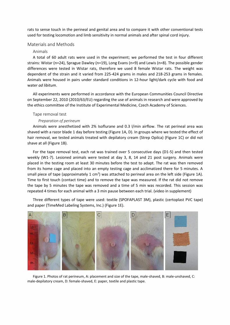
Animals were anesthetized with 2% Isoflurane and 0.3 l/min airflow. The rat perineal area was
shaved with a razor blade 1 day before testing (Figure 1A, D). In groups where we tested the effect of
hair removal, we tested animals treated with depilatory cream (Strep Opilca) (Figure 1C) or did not
shave at all (Figure 1B).
For the tape removal test, each rat was trained over 5 consecutive days (D1-5) and then tested
weekly (W1-7). Lesioned animals were tested at day 3, 8, 14 and 21 post surgery. Animals were
placed in the testing room at least 30 minutes before the test to adapt. The rat was then removed
from its home cage and placed into an empty testing cage and acclimatized there for 5 minutes. A
small piece of tape (approximately 1 cm2) was attached to perineal area on the left side (Figure 1A).
Time to first touch (contact time) and to remove the tape was measured. If the rat did not remove
the tape by 5 minutes the tape was removed and a time of 5 min was recorded. This session was
repeated 4 times for each animal with a 3 min pause between each trial. (video in supplement)
Three different types of tape were used: textile (SPOFAPLAST 3M), plastic (certoplast PVC tape)
and paper (TimeMed Labeling Systems, Inc.) (Figure 1E).
Figure 1. Photos of rat perineum, A: placement and size of the tape, male-shaved, B: male-unshaved, C:
male-depilatory cream, D: female-shaved, E: paper, textile and plastic tape.

Lesion/spinal cord injury
A dorsal column lesion was performed as described previously (Bradbury et al., 1999). Animals
were anesthetized with 2% Isoflurane and 0.3 l/min airflow. The back was shaved and a skin incision
was made between the T7 and L1 vertebrae. A laminectomy was performed on the T10 vertebra. The
dorsal columns were cut with fine-tipped forceps (approximately 1 mm deep). Muscles and skin were
sutured.
BBB
Motor function was evaluated using Basso Beattie and Bresnahan (BBB) 21-point locomotor scale
(Basso et al., 1995). Rats were placed into open-field and scored according to the criteria including
hindlimb movement, weight support and forelimb-hindlimb coordination. Animals were tested from
2 and 3 weeks after injury.
Ladder
Sensory-motor coordination was tested on the horizontal ladder walking test. Each rat was tested
2 times before the lesion to get a baseline score and then in the second and third week after the
lesion. Animals were placed in the testing room at least 30 minutes before the test to adapt. The rat
was then removed from its home cage and placed onto a horizontal ladder in the MotoRater (TSE
Systems). They acclimatized there for 5 minutes and were returned to their home cage. Rats were
then recorded with a high-speed camera (CamRecord CL600x2) walking along 155 cm long, 10 cm
wide horizontal ladder with unevenly spaced rungs. Each step was given a score according to a 0-6
foot fault scoring system (Metz and Whishaw, 2009). The average score for each step from three
trials was calculated.
Plantar (Hargreaves) test
Changes in thermal sensation after SCI were measured by the Ugo Basile Plantar Heat test
apparatus (Comerio VA, Italy). Each rat was tested 2 times before the lesion to get a baseline latency
time and then second and third week after the lesion. Animals were placed in the testing room at
least 30 minutes before the test to adapt. Rats were then removed from their home cage and placed
into a plexiglass box on a glass surface and acclimatized there for 5 minutes. An infrared light beam
was then applied to the plantar surface of the hind paw. The light and timer were activated
simultaneously. When the rat withdrew its paw, the time was recorded. The infrared stimulus turned
off automatically after 30 s. Five trials were performed for each hind paw of each animal with at least
3 min pause between individual trials. Of the 5 values the lowest and the highest were deleted and
three rest were averaged.
Von Frey
In the lesioned animals touch sensitivity was measured with the electronic von Frey test (IITC Inc.,
Life Science Instruments, Woodland Hills, CA, USA) as described previously (Ferrier et al., 2016;
Martinov et al., 2013). Each rat was measured 2 times before the lesion to get a baseline withdrawal
threshold and then 7, 13 and 20 days after the lesion. Animals were placed in the testing room at
least 30 minutes before the test to adapt. The rat was then removed from its home cage and placed
into a plexiglass box on a mesh floor stand (IITC Inc., Life Science Instruments, Woodland Hills, CA,
USA) and acclimatized there for 15 minutes. The perineal area was then stimulated by slowly raising
the probe with rigid tip to touch the shaved perineal area. Pressure was increased until nociceptive
response or until the rat lifted up so it was not possible to go further with the tip. The value was
recorded and measurement repeated until 5 values were measured for each rat. Of the 5 values the
lowest and the highest were deleted and three rest were averaged.

Perfusion
Animals were anesthetized with Isoflurane and injected with 1 ml Chloral hydrate per 100 g
intraperitoneally. Animals were transcardially perfused with approximately 300 ml of PB and next
with the same volume of 4% paraformaldehyde (PFA). Spine was cut off and stored in 4% PFA.
Histology
Spinal cords were removed from spine and an approximately 2 cm long piece containing the
lesion was cut out. It was embedded in paraffin and cut to 14 µm cross sections. They were then
labelled using Luxol blue/cresyl violet staining. Images were taken with Axiocam (Zeiss) microscope.
Lesion size quantification
Lesions were reconstructed from the histological sections from all rats and lesion size was
measured using ImageJ. The percentage of preserved area on the left side of the lesion was then
correlated with behavioural deficit as described in the results section.
Statistical analysis
All data are presented as mean ± SEM. The GraphPad Prism 5 software was used to analyse the
data. To find differences we performed two-way repeated measures ANOVA. In case ANOVA found a
difference, it was followed by Bonferroni post hoc test to compare individual groups. If the P value
was less than 0.05, the difference was considered statistically significant.
Results of behavioural tests of animals that underwent a dorsal column lesion were evaluated in
two ways. In the first study we assessed relationship between lesion size and functional outcome.
The percentage of preserved area in the lesion was correlated with the results of behavioural tests
(tape removal, BBB, von Frey, ladder and plantar) and linear regression line was calculated. The
result of individual behavioural test was calculated as a difference of animal performance before and
after SCI.
Rats were then divided to three groups according to lesion size に small, medium, big. Differences
between individual groups and within groups in time were evaluated using two-way repeated
measures ANOVA in GraphPad Prism 5 software.
Results
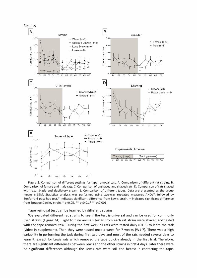
Figure 2. Comparison of different settings for tape removal test. A. Comparison of different rat strains. B.
Comparison of female and male rats. C. Comparison of unshaved and shaved rats. D. Comparison of rats shaved
with razor blade and depilatory cream. E. Comparison of different tapes. Data are presented as the group
means ± SEM. Statistical analysis was performed using two-way repeated measures ANOVA followed by
Bonferroni post hoc test.* indicates significant difference from Lewis strain. + indicates significant difference
from Sprague Dawley strain. * p<0.05, ** p<0.01,*** p<0.001
Tape removal test can be learned by different strains.
We evaluated different rat strains to see if the test is universal and can be used for commonly
used strains (Figure 2A). Eight to nine animals tested from each rat strain were shaved and tested
with the tape removal task. During the first week all rats were tested daily (D1-5) to learn the task
(video in supplement). Then they were tested once a week for 7 weeks (W1-7). There was a high
variability in performing the task during first two days and most of the rats needed several days to
learn it, except for Lewis rats which removed the tape quickly already in the first trial. Therefore,
there are significant differences between Lewis and the other strains in first 4 days. Later there were
no significant differences although the Lewis rats were still the fastest in contacting the tape.
However, all strains were able to learn the task during first week and their further performance was
stable.
Gender
We compared the performance in the tape removal test in both genders (Figure 2B). Eight female
and eight male Wistar rats were used in this comparison. During the first week both groups were
tested daily to learn the task. Then they were tested once a week for 5 weeks. There were no
significant differences between male and female rats.
Perineum exposure
The animals were shaved with a standard razor blade, but we also tested different ways of
preparing the perineum.
We investigated whether the test can be performed without shaving. Eight animals were not
shaved and were compared to 8 shaved rats (Figure 2C). Otherwise the design of the experiment
remained the same, i.e. during the first week all rats were tested daily (D1-5) to learn the task. Then
they were tested once a week for 7 weeks (W1-7). In this case the unshaved animals showed a non-
significant trend to slower time to contact the tape.
In the next experiment, we tested if the razor blade can be changed for depilatory cream (Figure
2D). One group of rats was shaved with a razor blade (n=5), while the other one was treated with
depilatory cream (n=6). There were no differences in contact time in the tape removal test, however,
sometimes the skin was visibly red and irritated after using the cream (Figure 1C).
Types of tape
The same group of animals used for the shaving experiment (razor blade versus depilatory cream)
was then shaved with a razor blade and tested with different types of tape (Figure 2E). Rats were
divided in 3 groups. Each group was tested with a different tape (textile, plastic and paper). Since
these animals had already learned the task, they were tested only weekly for 7 weeks. The animals
tested with paper tape showed better performance but this difference was not significant because of
the low number of animals in groups.
Lesion
Thirty-three animals from different rat strains (from the first task) underwent a T10 dorsal column
lesion.
The tape removal test was compared with additional sensory or motor tests before and after the
surgery. Since the lesions varied in size, we divided the rats according to the extent of the damaged
areas into three groups, small, medium and big lesion (Figure 3).
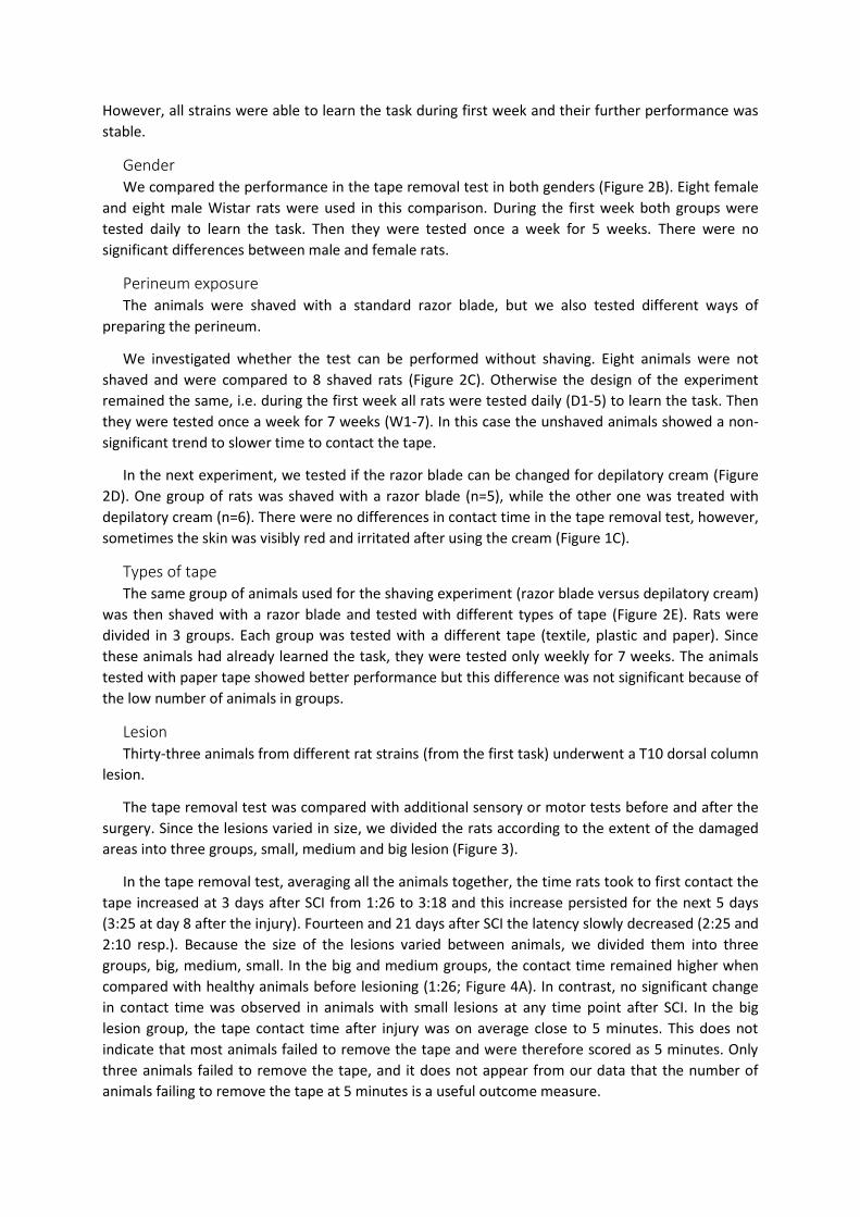
In the tape removal test, averaging all the animals together, the time rats took to first contact the
tape increased at 3 days after SCI from 1:26 to 3:18 and this increase persisted for the next 5 days
(3:25 at day 8 after the injury). Fourteen and 21 days after SCI the latency slowly decreased (2:25 and
2:10 resp.). Because the size of the lesions varied between animals, we divided them into three
groups, big, medium, small. In the big and medium groups, the contact time remained higher when
compared with healthy animals before lesioning (1:26; Figure 4A). In contrast, no significant change
in contact time was observed in animals with small lesions at any time point after SCI. In the big
lesion group, the tape contact time after injury was on average close to 5 minutes. This does not
indicate that most animals failed to remove the tape and were therefore scored as 5 minutes. Only
three animals failed to remove the tape, and it does not appear from our data that the number of
animals failing to remove the tape at 5 minutes is a useful outcome measure.
The von Frey test is frequently used to measure touch sensitivity. In all three groups of rats the
withdrawal threshold increased after SCI compared to baseline threshold. However, we were unable
to detect any differences between big, medium and small lesions and the increase in withdrawal
threshold was significant in comparison with animals before lesioning (Figure 4B).
For testing response to thermal stimuli, the plantar test was performed. No significant change
after spinal injury was detected in any of the lesion size groups (Figure 4E).
Possible motor dysfunction was assessed by the BBB open field test. A decrease in the BBB score
was detected in all lesioned animals and was dependent on the size of the lesion. Rats with big
lesions dropped their score down to 12.41 ± 1.45, which was significantly lower than values in
animals with medium size lesion (16.25 ± 0.5). The lowest deficit was recorded in animals with small
lesion, where the average value was 18.9 (± 0.66), and the decrease was significant only 14 days after
SCI, when compared with healthy animals before lesioning (Figure 4B).
As a sensory-motor coordination test, we applied the horizontal ladder walking test (Figure 4C). A
large decrease in foot fault score was detected in animals with big lesions which was maintained to
23 days. Animals with medium and small lesions scored similarly 5.03 ± 0.24 and 5.45 ± 0.11 resp. at
day 17 after the lesion and there was no statistical difference between these two groups. Although
the scores of these two groups remained similar (4.99 ± 0.23 and 5.63 ± 0.10) at 23 days after the
lesion, only the scores from animals with medium lesions were statistically significant from animals
before lesioning.
Because there were clear differences between the deficits depending on lesion size, we
performed plots of lesion size against the pre- and post-lesion changes in the behavioural test results
with a point for each individual animal (Figure 5). In the tape removal test, there was a strong linear
correlation between preserved tissue area and the results of the test (p<0.001) and the BBB test and
horizontal ladder walking test also showed significant changes correlated with lesion size. For the von
Frey and plantar tests there was no significant linear correlation with lesion size.
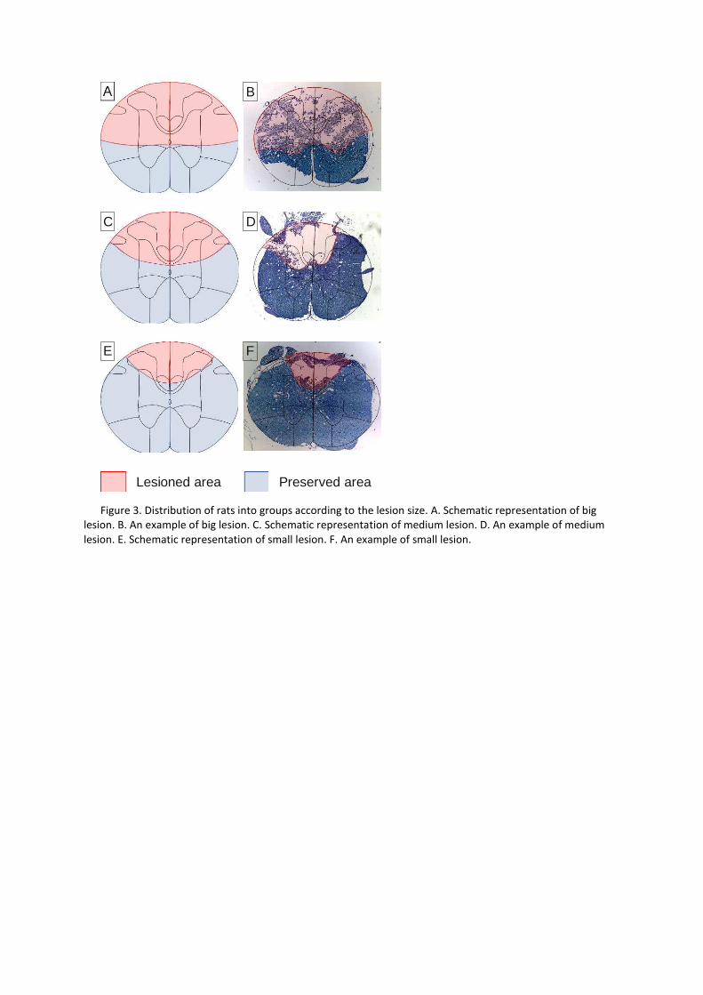
Figure 3. Distribution of rats into groups according to the lesion size. A. Schematic representation of big
lesion. B. An example of big lesion. C. Schematic representation of medium lesion. D. An example of medium
lesion. E. Schematic representation of small lesion. F. An example of small lesion.
A
C
E
B
D
F
Lesioned area Preserved area
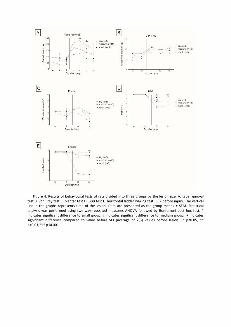
Figure 4. Results of behavioural tests of rats divided into three groups by the lesion size. A. tape removal
test B. von Frey test C. plantar test D. BBB test E. horizontal ladder waking test. BI = before injury. The vertical
line in the graphs represents time of the lesion. Data are presented as the group means ± SEM. Statistical
analysis was performed using two-way repeated measures ANOVA followed by Bonferroni post hoc test. *
indicates significant difference to small group. # indicates significant difference to medium group. + indicates
significant difference compared to value before SCI (average of 2(3) values before lesion). * p<0.05, **
p<0.01,*** p<0.001

Figure 5. Correlation between behavioural test results and percentage of preserved area on the left side of
spinal cord in the center of lesion. A. tape removal test B. BBB C. ladder D. von Frey E. plantar F. Schematic
representation of lesion on T10 cross section. Damaged region is highlighted in red. Percentage of preserved
tissue on the left side was calculated.
Discussion
Spinal cord injury is a complex event resulting in motor as well as sensory deficits. Tests have
been developed for the quantification of functional deficits and recovery of function after cervical or
thoracic lesions and for various motor and sensory behaviours. A priority for many patients is
restoration of sexual function including genital sensation. We therefore decided to find out if a
quantitative test for perineal sensation could be achieved by modification of the tape removal test.
Tape removal
0 20 40 60 80 100
5:00
4:00
3:00
2:00
1:00
0
-1:00
***y=-0.00003069x+0.002991R2=0.5155P<0.0001
% preserved area
Diff
eren
ce b
efor
e an
d af
ter
lesi
onC
onta
ct ti
me
(m:s
)BBB
0 20 40 60 80 1000
3
6
9
12
15
18
21
***y=-0.1341x+13.96R2=0.5387P<0.0001
% preserved area
Diff
eren
ce b
efor
e an
d af
ter
lesi
onB
BB
sco
re
Ladder
0 20 40 60 80 100-2
0
2
4
6
8***
y=-0.07706x+6.178R2=0.8582P<0.0001
% preserved area
Diff
eren
ce b
efor
e an
d af
ter
lesi
onF
oot f
ault
scor
e
Von Frey
0 20 40 60 80 100-5
0
5
10
15
20 y=-0.06976x+9.865R2=0.1461P=0.0962
% preserved area
Diff
eren
ce b
efor
e an
d af
ter
lesi
onW
ithdr
awal
thre
shol
d (g
)
Plantar
0 20 40 60 80 100-6
-4
-2
0
2
4
6 y=-0.01676x+1.645R2=0.03582P=0.4242
% preserved area
Diff
eren
ce b
efor
e an
d af
ter
lesi
onW
ithdr
awal
late
ncy
(s)
A B
C
E
D
F
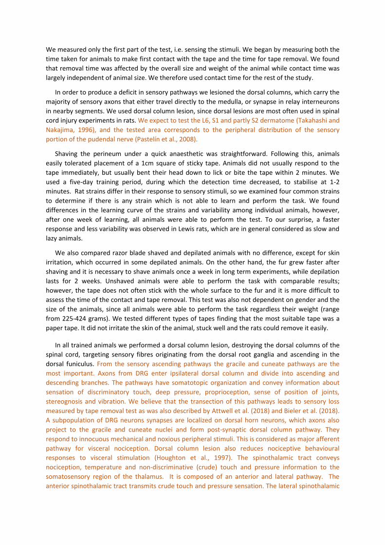
We measured only the first part of the test, i.e. sensing the stimuli. We began by measuring both the
time taken for animals to make first contact with the tape and the time for tape removal. We found
that removal time was affected by the overall size and weight of the animal while contact time was
largely independent of animal size. We therefore used contact time for the rest of the study.
In order to produce a deficit in sensory pathways we lesioned the dorsal columns, which carry the
majority of sensory axons that either travel directly to the medulla, or synapse in relay interneurons
in nearby segments. We used dorsal column lesion, since dorsal lesions are most often used in spinal
cord injury experiments in rats. We expect to test the L6, S1 and partly S2 dermatome (Takahashi and
Nakajima, 1996), and the tested area corresponds to the peripheral distribution of the sensory
portion of the pudendal nerve (Pastelín et al., 2008).
Shaving the perineum under a quick anaesthetic was straightforward. Following this, animals
easily tolerated placement of a 1cm square of sticky tape. Animals did not usually respond to the
tape immediately, but usually bent their head down to lick or bite the tape within 2 minutes. We
used a five-day training period, during which the detection time decreased, to stabilise at 1-2
minutes. Rat strains differ in their response to sensory stimuli, so we examined four common strains
to determine if there is any strain which is not able to learn and perform the task. We found
differences in the learning curve of the strains and variability among individual animals, however,
after one week of learning, all animals were able to perform the test. To our surprise, a faster
response and less variability was observed in Lewis rats, which are in general considered as slow and
lazy animals.
We also compared razor blade shaved and depilated animals with no difference, except for skin
irritation, which occurred in some depilated animals. On the other hand, the fur grew faster after
shaving and it is necessary to shave animals once a week in long term experiments, while depilation
lasts for 2 weeks. Unshaved animals were able to perform the task with comparable results;
however, the tape does not often stick with the whole surface to the fur and it is more difficult to
assess the time of the contact and tape removal. This test was also not dependent on gender and the
size of the animals, since all animals were able to perform the task regardless their weight (range
from 225-424 grams). We tested different types of tapes finding that the most suitable tape was a
paper tape. It did not irritate the skin of the animal, stuck well and the rats could remove it easily.
In all trained animals we performed a dorsal column lesion, destroying the dorsal columns of the
spinal cord, targeting sensory fibres originating from the dorsal root ganglia and ascending in the
dorsal funiculus. From the sensory ascending pathways the gracile and cuneate pathways are the
most important. Axons from DRG enter ipsilateral dorsal column and divide into ascending and
descending branches. The pathways have somatotopic organization and convey information about
sensation of discriminatory touch, deep pressure, proprioception, sense of position of joints,
stereognosis and vibration. We believe that the transection of this pathways leads to sensory loss
measured by tape removal test as was also described by Attwell et al. (2018) and Bieler et al. (2018).
A subpopulation of DRG neurons synapses are localized on dorsal horn neurons, which axons also
project to the gracile and cuneate nuclei and form post-synaptic dorsal column pathway. They
respond to innocuous mechanical and noxious peripheral stimuli. This is considered as major afferent
pathway for visceral nociception. Dorsal column lesion also reduces nociceptive behavioural
responses to visceral stimulation (Houghton et al., 1997). The spinothalamic tract conveys
nociception, temperature and non-discriminative (crude) touch and pressure information to the
somatosensory region of the thalamus. It is composed of an anterior and lateral pathway. The
anterior spinothalamic tract transmits crude touch and pressure sensation. The lateral spinothalamic
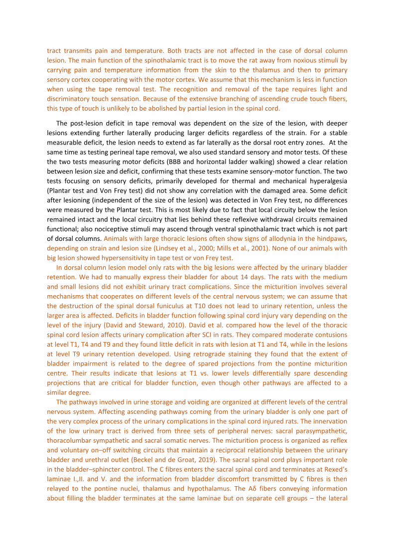
tract transmits pain and temperature. Both tracts are not affected in the case of dorsal column
lesion. The main function of the spinothalamic tract is to move the rat away from noxious stimuli by
carrying pain and temperature information from the skin to the thalamus and then to primary
sensory cortex cooperating with the motor cortex. We assume that this mechanism is less in function
when using the tape removal test. The recognition and removal of the tape requires light and
discriminatory touch sensation. Because of the extensive branching of ascending crude touch fibers,
this type of touch is unlikely to be abolished by partial lesion in the spinal cord.
The post-lesion deficit in tape removal was dependent on the size of the lesion, with deeper
lesions extending further laterally producing larger deficits regardless of the strain. For a stable
measurable deficit, the lesion needs to extend as far laterally as the dorsal root entry zones. At the
same time as testing perineal tape removal, we also used standard sensory and motor tests. Of these
the two tests measuring motor deficits (BBB and horizontal ladder walking) showed a clear relation
between lesion size and deficit, confirming that these tests examine sensory-motor function. The two
tests focusing on sensory deficits, primarily developed for thermal and mechanical hyperalgesia
(Plantar test and Von Frey test) did not show any correlation with the damaged area. Some deficit
after lesioning (independent of the size of the lesion) was detected in Von Frey test, no differences
were measured by the Plantar test. This is most likely due to fact that local circuity below the lesion
remained intact and the local circuitry that lies behind these reflexive withdrawal circuits remained
functional; also nociceptive stimuli may ascend through ventral spinothalamic tract which is not part
of dorsal columns. Animals with large thoracic lesions often show signs of allodynia in the hindpaws,
depending on strain and lesion size (Lindsey et al., 2000; Mills et al., 2001). None of our animals with
big lesion showed hypersensitivity in tape test or von Frey test.
In dorsal column lesion model only rats with the big lesions were affected by the urinary bladder
retention. We had to manually express their bladder for about 14 days. The rats with the medium
and small lesions did not exhibit urinary tract complications. Since the micturition involves several
mechanisms that cooperates on different levels of the central nervous system; we can assume that
the destruction of the spinal dorsal funiculus at T10 does not lead to urinary retention, unless the
larger area is affected. Deficits in bladder function following spinal cord injury vary depending on the
level of the injury (David and Steward, 2010). David et al. compared how the level of the thoracic
spinal cord lesion affects urinary complication after SCI in rats. They compared moderate contusions
at level T1, T4 and T9 and they found little deficit in rats with lesion at T1 and T4, while in the lesions
at level T9 urinary retention developed. Using retrograde staining they found that the extent of
bladder impairment is related to the degree of spared projections from the pontine micturition
centre. Their results indicate that lesions at T1 vs. lower levels differentially spare descending
projections that are critical for bladder function, even though other pathways are affected to a
similar degree.
The pathways involved in urine storage and voiding are organized at different levels of the central
nervous system. Affecting ascending pathways coming from the urinary bladder is only one part of
the very complex process of the urinary complications in the spinal cord injured rats. The innervation
of the low urinary tract is derived from three sets of peripheral nerves: sacral parasympathetic,
thoracolumbar sympathetic and sacral somatic nerves. The micturition process is organized as reflex
and voluntary onにoff switching circuits that maintain a reciprocal relationship between the urinary
bladder and urethral outlet (Beckel and de Groat, 2019). The sacral spinal cord plays important role
in the bladderにsphincter control. The C fibres enters the sacral spinal cord and terminates at Rexedげs
laminae I.,II. and V. and the information from bladder discomfort transmitted by C fibres is then
ヴWノ;┞WS デラ デエW ヮラミデキミW ミ┌IノWキが デエ;ノ;マ┌ゲ ;ミS エ┞ヮラデエ;ノ;マ┌ゲく TエW A~ aキHWヴゲ Iラミ┗W┞キミェ キミaラヴマ;デキラミ about filling the bladder terminates at the same laminae but on separate cell groups に the lateral

collateral pathway of the Lissauerげs tract. At the thoracic level where our lesion is performed the
bundles of this tract are located around the lateral edge of the dorsal horn and through dorsolateral
funiculus and we expect that are destroyed especially in big lesions.
The perineal tape removal test measured a clear functional deficit in perineal sensation for the
three-week post-lesion duration of our experiments. However, the size of the deficit and its duration
were dependent on lesion size. The classic tape removal test for limb sensation has been used in
various studies of dorsal spinal cord injury. In one study for hindlimb sensation after a T7 thoracic
injury the deficit was small and only significant at some time points (Fagoe et al., 2016). However
after cervical lesions a stable deficit in forepaw tape removal to 8 and 12 weeks was seen in several
studies (Agudo et al., 2008; Bradbury et al., 2002; Fagoe et al., 2016; Lu et al., 2005; Moreno-Flores
et al., 2006).
We conclude that the modified tape removal test is suitable for testing sensation in the area of
the perineum and gives similar results across strains and for both genders. Deficits in the perineal
sensation after T10 dorsal column lesions can be detected for at least 3 weeks after lesioning and
correlate with the size of the lesion. The test is therefore suitable for testing treatments for sensory
restoration in the genital region in dorsal spinal cord injuries of sufficient size.
Declaration of interest
Authors have nothing to declare.
Funding
Supported by Czech Science Foundation GACR 19-10365S, by Operational Programme Research,
DW┗WノラヮマWミデ ;ミS ES┌I;デキラミ キミ デエW aヴ;マW┘ラヴニ ラa デエW ヮヴラテWIデ さCWミデWヴ ラa RWIラミゲデヴ┌Iデキ┗W NW┌ヴラゲIキWミIWざが ヴWェキゲデヴ;デキラミ ミ┌マHWヴ CZくヰヲくヱくヰヱっヰくヰくっヰくヰっヱヵぱヰヰンっヰヰヰヰヴヱΓ, IRP Grant P172,
Reconstruction of the spinal cord sensory pathway, MRC grant MR/R004463, Wings for Life WFL-UK-
008/15, International Spinal Research Trust NRB119.
Supplementary material
Video: https://youtu.be/fqtn51dwlDE
References
Aehling, C., Weber, N.C., Zuurbier, C.J., Preckel, B., Galmbacher, R., Stefan, K., Hollmann, M.W., Popp,
E., Knapp, J., 2018. Effects of combined helium pre/post-conditioning on the brain and heart in
a rat resuscitation model. Acta Anaesthesiol. Scand. 62, 63に74.
https://doi.org/10.1111/aas.13041
Agudo, M., Woodhoo, A., Webber, D., Mirsky, R., Jessen, K.R., McMahon, S.B., 2008. Schwann cell
precursors transplanted into the injured spinal cord multiply, integrate and are permissive for
axon growth. Glia 56, 1263に70. https://doi.org/10.1002/glia.20695
Albertsmeier, M., Teschendorf, P., Popp, E., Galmbacher, R., Vogel, P., Böttiger, B.W., 2007.
Evaluation of a tape removal test to assess neurological deficit after cardiac arrest in rats.
Resuscitation 74, 552に558. https://doi.org/10.1016/j.resuscitation.2007.01.040
Anderson, K.D., 2004. Targeting Recovery: Priorities of the Spinal Cord-Injured Population. J.
Neurotrauma 21, 1371に1383. https://doi.org/10.1089/neu.2004.21.1371

Annett, L.E., Martel, F.L., Rogers, D.C., Ridley, R.M., Baker, H.F., Dunnett, S.B., 1994. Behavioral
assessment of the effects of embryonic nigral grafts in marmosets with unilateral 6-OHDA
lesions of the nigrostriatal pathway. Exp. Neurol. 125, 228に46.
https://doi.org/10.1006/exnr.1994.1026
Arakawa, H., Akkentli, F., Erzurumlu, R.S., 2014. Region-Specific Disruption of Adenylate Cyclase Type
1 Gene Differentially Affects Somatosensorimotor Behaviors in Mice. eNeuro 1, 1に12.
https://doi.org/10.1523/ENEURO.0007-14.2014
Attwell, C.L., van Zwieten, M., Verhaagen, J., Mason, M.R.J., 2018. The dorsal column lesion model of
spinal cord injury and its use in deciphering the neuron-intrinsic injury response. Dev.
Neurobiol. https://doi.org/10.1002/dneu.22601
Basso, D.M., Beattie, M.S., Bresnahan, J.C., 1995. A Sensitive and Reliable Locomotor Rating Scale for
Open Field Testing in Rats. J. Neurotrauma 12, 1に21. https://doi.org/10.1089/neu.1995.12.1
Beckel, J.M., de Groat, W.C., 2019. Neural Control of Lower Urinary Tract Function, in: Oxford
Research Encyclopedia of Neuroscience. Oxford University Press.
https://doi.org/10.1093/acrefore/9780190264086.013.270
Bieler, L., Grassner, L., Zaunmair, P., Kreutzer, C., Lampe, L., Trinka, E., Marschallinger, J., Aigner, L.,
Couillard-Despres, S., 2018. Motor deficits following dorsal corticospinal tract transection in
rats: voluntary versus skilled locomotion readouts. Heliyon 4, e00540.
https://doi.org/10.1016/j.heliyon.2018.e00540
Bouet, V., Boulouard, M., Toutain, J., Divoux, D., Bernaudin, M., Schumann-Bard, P., Freret, T., 2009.
The adhesive removal test: a sensitive method to assess sensorimotor deficits in mice. Nat.
Protoc. 4, 1560に1564. https://doi.org/10.1038/nprot.2009.125
Bradbury, E.J., Khemani, S., King, V.R., Priestley, J. V., McMahon, S.B., Von R, ., King, ., Priestley, J. V.,
McMahon, S.B., 1999. NT-3 promotes growth of lesioned adult rat sensory axons ascending in
the dorsal columns of the spinal cord. Eur. J. Neurosci. 11, 3873に3883.
https://doi.org/10.1046/j.1460-9568.1999.00809.x
Bradbury, E.J., Moon, L.D.F., Popat, R.J., King, V.R., Bennett, G.S., Patel, P.N., Fawcett, J.W.,
McMahon, S.B., 2002. Chondroitinase ABC promotes functional recovery after spinal cord
injury. Nature 416, 636に640. https://doi.org/10.1038/416636a
David, B.T., Steward, O., 2010. Deficits in bladder function following spinal cord injury vary depending
on the level of the injury. Exp. Neurol. 226, 128.
https://doi.org/10.1016/J.EXPNEUROL.2010.08.014
Duricki, D.A., Hutson, T.H., Kathe, C., Soleman, S., Gonzalez-Carter, D., Petruska, J.C., Shine, H.D.,
Chen, Q., Wood, T.C., Bernanos, M., Cash, D., Williams, S.C.R., Gage, F.H., Moon, L.D.F., 2016.
Delayed intramuscular human neurotrophin-3 improves recovery in adult and elderly rats after
stroke. Brain 139, 259に75. https://doi.org/10.1093/brain/awv341
Fagoe, N.D., Attwell, C.L., Eggers, R., Tuinenbreijer, L., Kouwenhoven, D., Verhaagen, J., Mason,
M.R.J., 2016. Evaluation of Five Tests for Sensitivity to Functional Deficits following Cervical or
Thoracic Dorsal Column Transection in the Rat. PLoS One 11, e0150141.
https://doi.org/10.1371/journal.pone.0150141
Ferrier, J., Marchand, F., Balayssac, D., 2016. Assessment of Mechanical Allodynia in Rats Using the
Electronic Von Frey Test. BIO-PROTOCOL 6. https://doi.org/10.21769/BioProtoc.1933
Fleming, S.M., Ekhator, O.R., Ghisays, V., 2013. Assessment of sensorimotor function in mouse
マラSWノゲ ラa P;ヴニキミゲラミげゲ SキゲW;ゲWく Jく Vキゲく E┝ヮく エデデヮゲぎっっSラキくラヴェっヱヰくンΑΓヱっヵヰンヰン

Houghton, A.K., Kadura, S., Westlund, K.N., 1997. Dorsal column lesions reverse the reduction of
homecage activity in rats with pancreatitis. Neuroreport 8, 3795に3800.
https://doi.org/10.1097/00001756-199712010-00028
Levine, A.J., Hinckley, C.A., Hilde, K.L., Driscoll, S.P., Poon, T.H., Montgomery, J.M., Pfaff, S.L., 2014.
Identification of a cellular node for motor control pathways. Nat. Neurosci. 17, 586に93.
https://doi.org/10.1038/nn.3675
Lindsey, A.E., LoVerso, R.L., Tovar, C.A., Hill, C.E., Beattie, M.S., Bresnahan, J.C., 2000. An analysis of
changes in sensory thresholds to mild tactile and cold stimuli after experimental spinal cord
injury in the rat. Neurorehabil. Neural Repair 14, 287に300.
https://doi.org/10.1177/154596830001400405
Lu, P., Jones, L.L., Tuszynski, M.H., 2005. BDNF-expressing marrow stromal cells support extensive
axonal growth at sites of spinal cord injury. Exp. Neurol. 191, 344に60.
https://doi.org/10.1016/j.expneurol.2004.09.018
Martinov, T., Mack, M., Sykes, A., Chatterjea, D., 2013. Measuring changes in tactile sensitivity in the
hind paw of mice using an electronic von Frey apparatus. J. Vis. Exp. e51212.
https://doi.org/10.3791/51212
Metz, G. a, Whishaw, I.Q., 2009. The ladder rung walking task: a scoring system and its practical
application. J. Vis. Exp. 2に5. https://doi.org/10.3791/1204
Mills, C.D., Hains, B.C., Johnson, K.M., Hulsebosch, C.E., 2001. Strain and Model Differences in
Behavioral Outcomes after Spinal Cord Injury in Rat. J. Neurotrauma 18, 743に756.
https://doi.org/10.1089/089771501316919111
Moreno-Flores, M.T., Bradbury, E.J., Martín-Bermejo, M.J., Agudo, M., Lim, F., Pastrana, E., Avila, J.,
Díaz-Nido, J., McMahon, S.B., Wandosell, F., 2006. A clonal cell line from immortalized olfactory
ensheathing glia promotes functional recovery in the injured spinal cord. Mol. Ther. 13, 598に608. https://doi.org/10.1016/j.ymthe.2005.11.014
Onifer, S.M., Zhang, Y.P., Burke, D.A., Brooks, D.L., Decker, J.A., McClure, N.J., Floyd, A.R., Hall, J.,
Proffitt, B.L., Shields, C.B., Magnuson, D.S.K., 2005. Adult rat forelimb dysfunction after dorsal
cervical spinal cord injury. Exp. Neurol. 192, 25に38.
https://doi.org/10.1016/j.expneurol.2004.10.016
Pastelín, C.F., Zempoalteca, R., Pacheco, P., Downie, J.W., Cruz, Y., 2008. Sensory and somatomotor
comヮラミWミデゲ ラa デエW さゲWミゲラヴ┞ Hヴ;ミIエざ ラa デエW ヮ┌SWミS;ノ ミWヴ┗W キミ デエW マ;ノW ヴ;デく Bヴ;キミ RWゲく ヱヲヲヲが 149に155. https://doi.org/10.1016/j.brainres.2008.05.012
Quaranta, A., Siniscalchi, M., Frate, A., Vallortigara, G., 2004. Paw preference in dogs: relations
between lateralised behaviour and immunity. Behav. Brain Res. 153, 521に5.
https://doi.org/10.1016/j.bbr.2004.01.009
Schallert, T., Fleming, S.M., Leasure, J.L.L., Tillerson, J.L., Bland, S.T., 2000. CNS plasticity and
assessment of forelimb sensorimotor outcome in unilateral rat models of stroke, cortical
ablation, parkinsonism and spinal cord injury. Neuropharmacology 39, 777に787.
https://doi.org/10.1016/S0028-3908(00)00005-8
Schallert, T., Upchurch, M., Lobaugh, N., Farrar, S.B., Spirduso, W.W., Gilliam, P., Vaughn, D., Wilcox,
R.E., 1982. Tactile extinction: Distinguishing between sensorimotor and motor asymmetries in
rats with unilateral nigrostriatal damage. Pharmacol. Biochem. Behav. 16, 455に462.
https://doi.org/10.1016/0091-3057(82)90452-X
Sengul, G., Watson, C., 2015. Ascending and Descending Pathways in the Spinal Cord. Rat Nerv. Syst.

115に130. https://doi.org/10.1016/B978-0-12-374245-2.00008-5
Takahashi, Y., Nakajima, Y., 1996. Dermatomes in the rat limbs as determined by antidromic
stimulation of sensory C-fibers in spinal nerves. Pain 67, 197に202.
https://doi.org/10.1016/0304-3959(96)03116-8
Tsytsarev, V., Arakawa, H., Zhao, S., Chédotal, A., Erzurumlu, R.S., 2017. Behavioral Consequences of
a Bifacial Map in the Mouse Somatosensory Cortex. J. Neurosci. 37, 7209に7218.
https://doi.org/10.1523/JNEUROSCI.0598-17.2017
Wang, L., Kang, Y., Zhang, G., Zhang, Y., Cui, R., Yan, W., Tan, H., Li, S., Wu, B., Cui, H., Shi, G., 2016.
Deficits in coordinated motor behavior and in nigrostriatal dopaminergic system ameliorated
and VMAT2 expression up-regulated in aged male rats by administration of testosterone
propionate. Exp. Gerontol. 78, 1に11. https://doi.org/10.1016/J.EXGER.2016.03.003
Zhang, L., Chen, J., Li, Y., Zhang, Z.G., Chopp, M., 2000. Quantitative measurement of motor and
somatosensory impairments after mild (30 min) and severe (2 h) transient middle cerebral
artery occlusion in rats. J. Neurol. Sci. 174, 141に146. https://doi.org/10.1016/S0022-
510X(00)00268-9