adaptive & innate immunity. the immune response and immunity immune response ▫ innate...
TRANSCRIPT

Adaptive & Innate Immunity
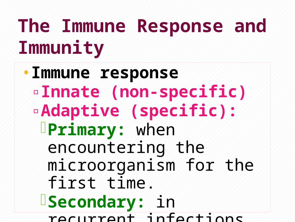
The Immune Response and Immunity•Immune response▫Innate (non-specific)▫Adaptive (specific):Primary: when encountering the microorganism for the first time.
Secondary: in recurrent infections (memory)

Acquisition of Immunity▫Natural: - active - passive▫Artificial: - active - passive

Naturally Acquired Immunity
•Active:▫Acquired through contact with
microorganisms (infection). ▫Provides long term protection.
•Passive:▫Antibodies pass from mother to fetus
across placenta or in breast milk (IgG)▫Provides immediate short term protection
(few months)

Artificially Acquired Immunity•Active:▫Antigens introduced through
vaccination.▫Provides long term protection.
•Passive:▫Induced by the transfer of antibodies.▫Referred to as: Immune serum.
globulins(ISG), immune globulins (IG) or gamma globulins▫Provides immediate short term protection

immunity
adaptive
natural
active
passive
artificial
active
passiveinnate

Soluble mediators of the adaptive immune system: Immunoglobulins. Cytokines and interferons. Complement.

Immunoglobulins
Immunoglobulins (Ig):• Are gamma globulins (proteins) of
defined specificity for different epitopes that make up antigens.• They are produced by plasma cells
(differentiated B cells).

N
epitopes

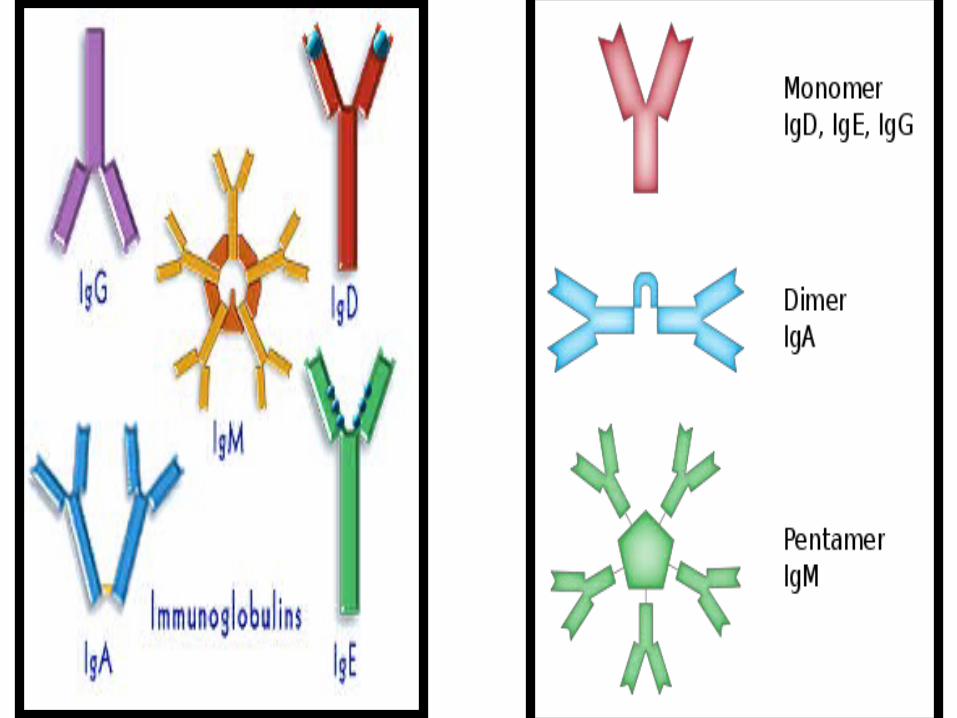
Immunoglobulins are divided into five classes (isotypes): •Three major classes ( IgG, IgM,
IgA).•Two minor classes (IgD and IgE).

N
Basic Structure of Immunoglobulin: •The immunoglobulin molecule consists
of four polypeptides chains: two heavy (H) and two light (L) chains fastened together by disulfide bonds.


•Heavy and light chains consist of two different domains (constant, and variable).•A light chain variable domain and a
heavy chain variable domain together form a pocket that constitutes the antigen-binding region (Fab).•The flexibility of Ig is associated with
the presence of the hinge region.

•Heavy chains are designated by
using of Greek letters (α, γ, δ, Є, and μ), and the immunoglobulins produced are IgA, IgG, IgD, IgE, and IgM, respectively.
•Each immunoglobulin isotype carry
one type of light chain kappa(κ) or lambda(λ) chains.

Immunoglobulins Isotypes: IgG• IgG accounts for approximately 75-85%
of the total serum Ig• It is the most abundant antibody
produced during secondary humoral immune response. • Have monovalent affinity.• It is the only class that can cross the
placenta.

IgG structure:
• n

IgM: • Form ~ 5-8 % of serum immunoglobulins. • Present on B lymphocyte surfaces.• Normally secreted as a J-chain containing
pentamer. (either as a B cell bound monomer or as a secreted pentamer).•Dominates in early primary immune
response. • Anti-A and Anti-B blood groups. • Complement fixation. • Multivalent avidity (10 antigens).

IgM Structure:
• n

IgA: It accounts for 10-16% of serum Igs. Abundant in saliva, tears, intestinal mucus,
bronchial secretions, milk, prostatic fluid (body fluids & secretions, secretory IgA).
The predominant Immunoglobulin produced in Peyer’s patches (illume submucosa), tonsils and other submucosal lymphoid tissue.
It has two subclass: in
- Monomer in the serum.
- Dimer when secreted (secretory IgA). IgA1: IgA2 ratio in blood is 5:1


IgD•Has a molecular weight of 180 KD. •Present as monomer on B-cell surface.

IgE: Form less than 1% of total serum Igs. A unique high affinity Fc receptor on
mast cell and basophils. (bounded) Great role in allergy, through cross
linking and release of histamine from mast cells and basophils.
Play a role in helminths infection.


Summary
• There are 5 isotypes of immunoglobulins.• IgG is the most abundant in serum,
the most important in secondary infections and the only one that can cross the placenta.• IgM is the most important in the
primary exposure and in complement fixation.

•The secretory IgA is most important in immunity in body secretions, submucosa and lumens.• IgD is bounded to the surface of B
cells.•IgE is bounded to the mast cells and
basophils and is the most important in allergic reactions and helminths infestation.

Second Lecture

Primary & Secondary Antibody Response•Primary Response▫Following the first exposure to an
antigen, there is a slow rise in IgM followed by a slow rise in IgG
•Secondary Response▫Following exposure to previously
encountered antigen, there is a rapid rise in IgG and slow or no rise in IgM


Molecules of Cellular Interaction: Cytokines: low-molecular weight soluble
protein messengers that are involved in:▫ Cellular interaction; inflammatory
response in innate and adaptive immunity.▫ Cellular growth, differentiation, and
repair mechanism. Cytokines are produced by a wide variety
of immune and non-immune somatic cells. Cytokines produced by lymphocytes are
known as lymphokines.

N
• N
Cytokine Cellular Source
Targets Function
IL-1 Macrophage, B cell
T cell, B cell, Endothelial cells.
Leukocyte activation, endothelial adhesion
IL-2 CD4 cell (TH1)
T cell, B cell, NK cell, macrophages
T cell proliferation.
IL-4, IL-5 CD4 cell (TH2)
B cell, T cell, Eos Differentiation of TH2 and B cell
IL-10, IL-13 TH2, CD8 cells B cell, TH2, Macrophage.
Inhibits IL-2, and INFγ.Down reg. IL-12
TNF-α Mac, PMN, T, B cells.
Mac, PMN, T, End.
InflammatoryMediator.
TNF-β Lymphocytes Wide Variety of cells
InflammatoryMediator.

Cytokines and Immune cells interaction: N
- Macrophages (phagocytosis)- CD8 T cytotoxic - Intracellular pathogens (cell mediated immunity)
- B cell- Immunoglobulin- Extracellular pathogens (humoral mediated immunity; phagocytosis independent)
IL-4, IL-10, IL-13

Interferons (IFNs): are low molecular weight soluble proteins.• Activated by presence of intracellular
pathogens such as viruses ,bacteria, parasites or tumor cells. •Released by lymphocytes and other somatic
non-immune cells.•Major action: - anti-viral infection. - Fight tumors.

N
N
Interferons Cellular Source Targets Function
INF-α Lymphocytes, Epithelium, DC fibroblasts.
Wide variety of cells.
-Up-regulates MHC Class I.-Inhibit viral proliferation
INF-β Epithelium, fibroblasts.
Wide variety of cells.
Up-regulates MHC Class I.Inhibit viral proliferation
INF-γ CD8*& CD4*T cells, and NK cells.
T , B, macrophages, NK, endothelial cells.
Anti-viral.Anti-parasitic.Enhances MHC Class I and II expression.

Complement system: Complement system: series of soluble
enzymes and proteins (C1,C2…….C9) + other actors that function in both innate and adaptive immune response against pathogens.
Complement activation can be initiated via:▫ Classical pathway. ▫Alternative pathway.▫ Mannan lectin pathway.

Classical pathway of complement:Starts by antigen - antibody interaction.C1 (q, r, s) binding to the Ag- Ab complex.C1qrs will split C2 and C4. C4b2b will split C3. (C4b2b = C3
convertase).C4b2b3b = C5 convertase.C5b will adhere to the microbe.continue tell MAC formation (C5, C6, C7,
C8 and multiple C9).

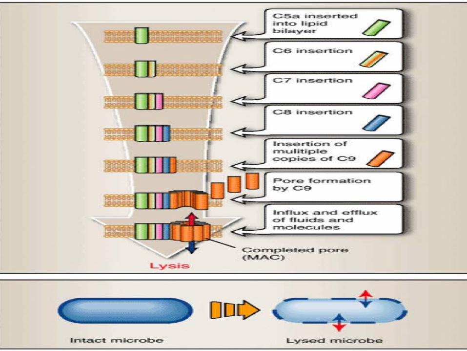

Alternative pathway: •Activation of the alternate pathway
started from C3 in the presence of the microbe.•C3 splits into C3a and C3b.•C3b bind to the microbe surface with
factor B.( C3bBb= C3 convertase)• C3Bb3b = C5 convertase, this will split
C5 and the process will continue tell formation of the membrane attack complex (MAC).


Mannan lectin pathway• Mannan is polymer of the sugar
mannose (part of the bacterial cell wall).• Lectins are serum proteins that bind
to mannan.• This pathway is activated by binding
of lectin to mannan on certain microbes, and continue tell MAC formation.

• N

The Complement Anaphylatoxins:
•The small fragments (C3a, C4a, C5a) act as anaphylotoxins. •They attract and activate neutrophils,
phagocytes and mast cells to the site of infection.•Produce an inflammatory reaction.

Mechanism of Inflammation: N