adeno-associated virus replication induces a dna …jvi.asm.org/content/83/12/6269.full.pdf ·...
TRANSCRIPT
JOURNAL OF VIROLOGY, June 2009, p. 6269–6278 Vol. 83, No. 120022-538X/09/$08.00�0 doi:10.1128/JVI.00318-09Copyright © 2009, American Society for Microbiology. All Rights Reserved.
Adeno-Associated Virus Replication Induces a DNA DamageResponse Coordinated by DNA-Dependent Protein Kinase�
Rachel A. Schwartz, Christian T. Carson,† Christine Schuberth,‡ and Matthew D. Weitzman*Laboratory of Genetics, The Salk Institute for Biological Studies, La Jolla, California 92037
Received 12 February 2009/Accepted 26 March 2009
The parvovirus adeno-associated virus (AAV) contains a small single-stranded DNA genome with invertedterminal repeats that form hairpin structures. In order to propagate, AAV relies on the cellular replicationmachinery together with functions supplied by coinfecting helper viruses such as adenovirus (Ad). Here, weexamined the host cell response to AAV replication in the context of Ad or Ad helper proteins. We show thatAAV and Ad coinfection activates a DNA damage response (DDR) that is distinct from that seen during Ad orAAV infection alone. The DDR was also triggered when AAV replicated in the presence of minimal Ad helperproteins. We detected autophosphorylation of the kinases ataxia telangiectasia mutated (ATM) and DNA-dependent protein kinase catalytic subunit (DNA-PKcs) and signaling to downstream targets SMC1, Chk1,Chk2, H2AX, and XRCC4 and multiple sites on RPA32. The Mre11 complex was not required for activationof the DDR to AAV infection. Additionally, we found that DNA-PKcs was the primary mediator of damagesignaling in response to AAV replication. Immunofluorescence revealed that some activated damage proteinswere found in a pan-nuclear pattern (phosphorylated ATM, SMC1, and H2AX), while others such as DNA-PKcomponents (DNA-PKcs, Ku70, and Ku86) and RPA32 accumulated at AAV replication centers. Althoughexpression of the large viral Rep proteins contributed to some damage signaling, we observed that the fullresponse required replication of the AAV genome. Our results demonstrate that AAV replication in thepresence of Ad helper functions elicits a unique damage response controlled by DNA-PK.
Replication of viral genomes produces a large amount ofextrachromosomal DNA that may be recognized by the cellu-lar DNA damage machinery. This is often accompanied byactivation of DNA damage response (DDR) signaling path-ways and recruitment of cellular repair proteins to sites of viralreplication. Viruses therefore provide good model systems tostudy the recognition and response to DNA damage (reviewedin reference 48). The Mre11/Rad50/Nbs1 (MRN) complexfunctions as a sensor of chromosomal DNA double-strandbreaks (DSBs) and is involved in activation of damage signal-ing (reviewed in reference 41). The MRN complex also local-izes to DNA DSBs and is found at viral replication compart-ments during infection with a number of DNA viruses (6, 40,47, 70, 75, 77, 87, 93). The phosphatidylinositol 3-kinase-likekinases (PIKKs) ataxia telangiectasia mutated (ATM), ATMand Rad3-related kinase (ATR), and the catalytic subunit ofthe DNA-dependent protein kinase (DNA-PKcs) are involvedin the signal transduction cascades activated by DNA damage(reviewed in references 43, 51, and 71). These kinases respondto distinct types of damage and regulate DSB repair duringdifferent phases of the cell cycle (5), either through nonho-mologous end-joining (NHEJ) or homologous recombinationpathways (reviewed in references 63, 81, and 86). TheDNA-PK holoenzyme is composed of DNA-PKcs and two
regulatory subunits, the Ku70 and Ku86 heterodimer.DNA-PK functions with XRCC4/DNA ligase IV to repairbreaks during NHEJ, and works with Artemis to process DNAhairpin structures during VDJ recombination and during asubset of DNA DSB events (46, 50, 86). While the kinaseactivity of DNA-PKcs leads to phosphorylation of a large num-ber of substrates in vitro as well as autophosphorylation ofspecific residues (reviewed in references 16 and 85), it is cur-rently unclear how DNA-PKcs contributes to signaling in cellsupon different types of damage.
The adeno-associated virus (AAV) genome consists of amolecule of single-stranded DNA with inverted terminalrepeats (ITRs) at both ends that form double-hairpin struc-tures due to their palindromic sequences (reviewed in ref-erence 52). The ITRs are important for replication andpackaging of the viral genome and for integration into thehost genome. Four viral Rep proteins (Rep78, Rep68,Rep52, and Rep40) are also required for replication andpackaging of the AAV genome into virions assembled fromthe Cap proteins. Although the Rep and Cap genes arereplaced in recombinant AAV vectors (rAAV) that retainonly the ITRs flanking the gene of interest, these vectors canbe replicated by providing Rep in trans (reviewed in refer-ence 7). Productive AAV infection requires helper functionssupplied by adenovirus (Ad) or other viruses such as herpessimplex virus (HSV) (reviewed in reference 27), togetherwith components of the host cell DNA replication machin-ery (54, 55, 58). In the presence of helper viruses or minimalhelper proteins from Ad or HSV, AAV replicates in thenucleus at centers where the viral DNA and Rep proteinsaccumulate (35, 76, 84, 89). Cellular and viral proteins in-volved in AAV replication, including replication protein A
* Corresponding author. Mailing address: Laboratory of Genetics,The Salk Institute for Biological Studies, 10010 N. Torrey Pines Rd.,La Jolla, CA 92037. Phone: (858) 453-4100, ext. 2037. Fax: (858)558-7454. E-mail: [email protected].
† Present address: Becton Dickinson Biosciences, San Diego, CA.‡ Present address: Division of Clinical Pharmacology, Department
of Medicine, University of Bonn, Bonn, Germany.� Published ahead of print on 1 April 2009.
6269
on Septem
ber 16, 2018 by guesthttp://jvi.asm
.org/D
ownloaded from

(RPA), Ad DNA-binding protein (DBP), and HSV ICP8,localize with Rep proteins at these viral centers (29, 33, 76).
A number of published reports suggest associations betweenAAV and the cellular DNA damage machinery. For example,transduction by rAAV vectors is increased by genotoxic agentsand DNA damaging treatments (1, 62, 91) although the mech-anisms involved remain unclear. Additionally, the ATM kinasenegatively regulates rAAV transduction (64, 92), and we haveshown that the MRN complex poses a barrier to both rAAVtransduction and wild-type AAV replication (11, 67). UV-in-activated AAV particles also appear to activate a DDR involv-ing ATM and ATR kinases that perturbs cell cycle progression(39, 60, 88). It has been suggested that this response is pro-voked by the AAV ITRs (60) and that UV-treated particlesmimic stalled replication forks in infected cells (39). In addi-tion to AAV genome components, the viral Rep proteins havebeen observed to exhibit cytotoxicity and induce S-phase arrest(3, 65).
The role of cellular repair proteins in AAV genome pro-cessing has also been explored by examining the molecular fateof rAAV vectors, which are converted into circular and con-catemeric forms that persist episomally (18, 19, 66). Proteinsshown to regulate circularization in cell culture include ATMand the MRN complex (14, 64), while in vivo experimentsusing mouse models have implicated ATM and DNA-PK inthis process (14, 20, 72). Additionally, DNA-PKcs and Artemishave recently been shown to cleave the ITR hairpins of rAAVvectors in vivo in a tissue-dependent manner (36). Despitethese studies, it is not clear how damage response factorsfunction together and how they impact AAV transduction andreplication in human cells.
In this study we examined the cellular response to AAVreplication in the context of Ad infection or helper proteins.We show that coinfection with AAV and Ad activates a DDRthat is distinct from that seen during infection with Ad alone.The ATM and DNA-PKcs damage kinases are activated andsignal to downstream substrates, but the response does notrequire the MRN complex and is primarily mediated by DNA-PKcs. Although expression of the large Rep proteins inducedsome DDR events, full signaling appeared to require AAVreplication and was accompanied by accumulation of DNA-PKat viral replication compartments. Our results demonstratethat AAV replication induces a unique DNA damage signaltransduction response and provides a model system for study-ing DNA-PK.
MATERIALS AND METHODS
Plasmids and transfections. The wild-type full-length AAV type 2 genome wassupplied by the plasmid pNTC244 (12), and virus was produced by transfectionin 293T cells. For production of rAAV vectors, 293T cells were transfected withthree plasmids: pXX2, which supplied Rep and Cap proteins (90); pXX6, whichcontained the Ad helper functions (90); and the vector plasmid pAAV.GFP inwhich green fluorescent protein (GFP) under the control of the cytomegalovirus(CMV) promoter is cloned between viral ITRs. The Ad helper proteins Ad-DBPand E4orf6 were expressed from the CMV promoter in expression vectors pRK5or pcDNA3.1 (Clontech). The FLAG-Rep constructs were previously described(59) and were provided by M. Giacca. Rep78 was expressed under the control ofthe CMV promoter in the pcDNA3.1 plasmid (8). pGL2- and pGL3-basedplasmids containing the AAV ITR, p5 promoter, or both elements have beenpreviously described (9). Subconfluent monolayers of cells were transfected bycalcium phosphate precipitation according to standard protocols or with Lipo-fectamine 2000 (Invitrogen) according to the manufacturer’s recommendations.
Cell lines and drug treatments. HeLa, U2OS, and 293 cells were purchasedfrom the American Tissue Culture Collection. A stable cell line derived fromHeLa cells that expresses wild-type E1b55K from a retrovirus vector has beendescribed previously (6). A-TLD1 cells and complemented counterparts werepreviously described (6). NBS cells and complemented counterparts (10) werefrom P. Concannon. The HCT116-based cell lines (lacking DNA-PKcs or ex-pressing one copy of DNA-PKcs) were provided by E. Hendrickson (61). A-Tcells and their complemented counterparts were provided by Y. Shiloh. U2OScells expressing inducible FLAG-tagged ATR that was kinase dead or wild-typewere previously described (57) and were provided by S. Schreiber. All cells,except the MO59J fusion cells, were cultured in Dulbecco’s modified Eagle’smedium (DMEM) supplemented with 10 or 20% fetal bovine serum (FBS) andpenicillin/streptomycin, with appropriate selection at 37°C in a humidified atmo-sphere containing 5% CO2. MO59J fusion cells, Fus9 (lacking DNA-PKcs) andFus1 (expressing DNA-PKcs), were previously described (34) and were providedby T. Melendy. MO59J fusion cells were maintained in a 1:1 mixture of F10medium and DMEM supplemented with 10% FBS and penicillin/streptomycin.Where indicated in the figure legends, cells were treated with the ATM inhibitorKU55933, the DNA-PK inhibitor NU7026 (both purchased from Calbiochemand used at a final concentration of 10 �M), or an equal volume of dimethylsulfoxide (DMSO). Cells were pretreated for 1 h with inhibitors or DMSO beforeinfection and for the duration of infection.
Viruses and infections. Ad serotype 5 (Ad5) was propagated on 293 cells,purified, and titrated as previously described (6). Wild-type and rAAV vectorrAAV.GFP were produced in 293T cells and purified as previously described (30,90). All AAV titers were determined by quantitative PCR using SYBR Green Idouble-stranded DNA binding dye and an ABI Prism 7700 Sequence detectionsystem (PE Biosystems). All infections were performed on monolayers of cul-tured cells in DMEM supplemented with 2% FBS. After 2 h at 37°C, infectionmedium was replaced with DMEM with 10% or 20% FBS. Infections of Fus cellswere performed in a 1:1 mixture of F10 medium and DMEM. Multiplicities ofinfection (MOIs) are detailed in the figure legends.
Antibodies, immunofluorescence, and immunoblotting. Commercially avail-able antibodies used in this study were purchased from Abcam (anti-RPA32phosphorylated at S33 [RPA32-P-533] and RPA32-P-T21), American ResearchProducts, Inc. (Rep clone 303.9), Bethyl (RPA32-P-S4/S8, and Chk1-P-S317),Cell Signaling (Chk2-P-T68 and Chk1-P-S345), Epitomics (ATM), Genetex(Mre11-12D7 and Rad50-13B3), NeoMarkers (DNA-PKcs and Ku70), Novus(Nbs1), Research Diagnostics Inc. (glyceraldehyde-3-phosphate dehydrogenase[GAPDH]), Rockland (ATM phosphorylated at S1981 [ATM-P-S1981] andSMC1 phosphorylated at S957 [SMC1-P-S957]), Santa Cruz (Ku86, ATR, andChk2), Serotech (XRCC4), Sigma (FLAG-M2), and Upstate (�-H2AX). Theother primary antibodies used in this study were the following: Rep (rabbitpolyclonal, a gift from J. Trempe), Rep (IF11, mouse monoclonal; a gift from J.Samulski), Ad-DBP (mouse monoclonal, a gift from A. Levine; and rabbitpolyclonal, a gift from P. van der Vliet), E1b55K (mouse monoclonal B-6, a giftfrom A. Levine), RPA32 (mouse monoclonal, a gift from T. Melendy), andDNA-PK-P-S2056 (DNA-PK phosphorylated at S2056; rabbit polyclonal; a giftfrom B. Chen). Secondary antibodies for immunofluorescence were coupled toAlexa fluorophores (Invitrogen). Cells grown on coverslips in 24-well plates wereprocessed for immunofluorescence as previously described (6, 75). Images wereobtained on a Nikon microscope in conjunction with a charge-coupled-devicecamera (Cooke Sensicam) in double or triple excitation mode and processedusing SlideBook and Adobe Photoshop. Immunoblotting was performed as pre-viously described (6).
RESULTS
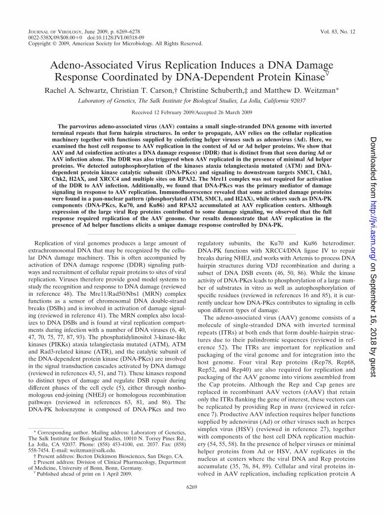
AAV and Ad coinfection induces an MRN-independentDDR. We examined the cellular DDR to replicating AAV inthe context of Ad helper virus. U2OS cells were infected withAAV and Ad, either alone or in combination (Fig. 1). Phos-phorylation of damage response proteins was detected by im-munoblotting (Fig. 1A) and visualized by immunofluorescence(Fig. 1B). Infections were confirmed by immunoblotting withantibodies to the DBP of Ad and Rep proteins of AAV. Wehave previously demonstrated that wild-type Ad5 infectiondoes not induce significant signaling by the cellular damagemachinery due to inactivation of the MRN complex, a sensorof DNA DSBs (6). Infection with Ad or AAV alone did not
6270 SCHWARTZ ET AL. J. VIROL.
on Septem
ber 16, 2018 by guesthttp://jvi.asm
.org/D
ownloaded from
significantly activate DNA damage signaling. In contrast, wefound that AAV and Ad coinfection generated robust signal-ing to DDR substrates, as revealed by phospho-specific anti-bodies to SMC1, Chk1, Chk2, H2AX, and RPA32. Consistentwith their phosphorylation (26, 42, 49), XRCC4 and Nbs1
exhibited gel mobility shifts. Additionally, we noted the acti-vation of ATM using a phospho-specific antibody recognizingthe autophosphorylation site at S1981 (2). Interestingly, immu-nofluorescence revealed that phosphorylated ATM, SMC1,and H2AX were not localized to AAV replication centers butexhibited diffusely nuclear staining patterns (Fig. 1B). We alsofound that AAV infection alone induced some H2AX phos-phorylation (�-H2AX) by immunofluorescence although theintensity was much less than that seen during coinfection. To-gether, these data show that AAV and Ad coinfection elicits arobust DDR not seen significantly with either virus alone.
Since Ad degrades the MRN complex to prevent a DDRduring virus infection (6), we examined MRN during coinfec-tion with AAV. We found that AAV coinfection did not affectthe ability of Ad to degrade the MRN complex, as revealed bya decrease in total levels of Rad50 and Nbs1 proteins (Fig. 1A).This also suggests that MRN is not required for the AAV-induced damage response. To confirm this observation, weanalyzed the damage signaling in response to AAV and Adcoinfection in cells lacking functional MRN. Cells with mutantMre11 (A-TLD1 cells) and Nbs1 (ILB1-NBS cells) were in-fected with both viruses and compared to infected cells thatwere complemented with the respective wild-type cDNAs (Fig.1C). Immunoblotting demonstrated that signaling to Chk2 (de-tected with a phospho-specific antibody) and RPA32 (shownby a mobility shift) was observed in mutant cells to a similarextent as in complemented cells. This shows that the DDR toAAV and Ad coinfection occurs independently of a functionalMRN complex.
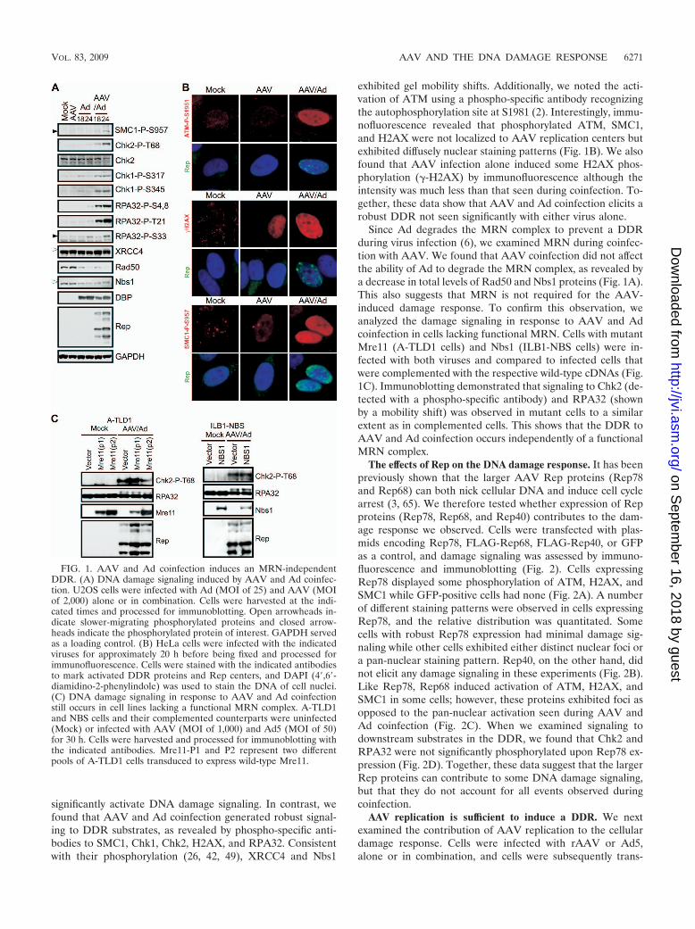
The effects of Rep on the DNA damage response. It has beenpreviously shown that the larger AAV Rep proteins (Rep78and Rep68) can both nick cellular DNA and induce cell cyclearrest (3, 65). We therefore tested whether expression of Repproteins (Rep78, Rep68, and Rep40) contributes to the dam-age response we observed. Cells were transfected with plas-mids encoding Rep78, FLAG-Rep68, FLAG-Rep40, or GFPas a control, and damage signaling was assessed by immuno-fluorescence and immunoblotting (Fig. 2). Cells expressingRep78 displayed some phosphorylation of ATM, H2AX, andSMC1 while GFP-positive cells had none (Fig. 2A). A numberof different staining patterns were observed in cells expressingRep78, and the relative distribution was quantitated. Somecells with robust Rep78 expression had minimal damage sig-naling while other cells exhibited either distinct nuclear foci ora pan-nuclear staining pattern. Rep40, on the other hand, didnot elicit any damage signaling in these experiments (Fig. 2B).Like Rep78, Rep68 induced activation of ATM, H2AX, andSMC1 in some cells; however, these proteins exhibited foci asopposed to the pan-nuclear activation seen during AAV andAd coinfection (Fig. 2C). When we examined signaling todownstream substrates in the DDR, we found that Chk2 andRPA32 were not significantly phosphorylated upon Rep78 ex-pression (Fig. 2D). Together, these data suggest that the largerRep proteins can contribute to some DNA damage signaling,but that they do not account for all events observed duringcoinfection.
AAV replication is sufficient to induce a DDR. We nextexamined the contribution of AAV replication to the cellulardamage response. Cells were infected with rAAV or Ad5,alone or in combination, and cells were subsequently trans-
FIG. 1. AAV and Ad coinfection induces an MRN-independentDDR. (A) DNA damage signaling induced by AAV and Ad coinfec-tion. U2OS cells were infected with Ad (MOI of 25) and AAV (MOIof 2,000) alone or in combination. Cells were harvested at the indi-cated times and processed for immunoblotting. Open arrowheads in-dicate slower-migrating phosphorylated proteins and closed arrow-heads indicate the phosphorylated protein of interest. GAPDH servedas a loading control. (B) HeLa cells were infected with the indicatedviruses for approximately 20 h before being fixed and processed forimmunofluorescence. Cells were stained with the indicated antibodiesto mark activated DDR proteins and Rep centers, and DAPI (4�,6�-diamidino-2-phenylindole) was used to stain the DNA of cell nuclei.(C) DNA damage signaling in response to AAV and Ad coinfectionstill occurs in cell lines lacking a functional MRN complex. A-TLD1and NBS cells and their complemented counterparts were uninfected(Mock) or infected with AAV (MOI of 1,000) and Ad5 (MOI of 50)for 30 h. Cells were harvested and processed for immunoblotting withthe indicated antibodies. Mre11-P1 and P2 represent two differentpools of A-TLD1 cells transduced to express wild-type Mre11.
VOL. 83, 2009 AAV AND THE DNA DAMAGE RESPONSE 6271
on Septem
ber 16, 2018 by guesthttp://jvi.asm
.org/D
ownloaded from
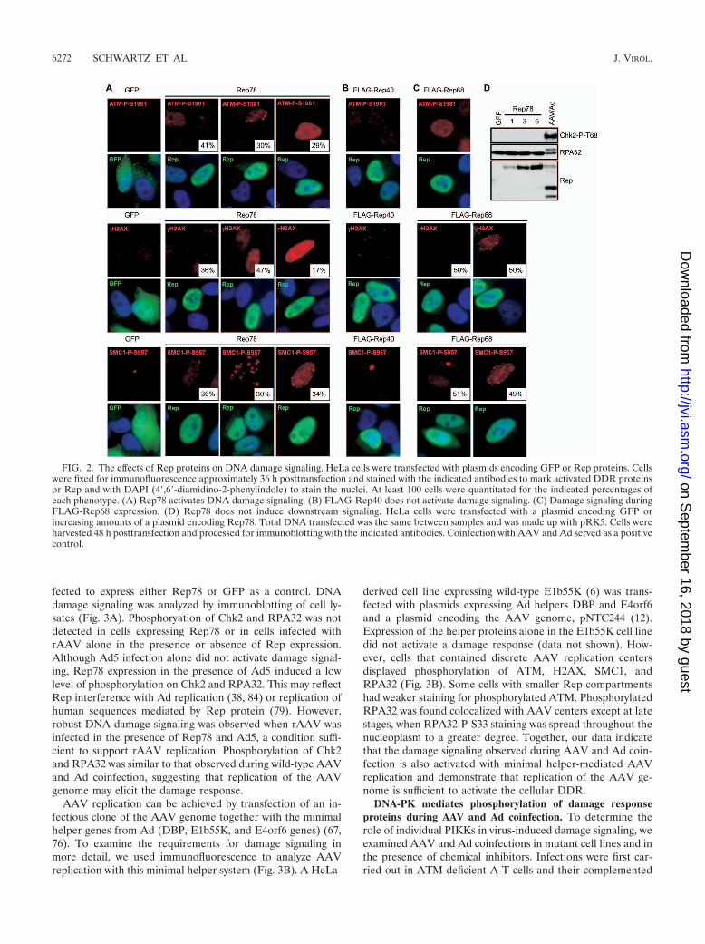
fected to express either Rep78 or GFP as a control. DNAdamage signaling was analyzed by immunoblotting of cell ly-sates (Fig. 3A). Phosphoryation of Chk2 and RPA32 was notdetected in cells expressing Rep78 or in cells infected withrAAV alone in the presence or absence of Rep expression.Although Ad5 infection alone did not activate damage signal-ing, Rep78 expression in the presence of Ad5 induced a lowlevel of phosphorylation on Chk2 and RPA32. This may reflectRep interference with Ad replication (38, 84) or replication ofhuman sequences mediated by Rep protein (79). However,robust DNA damage signaling was observed when rAAV wasinfected in the presence of Rep78 and Ad5, a condition suffi-cient to support rAAV replication. Phosphorylation of Chk2and RPA32 was similar to that observed during wild-type AAVand Ad coinfection, suggesting that replication of the AAVgenome may elicit the damage response.
AAV replication can be achieved by transfection of an in-fectious clone of the AAV genome together with the minimalhelper genes from Ad (DBP, E1b55K, and E4orf6 genes) (67,76). To examine the requirements for damage signaling inmore detail, we used immunofluorescence to analyze AAVreplication with this minimal helper system (Fig. 3B). A HeLa-
derived cell line expressing wild-type E1b55K (6) was trans-fected with plasmids expressing Ad helpers DBP and E4orf6and a plasmid encoding the AAV genome, pNTC244 (12).Expression of the helper proteins alone in the E1b55K cell linedid not activate a damage response (data not shown). How-ever, cells that contained discrete AAV replication centersdisplayed phosphorylation of ATM, H2AX, SMC1, andRPA32 (Fig. 3B). Some cells with smaller Rep compartmentshad weaker staining for phosphorylated ATM. PhosphorylatedRPA32 was found colocalized with AAV centers except at latestages, when RPA32-P-S33 staining was spread throughout thenucleoplasm to a greater degree. Together, our data indicatethat the damage signaling observed during AAV and Ad coin-fection is also activated with minimal helper-mediated AAVreplication and demonstrate that replication of the AAV ge-nome is sufficient to activate the cellular DDR.
DNA-PK mediates phosphorylation of damage responseproteins during AAV and Ad coinfection. To determine therole of individual PIKKs in virus-induced damage signaling, weexamined AAV and Ad coinfections in mutant cell lines and inthe presence of chemical inhibitors. Infections were first car-ried out in ATM-deficient A-T cells and their complemented
FIG. 2. The effects of Rep proteins on DNA damage signaling. HeLa cells were transfected with plasmids encoding GFP or Rep proteins. Cellswere fixed for immunofluorescence approximately 36 h posttransfection and stained with the indicated antibodies to mark activated DDR proteinsor Rep and with DAPI (4�,6�-diamidino-2-phenylindole) to stain the nuclei. At least 100 cells were quantitated for the indicated percentages ofeach phenotype. (A) Rep78 activates DNA damage signaling. (B) FLAG-Rep40 does not activate damage signaling. (C) Damage signaling duringFLAG-Rep68 expression. (D) Rep78 does not induce downstream signaling. HeLa cells were transfected with a plasmid encoding GFP orincreasing amounts of a plasmid encoding Rep78. Total DNA transfected was the same between samples and was made up with pRK5. Cells wereharvested 48 h posttransfection and processed for immunoblotting with the indicated antibodies. Coinfection with AAV and Ad served as a positivecontrol.
6272 SCHWARTZ ET AL. J. VIROL.
on Septem
ber 16, 2018 by guesthttp://jvi.asm
.org/D
ownloaded from
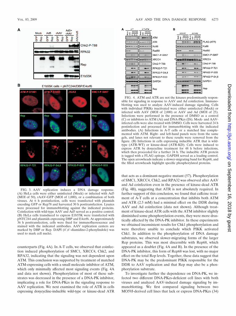
counterparts (Fig. 4A). In A-T cells, we observed that coinfec-tion induced phosphorylation of SMC1, XRCC4, Chk2, andRPA32, indicating that the signaling was not dependent uponATM. This conclusion was supported by treatment of matchedATM-expressing cells with a small molecule inhibitor of ATM,which only minimally affected most signaling events (Fig. 4Aand data not shown). Phosphorylation of most of these sub-strates was decreased in the presence of a DNA-PK inhibitor,implicating a role for DNA-PKcs in the signaling response toAAV replication. We next examined the role of ATR in cellsexpressing doxycycline-inducible wild-type or kinase-dead ATR
that acts as a dominant-negative mutant (57). Phosphorylationof SMC1, XRCC4, Chk2, and RPA32 was observed after AAVand Ad coinfection even in the presence of kinase-dead ATR(Fig. 4B), suggesting that ATR is not absolutely required. Infurther support of this conclusion, we found that caffeine treat-ment of A-T cells at a concentration that inhibits both ATMand ATR (2.5 mM) had a minimal effect on the DDR duringAAV and Ad coinfection (data not shown). Although treat-ment of kinase-dead ATR cells with the ATM inhibitor slightlydiminished some phosphorylation events, they were more dras-tically affected by the DNA-PK inhibitor. In these experimentswe obtained inconsistent results for Chk1 phosphorylation andwere therefore unable to conclude which PIKK activatedChk1. In addition to the phosphorylation of DNA damagesubstrates, we observed slower-migrating forms of the largerRep proteins. This was most discernible with Rep68, whichappeared as a doublet (Fig. 4A and B). In the presence of theDNA-PK inhibitor, this form of Rep68 was lost, with no majoreffect on the total Rep levels. Together, these data suggest thatDNA-PK may be the predominant PIKK responsible for theDDR to AAV replication and that Rep may also be a phos-phorylation substrate.
To investigate further the dependence on DNA-PK, we in-fected two different DNA-PKcs-deficient cell lines with bothviruses and analyzed AAV-induced damage signaling by im-munoblotting. We first compared signaling between twoMO59J-derived cell lines that lack or express DNA-PKcs (34)
FIG. 3. AAV replication induces a DNA damage response.(A) HeLa cells were either uninfected (Mock) or infected with Ad5(MOI of 50), rAAV-GFP (MOI of 1,000), or a combination of bothviruses. At 6 h postinfection, cells were transfected with plasmidsencoding GFP or Rep78 and harvested 30 h posttransfection. Lysateswere processed for immunoblotting against the indicated proteins.Coinfection with wild-type AAV and Ad5 served as a positive control.(B) HeLa cells transduced to express E1b55K were transfected withpNTC244 and plasmids expressing DBP and E4orf6. At approximately36 h posttransfection, cells were fixed for immunofluorescence andstained with the indicated antibodies. AAV replication centers aremarked by DBP or Rep. DAPI (4�,6�-diamidino-2-phenylindole) wasused to mark cell nuclei.
FIG. 4. ATM and ATR are not the kinases predominantly respon-sible for signaling in response to AAV and Ad coinfection. Immuno-blotting was used to analyze AAV-induced damage signaling. Cellswith individual PIKKs inactivated were either uninfected (Mock) orinfected with AAV (MOI of 2,000) or AAV and Ad (MOI of 25).Infections were performed in the presence of DMSO as a control(C) or inhibitors to ATM (Ai) and DNA-PKcs (Di). Mock- and AAV-infected cells were also treated with DMSO. Cells were harvested 24 hpostinfection and processed for immunoblotting with the indicatedantibodies. (A) Infections in A-T cells or a matched line comple-mented with ATM. Right- and left-hand panels were from the samegels, and lanes not relevant to these results were removed from thefigure. (B) Infections in cells expressing inducible ATR that is wild-type (ATR-WT) or kinase-dead (ATR-KD). Cells were induced toexpress ATR by doxycycline treatment for 48 h before infections,which then proceeded for a further 24 h. The inducible ATR proteinis tagged with a FLAG epitope. GAPDH served as a loading control.The open arrowheads indicate a slower-migrating band for Rep68, andthe filled arrowheads highlight specific phosphorylated proteins.
VOL. 83, 2009 AAV AND THE DNA DAMAGE RESPONSE 6273
on Septem
ber 16, 2018 by guesthttp://jvi.asm
.org/D
ownloaded from
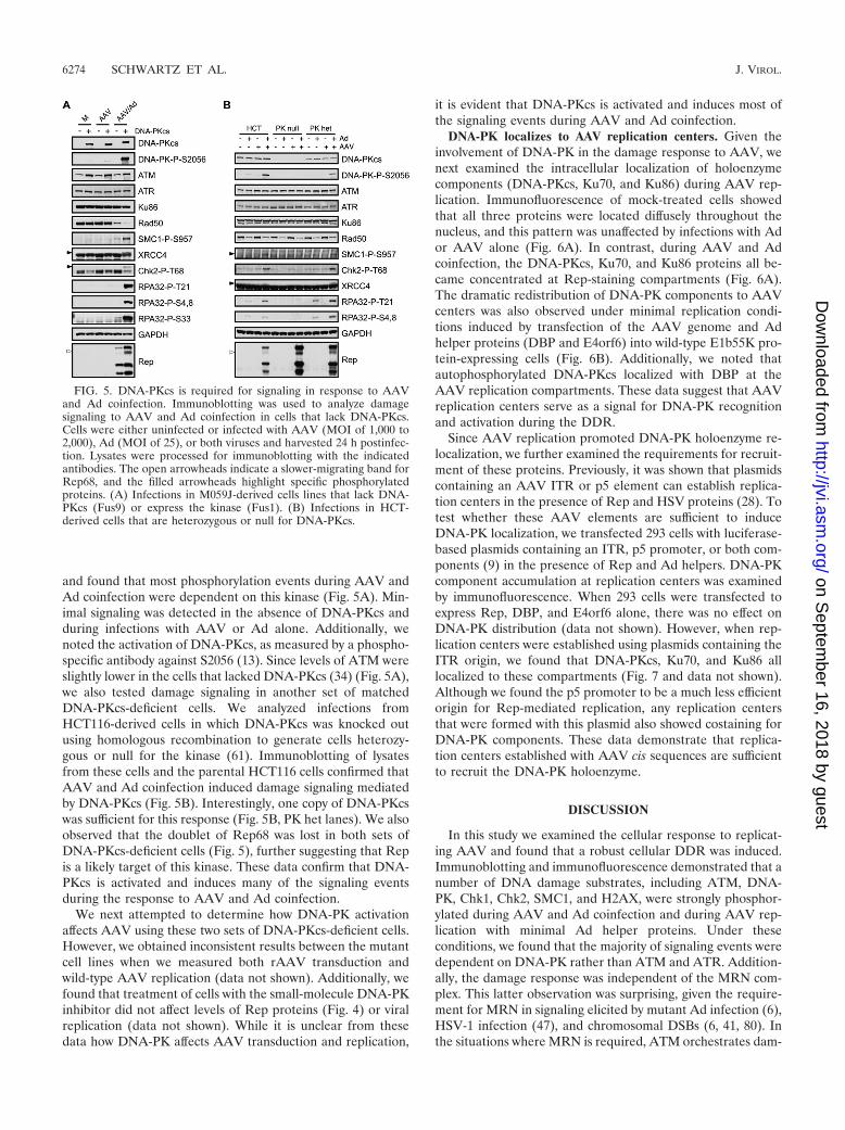
and found that most phosphorylation events during AAV andAd coinfection were dependent on this kinase (Fig. 5A). Min-imal signaling was detected in the absence of DNA-PKcs andduring infections with AAV or Ad alone. Additionally, wenoted the activation of DNA-PKcs, as measured by a phospho-specific antibody against S2056 (13). Since levels of ATM wereslightly lower in the cells that lacked DNA-PKcs (34) (Fig. 5A),we also tested damage signaling in another set of matchedDNA-PKcs-deficient cells. We analyzed infections fromHCT116-derived cells in which DNA-PKcs was knocked outusing homologous recombination to generate cells heterozy-gous or null for the kinase (61). Immunoblotting of lysatesfrom these cells and the parental HCT116 cells confirmed thatAAV and Ad coinfection induced damage signaling mediatedby DNA-PKcs (Fig. 5B). Interestingly, one copy of DNA-PKcswas sufficient for this response (Fig. 5B, PK het lanes). We alsoobserved that the doublet of Rep68 was lost in both sets ofDNA-PKcs-deficient cells (Fig. 5), further suggesting that Repis a likely target of this kinase. These data confirm that DNA-PKcs is activated and induces many of the signaling eventsduring the response to AAV and Ad coinfection.
We next attempted to determine how DNA-PK activationaffects AAV using these two sets of DNA-PKcs-deficient cells.However, we obtained inconsistent results between the mutantcell lines when we measured both rAAV transduction andwild-type AAV replication (data not shown). Additionally, wefound that treatment of cells with the small-molecule DNA-PKinhibitor did not affect levels of Rep proteins (Fig. 4) or viralreplication (data not shown). While it is unclear from thesedata how DNA-PK affects AAV transduction and replication,
it is evident that DNA-PKcs is activated and induces most ofthe signaling events during AAV and Ad coinfection.
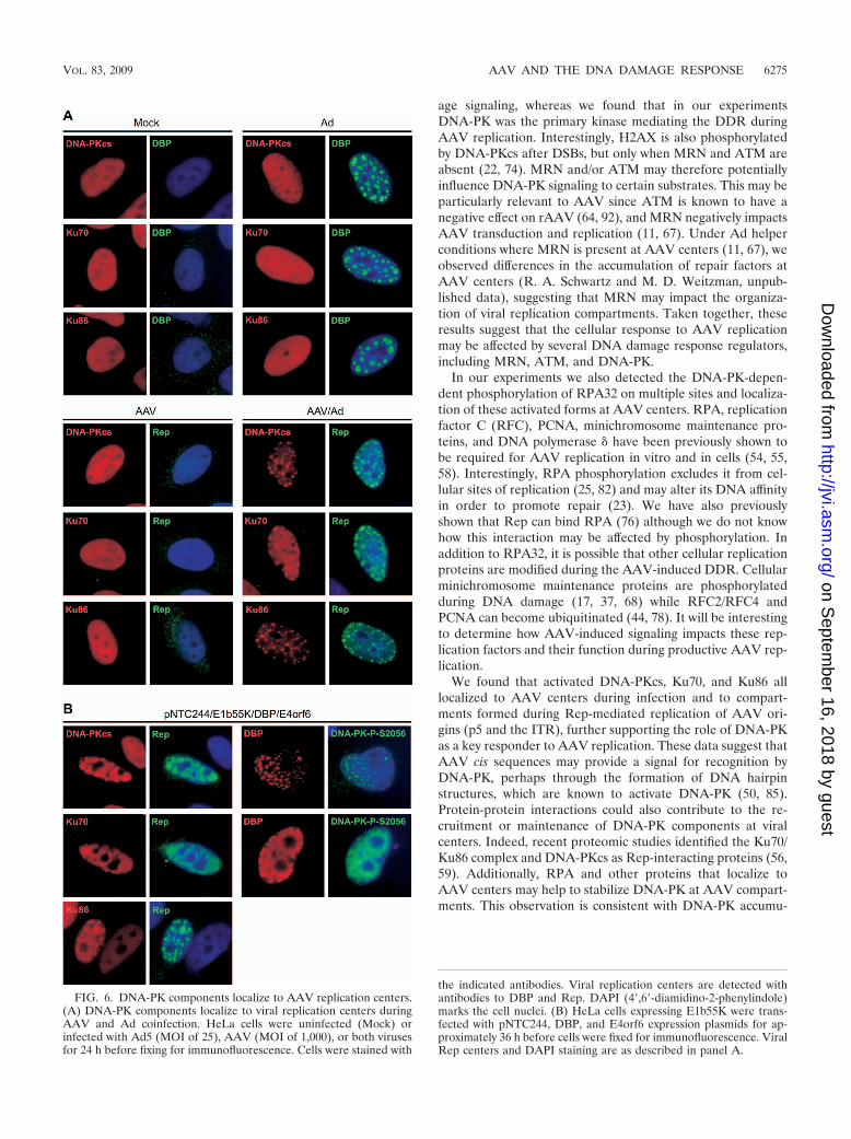
DNA-PK localizes to AAV replication centers. Given theinvolvement of DNA-PK in the damage response to AAV, wenext examined the intracellular localization of holoenzymecomponents (DNA-PKcs, Ku70, and Ku86) during AAV rep-lication. Immunofluorescence of mock-treated cells showedthat all three proteins were located diffusely throughout thenucleus, and this pattern was unaffected by infections with Ador AAV alone (Fig. 6A). In contrast, during AAV and Adcoinfection, the DNA-PKcs, Ku70, and Ku86 proteins all be-came concentrated at Rep-staining compartments (Fig. 6A).The dramatic redistribution of DNA-PK components to AAVcenters was also observed under minimal replication condi-tions induced by transfection of the AAV genome and Adhelper proteins (DBP and E4orf6) into wild-type E1b55K pro-tein-expressing cells (Fig. 6B). Additionally, we noted thatautophosphorylated DNA-PKcs localized with DBP at theAAV replication compartments. These data suggest that AAVreplication centers serve as a signal for DNA-PK recognitionand activation during the DDR.
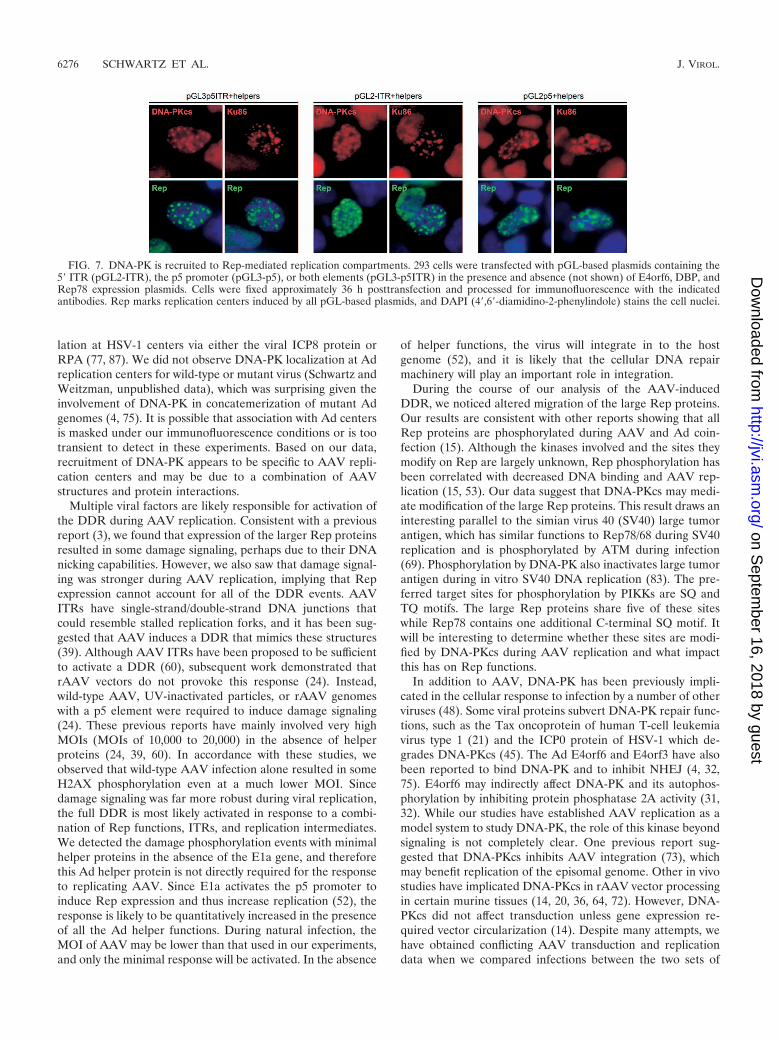
Since AAV replication promoted DNA-PK holoenzyme re-localization, we further examined the requirements for recruit-ment of these proteins. Previously, it was shown that plasmidscontaining an AAV ITR or p5 element can establish replica-tion centers in the presence of Rep and HSV proteins (28). Totest whether these AAV elements are sufficient to induceDNA-PK localization, we transfected 293 cells with luciferase-based plasmids containing an ITR, p5 promoter, or both com-ponents (9) in the presence of Rep and Ad helpers. DNA-PKcomponent accumulation at replication centers was examinedby immunofluorescence. When 293 cells were transfected toexpress Rep, DBP, and E4orf6 alone, there was no effect onDNA-PK distribution (data not shown). However, when rep-lication centers were established using plasmids containing theITR origin, we found that DNA-PKcs, Ku70, and Ku86 alllocalized to these compartments (Fig. 7 and data not shown).Although we found the p5 promoter to be a much less efficientorigin for Rep-mediated replication, any replication centersthat were formed with this plasmid also showed costaining forDNA-PK components. These data demonstrate that replica-tion centers established with AAV cis sequences are sufficientto recruit the DNA-PK holoenzyme.
DISCUSSION
In this study we examined the cellular response to replicat-ing AAV and found that a robust cellular DDR was induced.Immunoblotting and immunofluorescence demonstrated that anumber of DNA damage substrates, including ATM, DNA-PK, Chk1, Chk2, SMC1, and H2AX, were strongly phosphor-ylated during AAV and Ad coinfection and during AAV rep-lication with minimal Ad helper proteins. Under theseconditions, we found that the majority of signaling events weredependent on DNA-PK rather than ATM and ATR. Addition-ally, the damage response was independent of the MRN com-plex. This latter observation was surprising, given the require-ment for MRN in signaling elicited by mutant Ad infection (6),HSV-1 infection (47), and chromosomal DSBs (6, 41, 80). Inthe situations where MRN is required, ATM orchestrates dam-
FIG. 5. DNA-PKcs is required for signaling in response to AAVand Ad coinfection. Immunoblotting was used to analyze damagesignaling to AAV and Ad coinfection in cells that lack DNA-PKcs.Cells were either uninfected or infected with AAV (MOI of 1,000 to2,000), Ad (MOI of 25), or both viruses and harvested 24 h postinfec-tion. Lysates were processed for immunoblotting with the indicatedantibodies. The open arrowheads indicate a slower-migrating band forRep68, and the filled arrowheads highlight specific phosphorylatedproteins. (A) Infections in M059J-derived cells lines that lack DNA-PKcs (Fus9) or express the kinase (Fus1). (B) Infections in HCT-derived cells that are heterozygous or null for DNA-PKcs.
6274 SCHWARTZ ET AL. J. VIROL.
on Septem
ber 16, 2018 by guesthttp://jvi.asm
.org/D
ownloaded from
age signaling, whereas we found that in our experimentsDNA-PK was the primary kinase mediating the DDR duringAAV replication. Interestingly, H2AX is also phosphorylatedby DNA-PKcs after DSBs, but only when MRN and ATM areabsent (22, 74). MRN and/or ATM may therefore potentiallyinfluence DNA-PK signaling to certain substrates. This may beparticularly relevant to AAV since ATM is known to have anegative effect on rAAV (64, 92), and MRN negatively impactsAAV transduction and replication (11, 67). Under Ad helperconditions where MRN is present at AAV centers (11, 67), weobserved differences in the accumulation of repair factors atAAV centers (R. A. Schwartz and M. D. Weitzman, unpub-lished data), suggesting that MRN may impact the organiza-tion of viral replication compartments. Taken together, theseresults suggest that the cellular response to AAV replicationmay be affected by several DNA damage response regulators,including MRN, ATM, and DNA-PK.
In our experiments we also detected the DNA-PK-depen-dent phosphorylation of RPA32 on multiple sites and localiza-tion of these activated forms at AAV centers. RPA, replicationfactor C (RFC), PCNA, minichromosome maintenance pro-teins, and DNA polymerase � have been previously shown tobe required for AAV replication in vitro and in cells (54, 55,58). Interestingly, RPA phosphorylation excludes it from cel-lular sites of replication (25, 82) and may alter its DNA affinityin order to promote repair (23). We have also previouslyshown that Rep can bind RPA (76) although we do not knowhow this interaction may be affected by phosphorylation. Inaddition to RPA32, it is possible that other cellular replicationproteins are modified during the AAV-induced DDR. Cellularminichromosome maintenance proteins are phosphorylatedduring DNA damage (17, 37, 68) while RFC2/RFC4 andPCNA can become ubiquitinated (44, 78). It will be interestingto determine how AAV-induced signaling impacts these rep-lication factors and their function during productive AAV rep-lication.
We found that activated DNA-PKcs, Ku70, and Ku86 alllocalized to AAV centers during infection and to compart-ments formed during Rep-mediated replication of AAV ori-gins (p5 and the ITR), further supporting the role of DNA-PKas a key responder to AAV replication. These data suggest thatAAV cis sequences may provide a signal for recognition byDNA-PK, perhaps through the formation of DNA hairpinstructures, which are known to activate DNA-PK (50, 85).Protein-protein interactions could also contribute to the re-cruitment or maintenance of DNA-PK components at viralcenters. Indeed, recent proteomic studies identified the Ku70/Ku86 complex and DNA-PKcs as Rep-interacting proteins (56,59). Additionally, RPA and other proteins that localize toAAV centers may help to stabilize DNA-PK at AAV compart-ments. This observation is consistent with DNA-PK accumu-
FIG. 6. DNA-PK components localize to AAV replication centers.(A) DNA-PK components localize to viral replication centers duringAAV and Ad coinfection. HeLa cells were uninfected (Mock) orinfected with Ad5 (MOI of 25), AAV (MOI of 1,000), or both virusesfor 24 h before fixing for immunofluorescence. Cells were stained with
the indicated antibodies. Viral replication centers are detected withantibodies to DBP and Rep. DAPI (4�,6�-diamidino-2-phenylindole)marks the cell nuclei. (B) HeLa cells expressing E1b55K were trans-fected with pNTC244, DBP, and E4orf6 expression plasmids for ap-proximately 36 h before cells were fixed for immunofluorescence. ViralRep centers and DAPI staining are as described in panel A.
VOL. 83, 2009 AAV AND THE DNA DAMAGE RESPONSE 6275
on Septem
ber 16, 2018 by guesthttp://jvi.asm
.org/D
ownloaded from
lation at HSV-1 centers via either the viral ICP8 protein orRPA (77, 87). We did not observe DNA-PK localization at Adreplication centers for wild-type or mutant virus (Schwartz andWeitzman, unpublished data), which was surprising given theinvolvement of DNA-PK in concatemerization of mutant Adgenomes (4, 75). It is possible that association with Ad centersis masked under our immunofluorescence conditions or is tootransient to detect in these experiments. Based on our data,recruitment of DNA-PK appears to be specific to AAV repli-cation centers and may be due to a combination of AAVstructures and protein interactions.
Multiple viral factors are likely responsible for activation ofthe DDR during AAV replication. Consistent with a previousreport (3), we found that expression of the larger Rep proteinsresulted in some damage signaling, perhaps due to their DNAnicking capabilities. However, we also saw that damage signal-ing was stronger during AAV replication, implying that Repexpression cannot account for all of the DDR events. AAVITRs have single-strand/double-strand DNA junctions thatcould resemble stalled replication forks, and it has been sug-gested that AAV induces a DDR that mimics these structures(39). Although AAV ITRs have been proposed to be sufficientto activate a DDR (60), subsequent work demonstrated thatrAAV vectors do not provoke this response (24). Instead,wild-type AAV, UV-inactivated particles, or rAAV genomeswith a p5 element were required to induce damage signaling(24). These previous reports have mainly involved very highMOIs (MOIs of 10,000 to 20,000) in the absence of helperproteins (24, 39, 60). In accordance with these studies, weobserved that wild-type AAV infection alone resulted in someH2AX phosphorylation even at a much lower MOI. Sincedamage signaling was far more robust during viral replication,the full DDR is most likely activated in response to a combi-nation of Rep functions, ITRs, and replication intermediates.We detected the damage phosphorylation events with minimalhelper proteins in the absence of the E1a gene, and thereforethis Ad helper protein is not directly required for the responseto replicating AAV. Since E1a activates the p5 promoter toinduce Rep expression and thus increase replication (52), theresponse is likely to be quantitatively increased in the presenceof all the Ad helper functions. During natural infection, theMOI of AAV may be lower than that used in our experiments,and only the minimal response will be activated. In the absence
of helper functions, the virus will integrate in to the hostgenome (52), and it is likely that the cellular DNA repairmachinery will play an important role in integration.
During the course of our analysis of the AAV-inducedDDR, we noticed altered migration of the large Rep proteins.Our results are consistent with other reports showing that allRep proteins are phosphorylated during AAV and Ad coin-fection (15). Although the kinases involved and the sites theymodify on Rep are largely unknown, Rep phosphorylation hasbeen correlated with decreased DNA binding and AAV rep-lication (15, 53). Our data suggest that DNA-PKcs may medi-ate modification of the large Rep proteins. This result draws aninteresting parallel to the simian virus 40 (SV40) large tumorantigen, which has similar functions to Rep78/68 during SV40replication and is phosphorylated by ATM during infection(69). Phosphorylation by DNA-PK also inactivates large tumorantigen during in vitro SV40 DNA replication (83). The pre-ferred target sites for phosphorylation by PIKKs are SQ andTQ motifs. The large Rep proteins share five of these siteswhile Rep78 contains one additional C-terminal SQ motif. Itwill be interesting to determine whether these sites are modi-fied by DNA-PKcs during AAV replication and what impactthis has on Rep functions.
In addition to AAV, DNA-PK has been previously impli-cated in the cellular response to infection by a number of otherviruses (48). Some viral proteins subvert DNA-PK repair func-tions, such as the Tax oncoprotein of human T-cell leukemiavirus type 1 (21) and the ICP0 protein of HSV-1 which de-grades DNA-PKcs (45). The Ad E4orf6 and E4orf3 have alsobeen reported to bind DNA-PK and to inhibit NHEJ (4, 32,75). E4orf6 may indirectly affect DNA-PK and its autophos-phorylation by inhibiting protein phosphatase 2A activity (31,32). While our studies have established AAV replication as amodel system to study DNA-PK, the role of this kinase beyondsignaling is not completely clear. One previous report sug-gested that DNA-PKcs inhibits AAV integration (73), whichmay benefit replication of the episomal genome. Other in vivostudies have implicated DNA-PKcs in rAAV vector processingin certain murine tissues (14, 20, 36, 64, 72). However, DNA-PKcs did not affect transduction unless gene expression re-quired vector circularization (14). Despite many attempts, wehave obtained conflicting AAV transduction and replicationdata when we compared infections between the two sets of
FIG. 7. DNA-PK is recruited to Rep-mediated replication compartments. 293 cells were transfected with pGL-based plasmids containing the5� ITR (pGL2-ITR), the p5 promoter (pGL3-p5), or both elements (pGL3-p5ITR) in the presence and absence (not shown) of E4orf6, DBP, andRep78 expression plasmids. Cells were fixed approximately 36 h posttransfection and processed for immunofluorescence with the indicatedantibodies. Rep marks replication centers induced by all pGL-based plasmids, and DAPI (4�,6�-diamidino-2-phenylindole) stains the cell nuclei.
6276 SCHWARTZ ET AL. J. VIROL.
on Septem
ber 16, 2018 by guesthttp://jvi.asm
.org/D
ownloaded from
DNA-PKcs-deficient cells employed in the experiment shownin Fig. 5 (data not shown). The impact of DNA-PK signalingon AAV replication and transduction may depend on the cel-lular context and the viral genome structure. In this report wehave uncovered a unique role for DNA-PK in AAV-induceddamage signaling, expanding our knowledge of the links be-tween viral replication and cellular DDR pathways.
ACKNOWLEDGMENTS
We thank A. Berk, B. Chen, D. Chen, P. Concannon, M. Giacca, E.Hendrickson, J. Karlseder, A. Levine, T. Melendy, J. Petrini, R. J. Sam-ulski, S. Schreiber, Y. Shiloh, J. Trempe, and P. van der Vliet for reagents.We are grateful to D. Lee and D. Linfesty for technical support. We thankmembers of the Weitzman lab for helpful discussions and critical readingsof the manuscript. We acknowledge the James B. Pendleton CharitableTrust for providing the microscopy facility.
Work in the Weitzman lab is partially supported by a Pioneer Devel-opmental Chair. This work was supported in part by NIH grants CA97093and AI43341 (M.D.W.) and by gifts from the Joe W. & Dorothy DorsettBrown Foundation and the Lebensfeld Foundation to M.D.W. R.A.S.and C.T.C. were supported in part by NIH Training Grants to UCSD andthe Salk Institute and by scholarships from the ARCS Foundation.
ADDENDUM IN PROOF
After this paper was accepted, an independent study also ob-served a cellular DNA damage response activated by AAV andAd coinfection with DNA-PD as the most prominent kinase me-diating these effects (R. F. Collaco, J. M. Bevington, V. Bhrigu, V.Kalman-Maltese, and J. P. Trempe, Virology, in press).
REFERENCES
1. Alexander, I. E., D. W. Russell, and A. D. Miller. 1994. DNA-damagingagents greatly increase the transduction of nondividing cells by adeno-asso-ciated virus vectors. J. Virol. 68:8282–8287.
2. Bakkenist, C. J., and M. B. Kastan. 2003. DNA damage activates ATMthrough intermolecular autophosphorylation and dimer dissociation. Nature421:499–506.
3. Berthet, C., K. Raj, P. Saudan, and P. Beard. 2005. How adeno-associatedvirus Rep78 protein arrests cells completely in S phase. Proc. Natl. Acad. Sci.USA 102:13634–13639.
4. Boyer, J., K. Rohleder, and G. Ketner. 1999. Adenovirus E4 34k and E4 11kinhibit double strand break repair and are physically associated with thecellular DNA-dependent protein kinase. Virology 263:307–312.
5. Branzei, D., and M. Foiani. 2008. Regulation of DNA repair throughout thecell cycle. Nat. Rev. Mol. Cell Biol. 9:297–308.
6. Carson, C. T., R. A. Schwartz, T. H. Stracker, C. E. Lilley, D. V. Lee, andM. D. Weitzman. 2003. The Mre11 complex is required for ATM activationand the G2/M checkpoint. EMBO J. 22:6610–6620.
7. Carter, B. J. 2004. Adeno-associated virus and the development of adeno-associated virus vectors: a historical perspective. Mol. Ther. 10:981–989.
8. Cassell, G. D., and M. D. Weitzman. 2004. Characterization of a nuclearlocalization signal in the C-terminus of the adeno-associated virus Rep68/78proteins. Virology 327:206–214.
9. Cathomen, T., T. H. Stracker, L. B. Gilbert, and M. D. Weitzman. 2001. Agenetic screen identifies a cellular regulator of adeno-associated virus. Proc.Natl. Acad. Sci. USA 98:14991–14996.
10. Cerosaletti, K. M., A. Desai-Mehta, T. C. Yeo, M. Kraakman-Van Der Zwet,M. Z. Zdzienicka, and P. Concannon. 2000. Retroviral expression of theNBS1 gene in cultured Nijmegen breakage syndrome cells restores normalradiation sensitivity and nuclear focus formation. Mutagenesis 15:281–286.
11. Cervelli, T., J. A. Palacios, L. Zentilin, M. Mano, R. A. Schwartz, M. D.Weitzman, and M. Giacca. 2008. Processing of recombinant AAV genomesoccurs in specific nuclear structures that overlap with foci of DNA-damage-response proteins. J. Cell Sci. 121:349–357.
12. Chejanovsky, N., and B. J. Carter. 1989. Replication of a human parvovirusnonsense mutant in mammalian cells containing an inducible amber sup-pressor. Virology 171:239–247.
13. Chen, B. P., D. W. Chan, J. Kobayashi, S. Burma, A. Asaithamby, K.Morotomi-Yano, E. Botvinick, J. Qin, and D. J. Chen. 2005. Cell cycledependence of DNA-dependent protein kinase phosphorylation in responseto DNA double strand breaks. J. Biol. Chem. 280:14709–14715.
14. Choi, V. W., D. M. McCarty, and R. J. Samulski. 2006. Host cell DNA repairpathways in adeno-associated viral genome processing. J. Virol. 80:10346–10356.
15. Collaco, R., K. M. Prasad, and J. P. Trempe. 1997. Phosphorylation of theadeno-associated virus replication proteins. Virology 232:332–336.
16. Collis, S. J., T. L. DeWeese, P. A. Jeggo, and A. R. Parker. 2005. The life anddeath of DNA-PK. Oncogene 24:949–961.
17. Cortez, D., G. Glick, and S. J. Elledge. 2004. Minichromosome maintenanceproteins are direct targets of the ATM and ATR checkpoint kinases. Proc.Natl. Acad. Sci. USA 101:10078–10083.
18. Duan, D., P. Sharma, L. Dudus, Y. Zhang, S. Sanlioglu, Z. Yan, Y. Yue, Y.Ye, R. Lester, J. Yang, K. J. Fisher, and J. F. Engelhardt. 1999. Formationof adeno-associated virus circular genomes is differentially regulated byadenovirus E4 ORF6 and E2a gene expression. J. Virol. 73:161–169.
19. Duan, D., P. Sharma, J. Yang, Y. Yue, L. Dudus, Y. Zhang, K. J. Fisher, andJ. F. Engelhardt. 1998. Circular intermediates of recombinant adeno-asso-ciated virus have defined structural characteristics responsible for long-termepisomal persistence in muscle tissue. J. Virol. 72:8568–8577.
20. Duan, D., Y. Yue, and J. F. Engelhardt. 2003. Consequences of DNA-dependent protein kinase catalytic subunit deficiency on recombinant adeno-associated virus genome circularization and heterodimerization in muscletissue. J. Virol. 77:4751–4759.
21. Durkin, S. S., X. Guo, K. A. Fryrear, V. T. Mihaylova, S. K. Gupta, S. M.Belgnaoui, A. Haoudi, G. M. Kupfer, and O. J. Semmes. 2008. HTLV-1 Taxoncoprotein subverts the cellular DNA damage response via binding toDNA-dependent protein kinase. J. Biol. Chem. 283:36311–36320.
22. Falck, J., J. Coates, and S. P. Jackson. 2005. Conserved modes of recruit-ment of ATM, ATR and DNA-PKcs to sites of DNA damage. Nature434:605–611.
23. Fanning, E., V. Klimovich, and A. R. Nager. 2006. A dynamic model forreplication protein A (RPA) function in DNA processing pathways. NucleicAcids Res. 34:4126–4137.
24. Fragkos, M., M. Breuleux, N. Clement, and P. Beard. 2008. Recombinantadeno-associated viral vectors are deficient in provoking a DNA damageresponse. J. Virol. 82:7379–7387.
25. Francon, P., J. M. Lemaitre, C. Dreyer, D. Maiorano, O. Cuvier, and M.Mechali. 2004. A hypophosphorylated form of RPA34 is a specific compo-nent of pre-replication centers. J. Cell Sci. 117:4909–4920.
26. Gatei, M., D. Young, K. M. Cerosaletti, A. Desai-Mehta, K. Spring, S.Kozlov, M. F. Lavin, R. A. Gatti, P. Concannon, and K. Khanna. 2000.ATM-dependent phosphorylation of nibrin in response to radiation expo-sure. Nat. Genet. 25:115–119.
27. Geoffroy, M. C., and A. Salvetti. 2005. Helper functions required for wildtype and recombinant adeno-associated virus growth. Curr. Gene Ther.5:265–271.
28. Glauser, D. L., O. Saydam, N. A. Balsiger, I. Heid, R. M. Linden, M.Ackermann, and C. Fraefel. 2005. Four-dimensional visualization of thesimultaneous activity of alternative adeno-associated virus replication ori-gins. J. Virol. 79:12218–12230.
29. Glauser, D. L., R. Strasser, A. S. Laimbacher, O. Saydam, N. Clement, R. M.Linden, M. Ackermann, and C. Fraefel. 2007. Live covisualization of com-peting adeno-associated virus and herpes simplex virus type 1 DNA replica-tion: molecular mechanisms of interaction. J. Virol. 81:4732–4743.
30. Grifman, M., M. Trepel, P. Speece, L. B. Gilbert, W. Arap, R. Pasqualini,and M. D. Weitzman. 2001. Incorporation of tumor-targeting peptides intorecombinant adeno-associated virus capsids. Mol. Ther. 3:964–975.
31. Hart, L. S., D. Ornelles, and C. Koumenis. 2007. The adenoviral E4orf6protein induces atypical apoptosis in response to DNA damage. J. Biol.Chem. 282:6061–6067.
32. Hart, L. S., S. M. Yannone, C. Naczki, J. S. Orlando, S. B. Waters, S. A.Akman, D. J. Chen, D. Ornelles, and C. Koumenis. 2005. The adenovirusE4orf6 protein inhibits DNA double strand break repair and radiosensitizeshuman tumor cells in an E1B-55K-independent manner. J. Biol. Chem.280:1474–1481.
33. Heilbronn, R., M. Engstler, S. Weger, A. Krahn, C. Schetter, and M.Boshart. 2003. ssDNA-dependent colocalization of adeno-associated virusRep. and herpes simplex virus ICP8 in nuclear replication domains. NucleicAcids Res. 31:6206–6213.
34. Hoppe, B. S., R. B. Jensen, and C. U. Kirchgessner. 2000. Complementationof the radiosensitive M059J cell line. Radiat. Res. 153:125–130.
35. Hunter, L. A., and R. J. Samulski. 1992. Colocalization of adeno-associatedvirus Rep and capsid proteins in the nuclei of infected cells. J. Virol. 66:317–324.
36. Inagaki, K., C. Ma, T. A. Storm, M. A. Kay, and H. Nakai. 2007. The role ofDNA-PKcs and Artemis in opening viral DNA hairpin termini in varioustissues in mice. J. Virol. 81:11304–11321.
37. Ishimi, Y., Y. Komamura-Kohno, H. J. Kwon, K. Yamada, and M. Nakan-ishi. 2003. Identification of MCM4 as a target of the DNA replication blockcheckpoint system. J. Biol. Chem. 278:24644–24650.
38. Jing, X. J., V. Kalman-Maltese, X. Cao, Q. Yang, and J. P. Trempe. 2001.Inhibition of adenovirus cytotoxicity, replication, and E2a gene expression byadeno-associated virus. Virology 291:140–151.
39. Jurvansuu, J., K. Raj, A. Stasiak, and P. Beard. 2005. Viral transport ofDNA damage that mimics a stalled replication fork. J. Virol. 79:569–580.
40. Kudoh, A., M. Fujita, L. Zhang, N. Shirata, T. Daikoku, Y. Sugaya, H.
VOL. 83, 2009 AAV AND THE DNA DAMAGE RESPONSE 6277
on Septem
ber 16, 2018 by guesthttp://jvi.asm
.org/D
ownloaded from
Isomura, Y. Nishiyama, and T. Tsurumi. 2005. Epstein-Barr virus lytic rep-lication elicits ATM checkpoint signal transduction while providing an S-phase-like cellular environment. J. Biol. Chem. 280:8156–8163.
41. Lavin, M. F. 2007. ATM and the Mre11 complex combine to recognize andsignal DNA double-strand breaks. Oncogene 26:7749–7758.
42. Leber, R., T. W. Wise, R. Mizuta, and K. Meek. 1998. The XRCC4 geneproduct is a target for and interacts with the DNA-dependent protein kinase.J. Biol. Chem. 273:1794–1801.
43. Lee, J. H., and T. T. Paull. 2007. Activation and regulation of ATM kinaseactivity in response to DNA double-strand breaks. Oncogene 26:7741–7748.
44. Lee, K. Y., and K. Myung. 2008. PCNA modifications for regulation ofpost-replication repair pathways. Mol. Cells 26:5–11.
45. Lees-Miller, S. P., M. C. Long, M. A. Kilvert, V. Lam, S. A. Rice, and C. A.Spencer. 1996. Attenuation of DNA-dependent protein kinase activity andits catalytic subunit by the herpes simplex virus type 1 transactivator ICP0.J. Virol. 70:7471–7477.
46. Lieber, M. R. 2008. The mechanism of human nonhomologous DNA endjoining. J. Biol. Chem. 283:1–5.
47. Lilley, C. E., C. T. Carson, A. R. Muotri, F. H. Gage, and M. D. Weitzman.2005. DNA repair proteins affect the lifecycle of herpes simplex virus 1. Proc.Natl. Acad. Sci. USA 102:5844–5849.
48. Lilley, C. E., R. A. Schwartz, and M. D. Weitzman. 2007. Using or abusing:viruses and the cellular DNA damage response. Trends Microbiol. 15:119–126.
49. Lim, D. S., S. T. Kim, B. Xu, R. S. Maser, J. Lin, J. H. Petrini, and M. B.Kastan. 2000. ATM phosphorylates p95/nbs1 in an S-phase checkpoint path-way. Nature 404:613–617.
50. Ma, Y., K. Schwarz, and M. R. Lieber. 2005. The Artemis:DNA-PKcs en-donuclease cleaves DNA loops, flaps, and gaps. DNA Repair 4:845–851.
51. McGowan, C. H., and P. Russell. 2004. The DNA damage response: sensingand signaling. Curr. Opin. Cell Biol. 16:629–633.
52. Muzyczka, N., and K. I. Berns. 2001. Parvoviridae: the viruses and theirreplication, p. 2327–2359. In D. M. Knipe, P. M. Howley, D. E. Griffin, R. A.Lamb, M. A. Martin, B. Roizman, and S. E. Straus (ed.), Fields virology, vol.2, 4th ed. Lippincott Williams & Wilkins, Philadelphia, PA.
53. Narasimhan, D., R. Collaco, V. Kalman-Maltese, and J. P. Trempe. 2002.Hyper-phosphorylation of the adeno-associated virus Rep78 protein inhibitsterminal repeat binding and helicase activity. Biochim. Biophys. Acta 1576:298–305.
54. Nash, K., W. Chen, W. F. McDonald, X. Zhou, and N. Muzyczka. 2007.Purification of host cell enzymes involved in adeno-associated virus DNAreplication. J. Virol. 81:5777–5787.
55. Nash, K., W. Chen, and N. Muzyczka. 2008. Complete in vitro reconstitutionof adeno-associated virus DNA replication requires the minichromosomemaintenance complex proteins. J. Virol. 82:1458–1464.
56. Nash, K., W. Chen, M. Salganik, and N. Muzyczka. 2009. Identification ofcellular proteins that interact with the adeno-associated virus Rep. protein.J. Virol. 83:454–469.
57. Nghiem, P., P. K. Park, Y. Kim, C. Vaziri, and S. L. Schreiber. 2001. ATRinhibition selectively sensitizes G1 checkpoint-deficient cells to lethal pre-mature chromatin condensation. Proc. Natl. Acad. Sci. USA 98:9092–9097.
58. Ni, T. H., W. F. McDonald, I. Zolotukhin, T. Melendy, S. Waga, B. Stillman,and N. Muzyczka. 1998. Cellular proteins required for adeno-associatedvirus DNA replication in the absence of adenovirus coinfection. J. Virol.72:2777–2787.
59. Pegoraro, G., A. Marcello, M. P. Myers, and M. Giacca. 2006. Regulation ofadeno-associated virus DNA replication by the cellular TAF-I/set complex.J. Virol. 80:6855–6864.
60. Raj, K., P. Ogston, and P. Beard. 2001. Virus-mediated killing of cells thatlack p53 activity. Nature 412:914–917.
61. Ruis, B. L., K. R. Fattah, and E. A. Hendrickson. 2008. The catalytic subunitof DNA-dependent protein kinase regulates proliferation, telomere length,and genomic stability in human somatic cells. Mol. Cell. Biol. 28:6182–6195.
62. Russell, D. W., I. E. Alexander, and A. D. Miller. 1995. DNA synthesis andtopoisomerase inhibitors increase transduction by adeno-associated virusvectors. Proc. Natl. Acad. Sci. USA 92:5719–5723.
63. Sancar, A., L. A. Lindsey-Boltz, K. Unsal-Kacmaz, and S. Linn. 2004. Mo-lecular mechanisms of mammalian DNA repair and the DNA damage check-points. Annu. Rev. Biochem. 73:39–85.
64. Sanlioglu, S., P. Benson, and J. F. Engelhardt. 2000. Loss of ATM functionenhances recombinant adeno-associated virus transduction and integrationthrough pathways similar to UV irradiation. Virology 268:68–78.
65. Saudan, P., J. Vlach, and P. Beard. 2000. Inhibition of S-phase progressionby adeno-associated virus Rep78 protein is mediated by hypophosphorylatedpRb. EMBO J. 19:4351–4361.
66. Schnepp, B. C., K. R. Clark, D. L. Klemanski, C. A. Pacak, and P. R.Johnson. 2003. Genetic fate of recombinant adeno-associated virus vectorgenomes in muscle. J. Virol. 77:3495–3504.
67. Schwartz, R. A., J. A. Palacios, G. D. Cassell, S. Adam, M. Giacca, and M. D.Weitzman. 2007. The Mre11/Rad50/Nbs1 complex limits adeno-associatedvirus transduction and replication. J. Virol. 81:12936–12945.
68. Shi, Y., G. E. Dodson, P. S. Mukhopadhyay, N. P. Shanware, A. T. Trinh, andR. S. Tibbetts. 2007. Identification of carboxyl-terminal MCM3 phosphory-
lation sites using polyreactive phosphospecific antibodies. J. Biol. Chem.282:9236–9243.
69. Shi, Y., G. E. Dodson, S. Shaikh, K. Rundell, and R. S. Tibbetts. 2005.Ataxia-telangiectasia-mutated (ATM) is a T-antigen kinase that controlsSV40 viral replication in vivo. J. Biol. Chem. 280:40195–40200.
70. Shirata, N., A. Kudoh, T. Daikoku, Y. Tatsumi, M. Fujita, T. Kiyono, Y.Sugaya, H. Isomura, K. Ishizaki, and T. Tsurumi. 2005. Activation of ataxiatelangiectasia-mutated DNA damage checkpoint signal transduction elicitedby herpes simplex virus infection. J. Biol. Chem. 280:30336–30341.
71. Shrivastav, M., L. P. De Haro, and J. A. Nickoloff. 2008. Regulation of DNAdouble-strand break repair pathway choice. Cell Res. 18:134–147.
72. Song, S., P. J. Laipis, K. I. Berns, and T. R. Flotte. 2001. Effect of DNA-dependent protein kinase on the molecular fate of the rAAV2 genome inskeletal muscle. Proc. Natl. Acad. Sci. USA 98:4084–4088.
73. Song, S., Y. Lu, Y. K. Choi, Y. Han, Q. Tang, G. Zhao, K. I. Berns, and T. R.Flotte. 2004. DNA-dependent PK inhibits adeno-associated virus DNA in-tegration. Proc. Natl. Acad. Sci. USA 101:2112–2116.
74. Stiff, T., M. O’Driscoll, N. Rief, K. Iwabuchi, M. Lobrich, and P. A. Jeggo.2004. ATM and DNA-PK function redundantly to phosphorylate H2AXafter exposure to ionizing radiation. Cancer Res. 64:2390–2396.
75. Stracker, T. H., C. T. Carson, and M. D. Weitzman. 2002. Adenovirusoncoproteins inactivate the Mre11-Rad50-NBS1 DNA repair complex. Na-ture 418:348–352.
76. Stracker, T. H., G. D. Cassell, P. Ward, Y. M. Loo, B. van Breukelen, S. D.Carrington-Lawrence, R. K. Hamatake, P. C. van der Vliet, S. K. Weller, T.Melendy, and M. D. Weitzman. 2004. The Rep protein of adeno-associatedvirus type 2 interacts with single-stranded DNA-binding proteins that en-hance viral replication. J. Virol. 78:441–453.
77. Taylor, T. J., and D. M. Knipe. 2004. Proteomics of herpes simplex virusreplication compartments: association of cellular DNA replication, repair,recombination, and chromatin remodeling proteins with ICP8. J. Virol. 78:5856–5866.
78. Tomida, J., Y. Masuda, H. Hiroaki, T. Ishikawa, I. Song, T. Tsurimoto, S.Tateishi, T. Shiomi, Y. Kamei, J. Kim, K. Kamiya, C. Vaziri, H. Ohmori, andT. Todo. 2008. DNA damage-induced ubiquitylation of RFC2 subunit ofreplication factor C complex. J. Biol. Chem. 283:9071–9079.
79. Urcelay, E., P. Ward, S. M. Wiener, B. Safer, and R. M. Kotin. 1995.Asymmetric replication in vitro from a human sequence element is depen-dent on adeno-associated virus Rep protein. J. Virol. 69:2038–2046.
80. Uziel, T., Y. Lerenthal, L. Moyal, Y. Andegeko, L. Mittelman, and Y. Shiloh.2003. Requirement of the MRN complex for ATM activation by DNAdamage. EMBO J. 22:5612–5621.
81. van Gent, D. C., and M. van der Burg. 2007. Non-homologous end-joining,a sticky affair. Oncogene 26:7731–7740.
82. Vassin, V. M., M. S. Wold, and J. A. Borowiec. 2004. Replication protein A(RPA) phosphorylation prevents RPA association with replication centers.Mol. Cell. Biol. 24:1930–1943.
83. Wang, Y., X. Y. Zhou, H. Wang, M. S. Huq, and G. Iliakis. 1999. Roles ofreplication protein A and DNA-dependent protein kinase in the regulation ofDNA replication following DNA damage. J. Biol. Chem. 274:22060–22064.
84. Weitzman, M. D., K. J. Fisher, and J. M. Wilson. 1996. Recruitment ofwild-type and recombinant adeno-associated virus into adenovirus replica-tion centers. J. Virol. 70:1845–1854.
85. Weterings, E., and D. J. Chen. 2007. DNA-dependent protein kinase innonhomologous end joining: a lock with multiple keys? J. Cell Biol. 179:183–186.
86. Weterings, E., and D. J. Chen. 2008. The endless tale of non-homologousend-joining. Cell Res. 18:114–124.
87. Wilkinson, D. E., and S. K. Weller. 2004. Recruitment of cellular recombi-nation and repair proteins to sites of herpes simplex virus type 1 DNAreplication is dependent on the composition of viral proteins within prerep-licative sites and correlates with the induction of the DNA damage response.J. Virol. 78:4783–4796.
88. Winocour, E., M. F. Callaham, and E. Huberman. 1988. Perturbation of thecell cycle by adeno-associated virus. Virology 167:393–399.
89. Wistuba, A., A. Kern, S. Weger, D. Grimm, and J. A. Kleinschmidt. 1997.Subcellular compartmentalization of adeno-associated virus type 2 assembly.J. Virol. 71:1341–1352.
90. Xiao, X., J. Li, and R. J. Samulski. 1998. Production of high-titer recombi-nant adeno-associated virus vectors in the absence of helper adenovirus.J. Virol. 72:2224–2232.
91. Yalkinoglu, A. O., R. Heilbronn, A. Burkle, J. R. Schlehofer, and H. zurHausen. 1988. DNA amplification of adeno-associated virus as a response tocellular genotoxic stress. Cancer Res. 48:3123–3129.
92. Zentilin, L., A. Marcello, and M. Giacca. 2001. Involvement of cellulardouble-stranded DNA break binding proteins in processing of the recombi-nant adeno-associated virus genome. J. Virol. 75:12279–12287.
93. Zhao, X., R. J. Madden-Fuentes, B. X. Lou, J. M. Pipas, J. Gerhardt, C. J.Rigell, and E. Fanning. 2008. Ataxia telangiectasia-mutated damage-signal-ing kinase- and proteasome-dependent destruction of Mre11-Rad50-Nbs1subunits in Simian virus 40-infected primate cells. J. Virol. 82:5316–5328.
6278 SCHWARTZ ET AL. J. VIROL.
on Septem
ber 16, 2018 by guesthttp://jvi.asm
.org/D
ownloaded from