advanced models for 3d screening: immune oncology applications · advanced models for 3d screening:...
TRANSCRIPT

Advanced Models for 3D Screening:
Immune Oncology Applications
Hannah Gitschier, M.S.
Applications Lab Manager
Corning Life Sciences
AACR | April 2017

2 © 2017 Corning incorporated
Common methods for 3D multicellular tumor spheroid
formation
• Physiological relevance
• Cell-cell interactions
• Cell-ECM interactions
• Complexity
• Uniformity and
reproducibility
• Scalability
• Compatibility with high-
throughput screening
• Equipment investment
• Reagent volume
• Cost
• Long term culture
Parameters to consider
Breslin & O’Driscoll, 2013, Drug Discov Today. 18(5-6), 240-9

3 © 2017 Corning incorporated
Multicellular tumor spheroids: importance of the
microenvironment
Hirschhaeuser et al., 2010, J. Biotechnol., 148(1), 3-15.
HT-29 monolayer
HT-29 multicellular spheroids
HT-29 single spheroid per well

4 © 2017 Corning incorporated
Challenges with solid tumor immune cell therapy: Hostile
tumor microenvironment
Newick et al., 2016, Mol. Ther. Oncolytics, 3, 16006
Overcoming counter-attack and local immunosuppression
• Oxidative stress,
nutrient depletion, low
pH, hypoxia
• Suppressive soluble
factors and cytokines
• Suppressive immune
cells (Tregs, MDSC, M2-
TAM / N2-TAN)
• “On target-off tumor”

5 © 2017 Corning incorporated
Advanced assay for detecting CAR-T mediated tumor spheroid
cytotoxicity
KILR Detection
Reagent added
Spheroid microplate
Flat-bottom microplate
2D Assay
3D Assay
24-48 hr. 24 hr. 1 hr. Detect
luminescence
CAR-T
cells
added
Target cells seeded
into 384-well
microplate

6 © 2017 Corning incorporated
CAR-T cell invasion of multicellular tumor spheroids
EGFR CAR-T E/T 40 EGFR CAR-T E/T 10
HCC827 spheroids with CAR-T cell invasion
EGFR CAR-T E/T 0
HCC827 spheroids formed using spheroid microplate for 48 hours. Twenty-four hours after ProMab
Biotechnologies EGFR scFv-CD28-CD3ζ CAR-T cell addition, spheroids were stained for cytokeratin-7
(green) and CD3ɛ (red), with Hoescht nuclei counterstain (blue). As E/T ratio is increased from 10:1 to
40:1, invasion of the CAR-T cells into the HCC827 tumor spheroid and subsequent tumor cell lysis is
visible. Images obtained using Thermo Fisher CellInsight CX7 in confocal mode using 10X objective.

7 © 2017 Corning incorporated
Sensitivity of tumor cells to CAR-T cell mediated cytotoxicity
shifted for 3D compared to 2D cell culture
• Affinity-tuned scFvs exhibit
higher anti-tumor efficacy to
cells with higher expression
of target receptor and no
anti-tumor efficacy to cells
exhibiting normal target
receptor levels.
• HCC827 cells (lung
adenocarcinoma) exhibit
EGFR copy number
amplification
• Second generation CAR-T
cells targeting EGFR were
used to target breast and
lung cancer cell lines. Mock
scFv Control CAR-T cells
were used as negative
control.
2D
3D
~93%
~54%
8 © 2017 Corning incorporated
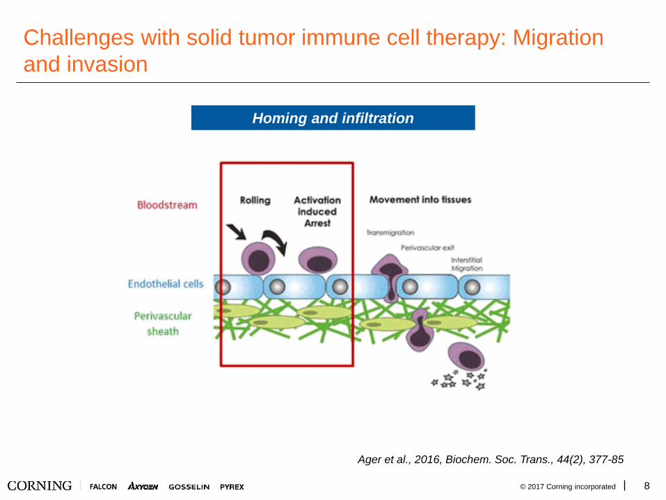
Challenges with solid tumor immune cell therapy: Migration
and invasion
Homing and infiltration
Ager et al., 2016, Biochem. Soc. Trans., 44(2), 377-85

9 © 2017 Corning incorporated
Combining technologies to enhance 3D microenvironments
A549 cells added to
spheroid microplate for
3D multicellular spheroid
formation
Transwell 96-well permeable
support is inserted into
spheroid microplate; NK cells
added to insert.
Plates incubated together
to allow for migration,
spheroid infiltration, and
cytotoxicity.

10 © 2017 Corning incorporated
Immune cell migration and tumor spheroid infiltration in a
single high-throughput screening amenable assay
• NK cell migration towards
A549 multicellular tumor
spheroids in the presence
and absence of SDF-1α
(SDF) and/or prostaglandin
E2 (PGE) in the medium
• NK cell induced cytotoxicity
of A549 multicellular tumor
spheroids after migration.
Percent cytotoxicity was
calculated via flow cytometry
by enumerating GFP positive
A549 cells.

11 © 2017 Corning incorporated
Acknowledgments
• Corning:
− Audrey Bergeron
− Hilary Sherman
• DiscoveRx:
− Abhishek Saharia
− Daniel Bassoni
− Gaurav Agrawal
• ProMab:
− Van Dang

Appendix
Supplementary figures and slides

13 © 2017 Corning incorporated
Clinical Trials of CAR-T Therapy
www.clincaltrials.gov, 3.14.2017
• 374 clinical trials all over the world
• CAR targets include cancer, diabetes, AIDS, vascular diseases, etc.

14 © 2017 Corning incorporated
CAR-T Cell Therapy
• T-cell therapy activates a patient's T cells against cancer with a chimeric antigen receptor (CAR) that
recognizes an antigen expressed on the cancer cells.
• CARs are single-chain antibodies coupled to a transmembrane region and an intracellular signaling
domain (e.g., from CD28 or 4-1BB).
• CARs against many tumor-associated antigens have been constructed and tested pre-clinically, and
some have entered clinical trials
https://www.mskcc.org/blog/car-t-cell-therapy-growing-area-
research
Front. Immunol., 14 November 2016 | https://doi.org/10.3389/fimmu.2016.00500

15 © 2017 Corning incorporated
CAR-T Cell Therapy
• CAR T cells face extra challenges with solid tumors:
− Have to be made specific for an antigen whose expression clearly identifies tumor
from normal tissue.
• “on-target, off-tumor” cytotoxicity
− Must be able to home and penetrate the fibrous connective tissue that surrounds the
tumor.
− Once within the tumor they must expand, persist and mediate cytotoxicity in a hostile
environment with immunosuppressive modulators.
http://www.nature.com/nbt/journal/v32/n7/full/nbt0714-604.html

16 © 2017 Corning incorporated
• ProMab Biotechnologies supplies second and third generation CAR-T cells
targeting a variety of cell-surface receptors
• This study used a second generation construct with affinity-tuned scFvs targeting
epidermal growth factor receptor (EGFR) and an empty vector Mock Control
Affinity-tuned CAR-T Cells from ProMab Biotechnologies
EGFR scFv CD28
TM CD28 CD3ζ
CD8
leader
CD8
hinge
Antigen
recognition
domain
Transmembrane
Domain
Costimulatory
Domain
T Cell
Activation
Domain
Front. Immunol., 14 November 2016
| https://doi.org/10.3389/fimmu.2016.00
500

17 © 2017 Corning incorporated
DiscoverX® KILR® Assay
• The KILR assay from DiscoverX is a highly specific, non-radioactive measure of target cell
death in a co-culture.
• KILR target cells are transduced to stably express a KILR reporter protein tagged with a β-
gal fragment. This KILR reporter protein is released into the media upon cell death and lysis.
Addition of detection reagents containing the other β-gal fragment results in a
chemiluminescent output.

18 © 2017 Corning incorporated
• This study used a second generation construct with affinity-tuned scFvs targeting epidermal
growth factor receptor (EGFR) and an empty vector Mock Control from ProMab Biotechnologies
Affinity-tuned CAR-T Cells & EGFR Targets
EGFR
scFv
CD28
TM CD28 CD3ζ
CD8
leader
CD8
hinge
Antigen
recognition
domain
Transmembrane
Domain
Costimulatory
Domain
• For this study, 2 cell lines from the ATCC® EGFR
Genetic Alteration Cell panel were selected:
− HCC827 contains high EGFR copy number
amplification
− NCI-H460 contains no EGFR copy number
amplification
T Cell
Activation
Domain
Data from ATCC® Brochure

19 © 2017 Corning incorporated
Corning® spheroid microplates enable improved spheroid
assays for screening
Standard ANSI/SBS
footprint dimensions for
96-well and 384-well
formats
Black sidewalls to reduce
cross-talk and background
noise in fluorescent- and
luminescent-based assays
Clear bottom for
visualization and imaging
• Corning Ultra-Low Attachment (ULA) surface and unique round well-bottom
design enable the formation and growth of a single, uniform spheroid per well
with reproducible size.
20 © 2017 Corning incorporated
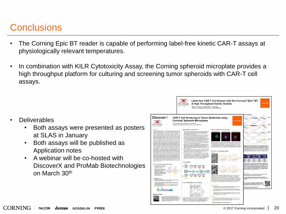
Conclusions
• The Corning Epic BT reader is capable of performing label-free kinetic CAR-T assays at
physiologically relevant temperatures.
• In combination with KILR Cytotoxicity Assay, the Corning spheroid microplate provides a
high throughput platform for culturing and screening tumor spheroids with CAR-T cell
assays.
• Deliverables
• Both assays were presented as posters
at SLAS in January
• Both assays will be published as
Application notes
• A webinar will be co-hosted with
DiscoverX and ProMab Biotechnologies
on March 30th

21 © 2017 Corning incorporated
Methods: Demonstrate Immune Cell Tumoricidal Activity
• Day 1: Seed 2,000 A549/GFP cells (cancer cells) per well of 96 well spheroid plate in
IMDM 10%FBS
• Day 2: Label effector cells with CellTracker Blue and add to A549 spheroids at various
concentrations
− NK92-MI: natural killer cell line derived from peripheral blood known to be cytotoxic
to a wide range of malignant cells
− MOLT-4: T-cell leukemia cell line with no known cytotoxic effect on other malignant
cells
• Day 3: Aspirate medium and replace with 150 µL TrypLE™ Select Enzyme (10X)
(Gibco™ Cat. No. A1217701) for 1 hour at 37°C or until spheroids could be broken up
into single cells with minimal pipetting. Single cells were then analyzed via flow
cytometry utilizing the Miltenyi Biotec MacsQuant®.

22 © 2017 Corning incorporated
Results: Demonstrate Immune Cell Tumoricidal Activity
Dose dependent effector function was demonstrated with NK cells and not MOLT-4 cells when
added at various concentrations to A549 spheroids.
Data represents the average of 2 independent studies. N=12 per concentration.

23 © 2017 Corning incorporated
Results: Image Immune Cell Infiltration (Confocal)
Representative photomicrographs of A549/GFP spheroids with (right) and without (left) NK
infiltration (200x). A549/GFP cells shown in green and NK-92MI cells shown in blue. Images
taken at a Z stack height of -125 µm via Thermo Scientific™ CellInsight™ CX7. Scale bar is
100 µm.

24 © 2017 Corning incorporated
Results: Image Immune Cell Infiltration (Histology)
200x CD45 (red) and e-cadherin stained (brown) sections of A549/GFP spheroids that were
infiltrated by NK-92MI cells. Spheroids were fixed in 4 % paraformaldehyde (Boston
Bioproducts Cat. No. BM-155) for cryostat sectioning and H&E staining (carried out at the
University of New England, Biddeford, Maine).
A549 only A549 infiltrated with NK
cells for 4 hours
A549 infiltrated with NK
cells for 18 hours

25 © 2017 Corning incorporated
Results: Demonstrate Immune Cell Chemotactic Response
Dose dependent migration of NK cells towards SDF over a period of 24 hours. Data represents
the average of 2 independent studies. N=24.

26 © 2017 Corning incorporated
Results: 2D versus 3D Immune Oncology Model
NK Migration towards 2D and 3D A549/GFP cells with or without SDF in the medium and with
and without prostaglandin E2 inhibition of NK cells. Horizontal lines indicate statistical
significance from a 1 way ANOVA with a Bonferroni's multiple comparison post test. ***
=p<0.0001 and ** =p<0.001. Data represents the average of 2 independent studies. N=24.

27 © 2017 Corning incorporated
Results: 2D versus 3D Immune Oncology Model
NK induced cytotoxicity of A549/GFP cells grown in 2D and 3D with or without SDF in the
medium and with and without prostaglandin E2 inhibition of NK cells. Horizontal lines indicate
statistical significance from a 1 way ANOVA with a Bonferroni's multiple comparison post test.
*** =p<0.0001, ** =p<0.001, and * =p<0.05. Data represents the average of 2 independent
studies. N=24.
28 © 2017 Corning incorporated
Summary
• Effector cell cytotoxicity and specificity can be assessed using a combination of
the spheroid microplate and flow cytometry.
• Transwell permeable supports can be utilized to assess NK migratory response
towards chemoattractants such as SDF.
• The combination of the spheroid microplate and HTS Transwell-96 Well
Permeable Supports allows for a novel 3D model that combines immune cell
migration, effector induced cytotoxicity, and immune cell evasion in one easy to
use model.
