advanced periodontal diagnostic aids
TRANSCRIPT
Advanced Periodontal Diagnostic Techniques(As an adjunct to conventional techniques)
Content of discussion
Introduction
Limitations of conventional periodontal diagnosis
Advances in Clinical diagnosis
Advances in Radiographic assessment
Advances in Microbiologic analysis
Advances in characterizing the Host response
Conclusion and future scopes
Introduction
Definition of DIAGNOSIS (Dx)* : Diagnosis is defined as; correct determination, discriminative estimation and logical appraisal of conditions found during examination as evidenced by distinctive marks, signs and characteristics of diseases
*Glossary of Periodontal terms
Recognizing a departure from health in the periodontium
and distinguishing one disease, disease categorization, or
etiology from another, Based on information obtained
from the medical and dental histories, clinical and
radiographic examination of the patient, and laboratory
findings.
Periodontal Dx*:
Purposes of Periodontal diagnostic procedures

According to Armitage GC(1996) Periodontal diagnostic procedures
can potentially serve 5 separate, but related purposes:
1. Screening
2. Diagnosis of specific periodontal disease
3. Identification of sites or subjects at an increased risk of experiencing the progression of periodontal destruction
4. Treatment planning and
5. Monitoring of therapy.
Current conventional techniques
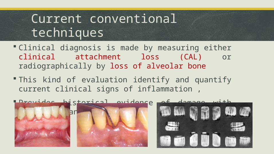
Clinical diagnosis is made by measuring either clinical attachment loss (CAL) or radiographically by loss of alveolar bone
This kind of evaluation identify and quantify current clinical signs of inflammation ,
Provides historical evidence of damage with its extent and severity
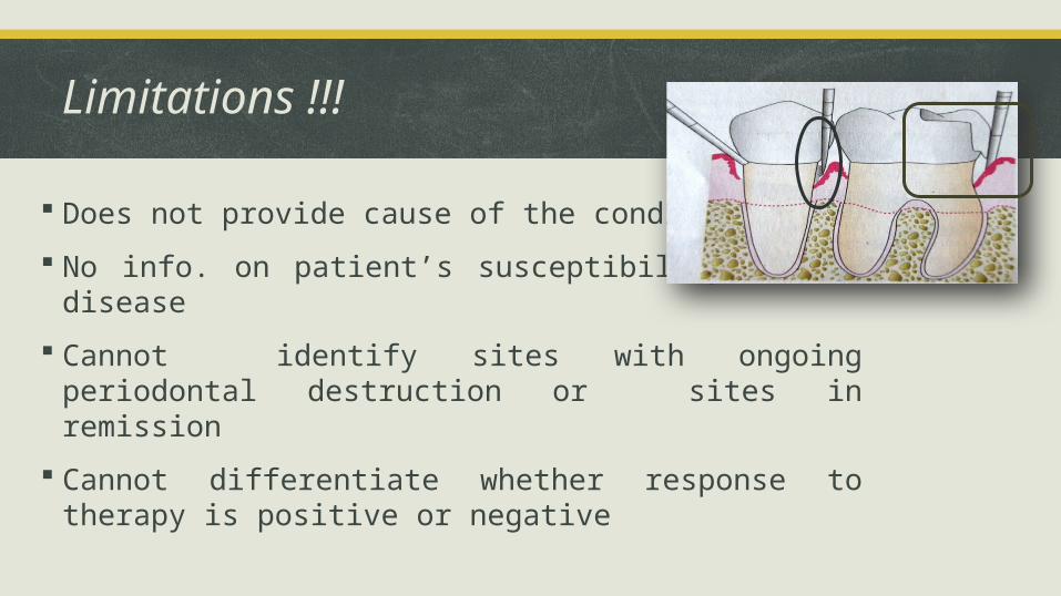
Limitations !!!
Does not provide cause of the condition
No info. on patient’s susceptibility to the disease
Cannot identify sites with ongoing periodontal destruction or sites in remission
Cannot differentiate whether response to therapy is positive or negative
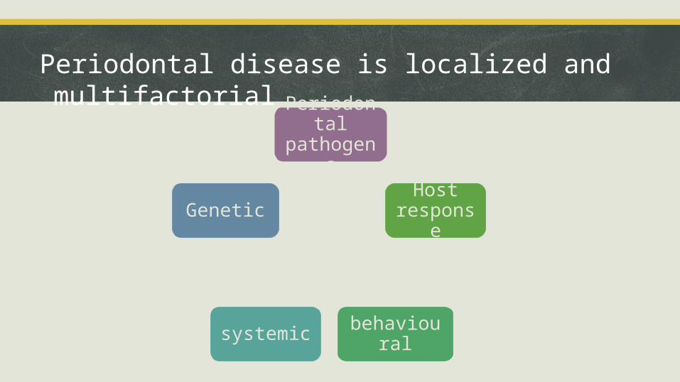
Periodontal disease is localized and multifactorial
Periodontal pathogens
Host response
behaviouralsystemic
Genetic

Advanced periodontal diagnostic techniques
Advances in Clinical diagnosis
Advances in Radiographic Assessment
Advances in Microbiologic Analysis
Advances in Characterizing the Host Response

Advances in Clinical Diagnosis
1. Gingival temperature
Kung et al (1990) claim that thermal probes are sensitive diagnostic
devices for measuring early inflammatory changes in gingival
tissue.
Subgingival temperature at diseased sites is increased as compared
to normal healthy sites
Commercially available system PerioTemp probe enables the
calculation of temperature differential (with sensitivity of 0.10C)
between the probed pocket and subgingival temperature
Possible explanation for ↑ temperature with increasing
probing depth is an increase in cellular and molecular activity
caused by increased periodontal inflammation
Haffajee et al. (1992): found that elevated subgingival site
temperature is related to attachment loss in shallow pockets
and elevated proportions of Pg, Pi, Tf, Aa.
2. Periodontal probing
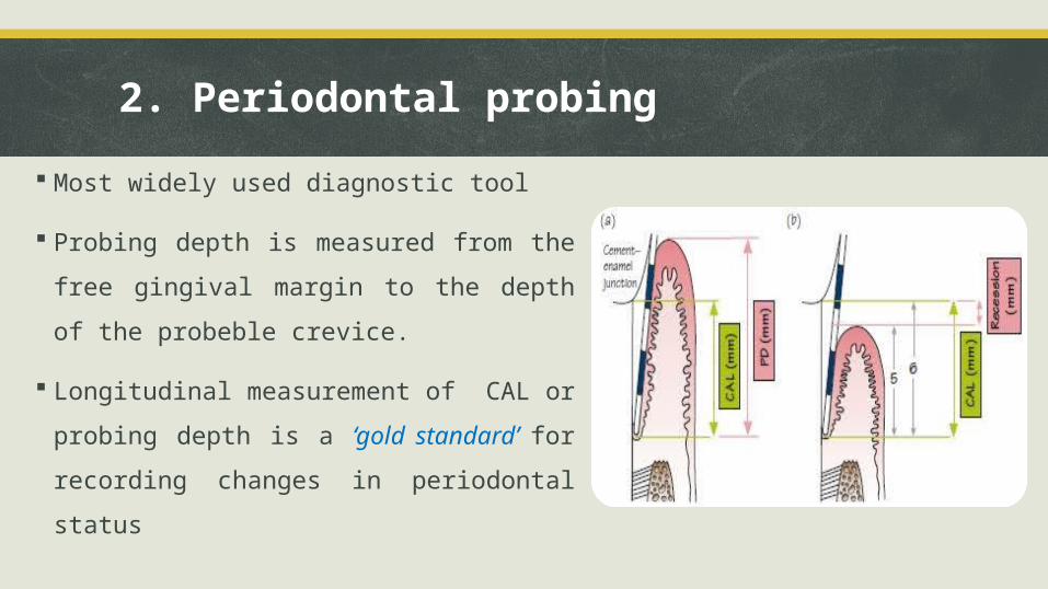
Most widely used diagnostic tool
Probing depth is measured from the free
gingival margin to the depth of the
probeble crevice.
Longitudinal measurement of CAL or
probing depth is a ‘gold standard’ for
recording changes in periodontal status
Limitation of conventional probing
Lack of sensitivity and reproducibility.
Disparity between measurement depends on:
probing technique, probing force, angle of insertion of probe, size
of probe, precision of calibration, presence of inflammation.
Readings of clinical pocket depth measured with probe does not
coinside with the histologic pocket depth.
All these variable contribute to the large standard deviations (0.5-
1.3 mm) in clinical probing results
Classification of periodontal probesdepending on generation
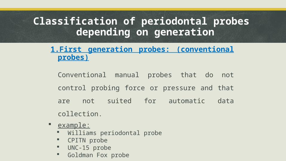
1.First generation probes: (conventional probes)
Conventional manual probes that do not control
probing force or pressure and that are not suited for
automatic data collection.
example: Williams periodontal probe CPITN probe UNC-15 probe Goldman Fox probe
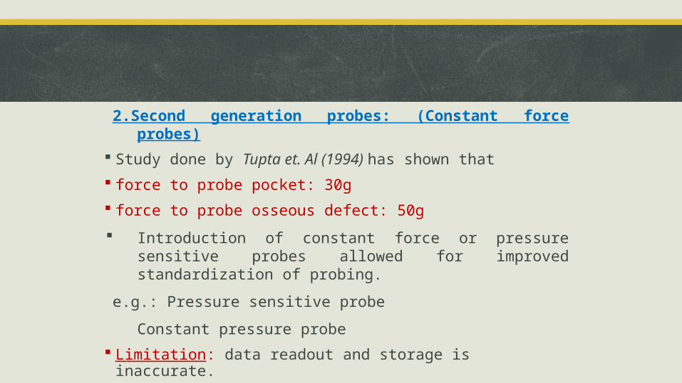
2.Second generation probes: (Constant force probes)
Study done by Tupta et. Al (1994) has shown that
force to probe pocket: 30g
force to probe osseous defect: 50g
Introduction of constant force or pressure sensitive probes allowed for improved standardization of probing.
e.g.: Pressure sensitive probe
Constant pressure probe
Limitation: data readout and storage is inaccurate.
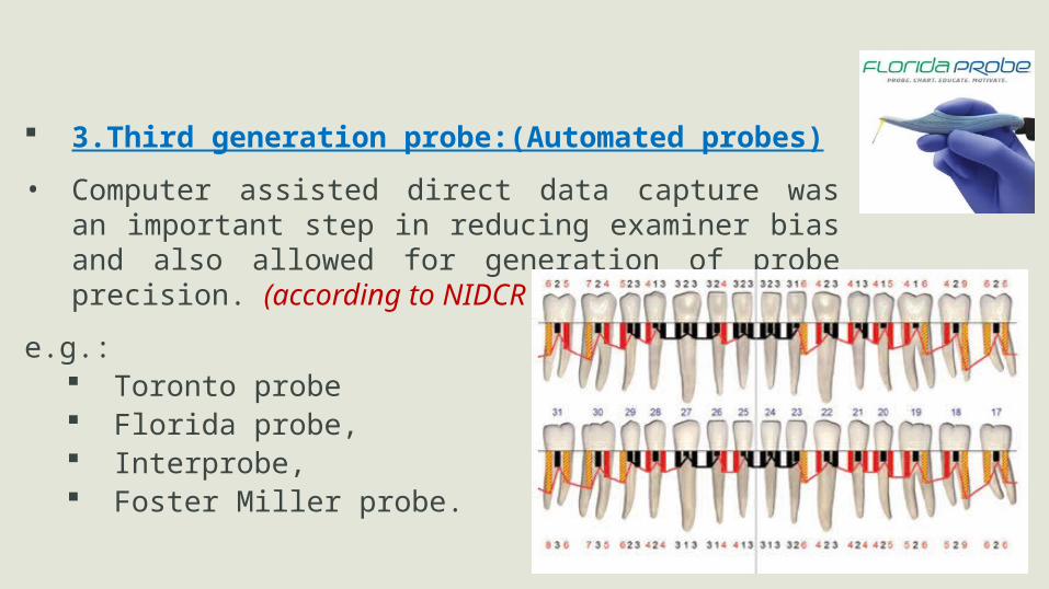
3.Third generation probe:(Automated probes)
• Computer assisted direct data capture was an important step in reducing examiner bias and also allowed for generation of probe precision. (according to NIDCR criteria)
e.g.: Toronto probe Florida probe, Interprobe, Foster Miller probe.
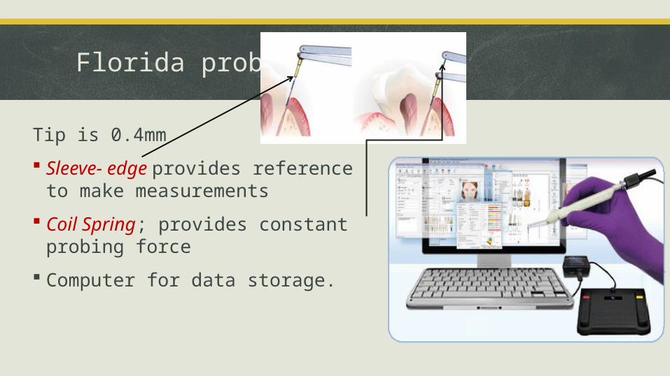
Florida probe
Tip is 0.4mm
Sleeve- edge provides reference to make measurements
Coil Spring; provides constant probing force
Computer for data storage.
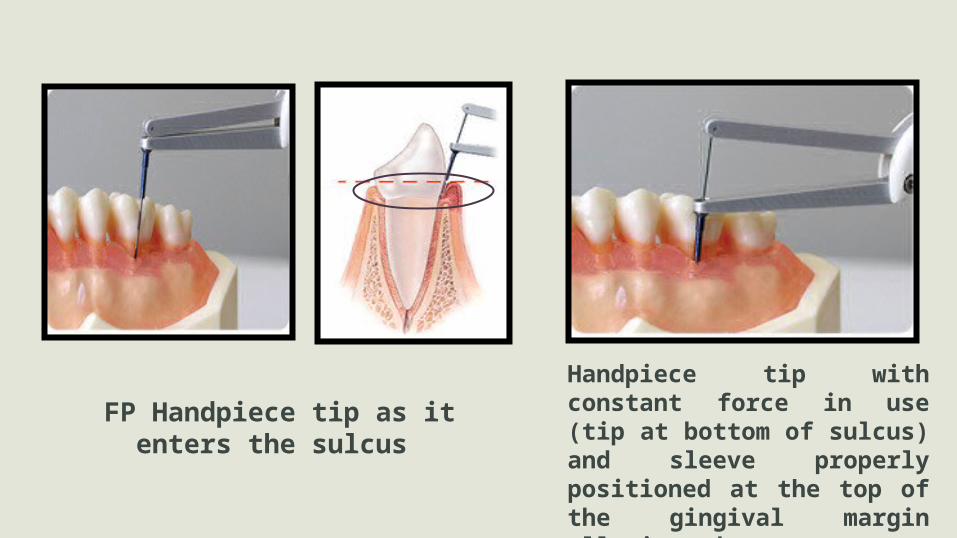
FP Handpiece tip as it enters the sulcus Handpiece tip with constant force in use (tip at bottom of sulcus) and sleeve properly positioned at the top of the gingival margin allowing the computer to measure the difference.
Clark and Yang (1992): trained operators and performing the ‘double pass’
method, the measurements taken with Florida probe system shows lower
standard deviation than those obtained with conventional probing.
Mean Standard Deviation for CAL of about 0.3mm, which is superior to an
average of 0.82mm reported by Haffajee et al. For conventional probing.
Limitations
Lack of tactile sensitivity
Fixed probing force
Underestimation of deep periodontal pockets.
4.Fourth generation probes: (Three dimensional probes)
• Currently under development, these are aimed at recording sequential probe positions along a gingival sulcus.
• An attempt to extend linear probing in a serial manner to take account of the continuous and three dimensional pocket that is being examined.
5.Fifth generation probe: (3D + Noninvasive)
• Basically these will add an ultrasound to a fourth generation probes.
• If the fourth generation can be made, it will aim in addition to identify the attachment level without penetrating it.
• e.g.: Ultra sonographic probe.
Advances inRadiographic Assessment

Dental Radiographs are traditional method to assess destruction of
alveolar bone.
“Conventional radiographs are very specific but lack sensitivity”
Primary criterion for bone loss is the distance from CEJ to the alveolar
crest and distance more than 2 mm is considered as the bone loss.
But variability affecting conventional radiographic technique are,
Variation in projection geometry
Variation in contrast and density
Masking by other anatomic structures.
1. Digital radiography
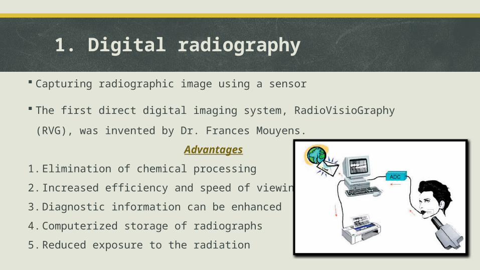
Capturing radiographic image using a sensor
The first direct digital imaging system, RadioVisioGraphy (RVG), was
invented by Dr. Frances Mouyens.
Advantages
1. Elimination of chemical processing
2. Increased efficiency and speed of viewing
3. Diagnostic information can be enhanced
4. Computerized storage of radiographs
5. Reduced exposure to the radiation
2. Subtraction radiography
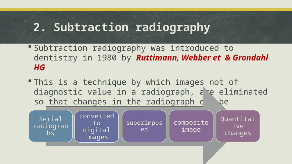
Subtraction radiography was introduced to dentistry in 1980 by Ruttimann, Webber et & Grondahl HG
This is a technique by which images not of diagnostic value in a radiograph, are eliminated so that changes in the radiograph can be precisely detected
Serial radiographs
converted to digital images
superimposed composite image
Quantitative changes
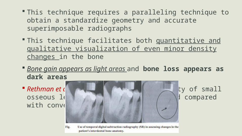
This technique requires a paralleling technique to obtain a standardize geometry and accurate superimposable radiographs
This technique facilitates both quantitative and qualitative visualization of even minor density changes in the bone
Bone gain appears as light areas and bone loss appears as dark areas
Rethman et al.(1985): increased detectability of small osseous lesions by substraction method compared with conventional radiography
Recent image subtraction:“diagnostic subtraction radiography” (DSR)
Modification
Use of a positioning device during film exposure
Image analysis software system applies an algorithm to correct angular
alignment discrepancies.
3. Computer Assisted Densitometric Image Analysis (CADIA)
Video camera measures the light transmitted through radiograph
and the signals form the camera is converted to gray scale image.
Advantage:
Measures quantitative changes in bone density longitudinally.
Higher sensitivity, reproducibility and accuracy as compared to
DSR.
4. Computed tomography (CT)
In 1972, Godfrey Hounsfield announced the invention of a
revolutionary imaging technique, which he referred to as
“computerized axial transverse scanning”
Fan shaped X-ray source is used
The computed tomographic image is reconstructed by computer, which
mathematically manipulates data obtained from multiple projections.
Computed tomography is a specialized radiographic technique that
allows visualization of planes or slices of interest
eliminates the super imposition of images of structures superficial or deep to the area of interest.
Because of inherent high contrast resolution, differences may be distinguished between tissues that differ in physical density by less than 1%.
multiple scans of a patient may be viewed as images in the axial, coronal, or sagittal planes depending on the diagnostic task, referred to as multiplanar imaging.
Advantages over conventional radiography
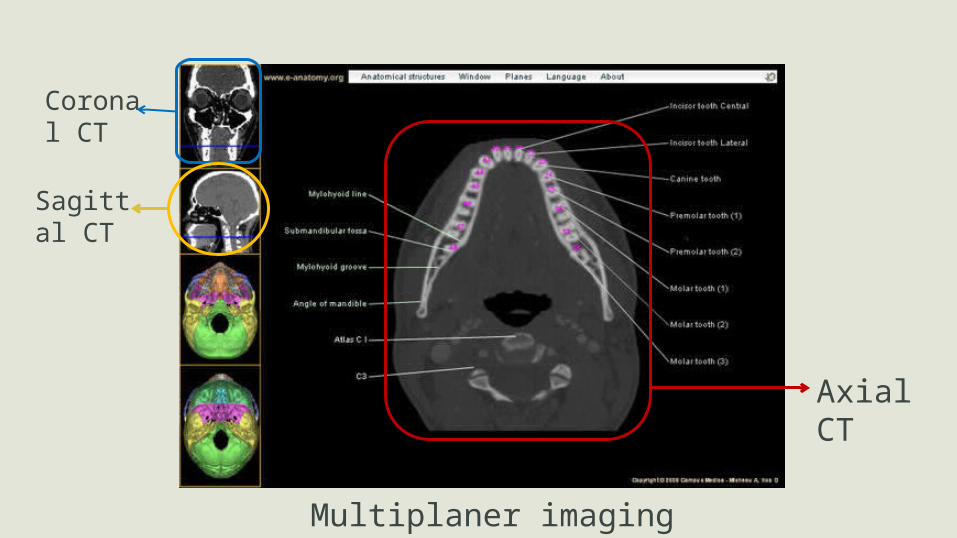
Axial CT
Coronal CT
Sagittal CT
Multiplaner imaging
Application of CT Used when accurate information regarding the topography of
osseous structure is needed
Soft tissue contour and dimension
To check continuity and density of the cortical plates
vertical height of the residual alveolar ridges
density of the medullary space and basilar bone
when determining how much space is available above the mandibular canal or amount of bone below maxillary sinus to receive a dental implant or whether there is a space occupying lesion in the maxillofacial region.
Disadvantages of Computed Tomography
specialized equipment and setting.
Radiologists and Technicians need to be knowledgeable of the
anatomy, anatomic variants and pathology of the jaws
higher radiation
Metallic Restorations can cause ring artifacts that impair the
diagnostic quality of the image
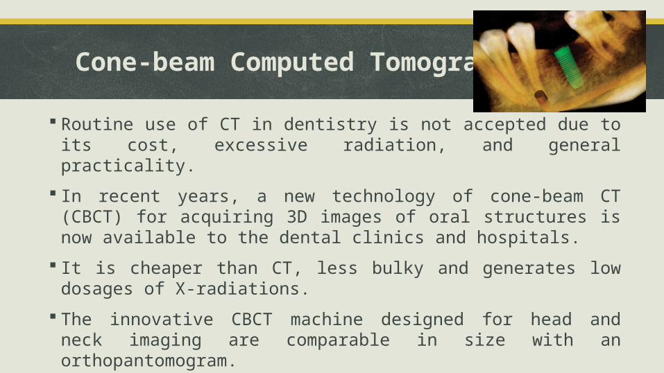
Cone-beam Computed Tomography
Routine use of CT in dentistry is not accepted due to its cost, excessive radiation, and general practicality.
In recent years, a new technology of cone-beam CT (CBCT) for acquiring 3D images of oral structures is now available to the dental clinics and hospitals.
It is cheaper than CT, less bulky and generates low dosages of X-radiations.
The innovative CBCT machine designed for head and neck imaging are comparable in size with an orthopantomogram.
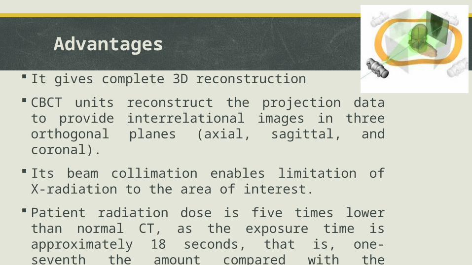
Advantages
It gives complete 3D reconstruction
CBCT units reconstruct the projection data to provide interrelational images in three orthogonal planes (axial, sagittal, and coronal).
Its beam collimation enables limitation of X-radiation to the area of interest.
Patient radiation dose is five times lower than normal CT, as the exposure time is approximately 18 seconds, that is, one-seventh the amount compared with the conventional medical CT.
Reduced image artefacts
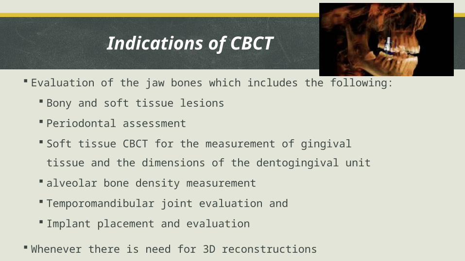
Evaluation of the jaw bones which includes the following:
Bony and soft tissue lesions
Periodontal assessment
Soft tissue CBCT for the measurement of gingival tissue and the dimensions
of the dentogingival unit
alveolar bone density measurement
Temporomandibular joint evaluation and
Implant placement and evaluation
Whenever there is need for 3D reconstructions
Indications of CBCT

Advances In Microbiologic Analysis
Uses of microbiologic analysis
1. support diagnosis of various Periodontal disease
2. Can tell about initiation & progression
3. To determine which periodontal sites are at high risk for active destruction
4. Can also be used to monitor Periodontal therapy
Advances In Microbiologic Analysis includes:
1. Immunohistodiagnostic methods
2. Enzymatic methods
3. Molecular biology techniques
Neucleic acid probes
Checkerboard DNA-DNA hybridization
PCR
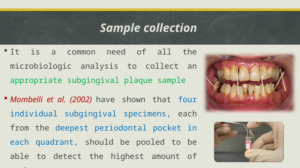
Sample collection
It is a common need of all the microbiologic analysis to
collect an appropriate subgingival plaque sample
Mombelli et al. (2002) have shown that four individual
subgingival specimens, each from the deepest periodontal
pocket in each quadrant, should be pooled to be able to
detect the highest amount of pathogens.
Transport the specimen in a anaerobic environment
“Immunodiagnostic methods”
Immunological assays use fluorescent conjugated antibodies that recognize specific bacterial antigens, and the identification of these specific antigen-antibody reactions allows the detection of target microorganisms.
This reaction can be visualized using a variety of techniques and reactions:
1. Direct (DFA) and indirect (IFA) immunofluorescent assays
2. Flow cytometry
3. Enzyme-linked immunosorbent assay (ELISA)
4. Latex agglutination
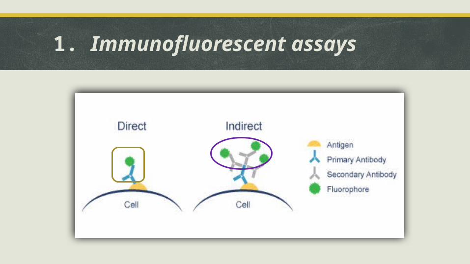
1. Immunofluorescent assays
IFA is used mainly to detect A.a and P.g
Zambon et. al (1986) showed that IFA is comparable to
bacterial culture in its ability to identify these pathogens
Zambon et. al (1995) sensitivity of these assays ranges from
82%-100% for A.a. and 91%-100% for P.g
Specificity values of 88%-92% and 87%-89% respectively
2. Flow cytometry
Rapid identification
Principle is labelling bacterial cells with both species-specific
antibody and a second fluorescein-conjugated antibody
This suspension is introduced into flowcytometer, which
separates bacterial cells into an almost single cell suspension
Limitation is sophistication and cost involved with this
procedure
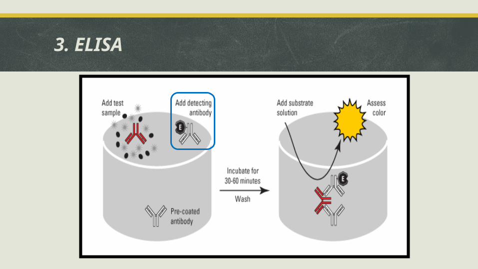
3. ELISA
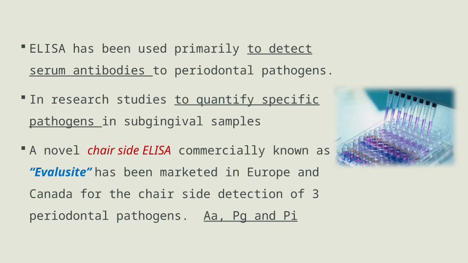
ELISA has been used primarily to detect serum antibodies
to periodontal pathogens.
In research studies to quantify specific pathogens in
subgingival samples
A novel chair side ELISA commercially known as
“Evalusite” has been marketed in Europe and Canada for
the chair side detection of 3 periodontal pathogens. Aa,
Pg and Pi
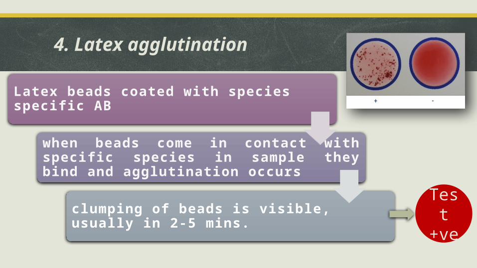
4. Latex agglutination
Latex beads coated with species specifi c AB
when beads come in contact with specifi c species in sample they bind and aggluti nati on occurs
clumping of beads is visible, usually in 2-5 mins.
Test +ve
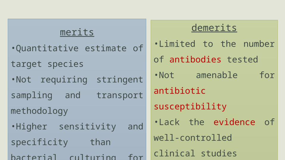
merits
•Quantitative estimate of target
species
•Not requiring stringent sampling and
transport methodology
•Higher sensitivity and specificity
than bacterial culturing for A.a, P.g
and T.f.
demerits
•Limited to the number of
antibodies tested
•Not amenable for antibiotic
susceptibility
•Lack the evidence of well-
controlled clinical studies
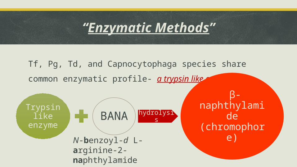
“Enzymatic Methods”
Tf, Pg, Td, and Capnocytophaga species share common enzymatic
profile- a trypsin like enzyme.
N-benzoyl-d L-arginine-2-naphthylamide
Trypsin like enzyme BANA hydrolysis
β-naphthylamide (chromophore)
PERIOSCAN uses this reaction for the identification of this bacterial
profile in plaque isolates
Loesh et al. (1986) detection of these pariodontal pathogens by BANA
reaction serves as a marker of disease activity
He also showed that shallow pockets exhibited 10% positive BANA
reaction, whereas deep pockets (7mm) exhibited 80%-90% +ve BANA
reaction
Beck et al. (1995) used BANA test as a risk indicator for periodontal
attachment loss
Disadvantage of BANA
May be positive in clinically healthy site
Can not detect sites undergoing periodontal destruction
Limited organisms detected
So that, negative results does not rule out the presence
of other important periodontal pathogens.
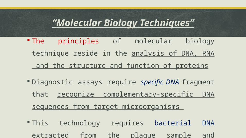
“Molecular Biology Techniques”
The principles of molecular biology technique reside in the
analysis of DNA, RNA and the structure and function of proteins
Diagnostic assays require specific DNA fragment that recognize
complementary-specific DNA sequences from target
microorganisms
This technology requires bacterial DNA extracted from the plaque
sample and amplification of the specific DNA sequence of the
target pathogen
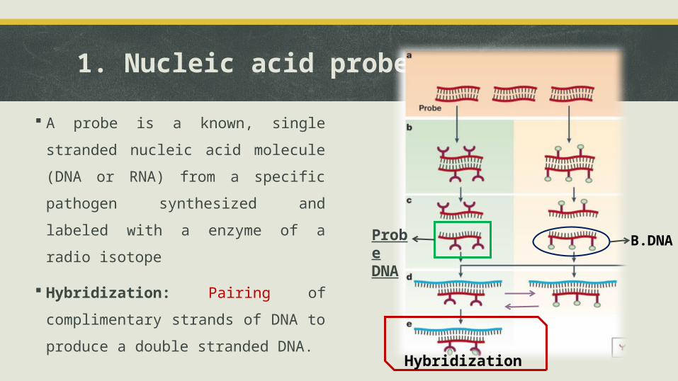
1. Nucleic acid probes
A probe is a known, single stranded
nucleic acid molecule (DNA or RNA)
from a specific pathogen
synthesized and labeled with a
enzyme of a radio isotope
Hybridization: Pairing of
complimentary strands of DNA to
produce a double stranded DNA.
Probe DNA
B.DNA
Hybridization
DMDx and Omnigene are commercially available genomic probes
for the detection of Aa, Pg, Pi and Td.
Van Steenberghe et al. (1999) reported a sensitivity of 96% and
specificity of 86% for Aa., and 60% and 82% respectively for Pg in
pure lab isolates.
In clinical specimens, both sensitivity and specificity were reduced
significantly, suggestive of cross reactivity with non target bacteria
in plaque sample because of the presence of homologues
sequences between different bacterial species
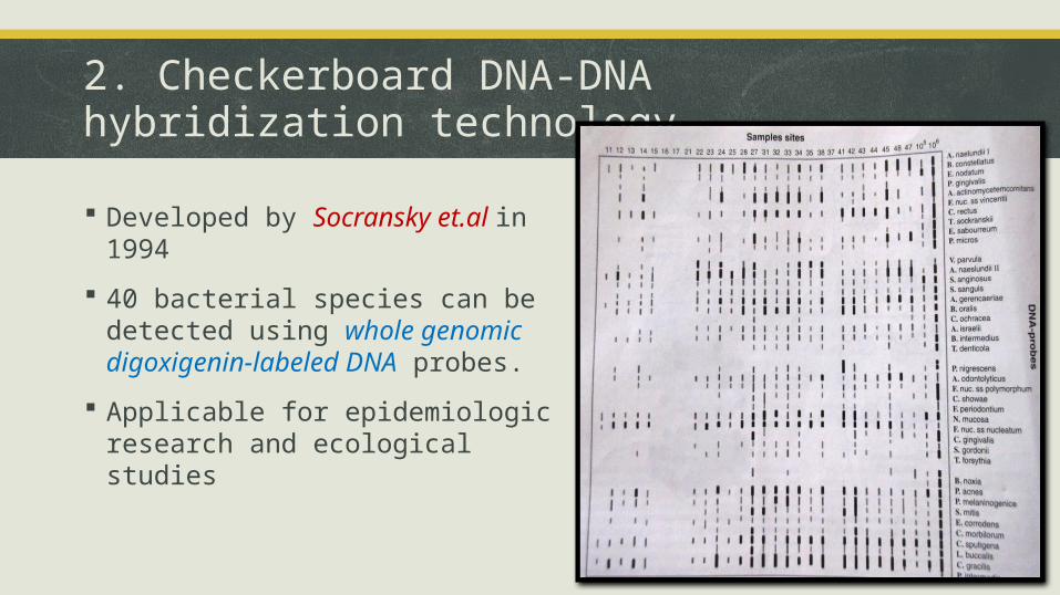
2. Checkerboard DNA-DNA hybridization technology
Developed by Socransky et.al in 1994
40 bacterial species can be detected using whole genomic digoxigenin-labeled DNA probes.
Applicable for epidemiologic research and ecological studies
3. Polymerase chain reaction (PCR)
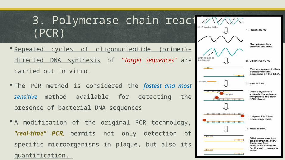
Repeated cycles of oligonucleotide (primer)–directed DNA
synthesis of “target sequences” are carried out in vitro.
The PCR method is considered the fastest and most
sensitive method available for detecting the presence of
bacterial DNA sequences
A modification of the original PCR technology, "real-time"
PCR, permits not only detection of specific
microorganisms in plaque, but also its quantification.
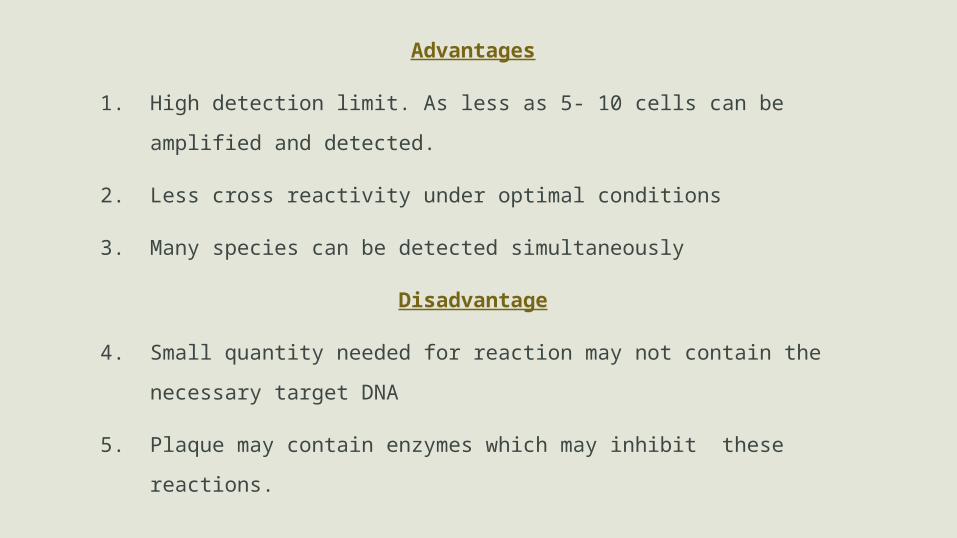
Advantages
1. High detection limit. As less as 5- 10 cells can be amplified and
detected.
2. Less cross reactivity under optimal conditions
3. Many species can be detected simultaneously
Disadvantage
4. Small quantity needed for reaction may not contain the necessary
target DNA
5. Plaque may contain enzymes which may inhibit these reactions.

Advances in characterizing
the Host response
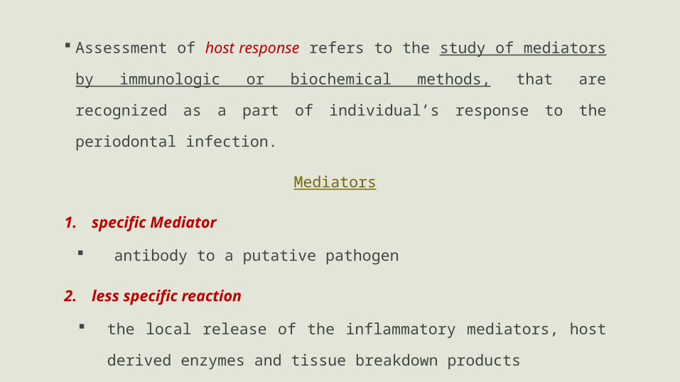
Assessment of host response refers to the study of mediators by
immunologic or biochemical methods, that are recognized as a part of
individual’s response to the periodontal infection.
Mediators
1. specific Mediator
antibody to a putative pathogen
2. less specific reaction
the local release of the inflammatory mediators, host derived
enzymes and tissue breakdown products
For that...
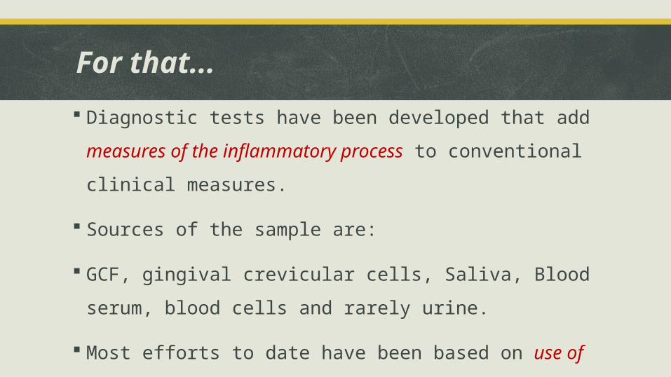
Diagnostic tests have been developed that add measures of the
inflammatory process to conventional clinical measures.
Sources of the sample are:
GCF, gingival crevicular cells, Saliva, Blood serum, blood cells and
rarely urine.
Most efforts to date have been based on use of components of GCF
and to a lesser extent, saliva and blood
Assessment of Host response
Inflammatory mediators and products
Host derived enzymes
Tissue breakdown products
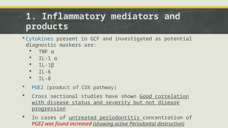
1. Inflammatory mediators and products
Cytokines present in GCF and investigated as potential diagnostic markers are: TNF α IL-1 α IL-1β IL-6 IL-8
PGE2 (product of COX pathway)
Cross sectional studies have shown Good correlation with disease status and severity but not disease progression
In cases of untreated periodontitis concentration of PGE2 was found increased (showing active Periodontal destruction)
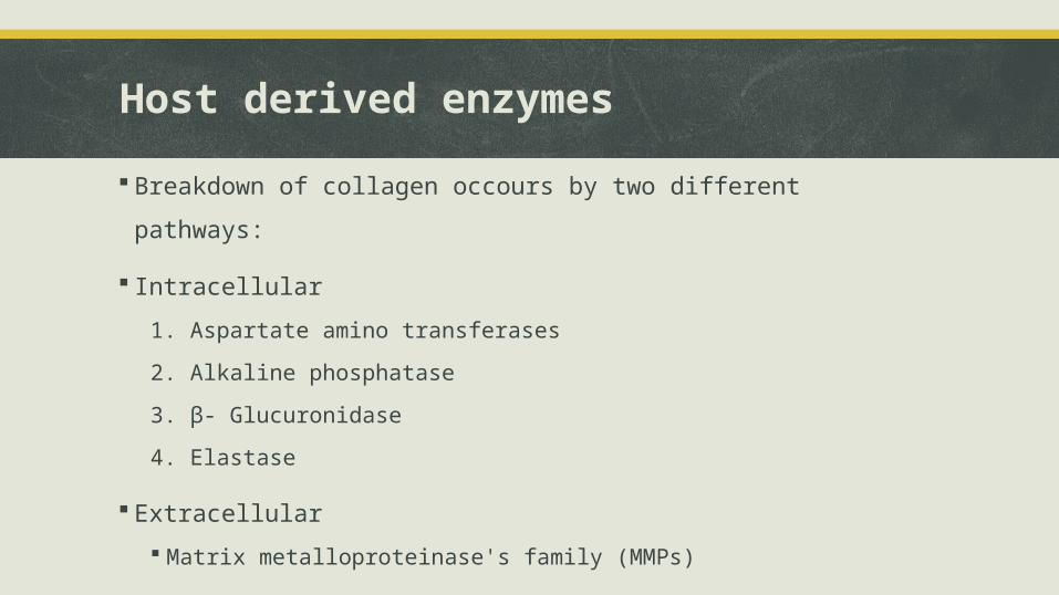
Host derived enzymes
Breakdown of collagen occours by two different pathways:
Intracellular
1. Aspartate amino transferases
2. Alkaline phosphatase
3. β- Glucuronidase
4. Elastase
Extracellular
Matrix metalloproteinase's family (MMPs)
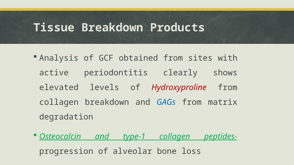
Tissue Breakdown Products
Analysis of GCF obtained from sites with active
periodontitis clearly shows elevated levels of
Hydroxyproline from collagen breakdown and GAGs from
matrix degradation
Osteocalcin and type-1 collagen peptides- progression of
alveolar bone loss

Conclusion
1. This discussion directly translates into improved periodontal
therapy by offering the clinician, the radiographic & laboratory
measure of periodontal infection as an adjunct to traditional
clinical indices of periodontal disease.
2. Future application of advanced diagnostic techniques will be of
value in documenting disease activity and treatment options
3. But, despite excellent progress in diagnostic
methodology,conventional efforts evaluating inflammation and
past evidence of tissue breakdown remain the standard for
disease evaluation
4. There is still a lack of a proven “gold standard” of disease
progression
5. After all these years of intensive research, we still lack a proven
diagnostic test that has demonstrated high predictive value for
disease progression, has a proven impact on disease incidence and
prevalence, and is safe, and cost-effective.
A tremendous amount of research is still required to
explore the role of advancements in diagnostic aids as
a possible medium for the future prediction and
prevention of periodontal disease.
1. Text book of Carranza's clinical periodontology, 2007, W.B. Saunders Co.
2. Armitage GC, Svanberg GK, Lde H: Microscopic evaluation of clinical measurements of connective tissue attachment levels. J Clin Periodontol 1977; 4:173.
3. Socransky SS, Haffajee AD, Smith C, Martin L, Haffajee JA, Uzel NG, Goodson JM. Use of checkerboard DNA-DNA hybridization to study complex microbial ecosystems. Oral Microbiol Immunol 2004;19:352-362
4. Clark WB, Yang MCK, Magnusson 1: Measuring clinical attachment: Reproducibility and relative measurements with an electronic probe. J Periodontol 1992; 63:831.
5. Goodson JM, Haffajee AD, Socransky SS: The relationship between attachment level loss and alveolar bone loss. J Clin Periodontol 1984; 11:348.
6. Grondahl HG, Grondahl K: Subtraction radiography for the diagnosis of periodontal bone lesions. Oral Surg 1983; 55:208.
References:
7. Haffajee AD, Socransky SS: Attachment level changes in destructive periodontal diseases. J Clin Periodontol 1986; 13:461.
8. Kung RT, Ochs B, Goodson JM: Temperature as a periodontal diagnostic. J Clin Periodontol 1990; 17:557.
9. Loesche WJ: The identification of bacteria associated with periodontal disease and dental caries by enzymatic methods. Oral Microbiol Immunol 1986; 1:65.
10. Mombelli A, Graf H: Depth force patterns in periodontal probing. J Clin Periodontol 1986; 13:126.
11. Page RC: Host response tests for diagnosing periodontal diseases. J Periodontol 1992; 63:356.
12. Papanou PN, Neiderud AM, Papadimitriou A, et al: Checkerboard assessments of Periodontal Microbiota and serum antibody responses: A case control study. J Periodontol 2000; 71:885.
13. Zambon JJ, Bochacki V, Genco RJ: Immunological assays for putative periodontal pathogens. Oral Microbiol Immunol 1986; 1:39.
14. Tupta-Veselicky L, Famili P, Ceravolo FJ et al: A clinical study of an electronic constant force periodontal probe. J Periodontol 1994; 65:616.

Thank you