advances in analysis of milk proteases activity at
TRANSCRIPT

sensors
Review
Advances in Analysis of Milk Proteases Activity atSurfaces and in a Volume by Acoustic Methods
Mark Dizon 1 , Marek Tatarko 2 and Tibor Hianik 2,*1 School of Chemistry, University College Dublin, Belfield, Dublin 4, Ireland; [email protected] Department of Nuclear Physics and Biophysics, Faculty of Mathematics, Physics and Informatics,
Comenius University, Mlynska dolina F1, 842 48 Bratislava, Slovakia; [email protected]* Correspondence: [email protected]
Received: 25 August 2020; Accepted: 19 September 2020; Published: 29 September 2020�����������������
Abstract: This review is focused on the application of surface and volume-sensitive acoustic methodsfor the detection of milk proteases such as trypsin and plasmin. While trypsin is an important proteinof human milk, plasmin is a protease that plays an important role in the quality of bovine, sheep andgoat milks. The increased activity of plasmin can cause an extensive cleavage of β-casein and, thus,affect the milk gelation and taste. The basic principles of surface-sensitive acoustic methods, as well ashigh-resolution ultrasonic spectroscopy (HR-US), are presented. The current state-of-the-art examplesof the application of acoustic sensors for protease detection in real time are discussed. The applicationof the HR-US method for studying the kinetics of the enzyme reaction is demonstrated. The sensitivityof the acoustics biosensors and HR-US methods for protease detection are compared.
Keywords: plasmin; trypsin; protease; casein; cleavage; acoustic sensor; thickness shear mode; quartzcrystal microbalance; high-resolution ultrasonic spectroscopy
1. Introduction
Milk proteases are of high importance in the digestion of milk proteins, mostly caseins.Among them, the plasmin has a crucial role, because its activity has a substantial effect on thequality of milk and milk products. The primary role of the plasmin consists in the cleavage of fibrin inblood and, thus, prevents thrombosis. However, it is also infiltrated in milk when it is responsible forcleavage, mostly of β-casein [1]. Plasmin in blood and milk originated from its zymogen plasminogen.The cleavage of plasminogen by urokinase activators results in plasmin [2]. The activity of plasminis regulated by a complex plasmin system that contains activators and inhibitors [3]. Due to thehigh temperature stability of plasmin, even after ultra-high temperature treatment (UHT) of the milk,this protease can be active. The uncontrolled activity of plasmin causes the extensive cleavage of caseinon small peptide fragments that are not desirable for milk storage because of inducing its gelation andbitter taste. It is therefore important to develop methods that can control the plasmin concentrationand their activity. The concentration of plasmin in raw milk is in the range of 4–12 nM [2]; therefore,the methods of plasmin determination should have sensitivity in order of 1 nM. It is also important foranalysis of the kinetics of casein cleavage by proteases and their time stability.
So far, plasmin and the other protease, trypsin, present in human milk were detected byconventional methods, such as the Enzyme-linked Immunosorbent Assay (ELISA), which is currentlythe only one commercially available test kit for plasmin detection [4]. In this assay, the specificantibodies to plasmin are used. However, this method does not allow monitoring the kinetics of thecleavage of the caseins. Other possible assays are based on optical methods, but they are limitedto only optically transparent liquids [5]. Other available methods include matrix-assisted laserdesorption/ionization time of flight mass spectrometry (MALDI-TOF-MS) [6], high-performance liquid
Sensors 2020, 20, 5594; doi:10.3390/s20195594 www.mdpi.com/journal/sensors

Sensors 2020, 20, 5594 2 of 32
chromatography (HPLC) [7] or capillary electrophoresis [8]. However, these methods determine onlythe peptide fragments and not the plasmin activity. Therefore, kinetic studies of proteases activity areimpossible by these methods.
It is evident that there is high demand on the development of effective methods for the detectionof proteases activity. Among the effective approaches are the acoustic methods. The advantage ishigh sensitivity and the capability to operate in nontransparent liquids. The acoustic methods canbe divided into two main groups—surface and volume-sensitive. In this review, we will present theadvantage of the thickness shear mode (TSM) acoustic method and its variations for studying theactivity of plasmin and trypsin at surfaces of piezoelectric crystals modified by short peptides or caseins.We already demonstrated the high sensitivity of acoustic sensors for the detection of the cleavage ofβ-caseins by trypsin and plasmin. The sensitivity of the TSM method is compared with those based onhigh-resolution ultrasonic spectroscopy (HR-US) that are convenient for studying the kinetics of thecleavage of caseins and other milk proteins by proteases in a small volume of approx. 1 mL.
The study of proteases activity is important also for obtaining information about the mechanismsof the enzymatic processes. Detection of the concentration of plasmin and its activity in milk isimportant for the dairy industry and, especially, for cheese makers. Among various methods ofdetection proteases, those based on acoustics principles are of a high advantage, because they do notneed any labeling of the substrate. In this review, we will focus on the explanation of the principles ofsurface-based acoustics methods and their use for the detection of plasmin and trypsin. We will alsoexplain the principles of detection of proteases activity in a volume using high-resolution ultrasonicspectroscopy (HR-US).
2. Milk Proteases
Bovine milk in its natural state is a white substance containing various components, mainlywater (86%), lactose (4.7%), lipids (4%), proteins (3.2%), citric acid and minerals [9]. Its composition isdependent on the species and breed of individuals. The absence of certain protease systems causeschanges in the amount of milk components and even can result in their absence. This can lead to theunique properties of milk in different mammal species [10]. Even some pathological states can supportchanges in milk due to effects on milk-producing cells. These circumstances are usually connectedwith inflammation processes [11].
Milk proteins are the most interesting and the most studied components of milk. They takepart in the various processes in milk, as their functionality varies. These proteins are surface-active,which allows them to form self-assembled structures at various interfaces and micelles in a volume.Milk proteins are divided into several groups. Mass spectroscopy analysis proved that caseins arethe most common proteins in milk. Caseins represent 78.3% of the total milk protein mass [9].Serum proteins, often called whey proteins, take approximately 19%. The rest of the milk mass ispresented by other protein groups, namely by membrane proteins and enzymes. Caseins are dividedinto several types, such as αs1, αs2, β and κ. Each of them has a different structure, functionality andlocation on the pseudo-micelles. Similar as with the protein group composition, the amount of eachcasein type differs in each species and breed of mammal. While in bovine milk, (Bos Taurus) β-caseinis only the second-most common casein, in sheep milk (Ovis aries), the amount of β-casein exceedsother casein types. This contributes to the bitterness of sheep cheese, which was already affected byproteolysis [12].
Proteolytic systems are inseparable part of all kinds of milk. They regulate the protein contentin milk, and they are also regulated. Like other proteins, they differ in all mammal species. It is notunique for certain protease systems to be absent in one species and to be the most prominent foranother. Proteases of these systems are excreted as inactive zymogens. They are later activated by thecleavage of specific peptide bonds. There are several ways how milk can obtain proteases (Figure 1).Proteases can either get into the milk via the blood stream or are released by the mammary epithelialcells. Lastly, milk contains immune cells that can also release proteases into the milk environment [13].
Sensors 2020, 20, 5594 3 of 32
Sensors 2020, 20, x FOR PEER REVIEW 3 of 32
cells. Lastly, milk contains immune cells that can also release proteases into the milk environment [13].
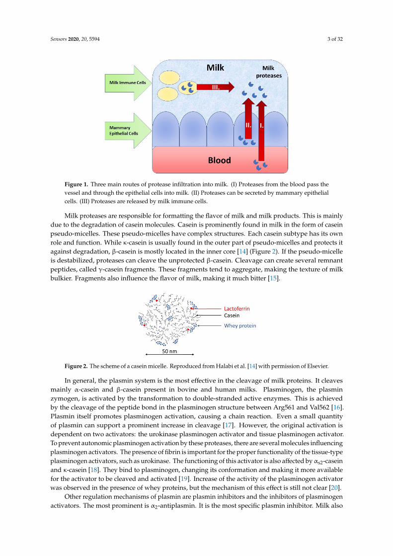
Figure 1. Three main routes of protease infiltration into milk. (I) Proteases from the blood pass the vessel and through the epithelial cells into milk. (II) Proteases can be secreted by mammary epithelial cells. (III) Proteases are released by milk immune cells.
Milk proteases are responsible for formatting the flavor of milk and milk products. This is mainly due to the degradation of casein molecules. Casein is prominently found in milk in the form of casein pseudo-micelles. These pseudo-micelles have complex structures. Each casein subtype has its own role and function. While κ-casein is usually found in the outer part of pseudo-micelles and protects it against degradation, β-casein is mostly located in the inner core [14] (Figure 2). If the pseudo-micelle is destabilized, proteases can cleave the unprotected β-casein. Cleavage can create several remnant peptides, called γ-casein fragments. These fragments tend to aggregate, making the texture of milk bulkier. Fragments also influence the flavor of milk, making it much bitter [15].
Figure 2. The scheme of a casein micelle. Reproduced from Halabi et al. [14] with permission of Elsevier.
In general, the plasmin system is the most effective in the cleavage of milk proteins. It cleaves mainly α-casein and β-casein present in bovine and human milks. Plasminogen, the plasmin zymogen, is activated by the transformation to double-stranded active enzymes. This is achieved by the cleavage of the peptide bond in the plasminogen structure between Arg561 and Val562 [16]. Plasmin itself promotes plasminogen activation, causing a chain reaction. Even a small quantity of plasmin can support a prominent increase in cleavage [17]. However, the original activation is dependent on two activators: the urokinase plasminogen activator and tissue plasminogen activator. To prevent autonomic plasminogen activation by these proteases, there are several molecules influencing plasminogen activators. The presence of fibrin is important for the proper functionality
Figure 1. Three main routes of protease infiltration into milk. (I) Proteases from the blood pass thevessel and through the epithelial cells into milk. (II) Proteases can be secreted by mammary epithelialcells. (III) Proteases are released by milk immune cells.
Milk proteases are responsible for formatting the flavor of milk and milk products. This is mainlydue to the degradation of casein molecules. Casein is prominently found in milk in the form of caseinpseudo-micelles. These pseudo-micelles have complex structures. Each casein subtype has its ownrole and function. While κ-casein is usually found in the outer part of pseudo-micelles and protects itagainst degradation, β-casein is mostly located in the inner core [14] (Figure 2). If the pseudo-micelleis destabilized, proteases can cleave the unprotected β-casein. Cleavage can create several remnantpeptides, called γ-casein fragments. These fragments tend to aggregate, making the texture of milkbulkier. Fragments also influence the flavor of milk, making it much bitter [15].
Sensors 2020, 20, x FOR PEER REVIEW 3 of 32
cells. Lastly, milk contains immune cells that can also release proteases into the milk environment [13].
Figure 1. Three main routes of protease infiltration into milk. (I) Proteases from the blood pass the vessel and through the epithelial cells into milk. (II) Proteases can be secreted by mammary epithelial cells. (III) Proteases are released by milk immune cells.
Milk proteases are responsible for formatting the flavor of milk and milk products. This is mainly due to the degradation of casein molecules. Casein is prominently found in milk in the form of casein pseudo-micelles. These pseudo-micelles have complex structures. Each casein subtype has its own role and function. While κ-casein is usually found in the outer part of pseudo-micelles and protects it against degradation, β-casein is mostly located in the inner core [14] (Figure 2). If the pseudo-micelle is destabilized, proteases can cleave the unprotected β-casein. Cleavage can create several remnant peptides, called γ-casein fragments. These fragments tend to aggregate, making the texture of milk bulkier. Fragments also influence the flavor of milk, making it much bitter [15].
Figure 2. The scheme of a casein micelle. Reproduced from Halabi et al. [14] with permission of Elsevier.
In general, the plasmin system is the most effective in the cleavage of milk proteins. It cleaves mainly α-casein and β-casein present in bovine and human milks. Plasminogen, the plasmin zymogen, is activated by the transformation to double-stranded active enzymes. This is achieved by the cleavage of the peptide bond in the plasminogen structure between Arg561 and Val562 [16]. Plasmin itself promotes plasminogen activation, causing a chain reaction. Even a small quantity of plasmin can support a prominent increase in cleavage [17]. However, the original activation is dependent on two activators: the urokinase plasminogen activator and tissue plasminogen activator. To prevent autonomic plasminogen activation by these proteases, there are several molecules influencing plasminogen activators. The presence of fibrin is important for the proper functionality
Figure 2. The scheme of a casein micelle. Reproduced from Halabi et al. [14] with permission of Elsevier.
In general, the plasmin system is the most effective in the cleavage of milk proteins. It cleavesmainly α-casein and β-casein present in bovine and human milks. Plasminogen, the plasminzymogen, is activated by the transformation to double-stranded active enzymes. This is achievedby the cleavage of the peptide bond in the plasminogen structure between Arg561 and Val562 [16].Plasmin itself promotes plasminogen activation, causing a chain reaction. Even a small quantityof plasmin can support a prominent increase in cleavage [17]. However, the original activation isdependent on two activators: the urokinase plasminogen activator and tissue plasminogen activator.To prevent autonomic plasminogen activation by these proteases, there are several molecules influencingplasminogen activators. The presence of fibrin is important for the proper functionality of the tissue-typeplasminogen activators, such as urokinase. The functioning of this activator is also affected byαs2-caseinand κ-casein [18]. They bind to plasminogen, changing its conformation and making it more availablefor the activator to be cleaved and activated [19]. Increase of the activity of the plasminogen activatorwas observed in the presence of whey proteins, but the mechanism of this effect is still not clear [20].
Other regulation mechanisms of plasmin are plasmin inhibitors and the inhibitors of plasminogenactivators. The most prominent is α2-antiplasmin. It is the most specific plasmin inhibitor. Milk also

Sensors 2020, 20, 5594 4 of 32
includes several nonspecific inhibitors, such as α2-macroglobulin [21]. α2-antiplasmin can createcomplexes with the plasmin, which even after separation, lead to a decrease of protease activity andinhibitor binding [17]. Plasminogen activator inhibitors belong to another specific regulatory system.Only the plasminogen activator inhibitors I and II were confirmed in milk [21]. UHT processing ofmilk must be therefore performed correctly (see [22] for the methods of UHT processing of milk).The inadequate heating of milk during UHT treatment, especially omitting the process of heatingat 140 ◦C for at least 15 s, causes damage to the thermally unstable inhibitors of the plasmin systemin milk [23,24]. Plasminogen activators in bovine milk are thermally more stable in the range oftemperatures from 60 ◦C to 140 ◦C [25]. The increased activity of plasmin activators was observed afterthe thermal treatment of milk with a high content of somatic cells, but this effect was not completelymonitored in the dairy industrial conditions [26]. The nonspecific inhibition of plasmin includes anothergroup of milk proteins, whey proteins, namely α-lactalbumin, β-lactoglobulin and bovine serumalbumin (BSA). The inhibition process of these proteins was not yet completely documented, as theirinhibition effect is strongly dependent on the specific substrate that is the target of plasmin-inducedproteolysis [27].
Plasmin is not an exclusive protease in milk. Another protease active in human milk is trypsin [28].It is, however, not active in bovine milk. Trypsin concentrations in human milk are in the range of2.9–5.6 µg/L, and its active form is present as an anion (trypsin-2). The activation of a trypsin anion isnot completely clear, because for its zymogen, trypsinogen, the ways of its activation in milk are notknown. Both trypsin activators, thrombin and enterokinase, are either absent or are found only inthe form of zymogens. Therefore, trypsinogen is probably activated when the ingested milk reachesthe duodenum. In the duodenum, it is cleaved and activated by a corresponding protein kinase.Trypsin activity is further inhibited by several inhibitors, namely α1-antitrypsin. The presence ofα1-antitrypsin is lowered at the beginning and during lactation. It is active only in the small intestinesof newborns. However, trypsin is there separated from α1-antitrypsin and other inhibitors [29].
The cathepsin protease system contains several cathepsins, and each of them differs in theirfunction and presence of certain mammal species [30]. One of the cathepsin present in bovine andhuman milk is cathepsin D. Even when discovered in the active from, most of cathepsin D is in theform of an inactive zymogen. Cathepsin B, similarly to other proteases, cleaves mainly αs1-casein andβ-casein. It, however, cleaves them at different peptide bonds. It is inactivated in basic pH, and itsgeneral purpose and activity in milk is unclear.
The kallikrein system has a prominent role for coagulation and fibrinolysis. Its active form isabsent in milk, but some kallikrein molecules were detected in milk by proteomics [31]. The Westernblot method was used to positively detect the plasma protease C1 inhibitor, which is important forblood clot formation.
In addition to the above-mentioned proteases, milk contains several other protease systems [13].The elastase protease system was discovered in the neutrocytes of bovine milk. However,elastase activators are missing in milk, but several inhibitors were found. The chymotrypsinsystem depends on the chymotrypsin activator—trypsin. It is, however, absent in bovine milk.Colostrum contains several peptide sequences with a thrombin structure, but it lacks activatorsto release them. The aminopeptidase and carboxypeptidase protease systems were discovered byproteomic methods in mammary glands, but their important activators, including thrombin, are missingin this environment. The last identified protease system was matrix metalloproteinase. It degradesthe extracellular matrix, allowing reconstruction of the tissue. Collagen and gelatin-degradingmetalloproteinases MMP-2 and MMP-9 were localized in human milk, and the inhibitors for MMP-4and MMP-1 were also discovered.
3. Surface Acoustic Methods
Piezoelectricity-based sensors are among most used sensing devices on the microscale andmacroscale levels, as is evident from an increasing number of scientific papers that further broaden their

Sensors 2020, 20, 5594 5 of 32
applications [32,33]. Piezoelectric devices are generally divided into two groups: surface acoustic waves(SAW) and bulk acoustic waves (BAW). SAW contain electrodes at one side of the crystal. Generatedwave deformation is defined by the crystal’s surface. While more sensitive, signals generated by devicessuffer significant attenuation in biological solutions. More suitable biosensors for measurements inliquids are BAW-based sensors, namely quartz crystal microbalance (QCM) and TSM [34].
3.1. The Principles of QCM and TSM
The first biosensor was reported in 1962, when Leland C. Clark and Champ Lyons suggestedthe concept of the oxygen electrode with the immobilized glucose oxidase at the electrode surfacefor the detection of glucose [35]. The IUPAC (International Union for Pure and Applied Chemistry)defines biosensors as an integrated receptor-transducer device that can provide selective quantitativeor semi-quantitative analytical information using a biological recognition element [36].
The first biosensors used were mostly electrochemical transducers. Other transducers, such asoptical, mass or thermal reaction-based, are also currently and widely used. Mass-sensitive acoustic(piezoelectric) biosensors are based on the measurements of the properties of the acoustic wavesthat travels through the volumes and surfaces of sensors. The changes in the trajectory that wavestravel causes changes in its velocity, amplitude and phase. Using these changes in gas or liquidenvironments, it is possible to measure changes of masses on surfaces caused by adsorbing or desorbingmolecules. It is also affected by viscoelastic properties of thin films and liquid on the transducersurface. Acoustic sensors are rather popular due to their high sensitivity to detect small changes ofmasses at the surface. The resonant frequency of the crystal is dependent on its thickness, density andmechanical properties [37].
In 1880, Jacques and Pierre Curie demonstrated piezoelectricity. They showed that mechanicalstress applied to certain materials (crystals, ceramics and bone) generated an electric differencepotential [38]. This direct piezoelectric effect is used even in the recent development of novel HeelStrike generators, converting the mechanical energy of walking into electrical energy [39]. In contrast,deformation caused by the application of an electric field is called the inverse piezoelectric effect.In 1959, Günter Sauerbrey proved that changes of a mass on the quartz crystal surface are related to thechanges of the oscillation frequency of quartz [40]. This resulted in his discovery of the quartz crystalmicrobalance (QCM) method. Sauerbrey’s work established the basis for the development of quartzoscillators and a sensitive microbalance for the measurement of thin film masses. Mechanical strainon the crystal surface induces the oscillation of certain frequencies in the crystal. This is electricallyrepresented by the Butterworth-Van Dyke equivalent circuit (Figure 3A). In this circuit, the C0 is thestatic electrical capacitance. Due to the piezoelectric properties of the quartz, the electromechanicalcoupling resulted in additional motional elements L, C and Rm [41]. The parameters of this circuitcan be determined by a network analyzer, as is schematically presented in Figure 4. Changes of theoscillation frequency, ∆f, of the crystal are related to the changes of the mass, ∆m, on the crystal surface,which is expressed by the Sauerbrey equation:
∆f = −2fo2∆m/A(µqρq)1/2 (1)
where fo is the fundamental resonant frequency (Hz), A is the active crystal area (usually 0.2 cm2), ρq isthe quartz density (2.648 g.cm−3), ∆m is the mass change (g) and ρq is the shear modulus of the crystal(2.947 × 10¹¹ g.cm−1 s−2).

Sensors 2020, 20, 5594 6 of 32Sensors 2020, 20, x FOR PEER REVIEW 6 of 32
Figure 3. (A) Butterworth-van-Dyke (BvD) equivalent circuit. C0 = εA/h is the parallel electrical capacitance, L = 1/ω2C is the motional inductance (proportional to the mass), C = 8K2C0/(Nπ)2 is the motional capacitance (inversely proportional to the stiffness) and Rm = ηq/μqC is the motional resistance related to the dissipative losses. A is the electrode area, ε and h are the dielectric permittivity and thickness of the crystal, respectively, ω = 2πf, where f is series resonant frequency, K is the electromechanical coupling coefficient and N is the integer. ηq is the effective viscosity, and μq is the shear stiffness. (B) Scheme of the propagation of the acoustic wave. ηL and ρL are the viscosity and density of the liquid, respectively. δ is the penetration depth.
Figure 4. Schematics representation of the AT-cut quartz crystal with gold electrodes (A) and its implementation into the thickness shear mode (TSM) setting (B). Crystal is placed into the flow cell, and the liquid is added at the crystal surface using a syringe pump (C).
Changes of the resonance frequency determine the mass of the material absorbed on the surface, usually in ng/cm2. The Sauerbrey equation is valid for elastic materials, such as metal coverings, metal oxides and adsorbed layers in vacuum. In this case, no loses in the energy during the oscillation occurred. It is, however, difficult to apply the Sauerbrey equation on the crystals covered by viscoelastic materials such as cells, polymers and complex biomolecular systems. The viscosity causes a loss of the oscillation energy, which affects the frequency changes. The viscosity contribution can be evaluated by determination of the motional resistance, Rm. If the adsorbed mass on the crystal surface is heavier than 2% of the crystal mass, the Sauerbrey equation becomes invalid, and the linearity between Δm and Δf is lost [42].
Figure 3. (A) Butterworth-van-Dyke (BvD) equivalent circuit. C0 = εA/h is the parallel electricalcapacitance, L = 1/ω2C is the motional inductance (proportional to the mass), C = 8K2C0/(Nπ)2 isthe motional capacitance (inversely proportional to the stiffness) and Rm = ηq/µqC is the motionalresistance related to the dissipative losses. A is the electrode area, ε and h are the dielectric permittivityand thickness of the crystal, respectively, ω = 2πf, where f is series resonant frequency, K is theelectromechanical coupling coefficient and N is the integer. ηq is the effective viscosity, and µq is theshear stiffness. (B) Scheme of the propagation of the acoustic wave. ηL and ρL are the viscosity anddensity of the liquid, respectively. δ is the penetration depth.
Sensors 2020, 20, x FOR PEER REVIEW 6 of 32
Figure 3. (A) Butterworth-van-Dyke (BvD) equivalent circuit. C0 = εA/h is the parallel electrical capacitance, L = 1/ω2C is the motional inductance (proportional to the mass), C = 8K2C0/(Nπ)2 is the motional capacitance (inversely proportional to the stiffness) and Rm = ηq/μqC is the motional resistance related to the dissipative losses. A is the electrode area, ε and h are the dielectric permittivity and thickness of the crystal, respectively, ω = 2πf, where f is series resonant frequency, K is the electromechanical coupling coefficient and N is the integer. ηq is the effective viscosity, and μq is the shear stiffness. (B) Scheme of the propagation of the acoustic wave. ηL and ρL are the viscosity and density of the liquid, respectively. δ is the penetration depth.
Figure 4. Schematics representation of the AT-cut quartz crystal with gold electrodes (A) and its implementation into the thickness shear mode (TSM) setting (B). Crystal is placed into the flow cell, and the liquid is added at the crystal surface using a syringe pump (C).
Changes of the resonance frequency determine the mass of the material absorbed on the surface, usually in ng/cm2. The Sauerbrey equation is valid for elastic materials, such as metal coverings, metal oxides and adsorbed layers in vacuum. In this case, no loses in the energy during the oscillation occurred. It is, however, difficult to apply the Sauerbrey equation on the crystals covered by viscoelastic materials such as cells, polymers and complex biomolecular systems. The viscosity causes a loss of the oscillation energy, which affects the frequency changes. The viscosity contribution can be evaluated by determination of the motional resistance, Rm. If the adsorbed mass on the crystal surface is heavier than 2% of the crystal mass, the Sauerbrey equation becomes invalid, and the linearity between Δm and Δf is lost [42].
Figure 4. Schematics representation of the AT-cut quartz crystal with gold electrodes (A) and itsimplementation into the thickness shear mode (TSM) setting (B). Crystal is placed into the flow cell,and the liquid is added at the crystal surface using a syringe pump (C).
Changes of the resonance frequency determine the mass of the material absorbed on the surface,usually in ng/cm2. The Sauerbrey equation is valid for elastic materials, such as metal coverings,metal oxides and adsorbed layers in vacuum. In this case, no loses in the energy during the oscillationoccurred. It is, however, difficult to apply the Sauerbrey equation on the crystals covered by viscoelasticmaterials such as cells, polymers and complex biomolecular systems. The viscosity causes a loss of theoscillation energy, which affects the frequency changes. The viscosity contribution can be evaluated bydetermination of the motional resistance, Rm. If the adsorbed mass on the crystal surface is heavierthan 2% of the crystal mass, the Sauerbrey equation becomes invalid, and the linearity between ∆mand ∆f is lost [42].

Sensors 2020, 20, 5594 7 of 32
Due to the above-mentioned problems in the determination of the adsorbed mass for the viscoelasticlayers at the quartz crystal in liquid surroundings, a more precise term for QCM was established—thethickness shear mode (TSM). It defines the mode of the oscillations. For the development of theacoustic biosensor, a thin quartz crystal of a circular shape is used, with sputtered gold electrodes atboth sides. Transducer geometry is a key factor [33]. The quartz disc must be cut under a specificangle in respect to the crystal lattice. AT or BT cuts are used to secure the perpendicular propagationof the wave. The AT cut is the most used and requires a 35◦15′ cut towards the z-axis of the crystal.The influence of the temperature on the resonance frequency for the AT-cut crystal is almost negligibleat 25 ◦C. The temperature coefficient causes minimal changes of the frequency at this temperature inthe case of small temperature fluctuations. BT cut quartz crystal is approx. 50% thicker in comparisonwith AT cut [43].
Typically, the increase in the mass at the TSM transducer is accompanied by a decrease of theresonant frequency, as well as by changes of the motional resistance. The mass sensitivity of thecrystal depends on its thickness. Thinner crystals have a higher resonance frequency and sensitivity.However, they are more difficult in manipulation due to their fragility. The relation between theresonant frequency, f 0, and the thickness, h, of the crystal is given by equation:
fo = u/2h (2)
where u = (µqρq)1/2 is the sound velocity [44]. Typically, the crystals with a fundamental frequency inthe range 5–10 MHz are used, which corresponds to their thickness in the range of 0.334–0.167 mm,respectively. Optically polished crystals are more suitable for work in the liquid phase due to lessdamping of the oscillations. Changes in the frequency and signal attenuations caused by the viscositycontribution make measurements more complicated and require advanced evaluation methods toseparate these effects [45]. The TSM usually works in the flow setup. Changes of the main characteristics,the resonance frequency and motional resistance, can be monitored in real time by PC-controlledmeasurements (Figure 4).
The TSM is known by several modifications. One of the most prominent is quartz crystalmicrobalance with dissipation (QCM-D). Except the traditional frequency and resonance measurements,QCM-D allows measurements of the energy dissipation. The reason is that, thanks to the viscousforces, the acoustic wave that is propagated from the crystal into the surrounding solution decays(Figure 3B). The decay length, δ, can be determined by the following equation:
δ = (2ηL/ωρL)1/2 (3)
where ηL is the viscosity, ρL is the density of the surrounding liquid and ω = 2πf is the circularfrequency of the oscillations. For crystal of a fundamental frequency of 5 MHz, the value of δ in wateris 0.25 µm [44]. Only the processes at the surface of the quartz crystal that proceeds in the frameworkof the decay length can affect the series resonant frequency and motional resistance. In the QCM-Dmethod, the crystals do not oscillate continuously. Oscillation of the frequencies close to the crystalresonance is induced with breaks. The oscillation generator produces quick jumps between severalhigher harmonic frequencies (see Jonsson et al. [46] for the principles of the QCM-D method). The levelof the frequency response ∆f is increased with the increasing of the harmonic number, n:
∆f = −2nfo2∆m/A(µqρq)1/2 (4)
The possible viscosity contribution was mathematically modeled and verified using theexperimental data of the protein adlayers of the bilirubin oxidase [47]. It has been shown that,for conditions where the Sauerbrey equation is not valid, the algorithm developed help to find theresponses that are best for detecting the viscoelastic changes of the adlayers. The algorithm alsoallowed increasing the time resolution of the experiments by recording the most important harmonics.

Sensors 2020, 20, 5594 8 of 32
QCM, TSM and QCM-D-based biosensors were developed for a large number of assays connectedwith analysis of the molecular interactions at surfaces (see [48] for a recent review), as well as forthe study of cell–surface interactions [49]. New trends consist in the application of DNA aptamersimmobilized at the surface of TSM transducers for cancer diagnosis by the detection of cancer cells [50].QCM sensors are suitable also for the detection of viruses [51].
3.2. The Principles of Electromagnetic Piezoelectric Sensors EMPAS
Another novel acoustic technique is electromagnetic piezoelectric sensors (EMPAS). This methodcombines the thickness shear mode (TSM) and magnetic acoustic resonance sensor (MARS) [52].In this method, an extremely thin (approximately 83 µm) AT-cut quartz crystal with a 13-mm-diameteris placed in tight contact with the planar electromagnetic copper coil. The spacing does not exceed30 µm. The current flowing in the coil produces an electromagnetic field, imitating the effect of theTSM. It generates a secondary electric field and, thus, mechanic oscillation on the piezoelectric material(Figure 5). The resonant frequency changes caused by the adsorption or desorption of the mass on thecrystal surface induce the change in the coil impedance. This effect allows the monitoring of the kineticsof the frequency changes. The measured frequency is selected from one of the discrete odd multiples ofthe fundamental frequency, around f = 20 MHz. The EMPAS working frequency is typically 0.94 GHz,representing a 47-harmonic frequency. It is possible to even reach a working frequency of 1.06 GHz,corresponding to a 53-harmonic frequency [53]. The electromagnetic excitation mechanism producesan oscillation without gold electrodes at the crystal surface. The sensitivity of the EMPAS is high andallows monitoring of the surface processes, even in the case of the low concentration of the compoundof the interest.
Sensors 2020, 20, x FOR PEER REVIEW 8 of 32
aptamers immobilized at the surface of TSM transducers for cancer diagnosis by the detection of cancer cells [50]. QCM sensors are suitable also for the detection of viruses [51].
3.2. The Principles of Electromagnetic Piezoelectric Sensors EMPAS
Another novel acoustic technique is electromagnetic piezoelectric sensors (EMPAS). This method combines the thickness shear mode (TSM) and magnetic acoustic resonance sensor (MARS) [52]. In this method, an extremely thin (approximately 83 μm) AT-cut quartz crystal with a 13-mm-diameter is placed in tight contact with the planar electromagnetic copper coil. The spacing does not exceed 30 μm. The current flowing in the coil produces an electromagnetic field, imitating the effect of the TSM. It generates a secondary electric field and, thus, mechanic oscillation on the piezoelectric material (Figure 5). The resonant frequency changes caused by the adsorption or desorption of the mass on the crystal surface induce the change in the coil impedance. This effect allows the monitoring of the kinetics of the frequency changes. The measured frequency is selected from one of the discrete odd multiples of the fundamental frequency, around f = 20 MHz. The EMPAS working frequency is typically 0.94 GHz, representing a 47-harmonic frequency. It is possible to even reach a working frequency of 1.06 GHz, corresponding to a 53-harmonic frequency [53]. The electromagnetic excitation mechanism produces an oscillation without gold electrodes at the crystal surface. The sensitivity of the EMPAS is high and allows monitoring of the surface processes, even in the case of the low concentration of the compound of the interest.
Figure 5. (A) The design and the typical thickness of the quartz crystals for the TSM and electromagnetic piezoelectric sensor (EMPAS) applications. (B) Schematic representation of the generation of the oscillation by the secondary electric field in the EMPAS.
The EMPAS has demonstrated wide range of applications since its invention. Firstly, the adsorption of neutravidin layers was analyzed to compare the TSM and EMPAS [52]. Later, more complex tasks were studied. In particular, DNA aptamers MN4 selective to the cocaine that were immobilized at the surface of the crystal by crosslinking with S-(11-trichlorosilyl-undecanyl)-benzenethiosulfonate. The detection of cocaine with immobilized aptamers by the EMPAS was possible with a limit of detection (LOD) of 0.9 μM [54]. Using the EMPAS and the crystal with immobilized antibodies against HIV, the label-free detection of HIV-1 and HIV-2 was performed. The results showed specificity of the biosensor for anti-HIV-2 antibodies with a LOD of 100 μg/mL [55]. Similarly, anti-PTHrP protein antibodies were tested using the EMPAS. The PTHrP protein is potential biomarker for breast and prostate cancer. The immobilization of aptamers and testing of the protein allowed to achieve a LOD of 61 ng/mL of the PTHrP protein [56]. The EMPAS has been also used for the analysis of the interaction of milk and a serum with the surfaces of the crystals covered by antifouling layer. It has been shown that the layer
Figure 5. (A) The design and the typical thickness of the quartz crystals for the TSM and electromagneticpiezoelectric sensor (EMPAS) applications. (B) Schematic representation of the generation of theoscillation by the secondary electric field in the EMPAS.
The EMPAS has demonstrated wide range of applications since its invention. Firstly, the adsorptionof neutravidin layers was analyzed to compare the TSM and EMPAS [52]. Later, more complex taskswere studied. In particular, DNA aptamers MN4 selective to the cocaine that were immobilized atthe surface of the crystal by crosslinking with S-(11-trichlorosilyl-undecanyl)-benzenethiosulfonate.The detection of cocaine with immobilized aptamers by the EMPAS was possible with a limit ofdetection (LOD) of 0.9 µM [54]. Using the EMPAS and the crystal with immobilized antibodies againstHIV, the label-free detection of HIV-1 and HIV-2 was performed. The results showed specificity ofthe biosensor for anti-HIV-2 antibodies with a LOD of 100 µg/mL [55]. Similarly, anti-PTHrP proteinantibodies were tested using the EMPAS. The PTHrP protein is potential biomarker for breast and

Sensors 2020, 20, 5594 9 of 32
prostate cancer. The immobilization of aptamers and testing of the protein allowed to achieve a LOD of61 ng/mL of the PTHrP protein [56]. The EMPAS has been also used for the analysis of the interactionof milk and a serum with the surfaces of the crystals covered by antifouling layer. It has been shownthat the layer composed of monolayer-forming surface linker 3-(3-(trichlorosilylpropyloxy) propanoylchloride (MEG-Cl) provides excellent antifouling properties [57].
3.3. Immobilization of the Proteins at the Piezoelectric Transducers
For the detection of proteases activity at surfaces, it is crucial to optimize the methods of preparationof short peptide or protein layers that serve as a substrate for proteases of interest. The preparation of theprotein layers on the surface of the transducers is among common applications of acoustic biosensors.For example, the preparation of casein layers is attractive for the study of their physicochemicalproperties and for applications in the pharmaceutical and food industries [58].
Interactions between the amyloid-β protein and different forms of vitamin D on the QCMsurface were studied by Matsunage at al. [59]. They showed different effects of vitamin D2 andD3 on the oligomerization of amyloid-β proteins. The obtained results correlated with an electronmicroscopy study.
The protein layers were used also for the immobilization of biotinylated DNA aptamers in thedevelopment of acoustic aptasensors. In this case, the neutravidin layers are immobilized at the goldsurface of the QCM transducer by chemisorption. This was accompanied by a strong decrease ofthe resonant frequency and rather small changes in the motional resistance, which is evidence ofthe formation of a rigid protein layer. In this case, the evaluation of mass changes and subsequentdetermination of the neutravidin surface density was made possible by the Sauerbrey equation.Changes in the frequency ∆f = −196 Hz caused by the immobilization of neutravidin (Mw = 66 kDa)yield to a surface density of 1.2 × 10¹² molecules/cm2 [60].
Protein immobilization was also used in the development of an EMPAS-based biosensor forthe detection of the hexa-histidine-tagged heat shock protein 10, which is a biomarker for the earlystage diagnosis of ovarian cancer. This protein was immobilized on the quartz discs surface bycrosslinking with trichlorosilane. It was shown that this protein interacts specifically with DNAaptamers immobilized at the surface of the quartz disc [61].
Milk proteins were also immobilized at the surfaces of acoustic transducers. A hydrophilic silicondioxide surface was used to immobilize several casein types. The adsorption of α-casein, β-caseinand κ-casein with concentrations below the critical micellar concentration (CMC) was monitored bymultifrequency QCM-D measurements. It was shown that β-casein and κ-casein were able to producestabile layers. This is demonstrated in Figure 6, where the changes of the resonant frequency followingadditions of the caseins on the SiO2 surface with subsequent washing by a buffer are shown.
Sensors 2020, 20, x FOR PEER REVIEW 9 of 32
composed of monolayer-forming surface linker 3-(3-(trichlorosilylpropyloxy) propanoyl chloride (MEG-Cl) provides excellent antifouling properties [57].
3.3. Immobilization of the Proteins at the Piezoelectric Transducers
For the detection of proteases activity at surfaces, it is crucial to optimize the methods of preparation of short peptide or protein layers that serve as a substrate for proteases of interest. The preparation of the protein layers on the surface of the transducers is among common applications of acoustic biosensors. For example, the preparation of casein layers is attractive for the study of their physicochemical properties and for applications in the pharmaceutical and food industries [58].
Interactions between the amyloid-β protein and different forms of vitamin D on the QCM surface were studied by Matsunage at al. [59]. They showed different effects of vitamin D2 and D3 on the oligomerization of amyloid-β proteins. The obtained results correlated with an electron microscopy study.
The protein layers were used also for the immobilization of biotinylated DNA aptamers in the development of acoustic aptasensors. In this case, the neutravidin layers are immobilized at the gold surface of the QCM transducer by chemisorption. This was accompanied by a strong decrease of the resonant frequency and rather small changes in the motional resistance, which is evidence of the formation of a rigid protein layer. In this case, the evaluation of mass changes and subsequent determination of the neutravidin surface density was made possible by the Sauerbrey equation. Changes in the frequency Δf = −196 Hz caused by the immobilization of neutravidin (Mw = 66 kDa) yield to a surface density of 1.2 × 10¹² molecules/cm2 [60].
Protein immobilization was also used in the development of an EMPAS-based biosensor for the detection of the hexa-histidine-tagged heat shock protein 10, which is a biomarker for the early stage diagnosis of ovarian cancer. This protein was immobilized on the quartz discs surface by crosslinking with trichlorosilane. It was shown that this protein interacts specifically with DNA aptamers immobilized at the surface of the quartz disc [61].
Milk proteins were also immobilized at the surfaces of acoustic transducers. A hydrophilic silicon dioxide surface was used to immobilize several casein types. The adsorption of α-casein, β-casein and κ-casein with concentrations below the critical micellar concentration (CMC) was monitored by multifrequency QCM-D measurements. It was shown that β-casein and κ-casein were able to produce stabile layers. This is demonstrated in Figure 6, where the changes of the resonant frequency following additions of the caseins on the SiO2 surface with subsequent washing by a buffer are shown.
Figure 6. Kinetics of casein adsorption and removal from the SiO2 surface, as measured by a frequency shift (Δf) of the 1st–11th odd harmonics of the QCM-D. Exposure and removal of α-casein (left panel), β-casein (middle panel) and κ-casein (right panel) are marked by the first and second vertical dashed lines, respectively. Reproduced with permission of Elsevier from Tatarko et al. [62].
The fundamental and several higher harmonic frequencies were later used for a machine-learning algorithm to distinguish the adsorption of different casein types more effectively. The β-casein layer was tested also for the pH and salt stability. The pH stability tests suggest that
Figure 6. Kinetics of casein adsorption and removal from the SiO2 surface, as measured by a frequencyshift (∆f) of the 1st–11th odd harmonics of the QCM-D. Exposure and removal of α-casein (left panel),β-casein (middle panel) and κ-casein (right panel) are marked by the first and second vertical dashedlines, respectively. Reproduced with permission of Elsevier from Tatarko et al. [62].

Sensors 2020, 20, 5594 10 of 32
The fundamental and several higher harmonic frequencies were later used for a machine-learningalgorithm to distinguish the adsorption of different casein types more effectively. The β-casein layerwas tested also for the pH and salt stability. The pH stability tests suggest that β-casein is stablein lower pH (5–7.4). Alkaline pH caused a significant desorption of β-casein, resulting in a ~10-Hzfrequency shift corresponding to a loss of ~25% of the film mass at pH 8 and a ~15-Hz frequency shiftcorresponding to a loss of ~35% of the film mass at pH 9. This information is important, as it couldimply that β-casein is suitable for drug delivery in acidic environments [62]. Except for the caseinlayers, the short peptide monolayers were also used as a substrate for plasmin detection. The peptideswith a primary structure: Lys-Thr-Phe-Lys-Gly-Gly-Gly-Gly-Gly-Gly-Cys that are cleaved by plasminat the Lys residue close to the Gly, are immobilized at the QCM transducer by chemisorption. This waspossible due to the Cys residue at the C end of the peptide. The applications of these layers for plasmindetection are discussed in Section 3.4. [63].
The EMPAS was also used for monitoring the adsorption of β-casein with concentrations belowthe CMC (<0.5 mg/mL) on hydrophilic and hydrophobic surfaces. It was shown that β-caseins werebetter adsorbed on hydrophobic surfaces in comparison with hydrophilic surfaces. In addition, thehydrophobic surfaces were much more stable. The orientation of β-casein molecules at hydrophilicsurfaces caused the aggregation of air microbubbles at the surface, which is not desirable in acousticmeasurements [64].
3.4. Application of Surface Acoustic Methods for Detection Proteases
The surface of the acoustic biosensor modified with a suitable substrate can be used for thedetection of protease proteolytic cleavage. One of the first methods that used QCM for proteasedetection was the work by Sabot and Krause [65]. They used thin polymer films as the substrate for thedetection of chymotrypsin and dextranase. This paper showed the differences in the mechanisms ofdegradation caused by proteases and other factors, namely pH. Human neutrophil elastase (HNE) andcathepsin detection were the aim of the paper by Stair et al. [66]. High levels of HNE are associatedwith the inflammatory states of several acute and chronic diseases. They used peptide crosslinkeddextran hydrogels as substrates immobilized on the surface. They demonstrated a direct relationshipbetween the hydrogel degradation rate and HNE activities from 2.5 to 30 U/mL. Film degradationwas rapid and complete in less than 10 min for HNE activities ≥ 10 U/mL. With increasing thecrosslinking density from 25% to 75%, the rate of degradation increased by 3.5-fold. Another peptidecrosslinked poly(ethylene glycol) hydrogel substrate was used for the detection of collagenase bymeans of QCM. This is important for the diagnosis of pathological diseases such as rheumatoidarthritis and osteoarthritis. The sensor allowed the detection of collagenase in the range from 2 nM to2 µM [67]. Huenerbein et al. [68] used β-casein layers covalently immobilized at the gold electrodesof piezocrystals for the detection of trypsin and pepsin. They were able to monitor the cleavage ofβ-casein by both proteases in real time. The cleavage was pH-dependent and accompanied by anincrease of the resonant frequency of the piezoelectric crystals. The LOD was, however, not reported inthis paper.
Acoustic methods were also used for the detection of bacterial proteases. The ClpYP protease canbe found in Escherichia coli. Bacterial protease-induced protein regulation can be related to antibioticresistance or to other important factors. The QCM method revealed a strong interaction betweenClpYP and YbaB proteins [69].
The TSM method was also used for the detection of milk proteases. The first research was focusedon the deposition and cleavage of specific peptide substrates chemisorbed at the piezoelectric crystal.The method allowed the detection of plasmin with a LOD of 0.65 nM (Figure 7) [63].

Sensors 2020, 20, 5594 11 of 32Sensors 2020, 20, x FOR PEER REVIEW 11 of 32
Figure 7. (A) Changes in the resonance frequency (Δf) and motional resistance (ΔRm) of the quartz crystal microbalance (QCM) transducer modified by a 1-mM peptide substrate and mercaptohexanol after the addition of 20-nM plasmin. (B) Plots of relative changes of the Δf and ΔRm, a function of plasmin concentration (Δf = f − f0 and ΔRm = Rm − R0); here, f0 and R0 are the frequency and resistance measured before the addition of plasmin, respectively. The results represent the mean ± S.D. obtained from 3 independent measurements performed for each concentration of plasmin. Reproduced with permission of Elsevier from Poturnayova et al. [63].
Considering that the concentration of plasmin in raw milk is in the range of 4–12 nM, this method is suitable for practical applications. The multi-harmonic QCM has was for the detection of trypsin and plasmin by monitoring frequency changes following the cleavage of β-casein layers immobilized at the SiO2 surface. This method allowed the detection of the proteases with high sensitivity: 0.2-nM trypsin and 0.5-nM plasmin. This paper also demonstrated an advantage of application of machine-learning algorithms to distinguish between trypsin and plasmin [62]. Similar layers were later used for the detection of plasmin by the EMPAS. Hydrophilic and hydrophobic β-casein layers were immobilized on the crystal discs, and different concentrations of plasmin were applied. As hydrophobic β-casein produced more β-casein on the crystal surface, a much stronger kinetic response of frequency changes during plasmin applications were observed. This method allowed to achieve a LOD of plasmin detection at a very low concentration: 32 pM [64].
4. High-Resolution Ultrasonic Spectroscopy (HR-US)
The direct real-time monitoring of biochemical processes at their natural state and native media, as well as in a wide range of processing conditions, is an important tool in research analytical laboratory work. There are a wide range of sensors available that provide direct insight into the bioprocess states in terms of physical, chemical and biological variables. They are also preferred for the efficient process control of the manufacturing of final products and process optimization. Traditionally, the real-time detection of bioprocesses in the volume phase has been the convenient approach for most of the routine analytical applications due to its feasibility. It still presents a powerful detection modality in most homogenous activity assays to characterize the protease action, as well as properties of the peptide products [70]. In addition, detection in the volume phase also diminishes the need for extended sample preparation, which may possibly lead to alterations of the structures of the proteins. It has been desirable to have a sensitive and selective analytical technique that is capable of detection and monitoring in the volume phase. Traditional methods employ optical methods due to remarkably high sensitivity towards optically active atomic groups. Optically inactive samples such as proteins require conjugations or derivatization with optically active marker. However, this often results to alteration in properties of the molecules. They are also limited sometimes with the stability of the molecular markers and structural complexity of the media.
Alternatively, within the last decades, low-intensity ultrasonic techniques, particularly ultrasonic velocimetry and spectroscopy, have been classified as nondestructive mechanical sensors for the online characterization of the state of biomolecules, as well as biochemical reactions, in native or processing media [71,72]. These techniques offer sensitivity measurements of the viscoelastic
Figure 7. (A) Changes in the resonance frequency (∆f ) and motional resistance (∆Rm) of the quartzcrystal microbalance (QCM) transducer modified by a 1-mM peptide substrate and mercaptohexanolafter the addition of 20-nM plasmin. (B) Plots of relative changes of the ∆f and ∆Rm, a function ofplasmin concentration (∆f = f − f 0 and ∆Rm = Rm − R0); here, f 0 and R0 are the frequency and resistancemeasured before the addition of plasmin, respectively. The results represent the mean ± S.D. obtainedfrom 3 independent measurements performed for each concentration of plasmin. Reproduced withpermission of Elsevier from Poturnayova et al. [63].
Considering that the concentration of plasmin in raw milk is in the range of 4–12 nM, this methodis suitable for practical applications. The multi-harmonic QCM has was for the detection of trypsin andplasmin by monitoring frequency changes following the cleavage of β-casein layers immobilized at theSiO2 surface. This method allowed the detection of the proteases with high sensitivity: 0.2-nM trypsinand 0.5-nM plasmin. This paper also demonstrated an advantage of application of machine-learningalgorithms to distinguish between trypsin and plasmin [62]. Similar layers were later used for thedetection of plasmin by the EMPAS. Hydrophilic and hydrophobic β-casein layers were immobilizedon the crystal discs, and different concentrations of plasmin were applied. As hydrophobic β-caseinproduced more β-casein on the crystal surface, a much stronger kinetic response of frequency changesduring plasmin applications were observed. This method allowed to achieve a LOD of plasmindetection at a very low concentration: 32 pM [64].
4. High-Resolution Ultrasonic Spectroscopy (HR-US)
The direct real-time monitoring of biochemical processes at their natural state and native media,as well as in a wide range of processing conditions, is an important tool in research analytical laboratorywork. There are a wide range of sensors available that provide direct insight into the bioprocess statesin terms of physical, chemical and biological variables. They are also preferred for the efficient processcontrol of the manufacturing of final products and process optimization. Traditionally, the real-timedetection of bioprocesses in the volume phase has been the convenient approach for most of the routineanalytical applications due to its feasibility. It still presents a powerful detection modality in mosthomogenous activity assays to characterize the protease action, as well as properties of the peptideproducts [70]. In addition, detection in the volume phase also diminishes the need for extendedsample preparation, which may possibly lead to alterations of the structures of the proteins. It hasbeen desirable to have a sensitive and selective analytical technique that is capable of detection andmonitoring in the volume phase. Traditional methods employ optical methods due to remarkably highsensitivity towards optically active atomic groups. Optically inactive samples such as proteins requireconjugations or derivatization with optically active marker. However, this often results to alterationin properties of the molecules. They are also limited sometimes with the stability of the molecularmarkers and structural complexity of the media.
Alternatively, within the last decades, low-intensity ultrasonic techniques, particularly ultrasonicvelocimetry and spectroscopy, have been classified as nondestructive mechanical sensors for the

Sensors 2020, 20, 5594 12 of 32
online characterization of the state of biomolecules, as well as biochemical reactions, in native orprocessing media [71,72]. These techniques offer sensitivity measurements of the viscoelastic propertiesof analyzed samples determined by the molecular forces and dynamics involved in the process.Since most of the biochemical samples are ultrasonically transparent, ultrasonic measurements can beperformed in a wider range of solutions without the need for a chemical probe, as well as in opaqueand high-concentration samples that are difficult to measure with optical methods.
Among the recent ultrasonic techniques, high-resolution ultrasonic spectroscopy (HR-US) displaysand offers numerous advantages over the optical methods and traditional ultrasonic technology,particularly for the real-time noninvasive precise monitoring of proteolysis reactions towards milkproteins by milk proteases in a volume. The capabilities of HR-US in monitoring a wide range ofbiocatalysts have been comprehensively described in reference [73]. However, examples of the HR-USapplication in a milk protein-protease system are very few. The HR-US monitoring of bioprocesses isbased on the simultaneous measurements of changes of the characteristics of two major ultrasonic waveparameters: ultrasonic velocity, u, and ultrasonic attenuation, α, during a wave propagation caused bya change in the molecular characteristic. These ultrasonic parameters have different sensitivities tothe physicochemical characteristics of molecules and their processes. Recent ultrasonic methodologyemphasizes the experimental methods and data algorithms on translating both ultrasonic parametersinto more useful quantitative biochemical transformations, such as real-time profiles of the evolutionof the number of peptide bonds hydrolyzed per unit of time by means of calibration methods [73].These real-time ultrasonic profiles are useful to provide kinetic and mechanistic descriptions of theproteolysis of milk proteins in terms of kinetic and thermodynamic parameters consistent with theenzyme kinetic models, as well as for process control and optimization.
4.1. Principles of HR-US
Ultrasonic spectroscopy (US) is a continuous ultrasonic wave-based analytical technique thatutilizes a low amplitude, longitudinal deformation of high-frequency (1–20 MHz) acoustic wavespropagating through the analyzed sample [71]. In contrast to traditional optical spectroscopictechniques, US probes the viscoelastic, rather than electric and magnetic, characteristics of the analyzedsamples. Thus, most liquids have ultrasonic transparency. Among the measuring methods of anultrasound, it belongs to a class of resonant ultrasonic-type techniques consisting of a fixed couple ofemitting–receiving piezoelectric transducers (LiNbO3) located at opposite side walls of a resonatorchamber with a pathlength, d (Figure 8A) [74]. In the resonance chamber, the analyzed sample istypically filled in. The emitting transducer generates and propagates the ultrasonic wave through thesample and towards the receiving transducer. Following this, the propagating waves are reflectedand forth through the sample multiple times, creating a positive interference. By principle, resonanceoccurs when d is an integer number, n, of the half wavelength, λ, i.e., d = nλ2 . The interference createsa higher amplitude resonance peak at frequency, fn, which is detected and measured. This providesthe high-precision measurements of ultrasonic wave characteristics such as the ultrasonic velocity,u
(= fn 2d
n
), determined from the resonance frequency of the solution, and ultrasonic attenuation, α,
which is the ultrasonic energy losses given by the bandwidth of the resonance peak.

Sensors 2020, 20, 5594 13 of 32Sensors 2020, 20, x FOR PEER REVIEW 13 of 32
Figure 8. (A) Ultrasonic measuring system using an ultrasonic resonator system. (B) The ultrasonic wave propagation induces the oscillation of temperature or pressure, which results in the compression and decompression of water molecules within the hydration shell surrounding the terminal α-amino group. (C) Overall scheme of the peptide bond hydrolysis and high-resolution ultrasonic spectroscopy (HR-US) measuring principles.
There are two kinds of low-frequency ultrasonic waves (in the MHz frequency range), longitudinal and shear wave, which can be generated by the vibrating surface of a piezoelectric transducer in contact with the analyzed medium [75]. Currently, a longitudinal wave has been solely utilized for the purpose of sensing biocatalytic reactions in a volume. In a longitudinal ultrasonic wave, the direction of the propagation of the ultrasonic wave along the x-axis is parallel to the direction of the oscillation of the molecules and their molecular arrangements. This results in an oscillation displacement, ( , )X x t , of the medium from position of the medium at unstrained state x and time t . The resulting acceleration of the medium and mechanical momentum give rise to the well-known wave Equation (5), describing the propagation of the longitudinal wave in an isotropic medium, which is homogenous within the scale of an ultrasonic wave of low amplitude:
2 2
2 2
( , ) M ( , )
I
X x t X x tt xρ
∂ ∂=∂ ∂
(5)
where M is the modulus of longitudinal deformation, and Iρ is the effective inertial density of the medium. The detailed mathematical formulation and physical description of the propagation of a longitudinal ultrasonic wave have been previously well-discussed in the references [71,76–78]. Furthermore, under oscillation conditions where there are time delays between the stress and the strain, the displacement X possess a complex characteristic [79]. Solving the differential equation
above yields the following complex solution: 0
xx i tuX X e
α ω − + − = , where 0X is the amplitude of the
wave at x = 0 and t = 0, ( )2 fω π≡ is the angular frequency and ( )1i ≡ − is the imaginary unit.
Consequently, M = ' ''M iM+ . In liquid and gel systems, the elastic nature is dominant; thus, the imaginary loss modulus, ''iM , is usually very small: '' 'iM M<< . Following this, from the mathematical relationships discussed above, the two determinants of the propagation of longitudinal waves through liquid mixtures are ultrasonic velocity and attenuation and are given as [78,80]:
2
' '';2I I
M Muu
ωαρ ρ
= = (6)
Figure 8. (A) Ultrasonic measuring system using an ultrasonic resonator system. (B) The ultrasonicwave propagation induces the oscillation of temperature or pressure, which results in the compressionand decompression of water molecules within the hydration shell surrounding the terminal α-aminogroup. (C) Overall scheme of the peptide bond hydrolysis and high-resolution ultrasonic spectroscopy(HR-US) measuring principles.
There are two kinds of low-frequency ultrasonic waves (in the MHz frequency range), longitudinaland shear wave, which can be generated by the vibrating surface of a piezoelectric transducer incontact with the analyzed medium [75]. Currently, a longitudinal wave has been solely utilized for thepurpose of sensing biocatalytic reactions in a volume. In a longitudinal ultrasonic wave, the directionof the propagation of the ultrasonic wave along the x-axis is parallel to the direction of the oscillationof the molecules and their molecular arrangements. This results in an oscillation displacement, X(x, t),of the medium from position of the medium at unstrained state x and time t. The resulting accelerationof the medium and mechanical momentum give rise to the well-known wave Equation (5), describingthe propagation of the longitudinal wave in an isotropic medium, which is homogenous within thescale of an ultrasonic wave of low amplitude:
∂2X(x, t)∂t2 =
MρI
∂2X(x, t)∂x2 (5)
where M is the modulus of longitudinal deformation, and ρI is the effective inertial density of themedium. The detailed mathematical formulation and physical description of the propagation ofa longitudinal ultrasonic wave have been previously well-discussed in the references [71,76–78].Furthermore, under oscillation conditions where there are time delays between the stress and thestrain, the displacement X possess a complex characteristic [79]. Solving the differential equation aboveyields the following complex solution: X = X0e−αx+iω( x
u−t), where X0 is the amplitude of the wave atx = 0 and t = 0, ω(≡ 2π f ) is the angular frequency and i
(≡√−1
)is the imaginary unit. Consequently,
M = M′ + iM′′ . In liquid and gel systems, the elastic nature is dominant; thus, the imaginary lossmodulus, iM′′ , is usually very small: iM′′ << M′. Following this, from the mathematical relationshipsdiscussed above, the two determinants of the propagation of longitudinal waves through liquidmixtures are ultrasonic velocity and attenuation and are given as [78,80]:
u =
√M′
ρI; α =
ωM′′
2ρIu2 (6)

Sensors 2020, 20, 5594 14 of 32
The ultrasonic parameters are related to the molecular characteristics governing the viscoelasticproperties of the materials by the following: M
ρI= u2
(1−i αλ2π )2 . The modulus M
(≡ KV + 4
3 G)
is
the superposition of the volume deformation, KV, and the modulus of the shear deformation, G.The term volume deformation KV is more convenient to express in terms of the coefficient of adiabaticcompressibility β(≡ 1
KV= − 1
VδVδP ), which represents a relative change of the volume, δV, of the system
per unit of pressure applied, δP. For a simple homogenous protein solution, e.g., in the buffer system,the parameter G is significantly smaller than KV and can be neglected. As a result, the ultrasonicvelocity in Equation (6) can be simplified into u = 1√
βρI. Since the radius size of the protein molecules
and particles, 1–20 nm, are normally smaller than the wavelength of the viscoelastic waves at anultrasonic frequency range, e.g., λ~100 µm at 15 MHz, the ρI shall be equal to the density of theprotein solution, ρ(= 1
v ), where v is the specific volume and βS = ρkS, where kS is the specific adiabaticcompressibility, and the subscript “S” refers to the entropy. This results in a known mathematicalrelationship for ultrasonic velocity in a homogenous protein system, u = v√
kS.
The major determinant of the ultrasonic velocity is the compressibility (viscoelasticity) propertyof the medium. Compressibility is extremely sensitive to the intermolecular interactions, as well as themolecular organization taking place during a biomolecular process. During the oscillation of the wave,there are fluctuations of the pressure and temperature that result in the compression and decompressiondynamics of molecules. An example of this, which is crucial for the detection of biocatalytic processes,is the movement of water molecules within the hydration layer surrounding the polar atomic groups(e.g., amino group in Figure 8B) during the ultrasonic oscillation. The intermolecular forces attributedto the degree of resistance in the associated volume change as a response to the temperature-pressurechange in the ultrasonic wave. The pressure-induced volume change produces a thermal fluctuation inthe solute and continuous medium. Due to the difference in the physical properties of both systems,the amplitude of the thermal oscillation shall be different in between. Consequently, the heat exchangeis generated from the effective boundary of the solute into the continuous medium. However, in theHR-US measurement, the periodic ultrasonic compressions are usually faster than the speed of theheat dissipation from the compressed volume, and the heat exchange in the interface is almost absent.In this case, the adiabatic condition in all ultrasonic measurements shall be presumed. In addition,the low-amplitude oscillation of the temperature and pressure provides the noninvasive characteristicsof the measurement.
For complex media having viscoelastic characteristics, the propagation of the longitudinal waveis also determined by the shear storage modulus (measurement of G = G′ + iG′′ in a complexmedia is possible). The loss modulus, G′′ , becomes significantly higher than G′ at higher frequency(ultrasonic range) in contrast to lower frequency (dynamic rheology range). It is attributed to energydissipation (viscous and thermal losses in these waves) in the form of scattered waves. In ultrasonicmeasurements, the energy losses in compressions and decompressions is represented as ultrasonicattenuation [81,82]. In a homogenous protein solution, ultrasonic attenuation is usually caused byheat dissipation to the medium dependent to its intrinsic properties (density and viscosity). In somecases, the ultrasonic wave is attenuated through the perturbation of fast chemical kinetic reactionsand gives rise to frequency-dependent relaxation contributions. In a more complex nonhomogeneoussample, the presence of a dispersed particle with a significant radius size gives rise to ultrasonicscattering. The information on the ultrasonic scattering phenomenon can be utilized in particlesize characterization.
4.2. Key Advancement and Attributes of HR-US
The recent advances in ultrasonic spectroscopy for monitoring biocatalysts utilized today arebased on the advanced principles of ultrasonic resonator techniques developed a couple of decadesago, as well as the optimal built-in resonator cell designs. The current resonator instrumentation iscapable of exceptionally high-resolution ultrasonic velocity, down to 0.2 mm/sec, and an ultrasonic

Sensors 2020, 20, 5594 15 of 32
attenuation measurement of 0.2% [71,73]. This allows the resolving details of small changes in themolecular characteristics in the multi-steps or side reactions of bioprocesses, e.g., ionization reactionsbetween titratable groups in protein solutions and the occurrence of protein aggregation concomitantwith hydrolysis. The lower sample volumes that require ranging from 0.03–2 mL offer an advantage,particularly in the use of expensive biological or chemical reagents. In addition, its spectroscopicclassification comes from the generation of an ultrasonic resonance spectrum and the analysis ofresonance peaks, within the range of 1–20 MHz, of the analyzed samples, as well as the transducer.Such broad frequency range measurements give rise to the analysis of ultrasonic relaxation andscattering effects concomitant with biocatalysts.
The current HR-US resonance cell composites and compartment geometry were optimized forthe use of end-users withstanding a broad range of temperatures of −40 ◦C to 150 ◦C and elevatedpressure (up to 20 bars). The cells are designed for easy filling and the retrieval of a wide range ofliquids and gel samples, as well as appropriate cleaning and drying procedures. A common protocol toactivate the hydrolysis of a protein in a volume is the direct addition of enzyme stock solution throughthe injection hole of the cap fitted with a rubber septum and followed by proper homogenous stirring.In addition, the cell is compatible for the use of nonpolar solvents. Thus, the study of biocatalysisin system like water-in-oil microemulsion [83] can be done. High-sensitivity measurements mayoperate in a clean sample at the absence of impurities, which can be eliminated through samplefiltration. Efficient degassing of a protein solution is part of the liquid sample preparation to minimizethe water-air interfaces created by solubilized gases in the sample. Furthermore, a high thermallycontrolled cell is also very crucial for high sensitivity of the measurements. The presence of temperaturefluctuations, particularly for the ultrasonic velocity, can lower the sensitivity of the measurements.For example, in water, the ultrasonic velocity varies approximately 3 m.s−1 K−1 [84]. In the currentinstrumentation, differential cell modality is often utilized to compensate the temperature noise, and itis thermally controlled by a fluid circulator providing a maximum temperature stability of ±0.005 ◦C.The additional functionality of HR-US; suitably for titration, static and flowthrough and kinetic andtemperature ramp measurements come with analytical accessories, such as precision temperaturecontrollers, titration accessories and pressure and vacuum pumps for the titration methods that can beequipped within. These accessories allow the different measurement modes of the chemical processes.
4.3. Kinetics of Protein Hydrolysis
The enzymatic hydrolysis of a protein is a complex process because of the high specificity ofprotease and the diversity of the substrates involved [85], accessibility of the peptide bonds [86] andsensitivity to hydrolysis conditions [87]. Regardless of the complex factors, the reaction pathway of theenzymatic hydrolysis of a protein may be simply presented as below [88]:
E + SkES→
←k−ES
ESkh→ E + P′ + P′′
+
E↓ kd
E + Ed + S
(7)
Initially, the enzyme E will bind to protein substrate S to reversibly form an enzyme–substratecomplex ES, where kES and k−ES are the rate constant of the complex formation and dissociation,respectively. This is followed by the irreversible hydrolysis of S into two products, P′ and P′′ .Under some conditions, either S, P′ and P′′ may act as a reversible or irreversible inhibitor to theenzyme, Ed. The parameter kh (min−1) and kd (kg mmol−1 min−1) are the rates of enzymatic hydrolysisand deactivation (assuming that the enzyme denaturation is a second-order reaction), respectively.For milk proteases (e.g., plasmin) that display a high degree of specificity, the specific/hydrolysable

Sensors 2020, 20, 5594 16 of 32
peptide bonds act as the true substrate rather than the intact protein [73,87,89]. Their proteolytic actionis on the peptide bonds, resulting in the liberation of C-terminal and N-terminal groups of proteinhydrolysates:
P1 −C(O) −NH − P2 + H2OkP1−C(O)−NH−P2
→ P1 −COO− + NH+3 − P2 (8)
where P1 −COO− and NH+3 − P2 are the protein hydrolysates with the C-terminal group and protein
hydrolysates with the N-terminal group, respectively, P1 − C(O) −NH − P2 represents the peptidebond and kP1−C(O)−NH−P2
is the reaction rate constant of a specific peptide bond. The reaction rateconstant may vary between specific cleavage sites due to the degree of accessibility, as well as theenergetics of peptide bonds [90].
The kinetic progress of the reaction of the hydrolysis of a protein can be represented by theevolution of a concentration of peptide bonds hydrolysed, cbh, which are measured by traditionalenzymatic assays employing optical spectroscopy, the pH stat technique, a chromatography methodand osmometry [73,90]. These variables require information on the substrate consumption or productformation with the time of hydrolysis. Thus, real-time and precision experimental techniques orprotease assays for both the detection and monitoring of the reaction are often required for suchdetailed characterizations of the proteolytic activity. The extent of the reaction of protein hydrolysisis commonly represented by the degree of hydrolysis. The degree of hydrolysis (dh) of proteins isdefined as the percentage of the number of peptide bonds cleaved over the total number of peptidebonds present in the protein. Traditional quantitative methods to determine the degree of hydrolysisof proteins were based on the optical determination of the chemically derivatized free amino groupsreleased during hydrolysis or the titration of protons released during a peptide bond hydrolysis.Optical assays are highly sensitive and reliable methods but are often discontinuous and invasive.They require the chemical derivatization of a terminal amino group with an appropriate opticallyactive reagent, such as trinitrobenzene sulfonic acid (TNBS), ninhydrin and o-phthalaldehyde (OPA).However, there are limitations on the structural complexity of the media and the multistep measuringprocedures, which restricts their precision and rises the cost of analysis [73]. On the other hand, thepH stat technique is simpler and a more direct assay, as it is based on the principle of pH titration.The quantification of the number of terminal end groups liberated from the hydrolysis is calculatedbased on the amount of NaOH or HCl consumed into the reaction mixture to keep the pH constantduring enzymatic hydrolysis [91–94]. The reliability of the pH stat technique is in a mathematicalrelationship between the consumption of the acid/base and the number of the terminal end groupsbeing analyzed. However, such a relationship exhibits complexity due to the presence of weakacids in the mixtures, as well as determining the actual value of the apparent constant of protondissociation, pKapp
A [90]. Other analytical methods that provide other descriptors of the extent reactionsinclude reverse-phase high-performance liquid chromatography (RP-HPLC) and Fourier-transforminfrared spectroscopy (FTIR). Both methods have been utilized to concomitantly determine the degree ofhydrolysis, protein conversion rate and molecular weight distribution and explore their complementarynatures [94–98]. Both methods are invasive, time-consuming and tedious in sample preparations anddata collection and interpretation. An alternative to these methods, the HR-US technique, is based onthe precision measurements of the significant hydration change concomitant with the evolution of thepeptide bonds hydrolyzed. Thus, it directly quantifies the end terminal groups released through thechange of hydration relative to the parent amide bond. HR-US overcomes most of the major limitationsof the current electromagnetic wave techniques, making it a very potential technology for proteolyticactivity profiling under different reaction conditions, as well as the development of a protease assayusing natural substrates in the native environment. In addition, the ultrasonic profiles of hydrolysis canbe translated into more useful biochemical information on hydrolysis [73,99]. The verification, as wellas calculation, algorithm of the ultrasonic methodology will be discussed in the following section.

Sensors 2020, 20, 5594 17 of 32
The application of ultrasonic measurements for the analysis of enzyme kinetics requires extremelyhigh resolution and the ability to work with small volume samples. The remarkably high precisionprofile of the concentration of the peptide bond is derived into ultrasonic reaction rates using thenumerical differentiation method or function differentiation. The resulting overall reaction rate isfitted with the enzyme model, which provides the kinetic and mechanistic descriptions of hydrolysis.One of the most widely accepted enzyme kinetic models is the classical Michaelis-Menten kinetic,which was originally developed to describe the enzymatic conversion of a single substrate to a singleproduct. The approach uses the substrate concentration dependence of the initial rate of hydrolysis todetermine its two important kinetic model parameters: the Michaelis–Menten constant, KM (≡ k−ES+kh
kES),
and the maximum rate of peptide bond hydrolysis, rmax. Up to now, many researchers still suggestthat protein hydrolysis obeys the Michaelis-Menten behavior in which the model parameters provideuseful kinetic characteristics [87,91,100,101]. The KM and rmax are linked to useful kinetic parameters,
such as the catalytic constant (also known as the “turnover number”), kcat
(≡
rmax[E]
), and is defined as
the maximum amount of substrate converted to a product per enzyme molecule per second and thecatalytic efficiency, KM
kcat, which describes how well the proteins are bound to the protease. It can be
argued that the standard Michaelis-Menten model cannot be applied to proteolysis due to the presenceof specific peptide bonds, and the model assumes that these peptide bonds are hydrolyzed with thesame kinetic constants and can be freely attacked by proteases. Consequently, many variants of theMichaelis-Menten model have been formulated, satisfying the description of the complex proteinhydrolysis system.
An alternative approach is to perform a regression analysis of the degree of hydrolysis profile ata single substrate concentration to describe both the initial and later stages of hydrolysis. One suchempirical kinetic model was proposed by Gonzalez-Tello et al. [102,103] that utilizes the exponentialfunction with two fitting parameters related to kh and kd. The exponential model was derived fromEquation (7), which highlights the empirical explanation of the deceleration of dh at the later stage ofhydrolysis, as well as the reduction of the maximum dh with increasing the substrate concentration.These could be attributed to three possible factors: (i) a decrease in the concentration of hydrolysablepeptide bonds, (ii) inhibition of the enzymes by peptide intermediates and (iii) the physicochemicalinactivation of enzymes. Furthermore, a comparison of the exponential fitting of the degree of hydrolysisobtained at different hydrolytic comparisons can be used to examine comprehensively the enzymeperformance. The use of the model with HR-US to characterize the hydrolysis of β-casein by trypsinwas previously reported [104]. The degree of the hydrolysis profile obtained was consistent with theexponential model description discussed above. The trypsin activity was characterized by comparingthe kinetic parameters at different pH, temperature and enzyme concentrations. The temperaturedependence of trypsin activity was shown to be exponential, thus obeying the classic Arrhenius model,at a small window of temperatures: 15–45 ◦C. Following this, the Eyring transition state theory (TST)was applied to understand the temperature dependence of the reaction rate constants and relate themto the thermodynamic parameters of the activation of proteolysis, such as the changes in entropy,∆S‡, enthalpy, ∆H‡, and Gibb’s free energy, ∆G‡. It is also important to note that the classic modelis somehow limited to a small temperature window. The consideration of the other thermodynamicvariables and more complex temperature-dependent factors, such as the presence of denaturation,as well as aggregations, deviates from the model [105,106].
In a more advanced approach, the detailed ultrasonic “reaction rate vs. concentration ofthe reactant/product” profiles can also be applied for the establishment or verification of kineticmodels, as well as the determination of the underlying kinetic parameters (relative activity rates,enzyme inhibition constants, maximum rate of hydrolysis and the Michaelis-Menten constants) andthermodynamic constants (activation and deactivation energies of the enzyme reaction), includingthose involved in complex mechanisms of inhibition [101]. This was successfully applied in thehydrolysis of disaccharides such as cellobiose [107] and lactose [108]. Resa et al. [107] used ultrasonicreaction rate profiles to propose a mechanistic model explaining the complex kinetics of the hydrolysis

Sensors 2020, 20, 5594 18 of 32
of cellobiose by β-glucosidase. In the model, the parameters of the inhibition effects, as well as themathematical term related to a specific mechanistic pathway, were included. However, in the proteinsystem, they are multiple substrate arrangements represented by peptide bonds. So far, the modelsdiscussed above assume that all intermediate peptides have a similar enzyme affinity and all thepeptide bonds are hydrolyzed with the same kinetic constants and can be freely attacked by proteases.
A two-state kinetic model was proposed to explain the variations of the hydrolysis constantof individual peptides influenced by enzyme selectivity and accessibility. The model assumes theconsecutive demasking and hydrolysis stages. The demasking process leads to the exposure of morepeptide bonds, and the polypeptide chains are extensively degraded to different intermediate peptidesin accordance with the kinetic characteristics of the enzyme. The ratio between the rates of the first andsecond stages of hydrolysis points out two proteolytic mechanisms proposed by Linderstrom–Lang:“one-by-one” (the rate constant of demasking is lower than hydrolysis) and “zipper” (the rate constantis higher than hydrolysis) types. In the “one-by-one” mechanism, masking of the peptide bonds waspointed out to be the main limiting factor of the hydrolysis of proteins. Vorob’ev et al. [109] reportedthat the kinetics of the initial stage of the hydrolysis of milk proteins by α-chymotrypsin and trypsinfollowed the so-called “one-by-one” mechanism. The authors verified of demasking process of β-caseinandβ-lactoglobulin by trypsin by the monitoring of tryptophan fluorescence during proteolysis. Buckinand Altas [73] also confirmed the “one-by-one” mechanism ofα-chymotrypsin towards the degradationof β-lactoglobulin using the correlation between the ultrasonic hydrolysis profile and concentration ofthe peptide bonds hydrolyzed. The observed linear dependence indicates that the polypeptides withintermediate degrees of hydrolysis are nearly absent in the reaction mixture. A similar observation wasreported in the ultrasonic analysis of the hydrolysis of β-casein by trypsin [104]. The two-step modelcontained the relevant kinetic parameters, the second order hydrolysis constant and Michaelis-Mentenconstant, which both were a function of the degree of hydrolysis due to the contribution of enzymedeactivation and variance in the productive binding of enzymes at different the substrates present.Therefore, these parameters can be quantitatively determined by fitting the reaction rate vs. The degreeof hydrolysis with the model equation [86]. However, information on the ratio of the rate constants ofdemasking and the hydrolysis of unmasked bonds and the initial degree of masked bonds are required.Such tasks are complex, as they may require either reference data or another set of kinetic experiments.
4.4. Detection of Proteases Activity by HR-US
The ultrasonic detection of protease activity is based on the changes of the compressibility anddensity of the mixture caused by the hydrolysis reaction, affecting the characteristic of the ultrasonicwave traversing the mixture [71,73,108]. The enzymatic hydrolysis of one peptide bond, Equation (8),is accompanied by a change in the intrinsic properties of the protein (loss of the native or aggregatestructure) and the interactions between the protein and their hydrolysates with the environment, whichincludes solvation effects, i.e., hydration. The released atomic groups, –NH+
3 and –COO−, subsequentlyinteract with bulk water molecules through charge dipole interactions, and these results in the formationof a more rigid molecular network of water molecules, i.e., a hydration shell, around these atomicpolar groups [107]. Therefore, the hydration level of the released atomic group experiences shall begreater than the parent amide bonds. Since the hydration layer is less “compressible” than that of thebulk water, this should result in a negative value of the change in hydration compressibility. The levelof influence exerted on water in the hydration shell, i.e., electrostriction, and, hence, the magnitudeof this decrease in compressibility depends significantly on the nature and the number of exposedatomic groups [107,109]. The contribution from the change in the intrinsic volumes due to the loss ofthe native structures of the proteins is relatively small compared to the hydration effect. Therefore, thenegative contribution of hydration compressibility shall lead to the overall decrease of compressibilityof the hydrolysis mixture.
The evolution of the concentration of polar end terminal groups during hydrolysis results ina greater hydration effect in the hydrolysis mixture, which can be precisely measured by HR-US.

Sensors 2020, 20, 5594 19 of 32
The decrease of compressibility during the hydrolytic processes leads to an increase in ultrasonicvelocity observed in Figure 9 in the trypsin hydrolysis of β-casein in 0.1M phosphate buffer, pH 7.8,performed at 25 ◦C [104]. Thus, the mathematical relationship between the change in ultrasonicvelocity in the reaction mixture, δu(t), during time interval δt at reaction time t and the change in theconcentration of bonds hydrolysed δcbh(t) is described below:
δcbh(t) =δu(t)u0∆ar
(9)
where ∆ar is the change in the concentration increment of the ultrasonic velocity of the hydrolysisreaction, defined as:
∆ar =1u0
dudcbh
(10)
u0 is the ultrasonic velocity in the reaction medium without reactants and products. In summary,the magnitude of the change of the ultrasonic velocity is mainly determined by the difference in thehydration characteristics and in the intrinsic volumes of the atomic groups of reactants and of theproducts affected by the reaction.
Sensors 2020, 20, x FOR PEER REVIEW 19 of 32
in the intrinsic volumes due to the loss of the native structures of the proteins is relatively small compared to the hydration effect. Therefore, the negative contribution of hydration compressibility shall lead to the overall decrease of compressibility of the hydrolysis mixture.
The evolution of the concentration of polar end terminal groups during hydrolysis results in a greater hydration effect in the hydrolysis mixture, which can be precisely measured by HR-US. The decrease of compressibility during the hydrolytic processes leads to an increase in ultrasonic velocity observed in Figure 9 in the trypsin hydrolysis of β-casein in 0.1M phosphate buffer, pH 7.8, performed at 25 °C [104]. Thus, the mathematical relationship between the change in ultrasonic velocity in the reaction mixture, ( )u tδ , during time interval tδ at reaction time t and the change in the concentration of bonds hydrolysed ( )bhc tδ is described below:
0
( )( )bhr
u tc tu aδδ =
Δ (9)
where raΔ is the change in the concentration increment of the ultrasonic velocity of the hydrolysis reaction, defined as:
0
1r
bh
duau dc
Δ = (10)
0u is the ultrasonic velocity in the reaction medium without reactants and products. In summary, the magnitude of the change of the ultrasonic velocity is mainly determined by the difference in the hydration characteristics and in the intrinsic volumes of the atomic groups of reactants and of the products affected by the reaction.
Figure 9. (A) Ultrasonic velocity profiles of the hydrolysis of β-casein treated with trypsin in 0.1-M phosphate buffer, pH 7, at 25 °C, measured at four different frequencies. (B) The corresponding hydrolytic profiles translated from ultrasonic velocity profiles in Figure 9A using
raΔ (= 0.115 kg mol−1) determined from ultrasonic calibration with the trinitrobenzene sulfonic acid
(TNBS) assay method. The concentration of the peptide bonds hydrolyzed (primary y-axis) is calculated using Equation (9), and the corresponding degree of hydrolysis (secondary y-axis) is calculated using the second Equation (13). The insert presents the reduction of the average degree of polymerization (primary y-axis) and average molar mass (secondary y-axis) calculated from the concentration of the peptide bonds hydrolyzed using Equation (14). The protein concentration is 5 mg mL−1, and the concentration of enzymes is 0.41 μM. Reproduced with permission of Elsevier from Melikishvili et al. [104].
The change of concentration increment of the ultrasonic velocity, raΔ , in Equation (10) acts as the proportionality coefficient between the ultrasonic velocity and the concentration of the peptide bonds hydrolyzed. In a chemical reaction undergoing transformation from reactants to products, it is expressed as the difference between the concentration increment of the products,
Figure 9. (A) Ultrasonic velocity profiles of the hydrolysis of β-casein treated with trypsin in 0.1-Mphosphate buffer, pH 7, at 25 ◦C, measured at four different frequencies. (B) The correspondinghydrolytic profiles translated from ultrasonic velocity profiles in Figure 9A using ∆ar (= 0.115 kgmol−1) determined from ultrasonic calibration with the trinitrobenzene sulfonic acid (TNBS) assaymethod. The concentration of the peptide bonds hydrolyzed (primary y-axis) is calculated usingEquation (9), and the corresponding degree of hydrolysis (secondary y-axis) is calculated using thesecond Equation (13). The insert presents the reduction of the average degree of polymerization (primaryy-axis) and average molar mass (secondary y-axis) calculated from the concentration of the peptidebonds hydrolyzed using Equation (14). The protein concentration is 5 mg mL−1, and the concentrationof enzymes is 0.41 µM. Reproduced with permission of Elsevier from Melikishvili et al. [104].
The change of concentration increment of the ultrasonic velocity, ∆ar, in Equation (10) acts asthe proportionality coefficient between the ultrasonic velocity and the concentration of the peptidebonds hydrolyzed. In a chemical reaction undergoing transformation from reactants to products, it is
expressed as the difference between the concentration increment of the products, aP (≡ uP(t)−u0
u0cP), and
of the reactant, ar (≡ uR(t)−u0
u0cR), i.e., ∆ar = aP − aR [73]. The physical definition of ∆ar is related to
the hydration characteristics and intrinsic properties of the atomic groups of the reactants and of theproducts affected by hydrolysis [107,108]. The detailed molecular interpretation of the concentrationincrements of ultrasonic velocity, ai, of hydrolysis reaction at a wider range of concentrations is
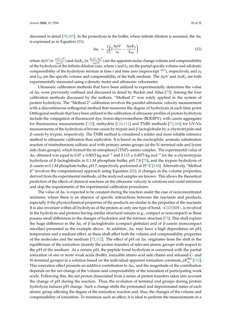
Sensors 2020, 20, 5594 20 of 32
discussed in detail [78,107]. In the proteolysis in the buffer, where infinite dilution is assumed, the ∆ar
is expressed as in Equation (11):
∆ar =12
(2
∆φVv0−
∆φKS
kS0
)(11)
where ∆φV (≡ v(t)−v0
cbh(t)) and ∆φKS (≡ kS(t)−kS
0
cbh(t)) are the apparent molar change volume and compressibility
of the hydrolysis at the infinite dilution case, where v and kS are the partial specific volume and adiabaticcompressibility of the hydrolysis mixture at time t and time zero (superscript “0”), respectively, and v0
and kS0 are the specific volume and compressibility of the bulk medium. The ∆φV and ∆φKS are bothexperimentally measured using a density meter and ultrasonic velocimeter.
Ultrasonic calibration methods that have been utilized to experimentally determine the valueof ∆ar were previously outlined and discussed in detail by Buckin and Altas [73]. Among the fourcalibration methods discussed by the authors, “Method 2” was solely applied in the system ofprotein hydrolysis. The “Method 2” calibration involves the parallel ultrasonic velocity measurementwith a discontinuous orthogonal method that measures the degree of hydrolysis at each time point.Orthogonal methods that have been utilized in the calibration of ultrasonic profiles of protein hydrolysisinclude the conjugation of fluorescent dye, boron-dipyrromethene (BODIPY), with casein aggregatesfor fluorescence measurements [110], ninhydrin [110,111] and TNBS methods [73,104] for UV-Vismeasurements of the hydrolysis of bovine casein by trypsin andβ-lactoglobulin byα-chymotrypsin andβ-casein by trypsin, respectively. The TNBS method is considered a milder and more reliable referencemethod to ultrasonic calibration than ninhydrin. It is based on the nucleophilic aromatic substitutionreaction of trinitrobenzene sulfonic acid with primary amino groups (at the N–terminal side and lysineside chain groups), which formed the tri-nitrophenyl (TNP)–amino complex. The experimental value of∆ar obtained was equal to 0.07 ± 0.0015 kg mol−1 and 0.115 ± 0.0075 kg mol−1 for the α-chymotrypsinhydrolysis of β-lactoglobulin in 0.1-M phosphate buffer, pH 7.8 [73], and the trypsin hydrolysis ofβ-casein in 0.1-M phosphate buffer, pH 7, respectively, performed at 25 ◦C [104]. Alternatively, “Method4” involves the computational approach using Equation (11), if changes in the volume properties,derived from the experimental methods, of the analyzed samples are known. This allows the theoreticalprediction of the effects of chemical reactions on the ultrasonic velocity in solutions and could minimizeand skip the requirements of the experimental calibration procedures.
The value of ∆ar is expected to be constant during the reaction under the case of nonconcentratedmixtures, where there is an absence of specific interactions between the reactants and products,especially if the physicochemical properties of the products are similar to the properties of the reactants.It is also invariant within all hydrolysis of the protein as only one type of bond, –C(O)NH–, is involvedin the hydrolysis and proteins having similar structural natures (e.g., compact or noncompact) as thesepossess small differences in the changes of hydration and the intrinsic structure [73]. This shall explainthe huge difference in the ∆ar of β-lactoglobulin (compact globular) and of β-casein (noncompactmicellar) presented as the example above. In addition, ∆ar may have a high dependence on pH,temperature and a medium effect, as these shall affect both the volume and compressibility propertiesof the molecules and the medium [73,112]. The effect of pH on ∆ar originates from the shift in theequilibrium of the ionization (mainly the proton transfer) of relevant atomic groups with respect tothe pH of the medium. At a certain pH, the peptide bond hydrolysis is concerned with the partialionization of one or more weak acids (buffer, ionizable amino acid side chains and released C- andN-terminal groups) in a solution based on the individual apparent ionization constants, pKapp
A [112].This ionization effect presents an additive contribution to ∆ar, and the magnitude of the contributiondepends on the net change of the volume and compressibility of the ionization of participating weakacids. Following this, the net proton dissociated from a series of proton transfers takes into accountthe change of pH during the reaction. Thus, the evolution of terminal end groups during proteinhydrolysis induces pH change. Such a change shifts the protonated and deprotonated states of eachatomic group affecting the degree of the ionization reaction and, thus, the changes of the volume andcompressibility of ionization. To minimize such an effect, it is ideal to perform the measurements in a

Sensors 2020, 20, 5594 21 of 32
buffered medium. The choice of a buffer varies with the weak acid present and responsible for thebuffering action. Usually, a relatively high concentration of the buffer is being utilized as a medium ofhydrolysis enough to compensate for the pH change. Therefore, the proton transfer between the bufferand the atomic groups in polypeptides dominates the ionization contribution. Likewise, the magnitudeof the contribution depends on the net change of the volume and compressibility, which is a functionof the pH. Finally, the temperature effect has been known to affect the hydration level through passingthe energy barrier of the breakage of the hydrogen-bonded network of the hydration, resulting in morerelaxed water. In addition, the dependence of pKapp
A on the temperature should also be considered.The frequency dependence measurement of the ultrasonic parameters is an effective tool to
characterize the microstructure rearrangement (e.g., protein aggregation and gelation), as well asthe fast kinetic reactions taken during the protein hydrolysis. As mentioned above, the protontransfer between weak acids present in the hydrolytic mixture takes place concomitant with hydrolysis.Depending on the ionization properties of the participating atomic groups, such a process gives rise tothe frequency-dependent relaxation contribution to ultrasonic velocity and attenuation. This affectsthe amplitude of the change of the ultrasonic parameters of hydrolysis, as observed in Figure 9A [104].In the given example, the dominant proton transfer takes place between the terminal α-amino groupof protein hydrolysates and the phosphate group with a rate of constant k:
–NH2 + H2PO−4k1
k−1–NH+
3 + HPO2−?4 (12)
The theoretical framework and mathematical relationship between the relaxation frequency, frel,was previously discussed in reference [73]. It indicates that the relaxation contribution has a positivecontribution to ultrasonic attenuation but a negative contribution to ultrasonic velocity close to frel,usually in the region of 0.5–3 MHz. The magnitude of the frequency dependence observed is attributedto three interrelated factors: (1) the significant volume effect of the proton transfer between participatingspecies, (2) the equilibrium concentration of the participating species and (3) the relaxation time of theprocess close to the relaxation frequency. The first factor is related to the thermodynamic property ofthe proton transfer, whereas the third factor is dependent on both the kinetics of the reaction, as well asthe ultrasonic frequency of the measurements. At low-MHz frequency measurements, the comparabletime scale of the oscillation of the temperature and pressure in ultrasonic waves relative to that ofthe relaxation process triggers perturbations of the equilibrium, giving rise to relaxation contribution.In contrast, at measurements above 10 MHz, the time scale of the oscillation of ultrasonic wavesis significantly fast relative to the time scale of the proton transfer. In this case, the process is inthe state of “frozen” relative to the timeframe of the ultrasonic wave oscillation, and the relaxationcontribution is absent [73]. Ultrasonic velocity profiles of hydrolysis presented at a high MHz frequencydo not take into account the relaxation effects but are directly attributed to changes in hydrationproportional to increasing the concentration of peptide bonds hydrolyzed. Ultrasonic attenuationexhibits a better sensitivity to the relaxation process. Since the relaxation contribution is dependent onthe concentration of participating species, particularly in Equation (12), ultrasonic attenuation maypresent a new ultrasonic methodology on the quantification of the concentration of peptide bondshydrolyzed in a nonaggregating system.
On the other hand, the loss of the native structure and subsequent exposure of hydrophobic regionssometimes may induce a self-assembly process into a large protein aggregate structure. Their interactionwith ultrasonic waves, particularly at a radius comparable to the wavelength of ultrasonic waves(including thermal and shear waves), results in a wave scattering contribution that is a function of theparticle radius, frequency and volume significant at a high frequency range >10 MHz. As aggregatesmay range from 10 nm up to microns size, the main scatterers are thermal and shear waves. Since ascattering contribution is likely to attenuate the ultrasonic signal, ultrasonic attenuation also exhibits abetter sensitivity to the process. However, its complex dependence to the frequency and radius exhibits

Sensors 2020, 20, 5594 22 of 32
a bell-shaped curve, but the scattering contribution to the ultrasonic velocity is positive, exhibiting aS-shaped profile.
The real-time ultrasonic profiles of cbh are utilized to calculate the real-time hydrolytic profiles ofthe extent of the chemical reaction, ζ, and degree of hydrolysis, dh, as well as the average degree ofpolymerization, DP, and average molar mass of protein, M, as shown below [73,99]:
ζ =cbh
c0PNhb
; dh =cbh
c0PNT
(13)
DP =DP
0
1 + cbhDP
0−1
c0PNT
; M =M
0−MH2O
1 + cbhM
0
w0P
+ MH2O (14)
where CP0 is the molar (mol per kg of mixture) concentration of the protein at time zero, Nhb and NT
are the number of hydrolysable peptide bonds and total peptide bonds in the native protein molecule,
respectively, DP0
and M0
are the average degree of polymerization and the average molar mass of theprotein at time zero, respectively, MH2O is the molar mass of water and w0
P is the weight fraction of
the protein (kg in one kg of the mixture) at reaction time zero. The ratios DP0−1
cb0 and M
0
w0P
are related to
each other [73]. An example of such profiles is presented in Figures 9B and 10B, calculated from the~15-MHz data of the ultrasonic velocity profile of the protein hydrolysis.Sensors 2020, 20, x FOR PEER REVIEW 23 of 32
Figure 10. (A) Ultrasonic velocity profiles of the hydrolysis of β-lactoglobulin treated with α-chymotrypsin in 0.1-M phosphate buffer, pH 7.8, at 25 °C. The concentration of the peptide bonds hydrolyzed (secondary y-axis) was translated from the ultrasonic velocity profile (primary y-axis) using Equation (9) and raΔ = 0.07 ± 0.0015 kg mol−1. (B) The corresponding profile of the reduction
of the average degree of polymerization (primary y-axis) and average molar mass (secondary y-axis), within 50 min of hydrolysis, calculated from the concentration of the peptide bonds hydrolyzed using Equation (14). The insert illustrates the profile at the full time range of the measurement. The concentration of enzyme is 4 μM. Adopted from Buckin and Altas [73].
Finally, it is important to examine that the precision of the ultrasonic measurements of the hydrolysis curve can be calculated in the concentration of bonds hydrolyzed. The precision of our ultrasonic measurement was a maximum of 0.0002 m s−1, which corresponds to the approximately ±2 μmol kg−1 concentrations of the peptide bonds hydrolyzed, based on our values of raΔ for the proteins [104].
4.5. Examples of Ultrasonic Measurements of Proteolysis
The feasibility of high-resolution ultrasonic measurements for the determination of the proteolytic activity has been demonstrated and widely discussed previously. Table 1 summarizes the references on the ultrasonic monitoring of the proteolytic activity of various proteases. The earliest examples of reported applications of high-resolution ultrasonic monitoring on the protease-milk system are on the rennet-induced gelation of milk components [113,114], though an ultrasonic measurement of enzyme-induced milk coagulation using the pulse-echo technique was reported earlier [115]. The detection of the protease activity in milk during the process is not achievable using an optical assay due to the opacity of the sample. However, low-frequency rheological measurements were utilized as the main complementary methods. Chymosin is the key component of the enzyme of rennet, which is responsible for the cleavage of the peptide bonds of κ-casein and which gives rise to the aggregation of casein proteins. Using high-resolution ultrasonic measurements, multi-stage processes can be distinguished in detail from the time profile with changes in the constituent gelling particles. Dwyer et al. [113] reported that the renneting process is attributed to the increase in ultrasonic velocity caused by the hydration effect during the hydrolysis and ultrasonic wave scattering during aggregation. In Figure 11A, the different rates of the increase illustrate the multi-substage molecular rearrangements. The first slope corresponds to the increase due to the hydrolysis of κ-casein. This is followed by aggregation and gelation, which further increase the ultrasonic velocity due to the scattering effects. On the other hand, the loss of the aggregate structure concomitant with the hydrolysis slightly decreased the attenuation, and the following aggregation and gelation process increased the ultrasonic attenuation (Figure 11B). The frequency dependence observed is attributed by the ultrasonic scattering effect. Similar findings were reported by Corredig et al. [114]. The author also applied a time derivative of the ultrasonic parameters to provide an additional explanation of the processes. Wang et al. [116] utilized a single-frequency ultrasound measurement at 7.8 MHz to assess the effects of preheatment at ultra-high temperatures on enzyme-induced gelation in milks.
Figure 10. (A) Ultrasonic velocity profiles of the hydrolysis of β-lactoglobulin treated withα-chymotrypsin in 0.1-M phosphate buffer, pH 7.8, at 25 ◦C. The concentration of the peptidebonds hydrolyzed (secondary y-axis) was translated from the ultrasonic velocity profile (primaryy-axis) using Equation (9) and ∆ar = 0.07 ± 0.0015 kg mol−1. (B) The corresponding profile of thereduction of the average degree of polymerization (primary y-axis) and average molar mass (secondaryy-axis), within 50 min of hydrolysis, calculated from the concentration of the peptide bonds hydrolyzedusing Equation (14). The insert illustrates the profile at the full time range of the measurement.The concentration of enzyme is 4 µM. Adopted from Buckin and Altas [73].
Lynch et al. [99] also previously formulated the calculation of the change of osmolality, m, using thevalues of ultrasonically measured cbh during the hydrolysis of lactose in milk:
m = Φln(1 + Mms)
M; where ms = (m0
s +cbh
ϕ0w)
1
1− cbhMϕ0
w
(15)
where Φ is the osmotic coefficient of the mixture; ms and m0s are the molality of solutes at time t
and zero, respectively, and ϕ0w is the weight fraction of the water molecules mixed with solutes in
unhydrolyzed media.
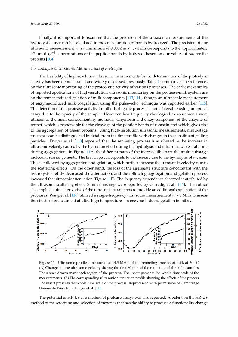
Sensors 2020, 20, 5594 23 of 32
Finally, it is important to examine that the precision of the ultrasonic measurements of thehydrolysis curve can be calculated in the concentration of bonds hydrolyzed. The precision of ourultrasonic measurement was a maximum of 0.0002 m s−1, which corresponds to the approximately±2 µmol kg−1 concentrations of the peptide bonds hydrolyzed, based on our values of ∆ar for theproteins [104].
4.5. Examples of Ultrasonic Measurements of Proteolysis
The feasibility of high-resolution ultrasonic measurements for the determination of the proteolyticactivity has been demonstrated and widely discussed previously. Table 1 summarizes the referenceson the ultrasonic monitoring of the proteolytic activity of various proteases. The earliest examplesof reported applications of high-resolution ultrasonic monitoring on the protease-milk system areon the rennet-induced gelation of milk components [113,114], though an ultrasonic measurementof enzyme-induced milk coagulation using the pulse-echo technique was reported earlier [115].The detection of the protease activity in milk during the process is not achievable using an opticalassay due to the opacity of the sample. However, low-frequency rheological measurements wereutilized as the main complementary methods. Chymosin is the key component of the enzyme ofrennet, which is responsible for the cleavage of the peptide bonds of κ-casein and which gives riseto the aggregation of casein proteins. Using high-resolution ultrasonic measurements, multi-stageprocesses can be distinguished in detail from the time profile with changes in the constituent gellingparticles. Dwyer et al. [113] reported that the renneting process is attributed to the increase inultrasonic velocity caused by the hydration effect during the hydrolysis and ultrasonic wave scatteringduring aggregation. In Figure 11A, the different rates of the increase illustrate the multi-substagemolecular rearrangements. The first slope corresponds to the increase due to the hydrolysis of κ-casein.This is followed by aggregation and gelation, which further increase the ultrasonic velocity due tothe scattering effects. On the other hand, the loss of the aggregate structure concomitant with thehydrolysis slightly decreased the attenuation, and the following aggregation and gelation processincreased the ultrasonic attenuation (Figure 11B). The frequency dependence observed is attributed bythe ultrasonic scattering effect. Similar findings were reported by Corredig et al. [114]. The authoralso applied a time derivative of the ultrasonic parameters to provide an additional explanation of theprocesses. Wang et al. [116] utilized a single-frequency ultrasound measurement at 7.8 MHz to assessthe effects of preheatment at ultra-high temperatures on enzyme-induced gelation in milks.
Sensors 2020, 20, x FOR PEER REVIEW 24 of 32
Figure 11. Ultrasonic profiles, measured at 14.5 MHz, of the renneting process of milk at 30 °C. (A) Changes in the ultrasonic velocity during the first 60 min of the renneting of the milk samples. The slopes drawn mark each region of the process. The insert presents the whole time scale of the measurements. (B) The corresponding ultrasonic attenuation profile showing the effects of the process. The insert presents the whole time scale of the process. Reproduced with permission of Cambridge University Press from Dwyer et al. [113].
The potential of HR-US as a method of protease assays was also reported. A patent on the HR-US method of the screening and selection of enzymes that has the ability to produce a functionality change in milk samples and wheat flour suspension was written by Dijk et al. [117]. The action of chymosin (Maxiren® 180) on milk samples and protease (Bakezyme B500®) on wheat flour suspension was detected through a change in the ultrasonic velocity but not kinetically measured. The analyses were rather qualitative, as no information on the enzyme reaction was reported. Niemeyer et al. [118] worked on the development of a novel ultrasonic enzyme assay for pharmacopoeia methods using HR-US. The author ultrasonically investigated the hydrolysis of the casein solution by pancreatin (mixture of enzymes) and trypsin (major component of pancreatin) under different hydrolytic conditions relevant to pharmacopoeia assays. Rather, the feasibility of the ultrasonic measurements as a potential process analytical technology (PAT) for such an online process control was examined and validated.
Table 1. Ultrasonic monitoring of thee proteolytic of the milk protease and milk proteins described in the literature.
Protease Reactions and Conditions Reference
Chymosin Ultrasonic renneting process of milk at 30 °C. Ultrasonic analysis of multistage structural rearrangement.
[113]
Chymosin Ultrasonic renneting process of milk at 30 °C. Comparison of effect of pH, temperature and enzyme-induced gelation in milks.
[114]
Chymosin Ultrasonic renneting process of milk at 30 °C. Effect of preheat treatment at ultra-high temperature.
[116]
Proteinase K Hydrolysis of Gly-Leu-Gly-Gly-Ala (synthetic pentapeptides) in 30-mM Tris buffer, pH 8, at 37 °C. Effect of substrate-enzyme ratio.
[109,119]
Proteinase K Hydrolysis of bovine serum albumin (BSA) in 30-mM Tris buffer, pH 8, at 37 °C. Effect of enzyme concentration.
[109,119]
Proteinase K Hydrolysis of bovine casein aggregates in 30-mM Tris buffer, pH 8, at 37 °C. Effect of substate concentration.
[109,119]
Chymosin (Maxiren® 180)
Proteolytic activity in milk samples at 30 °C. Screening method of enzyme activity.
[117]
Protease (Bakezyme
B500®)a
Proteolytic activity in wheat flour suspension at 30 °C. Screening method of enzyme activity.
[117]
Trypsin Hydrolysis of β-casein in 50-mM phosphate, pH 7.5, at 37 °C [111]
Pancreatin Proteolysis of casein solution at 37 °C. Effect of hydrolytic conditions relevant to pharmacopoeia assays.
[118]
Figure 11. Ultrasonic profiles, measured at 14.5 MHz, of the renneting process of milk at 30 ◦C.(A) Changes in the ultrasonic velocity during the first 60 min of the renneting of the milk samples.The slopes drawn mark each region of the process. The insert presents the whole time scale of themeasurements. (B) The corresponding ultrasonic attenuation profile showing the effects of the process.The insert presents the whole time scale of the process. Reproduced with permission of CambridgeUniversity Press from Dwyer et al. [113].
The potential of HR-US as a method of protease assays was also reported. A patent on the HR-USmethod of the screening and selection of enzymes that has the ability to produce a functionality change
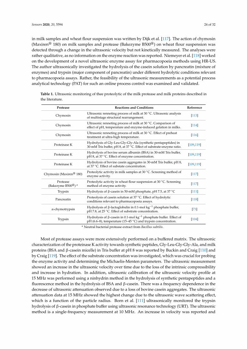
Sensors 2020, 20, 5594 24 of 32
in milk samples and wheat flour suspension was written by Dijk et al. [117]. The action of chymosin(Maxiren® 180) on milk samples and protease (Bakezyme B500®) on wheat flour suspension wasdetected through a change in the ultrasonic velocity but not kinetically measured. The analyses wererather qualitative, as no information on the enzyme reaction was reported. Niemeyer et al. [118] workedon the development of a novel ultrasonic enzyme assay for pharmacopoeia methods using HR-US.The author ultrasonically investigated the hydrolysis of the casein solution by pancreatin (mixture ofenzymes) and trypsin (major component of pancreatin) under different hydrolytic conditions relevantto pharmacopoeia assays. Rather, the feasibility of the ultrasonic measurements as a potential processanalytical technology (PAT) for such an online process control was examined and validated.
Table 1. Ultrasonic monitoring of thee proteolytic of the milk protease and milk proteins described inthe literature.
Protease Reactions and Conditions Reference
Chymosin Ultrasonic renneting process of milk at 30 ◦C. Ultrasonic analysisof multistage structural rearrangement. [113]
Chymosin Ultrasonic renneting process of milk at 30 ◦C. Comparison ofeffect of pH, temperature and enzyme-induced gelation in milks. [114]
Chymosin Ultrasonic renneting process of milk at 30 ◦C. Effect of preheattreatment at ultra-high temperature. [116]
Proteinase K Hydrolysis of Gly-Leu-Gly-Gly-Ala (synthetic pentapeptides) in30-mM Tris buffer, pH 8, at 37 ◦C. Effect of substrate-enzyme ratio. [109,119]
Proteinase K Hydrolysis of bovine serum albumin (BSA) in 30-mM Tris buffer,pH 8, at 37 ◦C. Effect of enzyme concentration. [109,119]
Proteinase K Hydrolysis of bovine casein aggregates in 30-mM Tris buffer, pH 8,at 37 ◦C. Effect of substate concentration. [109,119]
Chymosin (Maxiren® 180)Proteolytic activity in milk samples at 30 ◦C. Screening method ofenzyme activity. [117]
Protease(Bakezyme B500®) a
Proteolytic activity in wheat flour suspension at 30 ◦C. Screeningmethod of enzyme activity. [117]
Trypsin Hydrolysis of β-casein in 50-mM phosphate, pH 7.5, at 37 ◦C [111]
Pancreatin Proteolysis of casein solution at 37 ◦C. Effect of hydrolyticconditions relevant to pharmacopoeia assays. [118]
α-chymotrypsin Hydrolysis of β-lactoglobulin in 0.1-mol kg−1 phosphate buffer,pH 7.8, at 25 ◦C. Effect of substrate concentration.
[73]
Trypsin Hydrolysis of β-casein in 0.1-mol kg−1 phosphate buffer. Effect ofpH (6.6–8), temperature (15–45 ◦C) and trypsin concentration.
[104]
a Neutral bacterial protease extract from Bacillus subtilis.
Most of protease assays were more extensively performed on a buffered matrix. The ultrasoniccharacterization of the proteinase K activity towards synthetic peptides, Gly-Leu-Gly-Gly-Ala, and milkproteins (BSA and β-casein micelle) in Tris buffer at pH 8 was reported by Buckin and Craig [110] andby Craig [119]. The effect of the substrate concentration was investigated, which was crucial for probingthe enzyme activity and determining the Michaelis-Menten parameters. The ultrasonic measurementshowed an increase in the ultrasonic velocity over time due to the loss of the intrinsic compressibilityand increase in hydration. In addition, ultrasonic calibration of the ultrasonic velocity profile at15 MHz was performed using a ninhydrin method in the hydrolysis of synthetic pentapeptides and afluorescence method in the hydrolysis of BSA and β-casein. There was a frequency dependence in thedecrease of ultrasonic attenuation observed due to a loss of bovine casein aggregates. The ultrasonicattenuation data at 15 MHz showed the highest change due to the ultrasonic wave scattering effect,which is a function of the particle radius. Born et al. [111] ultrasonically monitored the trypsinhydrolysis of β-casein in phosphate buffer using ultrasonic resonance technology (URT). The ultrasonicmethod is a single-frequency measurement at 10 MHz. An increase in velocity was reported and

Sensors 2020, 20, 5594 25 of 32
measured orthogonally with the ninhydrin assay. However, a high salt concentration was reported tolimit the precision/sensitivity of the ultrasonic measurement.
The recent ultrasonic methodology, discussed in Section 4.4, has been hitherto applied on thehydrolysis of β-lactoglobulin by α-chymotrypsin [73] and β-casein by trypsin [104] in phosphatebuffers. The ultrasonic profiles provide information on the evolution of the concentrations of reactantsand products, which can be further derived into more advanced hydrolytic profiles, such as theaverage degree of polymerization and molar mass, as well as the ultrasonic osmolality. The currentmethodology can be extendedly applied, but not limited to, other protease systems that require asensitivity in the buffer or even in the complex media. The sensitivity of the HR-US technique indetecting the protease was reported to be within the sub-nM range [104], which is comparable withsurface-sensitive acoustic methods. This is evident from Table 2, which compares the sensitivityof surface-sensitive and HR-US methods for the detection of trypsin and plasmin. Such ultrasonicmonitoring in the buffer system is an important first step required to prove the detection concepts.In addition, most protease assays have been extensively studied under a buffered matrix to unravel theintrinsic behavior of the assays.
Table 2. Comparison of the sensitivity of acoustics biosensors and high-resolution ultrasonicspectroscopy (HR-US) methods for milk protease detection. QCM: quartz crystal microbalanceand EMPAS: electromagnetic piezoelectric sensors.
AcousticMethod
DetectionPhase Substrate Limit of
DetectionTime of
Detection Reference
QCM surface Short peptides at gold surface 0.65 nMPlasmin 30 min [63]
QCM surface β-casein at gold substrate 0.1 nM Trypsin1 nM Plasmin 30 min [62]
EMPAS surface β-casein at gold substrate 0.032 nMPlasmin 30 min [64]
HR-US volume 0.1–1% (w/w) β-casein in buffer 0.2 nMTrypsin 15–30 min [104]
HR-US volume 0.1–1% (w/w) β-casein in buffer 0.2 nMPlasmin 15–30 min [120]
4.6. Future Works and Limitations
Although the potential application of ultrasonic methodology has been described and reported,we strongly believe that there are plenty of unexplored concepts that can arise in the future. Sofar, the current ultrasonic methodology utilizes only one ultrasonic parameter, ultrasonic velocity,in monitoring the proteolytic activity in which its sensitivity to molecular processes, as well as dataalgorithms of ultrasonic calibration, have been comprehensively described. Making use of ultrasonicattenuation and multi-frequency measurements will provide the characterization of concomitantsecondary reactions, such as fast chemical kinetics (proton and structural relaxation processes) andstructural rearrangements (changes in the protein particle or aggregate size and phase separation).
The future applications of HR-US in the protease assay also lie in the extension of ultrasonicmethodology into different protease systems. For example, it can be applied with the targeting ofdisease-related proteases to develop direct activity assays with clear clinical relevance. Furthermore,the growing interest for understanding protease activities in complex systems such as real biologicalsystems or complex food matrices demand emerging methods. The ultrasonic transparency for mostof the liquid or semi-solid samples makes HR-US a promising technology for such purposes. To date,only the activity of lactases in milk samples have been investigated extensively by HR-US. The ultrasonicmethodology employed was rather fast and simple. Particular to the protein system, increasing thestructural complexity of the analyzed samples may give rise to limitations and challenges, includingthe necessity of ultrasonic calibrations at different reaction conditions, clean substrate formulations,the influence of air bubbles and temperature stability, which need to be addressed. For example,

Sensors 2020, 20, 5594 26 of 32
the unforeseen and unwanted additional interferences may require additional studies of the system toretain the assay reliability and sensitivity. In addition, the high concentrations of other compoundspresent shall constitute some dependence of the calibration parameters on concentrations of othercomponents that also require consideration.
The surface-sensitive acoustics methods are well-suited for the development of biosensors formonitoring food quality and safety. This has been already shown in the detection of plasmin inreal milk samples [121]. The comparative analysis of the sensitivity of plasmin detection by QCMand ELISA methods revealed a similar sensitivity. However, while ELISA cannot detect the plasminactivity and only the whole plasmin concentration in milk, the QCM method allowed the real-timemonitoring of the cleavage of β-casein at the surfaces. Thus, this method is useful for applications indairy laboratories for monitoring the plasmin activity.
5. Conclusions
It has been demonstrated that surface acoustic methods, such as QCM, QCM-D, TSM and EMPAS,as well as high-resolution ultrasonic spectroscopy (HR-US), are effective tools for the detection of theactivity of milk proteases. The methods are suitable for applications in nontransparent liquids, such asmilk, and allowing the real-time analysis of the cleavage of proteins. The sensitivity of the surfaceacoustics and HR-US methods are comparable and allow the detection of trypsin and plasmin in thesub-nanomolar range, which is enough for practical applications in the milk industry, as well as inbasic research focused on the study the mechanism of enzyme reactions.
Overall, we consider that HR-US may emerge as an alternative method platform for milk proteaseassays that provides fast, sensitive and direct sensing towards the native protein substrate in thenatural or desired environment in the volume. Since proteolysis is accompanied by hydration changes,the current methodology can be extendedly applied, but not limited to, other protease systems thatrequire sensitivity in the buffer or even in the complex media. Being a considerably valuable tool in thefield of the enzyme-based industry, we also envisage that HR-US could accelerate milk protease-basedresearch and intensively advance the field in which it will play a huge role in improving the dairyfood quality.
Author Contributions: Conceptualization, T.H.; writing—original draft preparation, T.H., M.D. and M.T.;writing—review and editing, T.H. and M.D.; supervision, T.H.; project administration, T.H. and fundingacquisition, T.H. All authors have read and agreed to the published version of the manuscript.
Funding: T.H. acknowledges funding from the European Union’s Horizon 2020 Research and Innovation Programunder the Marie Sklodowska-Curie grant agreement No. 690898 and from the Science Grant Agency VEGA,project No.: 1/0419/20.
Conflicts of Interest: The authors declare no conflict of interest.
References
1. Grufferty, M.B.; Fox, P.F. Milk alkaline proteinase. J. Dairy Res. 1988, 55, 609–630. [CrossRef] [PubMed]2. Bastian, E.D.; Brown, R.J. Plasmin in milk and dairy products: An update. Int. Dairy J. 1996, 6, 435–457.
[CrossRef]3. Abdul, S.; Leebeek, F.W.G.; Rijken, D.C.; de Willige, S.U. Natural heterogeneity of α2-antiplasmin: Functional
and clinical consequences. Blood 2016, 127, 538–545. [CrossRef]4. Dupont, D.; Bailly, C.; Grosclaude, J.; Collin, J.C. Differential titration of plasmin and plasminogen in milk
using sandwich ELISA with monoclonal antibodies. J. Dairy Res. 1997, 64, 77–86. [CrossRef]5. Rauh, V.M.; Johansen, L.B.; Ipsen, R.; Paulsson, M.; Larsen, L.B.; Hammershøj, M. Plasmin activity in
UHT milk: Relationship between proteolysis, age gelation, and bitterness. J. Agricult. Food Chem. 2014,62, 6852–6860. [CrossRef]
6. Arena, S.; Salzano, A.M.; Scalon, A. Identification of protein markers for the occurrence of defrosted materialin milk through a MALDI-TOF-MS profiling approach. J. Proteom. 2016, 147, 56–65. [CrossRef]

Sensors 2020, 20, 5594 27 of 32
7. Volitaki, A.; Kaminarides, S. Detection of bovine milk in ovine Halloumi cheese by HPLC analysis of cheesecaseins hydrolysed by plasmin. Milchwissenschaft 2001, 56, 207–210.
8. Miralles, B.; Ramos, M.; Amigo, L. Influence of proteolysis of milk on the whey protein to total protein ratioas determined by capillary electrophoresis. J. Dairy Sci. 2003, 86, 2813–2817. [CrossRef]
9. Walstra, P.; Wouters, J.T.M.; Geurts, T.J. Dairy Science and Technology, 2nd ed.; CRC Press: Boca Raton, FL,USA; Taylor and Francis Group: Oxford, UK, 2005; pp. 17–108.
10. Raynal-Ljutovac, K.; Pirisi, A.; Cremoux, R.; Gonzalo, C. Somatic cells of goat and sheep milk: Analytical,sanitary, productive and technological aspects. Small Rumin. Res. 2007, 68, 126–144. [CrossRef]
11. Schwarz, D.; Diesterbeck, U.S.; Konig, S.; Brugemann, K.; Schlez, K.; Zschock, M.; Wolter, W.; Czerny, C.P.Flow cytometric differential cell counts in milk for the evaluation of inflammatory reactions in clinicallyhealthy and subclinically infected bovine mammary glands. J. Dairy Sci. 2011, 94, 5033–5044. [CrossRef]
12. Muller-Renaud, S.; Dupont, D.; Dulieu, P. Quantification of β-casein in milk and cheese using an opticalimmunosensor. J. Agric. Food Chem. 2004, 52, 659–664. [CrossRef] [PubMed]
13. Dallas, D.C.; Murray, N.M.; Gan, J. Proteolytic systems in milk: Perspectives on the evolutionary functionwithin the mammary gland and the infant. J. Mammary Gland Biol. Neoplasia 2015, 20, 133–147. [CrossRef][PubMed]
14. Halabi, A.; Deglaire, A.; Hennetier, M.; Violleau, F.; Burel, A.; Bouhallab, S.; Dupond, D.; Croguennec, T.Structural characterization of heat-induced protein aggregates in model infant milk formulas. Food Hydrocol.2020, 107, 105928. [CrossRef]
15. Bumberger, E.; Belitz, H.D. Bitter taste of enzymic hydrolysates of casein. I. Isolation, structural and sensorialanalysis of peptides from tryptic hydrolysates of β-casein. Z. Lebensm. Unters Forsch. 1993, 197, 14–19.[CrossRef]
16. Drinane, M.; Sherman, J.A.; Hall, A.E.; Simons, M.; Mulligan-Kehoe, M.J. Plasminogen and plasmin activityin patients with coronary artery disease. J. Thromb. Haemost. 2006, 4, 1288–1295. [CrossRef]
17. Cesarman-Maus, G.; Hajjar, K.A. Molecular mechanisms of fibrinolysis. Br. J. Haematol. 2005, 129, 307–321.[CrossRef]
18. Heegaard, C.W.; Amdreasen, P.; Petersen, T.R.; Ramussen, L.K.I. Binding of plasminogen and tissue-typeplasminogen activator to dimeric αs2-casein accelerates plasmin generation. Fibrinolysis Proteolysis 1997,11, 29–36. [CrossRef]
19. Politis, I.; White, J.H.; Zavizion, B.; Goldberg, J.J.; Guo, M.R.; Kindstedt, P. Effect of individual caseins onplasminogen activation by bovine urokinase-type and tissue-type plasminogen activators. J. Dairy Sci. 1995,78, 484–490. [CrossRef]
20. Rippel, K.M.; Nielson, S.S.; Hayes, K.D. Effects of native and denatured whey proteins on plasminogenactivator activity. J. Dairy Sci. 2004, 87, 2344–2350. [CrossRef]
21. Kelly, A.L. Indigenous proteinases in milk. In Advanced Dairy Chemistry Volume 1, Proteins; Fox, P.F.,McSweeney, P.L.H., Eds.; Springer: Boston, MA, USA, 2003; pp. 495–521.
22. Deeth, H.C.; Datta, N. Heat Treatment of Milk|Ultra-High Temperature Treatment (UHT): Heating Systems.In Encyclopedia of Dairy Sciences, 2nd ed.; Fuquay, J.W., McSweeney, P.L.H., Fox, P.F., Eds.; Elsiever SciencePublishing Co Inc.: San Diego, CA, USA, 2011; pp. 732–737.
23. Newstead, D.F.; Paterson, G.; Anema, S.G.; Coker, C.J.; Wewala, A.R. Plasmin activity in direct-steam-injectionUHT-processed reconstituted milk: Effects of preheat treatment. Int. Dairy J. 2006, 16, 573–579. [CrossRef]
24. Saint Denis, T.; Humbert, G.; Gaillard, J. Heat inactivation of native plasmin, plasminogen and plasminogenactivators in bovine milk: A revisited study. Le Lait 2001, 81, 715–729. [CrossRef]
25. Lu, D.D.; Nielsen, S.S. Heat inactivation of native plasminogen activators in bovine milk. J. Food Sci. 1993,58, 1010–1016. [CrossRef]
26. Kennedy, A.; Kelly, A.L. The influence of somatic cell count on the heat stability of bovine milk plasminactivity. Int. Dairy J. 1997, 7, 717–721. [CrossRef]
27. Hayes, M.G.; McSweeney, P.L.H.; Kelly, A.L. The influence of native and heat denatured whey proteins onenzyme activity. 1. Plasmin. Milchwissenschaft 2002, 57, 208–211.
28. Monti, J.C.; Mermoud, A.F.; Jollès, P. Trypsin in human milk. Experientia 1986, 42, 39–41. [CrossRef]29. McGilligan, K.M.; Thomas, D.W.; Eckhert, C.D. Alpha-1-antitrypsin concentration in human milk.
Pediatric Res. 1987, 22, 268–270. [CrossRef]

Sensors 2020, 20, 5594 28 of 32
30. Magboul, A.A.; Larsen, L.B.; McSweeney, P.L.; Kelly, A.L. Cysteine protease activity in bovine milk. Int. Dairy J.2001, 11, 865–872. [CrossRef]
31. Palmer, D.J.; Kelly, V.C.; Smit, A.M.; Kuy, S.; Knight, C.G.; Cooper, G.J. Human colostrum: Identification ofminor proteins in the aqueous phase by proteomics. Proteomics 2006, 6, 2208–2216. [CrossRef]
32. Fiorillo, A.S.; Critello, C.D.; Pullano, S.A. Theory, technology and applications of piezoresistive sensors:A review. Sens. Actuators A 2018, 281, 156–175. [CrossRef]
33. Fiorillo, A.S.; Pullano, S.A.; Bianco, M.G.; Critello, C.D. Ultrasonic Transducers Shaped in Archimedean andFibonacci Spiral: A Comparison. Sensors 2020, 20, 2800. [CrossRef]
34. Bundle, R.L.; Jarvi, E.J.; Rosentreter, J.J. Piezoelectric quartz crystal biosensors. Talanta 1998, 46, 1223–1236.[CrossRef]
35. Clark, L.C.; Lyons, C. Electrode systems for continuous monitoring in cardiovascular surgery. Ann. N. Y.Acad. Sci. 1962, 102, 29–45. [CrossRef] [PubMed]
36. Thévenit, D.R.; Toth, K.; Durst, R.A.; Wolson, G.S. Electrochemical biosensors: Recommended definitionsand classifications. Biosens. Bioelectr. 2001, 16, 121–131. [CrossRef]
37. Fu, Y.Q.; Luo, J.K.; Nguyen, N.T.; Walton, A.J.; Flewitt, A.J.; Zu, X.T.; Li, Y.; McHale, G.; Matthews, A.;Iborra, E.; et al. Advances in piezoelectric thin films for acoustic biosensors, acousto fluidics and lab-on-chipapplications. Prog. Mat. Sci. 2017, 89, 31–91. [CrossRef]
38. Manbachi, A.; Cobbold, R.S.C. Development and application of piezoelectric materials for ultrasoundgeneration and detection. Ultrasound 2011, 19, 187–196. [CrossRef]
39. Howells, C.H. Piezoelectric energy harvesting. Energy Convers. Manag. 2009, 50, 1847–1850. [CrossRef]40. Sauerbrey, G. Verwendung von schwingquarzen zur wagung dunnerschichten und zur mikrowagung. Z.
Phys. 1959, 155, 206–222. [CrossRef]41. Zainuddin, A.A.; Nordin, A.N.; Rahim, R.A.; Ralib, A.A.M.; Khan, S.; Guines, C.; Chatras, M.; Pothier, A.
Verification of quartz crystal microbalance array using vector network analyzer and open QCM. Indonesian J.Electrical Eng. Comput. Sci. 2018, 10, 84–93. [CrossRef]
42. Buttry, A.; Ward, M.D. Measurement of interfacial processes at electrode surfaces with the electrochemicalquartz crystal microbalance. Chem. Rev. 1992, 6, 1355–1379. [CrossRef]
43. Lu, C. Theory and practice of the quartz crystal microbalance. In Applications of Piezoelectric Quartz CrystalMicrobalances, 2nd ed.; Lu, C., Czanderna, A.W., Eds.; Elsiever Science Publishers B.V.: Amsterdam,The Netherlands, 1984; Volume 7, pp. 19–62.
44. Ballantine, D.S.; White, R.M.; Martin, S.J.; Ricco, A.J.; Zellers, E.T.; Frye, G.C.; Wohltjen, H. Acoustic WaveSensors. Theory, Design, and Physico-Chemical Applications; Academic Press: San Diego, CA, USA, 1997.
45. Rodahl, M.; Kasemo, F.H. QCM operation in liquids: An explanation of measured variations in frequencyand Q factor with liquid conductivity. Anal. Chem. 1996, 68, 2219–2227. [CrossRef]
46. Jonsson, M.P.; Jönsson, P.; Höök, F. Simultaneous nanoplasmonic and quartz crystal microbalance sensing:Analysis of biomolecular conformational changes and quantification of the bound molecular mass. Anal. Chem.2008, 80, 7988–7995. [CrossRef] [PubMed]
47. McNamara, T.P.; Blandford, C.F. A sensitivity metric and software to guide the analysis of soft films measuredby a quartz crystal microbalance. Analyst 2016, 141, 2911–2919. [CrossRef] [PubMed]
48. Pohanka, M. Overview of piezoelectric biosensors, immunosensors and DNA sensors and their applications.Materials 2018, 11, 448. [CrossRef] [PubMed]
49. Saitakis, M.; Gizeli, E. Acoustic sensors as a biophysical tool for probing cell attachment and cell/surfaceinteractions. Cell. Molec. Life Sci. 2012, 69, 357–371. [CrossRef]
50. Elmlund, L.; Käck, C.; Aastrup, T.; Nicholls, I.A. Study of the interaction of trastuzumab and SKOV3 epithelialcancer cells using a quartz crystal microbalance sensor. Sensors 2015, 15, 5884–5894. [CrossRef]
51. Afzal, A.; Mujahid, A.; Schirhagl, R.; Bajwa, S.Z.; Latif, U.; Feroz, S. Gravimetric viral diagnostics: QCMbased biosensors for early detection of viruses. Chemosensors 2017, 5, 7. [CrossRef]
52. Thompson, M.; Ballantyne, S.M.; Cheran, L.E.; Stevenson, A.C.; Loweb, C.H.R. Electromagnetic excitation ofhigh frequency acoustic waves and detection in the liquid phase. Analyst 2003, 128, 1048–1055. [CrossRef]
53. Sheikh, S.; Blaszykowski, C.; Thompson, M. Sacrificial BSA to block non-specific adsorption on organosilaneadlayers in ultra-high frequency acoustic wave sensing. Surf. Interface Anal. 2013, 45, 1781–1784. [CrossRef]
54. Neves, M.A.D.; Blaszykowski, C.; Bokhari, S.; Thompson, M. Ultrahigh frequency piezoelectric aptasensorfor the label-free detection of cocaine. Biosens. Bioelectr. 2015, 72, 383–392. [CrossRef]

Sensors 2020, 20, 5594 29 of 32
55. Sheikh, S.; Blaszykowski, C.; Thompson, M. Label-free detection of HIV-2 antibodies in serum with anultra-high frequency acoustic wave sensor. Talanta 2011, 85, 816–819. [CrossRef]
56. Crivianu-Gaita, V.; Aamer, M.; Posaratnanathan, R.T.; Romaschin, A.; Thompson, M. Acoustic wave biosensorfor the detection of the breast and prostate cancer metastasis biomarker protein PTHrP. Biosens. Bioelectr.2016, 78, 92–99. [CrossRef] [PubMed]
57. Spagnolo, S.; De La Franier, B.; Hianik, T.; Thompson, M. Surface probe linker with tandem anti-foulingproperties for application in biosensor technology. Biosensors 2020, 10, 20. [CrossRef] [PubMed]
58. Strube, O.I.; Rüdiger, A.A.; Bremser, W. Buildup of biobased adhesive layers by enzymatically controlleddeposition on the example of casein. Int. J. Ahesion Ahesives 2015, 63, 9–13. [CrossRef]
59. Matsunage, Y.; Suenaga, M.; Takahashi, H.; Furuta, A. Vitamin D affects neuronal peptides inneurodegenerative disease: Differences of V-D2 and V-D3 for affinity to amyloid-β and scrapie prionprotein in vitro. In A Critical Evaluation of Vitamin D—Clinical Overview; Sivakumar, J.T.G., Ed.; IntechOpen:Rijeka, Croatia, 2017.
60. Poturnayova, A.; Castillo, G.; Subjakova, V.; Tatarko, M.; Snejdarkova, M.; Hianik, T. Optimization of thecytochrome c detection by acoustic and electrochemical methods based on aptamer sensors. Sens. Actuators B2017, 228, 817–827. [CrossRef]
61. Chen, J.B.; Neves, M.A.D.; Thompson, M. Biosensor surface attachment of the ovarian cancer biomarkerHSP10 via His-tag modification. Sens. Bio Sens. Res. 2016, 11, 107–112. [CrossRef]
62. Tatarko, M.; Muckley, E.; Šubjaková, V.; Goswami, M.; Sumpter, B.; Hianik, T.; Ivanov, I.N. Machinelearning enabled acoustic detection of subnanomolar concentration of trypsin and plasmin in solution.Sens. Actuators B 2018, 272, 282–288. [CrossRef]
63. Poturnayova, A.; Karpisova, I.; Castillo, G.; Mezo, G.; Kocsis, L.; Csámpai, A.; Keresztes, Z.; Hianik, T.Detection of plasmin based on specific peptide substrate usingacoustic transducer. Sens. Actuators B 2016,223, 591–598. [CrossRef]
64. Románski, L.; Tatarko, M.; Jiao, M.; Keresztes, Z.; Hianik, T.; Thompson, M. Casein probe-based fast plasmindetermination in the picomolar range by an ultra-high frequency acoustic wave biosensor. Sens. Actuators B2018, 275, 206–214. [CrossRef]
65. Sabot, A.; Krause, S. Simultaneous quartz crystal microbalance impedance and electrochemical impedancemeasurements. Investigation into the degradation of thin polymer films. Anal. Chem. 2002, 74, 3304–3311.[CrossRef]
66. Stair, L.J.; Watkinson, M.; Krause, S. Sensor materials for detection of proteases. Biosens. Bioelectr. 2009,24, 2113–2118. [CrossRef]
67. Ahmad, N.; Colak, B.; Zhang, D.-W.; Gibbs, M.J.; Watkinson, M.; Becer, C.R.; Gautrot, J.E.; Krause, S.Peptide cross-linked poly (ethylene glycol) hydrogel films as biosensor coatings for the detection ofcollagenase. Sensors 2019, 19, 1677. [CrossRef] [PubMed]
68. Huenerbein, A.; Schmelzer, C.E.H.; Neubert, R.H.H. Real-time monitoring of peptic and tryptic digestions ofbovine β-casein using quartz crystal microbalance. Anal. Chim. Acta 2007, 584, 72–77. [CrossRef] [PubMed]
69. Tsai, C.; Ho, Y.H.; Sung, T.C.; Wu, W.F.; Chen, C.S. Escherichia coli proteome microarrays identified thesubstrates of ClpYQ protease. Mol. Cell. Proteom. 2017, 16, 113–120. [CrossRef]
70. Ong, I.L.H.; Yang, K.L. Recent developments in protease activity assays and sensors. Analyst 2017,142, 1867–1881. [CrossRef] [PubMed]
71. Buckin, V. High-resolution ultrasonic spectroscopy. J. Sens. Sens. Syst. 2018, 7, 207–217. [CrossRef]72. Elvira, L.; Resa, P.; Castro, P.; Shukla, S.K.; Sierra, C.; Aparicio, C.; Durán, C.; de Espinosa, F.M. Bioprocess
Monitoring using Low-intensity Ultrasound. In Ultrasound in Food Processing: Recent Advances; Villamiel, M.,Montilla, A., García-Pérez, J.V., Cárcel, J.A., Benedito, J., Eds.; Wiley Online Library: Hoboken, NJ, USA,2017; pp. 146–174.
73. Buckin, V.; Altas, M.C. Ultrasonic monitoring of biocatalysis in solutions and complex dispersions. Catalysts2017, 7, 336. [CrossRef]
74. Kaatze, U.; Eggers, F.; Lautscham, K. Ultrasonic velocity measurements in liquids with highresolution—Techniques, selected applications and perspectives. Meas. Sci. Technol. 2008, 19, 062001.[CrossRef]

Sensors 2020, 20, 5594 30 of 32
75. Kudryashov, E.; Smyth, C.; Duffy, G.; Buckin, V. Ultrasonic high-resolution longitudinal and shear wavemeasurements in food colloids: Monitoring of gelation processes and detection of pathogens. Trends ColloidInterface Sci. Xiv 2000, 115, 287–294.
76. Povey, M.J.W. Ultrasonic Techniques for Fluids Characterization, 1st ed.; Academic Press: San Diego, CA, USA,1997; pp. 1–214.
77. Herzfeld, K.F.; Litovitz, T.A. Absorption and Dispersion of Ultrasonic Waves, 1st ed.; Academic Press: New York,NY, USA, 1959; pp. 1–536.
78. Buckin, V. Application of high-resolution ultrasonic spectroscopy for analysis of complex formulations.Compressibility of solutes and solute particles in liquid mixtures. IOP Conf. Ser. Mater. Sci. Eng. 2012,42, 012001. [CrossRef]
79. Smyth, C.; Kudryashov, E.D.; Buckin, V. High-frequency shear and volume viscoelastic moduli of caseinparticle gel. Colloids Surf. A 2001, 183, 517–526. [CrossRef]
80. Audebrand, M.; Doublier, J.L.; Durand, D.; Emery, J.R. Investigation of gelation phenomena of somepolysaccharides by ultrasonic spectroscopy. Food Hydrocoll. 1995, 9, 195–203. [CrossRef]
81. Buckin, V.; O’ Driscoll, B. Ultrasonic waves and material analysis: Recent advances and future trends.LabPlus Int. 2002, 16, 17–21.
82. Buckin, V.; O’ Driscoll, B.; Smyth, C. Ultrasonic spectroscopy for material analysis. Recent advances.Spectrosc. Eur. 2003, 15, 20–25.
83. Buckin, V.; Hallone, S. Ultrasonic characterisation of w/o microemulsions—Structure, phase diagrams, stateof water in nano-droplets, encapsulated proteins, enzymes. In Microemulsions—An Introduction to Propertiesand Applications; Najjar, R., Ed.; InTech: Rijeka, Croatia, 2012; pp. 33–66.
84. Del Grosso, V.A.; Mader, C.W. Speed of sound in pure water. J. Acoust. Soc. Am. 1972, 52, 1442–1446.[CrossRef]
85. Wubshet, S.G.; Måge, I.; Böcker, U.; Lindberg, D.; Knutsen, S.H.; Rieder, A.; Rodriguez, D.A.; Afseth, N.K.FTIR as a rapid tool for monitoring molecular weight distribution during enzymatic protein hydrolysis offood processing by-products. Anal. Methods 2017, 9, 4247–4254. [CrossRef]
86. Vorob’ev, M.M.; Kochetkov, K.A. Determination of kinetic parameters for casein hydrolysis by chymotrypsinusing two ranges of substrate concentration. Int. Dairy J. 2016, 61, 76–84. [CrossRef]
87. Valencia, P.; Pinto, M.; Almonacid, S. Identification of the key mechanisms involved in the hydrolysis of fishprotein by Alcalase. Process Biochem. 2014, 49, 258–264. [CrossRef]
88. Márquez, M.C.; Vázquez, M.A. Modeling of enzymatic protein hydrolysis. Process Biochem. 1999, 35, 111–117.[CrossRef]
89. Tardioli, P.W.; Sousa, R.; Giordano, R.C.; Giordano, R.L.C. Kinetic model of the hydrolysis of polypeptidescatalyzed by Alcalase® immobilized on 10% glyoxyl-agarose. Enzym. Microb. Technol. 2005, 36, 555–564.[CrossRef]
90. Vorob’ev, M.M. Kinetics of peptide bond demasking in enzymatic hydrolysis of casein substrates. J. Mol.Catal. B Enzym. 2009, 58, 146–152. [CrossRef]
91. Adler-Nissen, J. Enzymic Hydrolysis of Food Protein; Elsevier Applied Science: London, UK, 1986; Volume 8,pp. 1–427.
92. Rutherfurd, S.M. Methodology for determining degree of hydrolysis of proteins in hydrolysates: A Review.J. AOAC Int. 2010, 93, 1515–1522. [CrossRef] [PubMed]
93. Spellman, D.; McEvoy, E.; O’Cuinn, G.; FitzGerald, R. Proteinase and exopeptidase hydrolysis of wheyprotein: Comparison of the TNBS, OPA and pH stat methods for quantification of degree of hydrolysis.Int. Dairy J. 2003, 13, 447–453. [CrossRef]
94. Beaubier, S.; Framboisier, X.; Ioannou, I.; Galet, O.; Kapel, R. Simultaneous quantification of the degree ofhydrolysis, protein conversion rate and mean molar weight of peptides released in the course of enzymaticproteolysis. J. Chromatogr. B 2019, 1105, 1–9. [CrossRef] [PubMed]
95. Camacho, F.; González-Tello, P.; Páez-Dueñas, M.-P.; Guadix, E.-M.; Guadix, A. Correlation of baseconsumption with the degree of hydrolysis in enzymic protein hydrolysis. J. Dairy Res. 2001, 68, 251–265.[CrossRef] [PubMed]
96. Bodin, A.; Framboisier, X.; Alonso, D.; Marc, I.; Kapel, R. Size-exclusion HPLC as a sensitive and calibrationlessmethod for complex peptide mixtures quantification. J. Chromatogr. B 2015, 1006, 71–79. [CrossRef]

Sensors 2020, 20, 5594 31 of 32
97. Kristoffersen, K.A.; Afseth, N.K.; Böcker, U.; Lindberg, D.; de Vogel-van den Bosch, H.; Ruud, M.L.;Wubshet, S.G. Average molecular weight, degree of hydrolysis and dry-film FTIR fingerprint of milkprotein hydrolysates: Intercorrelation and application in process monitoring. Food Chem. 2020, 310, 125800.[CrossRef]
98. Kristoffersen, K.A.; Liland, K.H.; Böcker, U.; Wubshet, S.G.; Lindberg, D.; Horn, S.J.; Afseth, N.K. FTIR-basedhierarchical modeling for prediction of average molecular weights of protein hydrolysates. Talanta 2019,205, 120084. [CrossRef]
99. Lynch, R.; Burke, A.; Byrne, J.; Buckin, V. Osmolality and molar mass of oligosaccharides in breast milksand infant formula during hydrolysis of lactose. Application of high-resolution ultrasonic spectroscopy.Food Chem. 2020, 322, 126645. [CrossRef]
100. Luo, Q.; Chen, D.; Boom, R.M.; Janssen, A.E.M. Revisiting the enzymatic kinetics of pepsin using isothermaltitration calorimetry. Food Chem. 2018, 268, 94–100. [CrossRef]
101. Cheison, S.C.; Kulozik, U. Impact of the environmental conditions and substrate pre-treatment on wheyprotein hydrolysis: A review. Crit. Rev. Food Sci. Nutr. 2017, 57, 418–453. [CrossRef]
102. Gonzàlez-Tello, P.; Camacho, F.; Jurado, E.; Páez, M.P.; Guadix, E.M. Enzymatic hydrolysis of whey proteins:I. Kinetic models. Biotechnol. Bioeng. 1994, 44, 523–528. [CrossRef] [PubMed]
103. Marquez Moreno, M.C.; Fernandez Cuadrado, V. Enzymic hydrolysis of vegetable proteins: Mechanism andkinetics. Process Biochem. 1993, 28, 481–490. [CrossRef]
104. Melikishvili, S.; Dizon, M.; Hianik, T. Application of high-resolution ultrasonic spectroscopy for real-timemonitoring of trypsin activity in β-casein solution. Food Chem. 2021, 337, 127759. [CrossRef] [PubMed]
105. Daniel, R.M.; Danson, M.J. A new understanding of how temperature affects the catalytic activity of enzymes.Trends Biochem. Sci. 2010, 35, 584–591. [CrossRef]
106. Arcus, V.L.; Prentice, E.J.; Hobbs, J.K.; Mulholland, A.J.; Van der Kamp, M.W.; Pudney, C.R.; Parker, E.J.;Schipper, L.A. On the temperature dependence of enzyme-catalyzed rates. Biochemistry 2016, 55, 1681–1688.[CrossRef]
107. Resa, P.; Buckin, V. Ultrasonic analysis of kinetic mechanism of hydrolysis of cellobiose by beta-glucosidase.Anal. Biochem. 2011, 415, 1–11. [CrossRef]
108. Altas, M.C.; Kudryashov, E.; Buckin, V. Ultrasonic monitoring of enzyme catalysis; enzyme activity informulations for lactose-intolerant infants. Anal. Chem. 2016, 88, 4714–4723. [CrossRef]
109. Vorob’ev, M.M.; Vogel, V.; Güler, G.; Mäntele, W. Monitoring of demasking of peptide bonds duringproteolysis by analysis of the apparent spectral shift of intrinsic protein fluorescence. Food Biophys. 2011,6, 519–526. [CrossRef]
110. Buckin, V.; Craig, E. New applications of high-resolution ultrasonic spectroscopy for real-time analysis ofenzymatic proteolysis. Eur. Biopharm. Rev. (Summer) 2005, 50–53.
111. Born, K.; Manns, A.; Dzeyk, K.; Lutz-Wahl, S.; Gau, D.; Fischer, L. Evaluation of ultrasound velocitymeasurements for estimating protease activities using casein as substrate. Biotechnol. Lett. 2010, 32, 249–253.[CrossRef]
112. Taulier, N.; Chalikian, T.V. Characterization of pH-induced transitions of beta-lactoglobulin: Ultrasonic,densimetric, and spectroscopic studies. J. Mol. Biol. 2001, 314, 873–889. [CrossRef] [PubMed]
113. Dwyer, C.; Donnelly, L.; Buckin, V. Ultrasonic analysis of rennet-induced pre-gelation and gelation processesin milk. J. Dairy Res. 2005, 72, 303–310. [CrossRef] [PubMed]
114. Corredig, M.; Alexander, M.; Dalgleish, D.G. The application of ultrasonic spectroscopy to the study of thegelation of milk components. Food Res. Int. 2004, 37, 557–565. [CrossRef]
115. Bakkali, F.; Moudden, A.; Faiz, B.; Amghar, A.; Maze, G.; Espinosa, F.M.; Akhnak, M. Ultrasonic measurementof milk coagulation time. Meas. Sci. Technol. 2001, 12, 2154–2159. [CrossRef]
116. Wang, Q.; Bulca, S.; Kulozik, U. A comparison of low-intensity ultrasound and oscillating rheology to assessthe renneting properties of casein solutions after UHT heat pre-treatment. Int. Dairy J. 2007, 17, 50–58.[CrossRef]
117. Dijk, A.A.V.; Tilborg, M.W.E.M.V.; Gerritsma, J.S.J.; Olsthoorn, M.M.A.; Folkertsma, B.; Westerlaken, I.Novel Method for Screening and Selecting Enzymes. 2007. Patent. Available online: http://europepmc.org/
article/PAT/EP1761636 (accessed on 17 June 2020).
Sensors 2020, 20, 5594 32 of 32
118. Niemeyer, K. Ultrasonic Methods for Analytical Determination of Pancreatic Enzyme Activities inPharmaceutical Preparations. Ph.D. Thesis, Technische Universität Braunschweig, Braunschweig, Germany,2012.
119. Craig, E. Detection and Partial Characterization of Biopolymer Aggregates Using High Resolution UltrasonicSpectroscopy. Ph.D. Thesis, National University of Ireland, University College Dublin, Dublin, Ireland, 2004.
120. Dizon, M.; Tatarko, M.; Szabo, K.; Hianik, T. Comparative analysis of plasmin detection by high resolutionultrasonic spectroscopy and ELISA in milk samples. Food Chem. 2020. under review.
121. Poturnayova, A.; Szabo, K.; Hucker, A.; Kocsis, R.; Hianik, T. Determination of plasmin in milk using QCMand ELISA methods. Food Control 2020. under review.
© 2020 by the authors. Licensee MDPI, Basel, Switzerland. This article is an open accessarticle distributed under the terms and conditions of the Creative Commons Attribution(CC BY) license (http://creativecommons.org/licenses/by/4.0/).