advances in myelofibrosis: a clinical case approach · not meeting who criteria for polycythemia...
TRANSCRIPT
haematologica | 2013; 98(10)
REVIEW ARTICLE
1499
Introduction
Primary myelofibrosis (PMF) is a member of the myelopro-liferative neoplasms (MPN), a diverse group of bone marrowmalignancies that includes chronic myelogenous leukemia(CML), essential thrombocythemia (ET), polycythemia vera(PV), chronic neutrophilic leukemia (CNL), chroniceosinophilic leukemia, NOS (CEL-NOS), and systemic masto-cytosis (SM). ET and PV are capable of evolving into amyelofibrotic state (post ET-related MF and post PV-relatedMF) that resemble PMF, and these three entities are collective-ly termed MF. MF is characterized by a hyperproliferativebone marrow with dysmyelopoiesis and hypolobulatedmegakaryocytes, bone marrow fibrosis, cytopenias or cyto-sis, and progressive splenomegaly.1 Symptoms of myelofibro-sis, particularly those associated with splenomegaly (abdom-inal distention and pain, early satiety, dyspnea, and diarrhea)and constitutional symptoms, represent a substantial burdento patients. Most patients eventually die from the diseasewith a median survival ranging from approximately 5-7years.2,3 As shown in Table 1, the prognosis of PMF dependson several factors, including age, presence of constitutionalsymptoms, anemia, white blood cell (WBC) count, and per-centage of peripheral blood blasts.4
Treatment of MF is generally based on the severity of thepatient’s disease and often directed to address an individual’sclinical features (Table 2).5 Hematopoietic stem cell transplan-tation (HSCT) is the only potentially curative treatment;however, it is associated with significant morbidity and mor-tality, and may not be a viable option for many MF patientswho are advanced in age and who suffer from other signifi-cant competing comorbidities. In recent years, the molecular
abnormalities underlying the natural history of MF have beenextensively studied. Mutations in Janus kinase 2 (JAK2), akinase that is essential for the normal development of ery-throcytes, granulocytes, and platelets, notably the V617Fmutation, have been identified in approximately 50% ofpatients with MF.6 The FDA approval of JAK2 inhibitor ther-apy in 2011 has improved the outlook of many patients withMF. However, JAK2 tyrosine kinase inhibitor therapy doesnot cure MF.This article focuses on some of the important issues in cur-
rent MF treatment management, including differentiation ofMF from ET and PV, up-dated data on the results of JAK2inhibitor therapy, the role of epigenetic mechanisms in MFpathogenesis, investigational therapies for MF, and improve-ments in the approach of HSCT. Three MF cases are also pre-sented to underscore the issues in diagnosing and treating thisdisease.
Diagnosis of primary myelofibrosis and its relationship to essential thrombocythemiaAmong the MPNs, PMF shares similarities with ET and PV,
both with respect to its molecular basis and its clinical pres-entation.7 For example, JAK2 mutations are estimated to bepresent in approximately 95%, 50%-60%, and 50% of PV,ET, and PMF patients, respectively.8 Furthermore, isolatedthrombocytosis can be the presenting sign for all three con-ditions.7 However, PV, ET, and PMF are distinct clinical enti-ties with unique pathologies and varying prognoses. Thus,the use of precise diagnostic criteria that can distinguishamong these MPNs is essential for proper patient manage-ment. Case 1 illustrates the complexities of making a diagno-sis of PMF.
Advances in myelofibrosis: a clinical case approachJohn O. Mascarenhas,1 Attilio Orazi,2 Kapil N. Bhalla,3 Richard E. Champlin,4 Claire Harrison,5 and Ronald Hoffman1
1Tisch Cancer Institute, Mount Sinai School of Medicine, New York, USA; 2Weill Cornell Medical College/NY Presbyterian Hospital, NewYork, USA; 3The University of Kansas Medical Center, Kansas City, USA; 4The University of Texas MD Anderson Cancer Center, Houston,USA; and 5Guy’s and St. Thomas’ Hospitals, London, UK
©2013 Ferrata Storti Foundation. This is an open-access paper. doi:10.3324/haematol.2013.086348JOM, AO, KNB, REC, CH and RH contributed equally to this manuscript.Manuscript received on March 4, 2013. Manuscript accepted on July 2, 2013.Correspondence: [email protected]
Primary myelofibrosis is a member of the myeloproliferative neoplasms, a diverse group of bone marrow malignan-cies. Symptoms of myelofibrosis, particularly those associated with splenomegaly (abdominal distention and pain,early satiety, dyspnea, and diarrhea) and constitutional symptoms, represent a substantial burden to patients. Mostpatients eventually die from the disease, with a median survival ranging from approximately 5-7 years. Mutations inJanus kinase 2 (JAK2), a kinase that is essential for the normal development of erythrocytes, granulocytes, andplatelets, notably the V617F mutation, have been identified in approximately 50% of patients with myelofibrosis.The approval of a JAK2 inhibitor in 2011 has improved the outlook of many patients with myelofibrosis and haschanged the treatment landscape. This article focuses on some of the important issues in current myelofibrosis treat-ment management, including differentiation of myelofibrosis from essential thrombocythemia and polycythemiavera, up-dated data on the results of JAK2 inhibitor therapy, the role of epigenetic mechanisms in myelofibrosispathogenesis, investigational therapies for myelofibrosis, and advances in hematopoietic stem cell transplant. Threemyelofibrosis cases are included to underscore the issues in diagnosing and treating this complex disease.
ABSTRACT
Diagnosis of primary myelofibrosis versus essentialthrombocythemiaThe WHO 2008 diagnostic criteria for PMF have been
widely accepted (see Table 3 for a summary).9 To confirma diagnosis of PMF, patients must meet all three major cri-teria plus two minor criteria. The major criteria are largelyhistopathology-based. For example, the presence ofincreased megakaryopoiesis with a preponderance ofatypical megakaryocytes is an important characteristic ofPMF. Such findings are usually associated with increasedbone marrow cellularity. The latter, although less fre-quently, can also occur in cases of ET. The integration ofmajor and minor criteria makes the diagnosis specific.More commonly fulfilled minor criteria include the pres-ence of a high lactate dehydrogenase (LDH) level,splenomegaly, and/or anemia. Although reproducibility ofthe histological parameters of the WHO classification hasbeen questioned in some studies, there is some publishedevidence to support its validity.10 A gap in the literature isevidence that a broad spectrum of pathologists find thecriteria applicable and reproducible.The long-term survival of patients with ET is significant-
ly better than that for patients with prefibrotic PMF.11Patients with ET also have higher event-free survival (EFS)rates.12 The differences in survival, progression, and com-plications between prefibrotic PMFs and ET patients aresummarized in Table 4.13,14 Although some of the same
complications, especially evolution to overt MF, occur inET patients, their incidence and severity are generallymuch lower than in PMF patients.Prefibrotic PMF and ET are reported to have distinct
morphological characteristics.11 As illustrated in Figure 1,prefibrotic PMF is associated with a hypercellular marrowcharacterized by the presence of increased numbers ofhypolobulated megakaryocytes with atypical forms oftenpresent in clusters, increased granulopoiesis, anddecreased erythropoiesis.11 By contrast, ET bone marrowis notably less cellular with largely unremarkable erythro-
J.O. Mascarenhas et al.
1500 haematologica | 2013; 98(10)
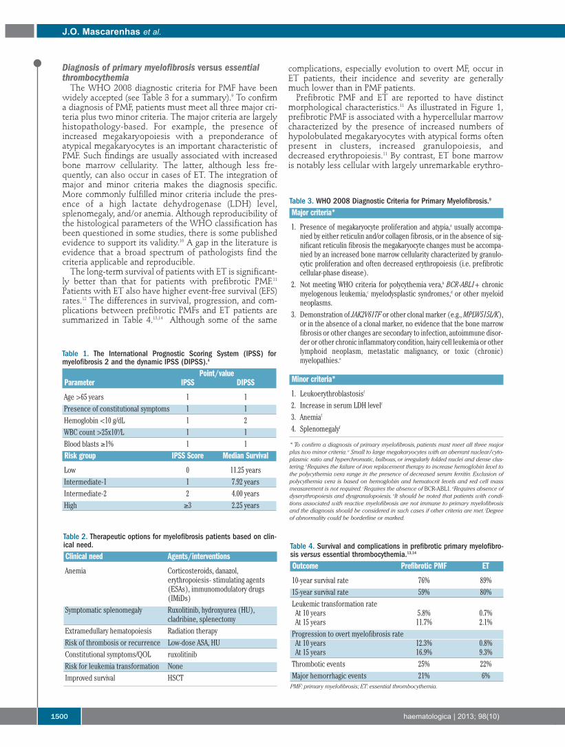
Table 1. The International Prognostic Scoring System (IPSS) formyelofibrosis 2 and the dynamic IPSS (DIPSS).4
Point/valueParameter IPSS DIPSS
Age >65 years 1 1Presence of constitutional symptoms 1 1Hemoglobin <10 g/dL 1 2WBC count >25x109/L 1 1Blood blasts ≥1% 1 1Risk group IPSS Score Median Survival
Low 0 11.25 yearsIntermediate-1 1 7.92 yearsIntermediate-2 2 4.00 yearsHigh ≥3 2.25 years
Table 3. WHO 2008 Diagnostic Criteria for Primary Myelofibrosis.9
Major criteria*
1. Presence of megakaryocyte proliferation and atypia,a usually accompa-nied by either reticulin and/or collagen fibrosis, or in the absence of sig-nificant reticulin fibrosis the megakaryocyte changes must be accompa-nied by an increased bone marrow cellularity characterized by granulo-cytic proliferation and often decreased erythropoiesis (i.e. prefibroticcellular-phase disease).
2. Not meeting WHO criteria for polycythemia vera,b BCR-ABL1+ chronicmyelogenous leukemia,c myelodysplastic syndromes,d or other myeloidneoplasms.
3. Demonstration of JAK2V617F or other clonal marker (e.g., MPLW515L/K),or in the absence of a clonal marker, no evidence that the bone marrowfibrosis or other changes are secondary to infection, autoimmune disor-der or other chronic inflammatory condition, hairy cell leukemia or otherlymphoid neoplasm, metastatic malignancy, or toxic (chronic)myelopathies.e
Minor criteria*
1. Leukoerythroblastosisf
2. Increase in serum LDH levelf
3. Anemiaf
4. Splenomegalyf
* To confirm a diagnosis of primary myelofibrosis, patients must meet all three majorplus two minor criteria. a Small to large megakaryocytes with an aberrant nuclear/cyto-plasmic ratio and hyperchromatic, bulbous, or irregularly folded nuclei and dense clus-tering. bRequires the failure of iron replacement therapy to increase hemoglobin level tothe polycythemia vera range in the presence of decreased serum ferritin. Exclusion ofpolycythemia vera is based on hemoglobin and hematocrit levels and red cell massmeasurement is not required. cRequires the absence of BCR-ABL1. dRequires absence ofdyserythropoiesis and dysgranulopoiesis. eIt should be noted that patients with condi-tions associated with reactive myelofibrosis are not immune to primary myelofibrosisand the diagnosis should be considered in such cases if other criteria are met. fDegreeof abnormality could be borderline or marked.
Table 4. Survival and complications in prefibrotic primary myelofibro-sis versus essential thrombocythemia.13,14
Outcome Prefibrotic PMF ET
10-year survival rate 76% 89%15-year survival rate 59% 80%Leukemic transformation rateAt 10 years 5.8% 0.7%At 15 years 11.7% 2.1%Progression to overt myelofibrosis rateAt 10 years 12.3% 0.8%At 15 years 16.9% 9.3%Thrombotic events 25% 22%Major hemorrhagic events 21% 6%PMF: primary myelofibrosis; ET: essential thrombocythemia.
Table 2. Therapeutic options for myelofibrosis patients based on clin-ical need.Clinical need Agents/interventions
Anemia Corticosteroids, danazol, erythropoiesis- stimulating agents(ESAs), immunomodulatory drugs(IMiDs)
Symptomatic splenomegaly Ruxolitinib, hydroxyurea (HU),cladribine, splenectomy
Extramedullary hematopoiesis Radiation therapyRisk of thrombosis or recurrence Low-dose ASA, HUConstitutional symptoms/QOL ruxolitinibRisk for leukemia transformation NoneImproved survival HSCT

poiesis and granulopoiesis. Megakaryopoiesis is increasedwith large hyperlobulated (staghorn-like) megakary-ocytes. Problems may arise when there is a mixture ofmegakaryocyte morphology.
Polycythemia vera versus essential thrombocythemiaand primary myelofibrosisIn contrast to ET, which has no specific distinguishing
clinical or laboratory features and, therefore, remains adiagnosis of exclusion,7 a unique set of PV diagnostic crite-ria has been developed.15 A diagnosis of PV requires thepresence of both major criteria and one minor criterion orthe presence of the first major together with two minorcriteria. Importantly, 50% of ET patients harborJAK2V617F. Although the diagnostic criteria for PV areuseful, they may not capture early phases of the disease.The so-called “pre-polycythemic” PV must be excluded inthrombocytotic patients, particularly those with microcyt-ic anemia, low ferritin, and/or who lack stainable bonemarrow iron. The first major WHO criterion for diagnos-ing PV is often not fulfilled in such patients and they maybe erroneously considered to have ET. Early stage PV can be distinguished from ET by bone
marrow histology. PV can be suspected in cases with ahypercellular panmyelotic marrow with pleomorphicmegakaryocytes. In addition, a marrow fibrosis grade 1-2on a scale of 4 is much more commonly seen in PV than inET.A diagnosis of prefibrotic PMF raises several questions
that still have to be answered. 1) Can IFN or JAK2
inhibitor therapy be used to prevent disease progression?2) Should low-dose aspirin or hydroxyurea (HU) be usedfor thrombosis prophylaxis? 3) Is observation and bloodcount monitoring sufficient? 4) Should bone marrow biop-sies be periodically repeated? Unfortunately, the therapeu-tic implications of distinguishing between ET and earlyPMF remain uncertain, and a therapeutic strategy that pre-vents or delays disease progression to overt MF has yet tobe identified. Cases 2 and 3 (see below) illustrate howsome of these questions are answered.
JAK2 inhibitorsBased on the observation that approximately 50% of
patients with MF harbor the JAK2V617Fmutation,6 devel-opment of JAK2 inhibitor therapies has been actively pur-sued. The first such agent to be developed and given topatients is ruxolitinib (Jakafi, Incyte; JAKAVI, Novartis), asmall-molecule inhibitor of JAK1/JAK2 that was approvedin 2011 for the treatment of patients with intermediate- orhigh-risk MF, including PMF, post-PV MF, and post-ETMF.16 Ruxolitinib was evaluated in a phase I/II study ofpatients with MF that established the safety and efficacyof this agent in terms of symptom improvement andspleen reduction, and provided the rationale for phase IIIstudies.17 This study also established what has subse-quently become a standard for determining response toJAK inhibitors in clinical studies, which is the use of mag-netic resonance imaging (MRI) to determine spleen vol-ume based on the correlation between a 35% reduction involume and a 50% reduction in palpable spleen length.
Advances in myelofibrosis
haematologica | 2013; 98(10) 1501
Figure 1. Prefibrotic PMF isassociated with a hypercellularmarrow characterized by thepresence of increased numbersof hypolobulated megakary-ocytes with atypical forms oftenpresent in clusters, increasedgranulopoiesis and decreasederythropoiesis (left). ET bonemarrow is less cellular withlargely unremarkable erythro-poiesis and granulopoiesis(right). Megakaryopoiesis isincreased with large hyperlobu-lated (staghorn-like) megakary-ocytes. (Top): republished with permission ofAmerican Society of Hematology.Permission conveyed throughCopyright Clearance Center, Inc.11

The COMFORT-I and COMFORT-II studies were amongthe first randomized phase III studies to be conducted inMF patients.18,19 These studies had similar designs; COM-FORT-I was conducted in the United States, Canada, andAustralia and compared ruxolitinib to placebo, whileCOMFORT-II was conducted in Europe and comparedruxolitinib to best available therapy. Up-dated safety and efficacy results for the COMFORT-
I and COMFORT-II studies were presented at theAmerican Society of Hematology (ASH) meeting inDecember 2012 (Table 5).20,21 Ruxolitinib treatment result-ed in a significant decrease in spleen reduction, with41.9% of patients in COMFORT-I achieving a 35% reduc-tion or more in spleen volume at Week 24 and 28.5% ofpatients in COMFORT-II achieving a 35% or more reduc-tion in spleen volume at Week 48. The degree of spleenreduction appeared to correlate with improved outcome.A subgroup analysis of COMFORT-II revealed that thevast majority of both JAK2V617F+ and JAK2V617F–patients experienced a reduction in spleen volume.22Overall, responses were observed for ruxolitinib-treatedpatients in all subgroups and were higher than those inpatients receiving best available therapy (BAT).Statistically significant differences in overall survival (OS)were observed between study arms in COMFORT-I(Figure 2), and a survival benefit was also observed inCOMFORT-II. The dose and duration of ruxolitinib thera-py, as well as the degree of spleen reduction, appear to becritical to survival outcomes.The most common adverse events in both studies were
anemia and thrombocytopenia, although it should benoted that the majority of patients had grade 1-2 anemiaand/or thrombocytopenia at baseline. To address the ane-
mia, a small number of patients (n=13) in the COMFORT-II study received recombinant erythropoiesis-stimulatingagents (ESAs), which appeared to be well tolerated and,interestingly, did not result in an increase in spleen size.The management of thrombocytopenic patients treatedwith ruxolitinib is currently being evaluated in theEXPAND phase Ib study and a study sponsored by Incyte.Non-hematologic adverse events associated with ruxoli-tinib therapy were not serious in nature and the most fre-quently reported were headache, dizziness, and easybruising unrelated to the platelet count. Both of thesestudies were also up-dated at the ASH 2012 meeting andappear to show equivalent efficacy and tolerability in thissubgroup of patients.23,24Although no specific pattern of return of symptoms
after withdrawal of ruxolitinib was reported in eitherCOMFORT study, an investigator at a single institutionhas reported the occurrence of life-threatening hemody-namic instability after abrupt cessation of ruxolitinib.25Attempts to taper ruxolitinib whenever possible, com-bined with the use of prednisone taper to blunt the acutereturn of symptoms, are generally recommended. In addition to ruxolitinib, there are a significant number
of other JAK inhibitors at different phases of development.Most of them show the same profile of efficacy as ruxoli-tinib, i.e. spleen and symptom reduction. However, asdata from early phase studies indicate, some of theseagents have different tolerability and efficacy. A phase I/IIstudy26 with SAR302503 reported that SAR302503 wasgenerally well tolerated with frequent grade 1 adverseevents, and significantly improved symptoms, such asanorexia and pruritus. However, in contrast to the findingswith ruxolitinib, these effects occurred in the absence of a
J.O. Mascarenhas et al.
1502 haematologica | 2013; 98(10)
Figure 2. Ruxolitinib-treatedpatients in COMFORT-I experi-enced a statistically significantincrease in overall survival (OS)compared to placebo. Reprinted with permission fromMassachusetts Medical Society.18
Table 5. COMFORT-I and COMFORT-II studies.18-21
COMFORT-I COMFORT-II
N. 309, randomized 1:1 219, randomized 2:1Primary end point ≥35% reduction in spleen volume at Week 24 ≥35% reduction in spleen volume at Week 48% Patients that achieved primary end point 41.9% 28.5%Survival Ruxolitinib associated with OS benefit Ruxolitinib associated with OS benefit
(HR=0.58; 95% CI: 0.36-0.95; P=0.028) (HR=0.54; 95% CI: 0.27-1.00)
Survival probability
1.0
0.8
0.6
0.4
0.2
0.0
HR=0.50 (0.25-0.98)
Ruxolitinib Placebo
Number of patients at risk--Ruxolitinib
Number of patients at risk--Placebo155 155 155 154 153 152 148 144 143 143 140 134 102 68 52 37 18 8 3
154 152 151 148 147 147 142 139 132 131 123 115 83 58 45 35 20 9 3
0 4 8 12 16 20 24 28 32 36 40 44 48 52 56 60 64 68 72 76Weeks
P=0.04
marked reduction in serum pro-inflammatory cytokinelevels (e.g. IL-2, IL-6, IL-8, TNF-α) and in the absence ofsignificant JAK1 inhibition. Two unique, potentially signif-icant aspects of response to this agent include a decreasein the JAK2V617F allele burden during therapy in themutation-positive subjects and the recently reportedreduction in marrow fibrosis scores.27 Following six cycles,16 of 20 (80%) patients with a base-line JAK2V617F alleleburden of more than 20% experienced a median reductionof 61%. These preliminary results reported withSAR302503 are promising and of great interest if con-firmed in phase III studies, although their clinical signifi-cance remains to be assessed, as the benefit is unlikely toequate to the reduction in the level of BCR/ABL1 burdenin CML. The phase III JAKARTA study has completedrecruitment and results are expected this year; in addition,JAKARTA 2, evaluating this agent in patients resistant orrefractory to ruxolitinib, is also open. In the phase II studywith SB1518 (pacritinib), rapid and sustained responses inspleen have been seen for MF at the 400 mg/die dose.28 Inan update presented at the ASH 2011 meeting, after amedian time on-study of 8.2 months (range 0.5-12.1), 50%of patients had discontinued treatment and response rateswere 44% on physical examination and 32% by MRI(35% reduction in volume). Two patients met criteria foranemia response,28 and a phase III study has beenlaunched. Pardanani recently presented data from a phaseI/II multicenter study with CYT387 demonstrating theanticipated improvements in splenomegaly and constitu-tional symptoms, as well as in transfusion requirements.29Subjects have now reached a minimum of nine months onstudy, and up-dated safety and efficacy results were pre-sented; 166 subjects were enrolled and the median dura-tion (range) of follow up is 16.1 months (0.7-31.0 months).Particular novel data of interest with this compound aretransfusion independence responses that were observed inmore than half of the RBC transfusion-dependent subjectswith a maximal transfusion-free period exceeding twoyears and still ongoing. In addition, the percentage of allsubjects requiring RBC transfusions substantiallydecreased over the treatment period. As has been previ-ously reported, treatment with CYT387 resulted in rapidand sustained reductions in splenomegaly, with maximalresponse duration now approaching two years.Concerning safety, the most common treatment-relatedAEs were thrombocytopenia, peripheral neuropathy,dizziness, diarrhea, nausea, and headache. Treatment-related peripheral neuropathy is a characteristic of thisagent. It was reported as sensory, and mainly grade 1.There were no treatment-related deaths. A phase III studywith this agent is also due to start shortly.Although JAK inhibition clearly reduces the sympto-
matic burden of MF, it is neither curative nor effective inreducing the risk of leukemic transformation. Strategies toimprove upon JAK inhibition therapy include explorationof other investigational small molecule JAK inhibitors,combining JAK inhibitors with other agents, and combin-ing JAK inhibitors with HSCT. The key to optimal JAKinhibitor therapy is likely to lie between patient selectionand proper dosing strategy.
The evolving role of histone deacetylase inhibitortherapy for myeloproliferative neoplasms Epigenetic changes are increasingly being recognized as
playing an important role in the progression of malignan-
cies, including MPNs. Such changes encompass biochemi-cal modifications to DNA or histones that are somaticallyheritable from mother cell to daughter cell. Epigeneticmodifications lead to changes in the expression of down-stream genes, and also contribute to genomic instabilityand drug resistance. Thus far, two of the most well-stud-ied epigenetic processes are DNA methylation and histonedeacetylation. DNA methyltransferases (DNMTs) cat-alyze the methylation of CpG islands within promotersites of DNA, thereby down-regulating transcription byblocking access to transcription factor complexes.Observations that support deregulation of DNA methyla-tion in MPNs include hypermethylation of key genesimportant for cell cycling, differentiation, and homing ofhematopoietic cells to the bone marrow,30-32 and hyperme-thylation of genes that negatively regulate the hyperactiveJAK/STAT signaling pathway.33Histone deacetylases (HDACs) remove acetyl groups
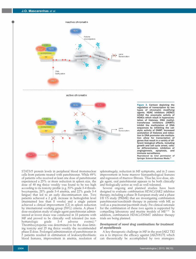
from lysine residues on histone tails, thereby inducing aninactive or closed conformation that restricts access oftranscription factors to DNA and thus downregulation oftranscription. HDACs fall into four classes that differ intheir subcellular localization, tissue distribution, sub-strates, and binding partners. Two HDAC inhibitors(HDACis) have been FDA-approved for the treatment ofcutaneous T-cell lymphoma, and several others are in clin-ical development for a wide array of cancers. HDACismodulate the acetylation status of histones as well asother non-histone proteins, resulting in a variety of differ-ent biological effects, including growth and cell cyclearrest, cellular differentiation, inhibition of angiogenesis,apoptosis, and immune surveillance (Figure 3). EnhancedHDAC expression has been observed in patients withPMF.35 Pre-clinical data support the use of HDACis inMPN.36-38 Characteristics and preliminary results for theuse of several HDAC inhibitors in MPNs are describedbelow.Pracinostat is an orally bioavailable pan-HDACi that has
1,000-fold selectivity for class 1 and 2 HDACs.39,40 In aphase II study of 22 patients with intermediate- and high-risk MF, single-agent pracinostat resulted in reductions insplenomegaly in 27% of patients. The most commonadverse event was fatigue, and grade 3-4 toxicities includ-ed neutropenia in 13% of patients and thrombocytopeniain 21% of patients; no grade 3-4 anemia was observed.41Givinostat is an orally bioavailable inhibitor of class 1
and 2 HDACs.42 The safety and activity of single-agentgivinostat were demonstrated in a phase II study inpatients with JAK2V617F+ MPNs (MF, n=16; ET, n=1; PV,n=12).43 Overall, 38% of patients with MF and 75% ofpatients with ET/PV experienced a reduction insplenomegaly, prompting phase II evaluation in patientswith ET/PV. In a separate study, combinationgivinostat/HU in patients with JAK2V617F+ PV who hadsuboptimal responses with single-agent HU resulted in aresponse rate of 45-50% and a notable reduction in pruri-tus in a majority of patients; no grade 3-4 toxicities wereobserved.44Panobinostat is a pan-HDACi with superior nanomolar
potency for inhibition of class 1, 2, and 4 HDACs com-pared to other HDAC inhibitors.45-47 A phase II study ofsingle-agent panobinostat was conducted in 35 patientswith intermediate/high risk MF.48 Molecular assaysshowed decreased JAK2 and PRV1 mRNA expression,increased histone acetylation, and decreased JAK2 and p-
Advances in myelofibrosis
haematologica | 2013; 98(10) 1503
STAT3/5 protein levels in peripheral blood mononuclearcells from patients treated with panobinostat. While 69%of patients who received at least one dose of panobinostatexperienced a 25% or more reduction in spleen size, thedose of 40 mg thrice weekly was found to be too highaccording to its toxicity profile (e.g. 53% grade 3-4 throm-bocytopenia, 28% grade 3-4 anemia, and 22% grade 3-4fatigue) that led to an early discontinuation rate. Twopatients achieved a 2 g/dL increase in hemoglobin level(maintained less than 8 weeks) and a single patientachieved a clinical improvement (CI) in spleen reductionby international working group (IWG) criteria. A phase Idose-escalation study of single-agent panobinostat admin-istered at lower doses was conducted in 18 patients withMF and proved to be clinically well tolerated (no non-hematologic grade 3-4 adverse events).49Thrombocytopenia was determined to be the dose-limit-ing toxicity and 25 mg thrice weekly the recommendedphase II dose. Prolonged administration of panobinostat in5 patients resulted in elimination of leukoerythroblasticblood features, improvement in anemia, resolution of
splenomegaly, reduction in MF symptoms, and in 2 casesimprovement in bone marrow histopathological featuresand regression of marrow fibrosis. Thus far, low-dose, sin-gle agent, oral panobinostat appears to be both clinicallyand biologically active as well as well tolerated. Several ongoing and planned studies have been
designed to evaluate combination HDACi/JAK2 inhibitortherapy, including a phase Ib European study and a phaseI/II US study (PRIME) that are investigating combinationpanobinostat/ruxolitinib therapy in patients with MF, aswell as a pracinostat/pacritinib study. Pre-clinical rationalefor the combination of these two agents is supported bycompelling laboratory and murine models of MF.37,50 Inaddition, combination HDACi/DNMT inhibitor therapytrials are being planned.
Development of novel drug combinations for treatmentof myelofibrosisA key therapeutic challenge in MF in the post-JAK2 TKI
era is to improve the efficacy against JAK2V617F, whichcan theoretically be accomplished by two strategies:
J.O. Mascarenhas et al.
1504 haematologica | 2013; 98(10)
Figure 3. Cartoon depicting theregulation of transcription by twotypes of chromatin modifyingagents. HDAC inhibitors (HDACI)inhibit the enzymatic activity ofHDACs which result in hyperacety-lation of histones. DNA methyl-transferase inhibitors (DNMTI)inhibit the methylation of DNAsequences by inhibiting the cat-alytic activity of DNMT. Increasedacetylation of histones and reduc-tion in DNA promoter site methyla-tion allow for transcription ofgenes that result in a variety of dif-ferent biological effects, includinggrowth and cell cycle arrest, cellu-lar differentiation, inhibition ofangiogenesis, apoptosis, andimmune surveillance. Reproduced by kind permission ofSpringer Science+Business Media.34

reducing JAK2V617F allelic burden and inactivating down-stream components of the JAK/STAT signal transductionpathway. Currently, four classes of investigational agentsare being evaluated that target JAK2V617F or downstreamsignaling molecules, as shown in Figure 4: HDACis/Hsp90inhibitors, PI3K/mTOR inhibitors, PIM kinase inhibitors,and MEK inhibitors.51
Heat-shock protein 90 (Hsp90) inhibitorsHsp90 is an ATP-dependent, dimeric molecular chaper-
one that folds and stabilizes its client proteins, includingJAK2 and STAT5, into their active conformations.52 Hsp90inhibitors bind to the N-terminal ATP-binding domain ofHsp90 and inhibit its chaperone function, which has beenshown to induce proteasome-mediated degradation ofclient proteins in MPN cells. Interestingly and importantly,pan-HDACis, such as vorinostat and panobinostat, induceHsp90 acetylation, inhibit Hsp90 chaperone function, andpromote proteasomal degradation of Hsp90 client proteinsin MPN cells.Hsp90 inhibitors have shown promise in both reducing
JAK2V617F allelic burden and overcoming resistance toJAK2 inhibitor therapy. Panobinostat is an HDACi thatalso mediates acetylation of Hsp90. This agent has beenshown to induce apoptosis in JAK2V617F+ MPN cells,likely via the inhibition of chaperone associationbetween JAK2 and Hsp90, thereby resulting in proteaso-mal degradation of JAK2.53 Co-treatment of MPNstem/progenitor cells with panobinostat and the JAK2
inhibitor TG101209 results in increased cell death com-pared to treatment with either compound alone, provid-ing the rationale for combination JAK2/Hsp90 inhibitortherapy as noted above.53AUY922 is an Hsp90 inhibitor that has been shown to
deplete JAK2 and to induce apoptosis in MPN cells. Thiscompound is also known to disrupt chaperone associationof JAK2 with Hsp90 and induces degradation of JAK2.Similar to the results observed with panobinostatdescribed above, co-treatment of primary MPN cells withAUY922 and the JAK2 inhibitor TG101209 enhancesapoptosis.54 Two lines of evidence suggest that Hsp90 inhibitors also
appear to have a potential role in overcoming resistance toJAK2 inhibition. First, JAK2 TKI-resistant MPN cells havebeen shown to exhibit greater sensitivity to AUY922 thannon-resistant cells.54 Second, the Hsp90 inhibitor PU-H71was demonstrated to abrogate heterodimeric JAK/STATactivation in JAK2 inhibitor therapy-resistant cell lines.55
PI3K/mTOR inhibitorsThe PI3K/mTOR signaling pathway plays an important
role in cell growth and proliferation in many malignancies,including MF.56-58 BEZ235 is a dual PI3K/mTOR inhibitorthat has been shown to induce apoptosis in MPN cells.Co-treatment of PMF cells with BEZ235 and the JAK2inhibitor SAR302503 enhances JAK2 inhibitor-mediatedloss of survival, and BEZ235 also induces apoptosis inJAK2-TKI–resistant MPN cells.59
Advances in myelofibrosis
haematologica | 2013; 98(10) 1505
Figure 4. HDACis/Hsp90inhibitors, PI3K/mTORinhibitors, PIM kinaseinhibitors, and MEKinhibitors targetJAK2V617F or down-stream signaling mole-cules.Reproduced by kind permis-sion of Elsevier 2012.51
Additional investigational agentsIn addition to agents that abrogate Hsp90 activity, sev-
eral other targeted therapies are currently being evaluatedfor their activity in MF. Agents that show activity alone orin combination with a JAK2 inhibitor include the MEKinhibitor AZD6244, the PIM kinase inhibitor SGI1776, theβ-catenin antagonist BC2059, and DNMT1 inhibitors.51
Hematopoietic stem cell transplantation for myelofibrosisAllogeneic HSCT remains the only potentially curative
therapy for MF. HSCT is an established treatment capableof eradication of the disease process, normalization ofbone marrow findings (including reversal of bone marrowreticulin and collagen fibrosis), and produces durable dis-ease-free survival (DFS). However, this “high-reward”treatment is also associated with high risk, and representsa major commitment for the patient. An important issue regarding the use of HSCT in MF
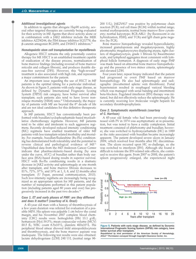
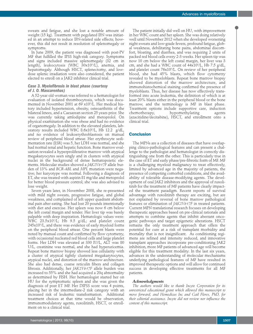
patients is the optimal timing for a particular individual.As shown in Figure 5, patients with early stage disease, asdefined by Dynamic International Prognostic ScoringSystem (DIPSS) risk category, have better survival aftertransplant; such patients also tend to have lower non-relapse mortality (NRM) rates.60 Unfortunately, the major-ity of patients with MF are beyond the 6th decade of lifeand are not ideal candidates for this definitive therapeuticapproach. In MF patients, HSCT has conventionally been per-
formed with busulfan/cyclophosphamide-based myeloab-lative chemotherapy regimens. However, MF patientstend to be older and intolerant to such aggressive regi-mens. Recently developed reduced intensity conditioning(RIC) regimens have enabled treatment of older MFpatients with less transplant-related morbidity and mortal-ity. For example, busulfan/fludarabine (Bu-Flu) condition-ing has demonstrated both tolerability and the potential toreverse clinical and pathological evidence of MF.61Unpublished data from the MD Anderson Cancer Centerindicate that pharmacokinetic-based dosing (by areaunder the curve, AUC) of busulfan rather than body sur-face area (BSA)-based dosing results in superior survival.HSCT with Bu-Flu conditioning results in a dramaticdecrease in JAK2 activity and splenomegaly at one monthafter transplant, and bone marrow fibrosis decreases to81%, 72%, 37%, and 19% at 1, 3, 6, and 12 months aftertransplant. (U Popat, personal communication, 2013).Such low-intensity regimens are increasingly being recog-nized as an appropriate option for MF patients, and thenumber of transplants performed in this patient popula-tion (including patients aged 60 years and over) has pro-gressively increased in the past two decades.62
Case 1. ET and early phases of PMF: are they differentand does it matter? (courtesy of A. Orazi)A 43-year old man with a history of thrombocytosis of
a few years duration was referred for evaluation of a pos-sible MPN. His spleen was palpable 1 cm below the costalmargin, and his November 2007 complete blood chem-istry (CBC) results were: hemoglobin (Hb) 13.1 g/dL;hematocrit (Hct) 39.5%; mean corpuscular volume (MCV)80.5 fL; WBC count 6.5x10⁹/L; platelets 483x109/L. Hisperipheral blood smear showed mild anisopoikilocytosisand thrombocytosis, and the bone marrow aspirate wasinadequate. The following test results were also obtained:lactate dehydrogenase (LDH) 248 U/L (normal range 96-
200 U/L); JAK2V617 was positive by polymerase chainreaction (PCR); red cell mass (RCM) within normal range;normal erythropoietin; normal bone marrow flow cytom-etry; normal karyotype; BCR-ABL1– (by fluorescent in situhybridization, FISH); and TCRγ and IgH chain gene nega-tive (by PCR). Bone marrow histopathology revealed hypercellularity,
increased granulopoiesis and megakaryopoiesis, highlypleomorphic megakaryocytes displaying atypia, tight clus-ters of megakaryoctyes, grade 1 fibrosis by Bauermeisterscale, and lymphoid aggregates most consistent with lym-phoid follicle formation. A diagnosis of early stage PMFwas made based on abnormal bone marrow histopatholo-gy and the presence of two minor criteria, splenomegalyand elevated LDH. Four years later, repeat biopsy indicated that the patient
had progressed to overt PMF based on marrowhistopathology. He also had splenomegaly and radi-ographic (documented splenic vein thrombosis). Portalhypertension resulted in esophageal variceal bleedingwhich was managed with serial banding and intermittentbeta-blockers. Pegylated interferon (IFN) therapy was ini-tiated, but did not effectively reduce the splenomegaly. Heis currently receiving low molecular weight heparin forsecondary thromboprophylaxis.
Case 2. Symptomatic myelofibrosis (courtesy of C. Harrison)A 65-year old female who had been previously diag-
nosed with PV in 1974 was asymptomatic at re-presenta-tion, but was noted to have a ruddy complexion. Initialtreatment consisted of phlebotomy and busulfan; howev-er, she was switched to hydroxycarbamide (HC) in 1989as the risks associated with busulfan became increasinglyapparent. The patient developed severe ulcers in January2009, which resolved four months after HC discontinua-tion. The ulcers recurred upon HC re-challenge, so shewas switched to interferon (IFN). Although she found itdifficult to tolerate the IFN-related side effects, she contin-ued to receive this agent. From 2007 to 2008, the patient’sspleen progressively enlarged, she experienced night
J.O. Mascarenhas et al.
1506 haematologica | 2013; 98(10)
Figure 5. Patients with early stage disease, as defined by DynamicInternational Prognostic Scoring System (DIPSS) risk category, havebetter survival after transplant.Republished by kind permission of the American Society of Hematology,2012.60 Permission conveyed through Copyright Clearance Center, Inc.
0 2 4 6 8 10 12 14 16 18 20Years after transplant
1.0
0.8
0.6
0.4
0.2
0.0
Probability of survival
sweats and fatigue, and she lost a notable amount ofweight (15 kg). Treatment with pegylated IFN was initiat-ed in an attempt to reduce IFN-related side effects; how-ever, this did not result in resolution of splenomegaly orsymptoms.In June 2009, the patient was diagnosed with post-PV
MF that fulfilled the IPSS high-risk category. Symptomsand signs included massive splenomegaly (32 cm inlength), leukocytosis (WBC 30x109/L), anemia, andhepatomegaly. Although HSCT, splenectomy, and low-dose splenic irradiation were also considered, the patientelected to enroll on a JAK2 inhibitor clinical trial.
Case 3. Myelofibrosis in blast phase (courtesy of J. O. Mascarenhas)A 52-year old woman was referred to a hematologist for
evaluation of isolated thrombocytosis, which was docu-mented in November 2001 at 69 x109/L. Her medical his-tory included hypertension, obesity, osteoarthritis of thebilateral knees, and a Caesarean section 25 years prior. Shewas currently taking amlodipine and metoprolol. Onphysical examination she was obese and had no evidenceof organomegaly. In addition to the elevated platelets, lab-oratory results included WBC 8.6x10⁹/L, Hb 12.2 g/dL,and no evidence of leukoerythroblastosis on manualreview of peripheral blood smear. Her erythrocyte sedi-mentation rate (ESR) was 5, her LDH was normal, and shehad normal renal and hepatic function. Bone marrow eval-uation revealed a hyperproliferative marrow with atypicalmegakaryocytes seen singly and in clusters with atypicalnuclei in the background of dense hematopoietic ele-ments. Molecular studies showed a JAK2V617F allele bur-den of 13% and BCR-ABL and MPL mutation were nega-tive; her karyotype was normal. Following a diagnosis ofET, she was treated with aspirin 81 mg/die and metoprololfor better blood pressure control; she was also advised tolose weight. Seven years later, in November 2008, she re-presented
with mild night sweats, progressive fatigue, and globalweakness, and complained of left upper quadrant abdom-inal pain after eating. She had lost 20 pounds intentionallywith diet and exercise. Her spleen was now 6 cm belowthe left costal margin and tender. Her liver tip was barelypalpable with deep inspiration. Hematologic values were:WBC 23.5x10⁹/L; Hb 9.5 g/dL; and platelet count245x109/L, and there was evidence of early myeloid formson the peripheral blood smear. One percent blasts werenoted by manual count and confirmed by flow cytometry,with occasional nucleated red blood cells and large plateletforms. Her LDH was elevated at 330 IU/L, ALT was 80U/L, creatinine was normal, and she had hyperuricemia.Repeat bone marrow biopsy showed less cellularity witha cluster of atypical tightly clustered megakarytocytes,atypical nuclei, and distortion of the marrow architecture.She also had dense, coarse reticulin fibers and collagenfibrosis. Additionally, her JAK2V617F allele burden wasincreased to 55% and she had acquired a 20q abnormalityas determined by FISH. Her hematologist started her onHU for the symptomatic spleen and she was given thediagnosis of post ET MF. Her DIPSS score was 4 points,placing her in the intermediate-2 risk category with anincreased risk of leukemic transformation. Additionaltreatment choices at that time would be observation,immunomodulatory agents, ruxolitinib, HSCT, or enroll-ment on to a clinical trial.
The patient initially did well on HU, with improvementin her WBC count and her spleen. She was doing relativelywell until November 2012 when she developed worseningnight sweats and low-grade fevers, profound fatigue, glob-al weakness, debilitating bone pains, abdominal discom-fort, bloating, and diarrhea, and was requiring 2 units ofpacked red blood cells every 2-3 weeks. Her spleen tip wasnow 10 cm below the left costal margin, her liver was 3cm, and she had a WBC count of 44x109/L, Hb 7.5 g/dL,and platelet count 76x109/L. On review of her peripheralblood, she had 45% blasts, which flow cytometryrevealed to be myeloblasts. Repeat bone marrow biopsyshowed distortion of the marrow architecture, andimmunohistochemical staining confirmed the presence ofmyeloblasts. Thus, her disease has now effectively trans-formed into acute leukemia, the definition of which is atleast 20% blasts either in the peripheral blood or the bonemarrow, and the terminology is MF in blast phase.Treatment options include supportive care, inductionchemotherapy, hypomethylating agents(azacitidine/decitabine), HSCT, and enrollment onto aclinical trial.
Conclusion
The MPNs are a collection of diseases that have overlap-ping clinico-pathological features and can present a chal-lenge to the pathologist and hematologist in correctly dis-tinguishing one from the other. This is particularly true inthe case of ET and early phase/pre-fibrotic form of MF. MFis a challenging myeloid malignancy to treat effectively,limited by advanced age in the majority of patients, thepresence of competing comorbid conditions, and the avail-ability of tolerable disease-modifying agents. The devel-opment of oral JAK2 inhibitors and the approval of ruxoli-tinib for the treatment of MF patients have clearly impact-ed the treatment paradigm. Recent reports of survivaladvantage with ruxolitinib therapy are exciting, but arenot explained by reversal of bone marrow pathologicalfeatures or elimination of JAK2V617F in treated patients.Current MPN translational research focuses on developingtherapeutic approaches based on pre-clinical rationale andattempts to combine agents that inhibit aberrant onco-genic pathways and target epigenetic alterations. HSCTremains the only treatment approach that offers thepotential for cure at a risk of transplant morbidity andmortality that is not insignificant. As conditioning regi-mens are refined and intensity reduced, and innovativetransplant approaches incorporate pre-conditioning JAK2inhibition, more MF patients of advanced age will becomeeligible for this treatment modality. In the last six years,advances in the understanding of molecular mechanismsunderlying pathological features of MF have resulted inimproved therapeutic options and will allow for continuedsuccess in developing effective treatments for all MFpatients.
AcknowledgmentsThe authors would like to thank Incyte Corporation for its
unrestricted educational grant which allowed this manuscript tomove forward, and Hemedicus Inc and Gail Flores, PhD, fortheir editorial assistance. Incyte did not review nor influence thecontent of this manuscript.
Advances in myelofibrosis
haematologica | 2013; 98(10) 1507

Authorship and DisclosuresInformation on authorship, contributions, and financial & other
disclosures was provided by the authors and is available with theonline version of this article at www.haematologica.org.
J.O. Mascarenhas et al.
1508 haematologica | 2013; 98(10)
References
1. Thiele J, Kvasnicka HM, Tefferi A, et al.Primary myelofibrosis. In: Swerdlow SH,Campo E, Harris NL et al, editors. WHOClassification of Tumours ofHaematopoietic and Lymphoid Tissues.IARC Lyon: World Health Organization,2008:44-7.
2. Cervantes F, Dupriez B, Pereira A,Passamonti F, Reilly JT, Morra E, et al. Newprognostic scoring system for primarymyelofibrosis based on a study of theInternational Working Group forMyelofibrosis Research and Treatment.Blood. 2009;113(13):2895-901.
3. Cervantes F, Dupriez B, Passamonti F,Vannucchi AM, Morra E, Reilly JT, et al.Improving survival trends in primarymyelofibrosis: an international study. J ClinOncol. 2012;30(24):2981-7.
4. Passamonti F, Cervantes F, Vannucchi AM,Morra E, Rumi E, Pereira A, et al. A dynamicprognostic model to predict survival in pri-mary myelofibrosis: a study by the IWG-MRT (International Working Group forMyeloproliferative Neoplasms Research andTreatment). Blood. 2010;115(9):1703-8.
5. Barbui T, Barosi G, Birgegard G, Cervantes F,Finazzi G, Griesshammer M, et al.Philadelphia-negative classical myeloprolif-erative neoplasms: critical concepts andmanagement recommendations fromEuropean LeukemiaNet. J Clin Oncol.2011;29(6):761-70.
6. Levine RL, Pardanani A, Tefferi A, GillilandDG. Role of JAK2 in the pathogenesis andtherapy of myeloproliferative disorders. NatRev Cancer. 2007;7(9):673-83.
7. Zhan H, Spivak JL. The diagnosis and man-agement of polycythemia vera, essentialthrombocythemia, and primary myelofibro-sis in the JAK2 V617F era. Clin Adv HematolOncol. 2009;7(5):334-42.
8. Jones AV, Kreil S, Zoi K, Waghorn K, CurtisC, Zhang L, et al. Widespread occurrence ofthe JAK2 V617F mutation in chronic myelo-proliferative disorders. Blood. 2005;106(6):2162-8.
9. Tefferi A, Vardiman JW. Classification anddiagnosis of myeloproliferative neoplasms:the 2008 World Health Organization criteriaand point-of-care diagnostic algorithms.Leukemia. 2008;22(1):14-22.
10. Barbui T, Thiele J, Vannucchi AM, Tefferi A.Problems and pitfalls regarding WHO-defined diagnosis of early/prefibrotic pri-mary myelofibrosis versus essential throm-bocythemia. Leukemia. 2013. [Epub aheadof print]
11. Thiele J, Kvasnicka HM, Mullauer L,Buxhofer-Ausch V, Gisslinger B, GisslingerH. Essential thrombocythemia versus earlyprimary myelofibrosis: a multicenter studyto validate the WHO classification. Blood.2011;117(21):5710-8.
12. Barbui T, Thiele J, Carobbio A, Passamonti F,Rumi E, Randi ML, et al. Disease character-istics and clinical outcome in young adultswith essential thrombocythemia versusearly/prefibrotic primary myelofibrosis.Blood. 2012;120(3):569-71.
13. Barbui T, Thiele J, Passamonti F, Rumi E,Boveri E, Ruggeri M, et al. Survival and dis-
ease progression in essential thrombo-cythemia are significantly influenced byaccurate morphologic diagnosis: an interna-tional study. J Clin Oncol. 2011;29(23):3179-84.
14. Finazzi G, Carobbio A, Thiele J, PassamontiF, Rumi E, Ruggeri M, et al. Incidence andrisk factors for bleeding in 1104 patientswith essential thrombocythemia or prefi-brotic myelofibrosis diagnosed according tothe 2008 WHO criteria. Leukemia. 2012;26(4):716-9.
15. Tefferi A, Thiele J, Orazi A, Kvasnicka HM,Barbui T, Hanson CA, et al. Proposals andrationale for revision of the World HealthOrganization diagnostic criteria for poly-cythemia vera, essential thrombocythemia,and primary myelofibrosis: recommenda-tions from an ad hoc international expertpanel. Blood. 2007;110(4):1092-7.
16. Incyte Corporation. Jakafi (R) (ruxolitinib)Full prescribing information. Available from:http://www.incyte.com/products/uspi_jakafi.pdf. Accessed 20 February 2013.
17. Verstovsek S, Kantarjian H, Mesa RA,Pardanani AD, Cortes-Franco J, ThomasDA, et al. Safety and efficacy ofINCB018424, a JAK1 and JAK2 inhibitor, inmyelofibrosis. N Engl J Med. 2010;363(12):1117-27.
18. Verstovsek S, Mesa RA, Gotlib J, Levy RS,Gupta V, DiPersio JF, et al. A double-blind,placebo-controlled trial of ruxolitinib formyelofibrosis. N Engl J Med.2012;366(9):799-807.
19. Harrison C, Kiladjian JJ, Al-Ali HK,Gisslinger H, Waltzman R, Stalbovskaya V,et al. JAK inhibition with ruxolitinib versusbest available therapy for myelofibrosis. NEngl J Med. 2012;366(9):787-98.
20. Verstovsek S, Mesa RA, Gotlib J, Levy RS,Gupta V, DiPersio JF, et al. Long-term out-come of ruxolitinib treatment in patientswith myelofibrosis: Durable reductions inspleen volume, improvements in quality oflife, and overall survival advantage in COM-FORT-I. Blood. 2012;120(21):Abstract 800.
21. Cervantes F, Kiladjian JJ, Niederwieser D,Sirulnik A, Stalbovskaya V, McQuity M, etal. Long-term safety, efficacy, and survivalfindings from Comfort-II, a phase 3 studycomparing ruxolitinib with best availabletherapy (BAT) for the treatment of myelofi-brosis (MF). Blood. 2012;120:(Abstract 801).
22. Harrison CN, Kiladjian JJ, Gisslinger H,Niederwieser D, Passamonti F, WaltzmanRJ, et al. Ruxolitinib provides reductions insplenomegaly across subgroups; An analysisof spleen response in the COMFORT-IIstudy. Blood. 2011;118:Abstract 279.
23. Talpaz M, Hamburg SI, Jamieson K,Terebelo HR, Afrin L, Winton EF, et al.Preliminary safety and eficiacy from a phaseII study of ruxolitinib in patients with pri-mary and secondary myelofibrosis withplatelets counts of 50-100 x 109/L.Haematologica. 2012;97:244 Abstract 597.
24. Harrison CN, Gisslinger H, Miller CB,Kiladjian JJ, Atienza E, Stalbovskaya V, et al.Expand: a phase 1b, open-label, dose-findingstudy of ruxolitinib in patients with myelofi-brosis and baseline platelet counts between5x109/L and 99x109/L. Blood. 2012;120(21):Abstract 177.
25. Tefferi A, Litzow MR, Pardanani A. Long-
term outcome of treatment with ruxolitinibin myelofibrosis. N Engl J Med.2011;365(15):1455-7.
26. Pardanani A, Gotlib J, Jamieson C, Cortes JE,Talpaz M, Stone R, et al. SAR302503:Interim safety, efficacy and long-termimpact on JAK2 V617F allele burden in aphase I/II study in patients with myelofibro-sis. Blood. 2011;118(21):Abstract 3838.
27. Talpaz M, Jamieson C, Gabrail NY,Lebedinsky C, Gao G, Liu F, et al. A phase IIrandomized dose-ranging study of the JAK2-selective inhibitor SAR302503 in patientswith intermediate-2 or high-risk primarymyelofibrosis (MF), or post-essential throm-bocythemia (ET) MF. Blood.2012;120(21):Abstract 2837.
28. Komrokji RS, Wadleigh M, Seymour JF,Roberts AW, To LB, Zhu HJ, et al. Results ofa phase 2 study of pacritinib (SB1518), anovel oral JAK2 inhibitor, in patients withprimary, post-polycythemia vera, and post-essential thrombocythemia myelofibrosis.Blood. 2011;118(21):Abstract 282.
29. Pardanani A, Gotlib J, Gupta V, Roberts AW,Wadleigh M, Sirhan S, et al. Phase I/II studyof CYT387, a JAK1/JAK2 inhibitor for thetreatment of myelofibrosis. Blood.2012;120(21):Abstract 178.
30. Wang JC, Chen W, Nallusamy S, Chen C,Novetsky AD. Hypermethylation of theP15INK4b and P16INK4a in agnogenicmyeloid metaplasia (AMM) and AMM inleukaemic transformation. Br J Haematol.2002;116(3):582-6.
31. Jones LC, Tefferi A, Idos GE, Kumagai T,Hofmann WK, Koeffler HP. RARbeta2 is acandidate tumor suppressor gene in myelofi-brosis with myeloid metaplasia. Oncogene.2004;23(47):7846-53.
32. Bogani C, Ponziani V, Guglielmelli P,Desterke C, Rosti V, Bosi A, et al.Hypermethylation of CXCR4 promoter inCD34+ cells from patients with primarymyelofibrosis. Stem Cells. 2008;26(8):1920-30.
33. Teofili L, Martini M, Cenci T, Guidi F, TortiL, Giona F, et al. Epigenetic alteration ofSOCS family members is a possible patho-genetic mechanism in JAK2 wild typemyeloproliferative diseases. Int J Cancer.2008;123(7):1586-92.
34. Mascarenhas J, Roper N, Chaurasia P,Hoffman R. Epigenetic abnormalities inmyeloproliferative neoplasms: a target fornovel therapeutic strategies. ClinEpigenetics. 2011;2(2):197-212.
35. Wang JC, Chen C, Dumlao T, Naik S, ChangT, Xiao YY, et al. Enhanced histone deacety-lase enzyme activity in primary myelofibro-sis. Leuk Lymphoma. 2008;49(12):2321-7.
36. Shi J, Zhao Y, Ishii T, Hu W, Sozer S, ZhangW, et al. Effects of chromatin-modifyingagents on CD34+ cells from patients withidiopathic myelofibrosis. Cancer Res.2007;67(13):6417-24.
37. Wang X, Zhang W, Ishii T, Sozer S, Wang J,Xu M, et al. Correction of the abnormal traf-ficking of primary myelofibrosis CD34+cells by treatment with chromatin-modify-ing agents. Cancer Res. 2009;69(19):7612-8.
38. Guerini V, Barbui V, Spinelli O, Salvi A,Dellacasa C, Carobbio A, et al. The histonedeacetylase inhibitor ITF2357 selectively tar-gets cells bearing mutated JAK2(V617F).

Leukemia. 2008;22(4):740-7.39. Garcia-Manero G, Chuah C, Wilding G,
Chang J, Verstovsek S, Faderl S, et al. Phase Istudy of the oral histone deacetylaseinhibitor SB939 in patients with advancedhematologic malignancies. Blood. 2010;116:Abstract 3292.
40. Wang H, Yu N, Chen D, Lee KC, Lye PL,Chang JW, et al. Discovery of (2E)-3-{2-butyl-1-[2-(diethylamino)ethyl]-1H-benzim-idazol-5-yl}-N-hydroxyacrylami de (SB939),an orally active histone deacetylase inhibitorwith a superior preclinical profile. J MedChem. 2011;54(13):4694-720.
41. Quintas-Cardama A, Kantarjian H, Estrov Z,Borthakur G, Cortes J, Verstovsek S.Therapy with the histone deacetylaseinhibitor pracinostat for patients withmyelofibrosis. Leuk Res. 2012;36(9):1124-7.
42. Golay J, Cuppini L, Leoni F, Mico C, BarbuiV, Domenghini M, et al. The histonedeacetylase inhibitor ITF2357 has anti-leukemic activity in vitro and in vivo andinhibits IL-6 and VEGF production by stro-mal cells. Leukemia. 2007;21(9):1892-900.
43. Rambaldi A, Dellacasa CM, Finazzi G,Carobbio A, Ferrari ML, Guglielmelli P, et al.A pilot study of the histone-deacetylaseinhibitor givinostat in patients withJAK2V617F positive chronic myeloprolifera-tive neoplasms. Br J Haematol. 2010;150(4):446-55.
44. Rambaldi A, Finazzi G, Vannucchi AM,Martinelli V, Rodeghiero F, Nobile F, et al. Aphase II study of the HDAC inhibitor givi-nostat in combination with hydroxyurea inpatients with polycythemia vera resistant tohydroxyurea monotherapy. Blood. 2011;118:Abstract 1748.
45. Prince HM, George D, Patnaik A, Mita M,Dugan M, Butterfoss D, et al. Phase I studyof oral LBH589, a novel deacetylase (DAC)inhibitor in advanced solid tumors and non-hodgkin's lymphoma. J Clin Oncol. 2007;25(18S):Abstract 3500.
46. Spencer A, Prince M, De Angelo DJ, FischerT, Bhalla KN, Giles FJ, et al. Phase IA/II study
of oral LBH589, a novel deacetylaseinhibitor (DACi), administered on 2 sched-ules, in patients with advanced hematologicmalignancies. Blood. 2007;110: Abstract 907.
47. Sharma S, Vogelzang NJ, Beck J, Patnaik A,Mita M, Dugan M, et al. Phase I pharmaco-kinetic and pharmacodynamic study ofonce-weekly IV LBH589. Eur J Cancer.2007;5(4):107: Abstract 702.
48. DeAngelo DJ, Tefferi A, Fiskus W, Mesa RA,Paley CS, Wadleigh M, et al. A phase II trialof panobinostat, an orally available deacety-lase inhibitor (DACi), in patients with pri-mary myelofibrosis (PMF), post essentialthrombocythemia (ET), and post poly-cythemia vera (PV) myelofibrosis. Blood.2010;116:Abstract 630.
49. Mascarenhas J, Lu M, Li T, Petersen B,Hochman T, Najfeld V, et al. A phase I studyof panobinostat (LBH589) in patients withprimary myelofibrosis (PMF) and post-poly-cythaemia vera/essential thrombo-cythaemia myelofibrosis (post-PV/ET MF).Br J Haematol. 2013;161(1):68-75.
50. Baffert F, Evrot E, Ebel N, Roelli C, AndraosR, Qian Z, et al. Improved efficacy uponcombined JAK1/2 and pan-deacetylase inhi-bition using ruxolitinib (INC424) andpanobinostat (LBH589) in preclinical mousemodels of JAK2V617-F-driven disease.Blood. 2011;118: Abstract 798.
51. Fiskus W, Ganguly S, Kambhampati S,Bhalla KN. Role of additional novel thera-pies in myeloproliferative neoplasms.Hematol Oncol Clin North Am.2012;26(5):959-80.
52. Whitesell L, Lindquist SL. HSP90 and thechaperoning of cancer. Nat Rev Cancer.2005;5(10):761-72.
53. Wang Y, Fiskus W, Chong DG, Buckley KM,Natarajan K, Rao R, et al. Cotreatment withpanobinostat and JAK2 inhibitor TG101209attenuates JAK2V617F levels and signalingand exerts synergistic cytotoxic effectsagainst human myeloproliferative neoplasticcells. Blood. 2009;114(24): 5024-33.
54. Fiskus W, Verstovsek S, Manshouri T, Rao R,
Balusu R, Venkannagari S, et al. Heat shockprotein 90 inhibitor is synergistic with JAK2inhibitor and overcomes resistance to JAK2-TKI in human myeloproliferative neoplasmcells. Clin Cancer Res. 2011;17(23):7347-58.
55. Koppikar P, Bhagwat N, Kilpivaara O,Manshouri T, Adli M, Hricik T, et al.Heterodimeric JAK-STAT activation as amechanism of persistence to JAK2 inhibitortherapy. Nature. 2012;489(7414):155-9.
56. Bjornsti MA, Houghton PJ. The TOR path-way: a target for cancer therapy. Nat RevCancer. 2004;4(5):335-48.
57. Guglielmelli P, Barosi G, Rambaldi A,Marchioli R, Masciulli A, Tozzi L, et al.Safety and efficacy of everolimus, a mTORinhibitor, as single agent in a phase 1/2 studyin patients with myelofibrosis. Blood.2011;118(8):2069-76.
58. Bogani C, Bartalucci N, Martinelli S, Tozzi L,Guglielmelli P, Bosi A, et al. mTORinhibitors alone and in combination withJAK2 inhibitors effectively inhibit cells ofmyeloproliferative neoplasms. PLoS One.2013;8(1):e54826.
59. Fiskus W, Verstovsek S, Manshouri T, SmithJE, Peth K, Abhyankar S, et al. Mol CancerTher. 2013;12(5):577-88.
60. Scott BL, Gooley TA, Sorror ML, RezvaniAR, Linenberger ML, Grim J, et al. TheDynamic International Prognostic ScoringSystem for myelofibrosis predicts outcomesafter hematopoietic cell transplantation.Blood. 2012;119(11):2657-64.
61. Kroger N, Holler E, Kobbe G, Bornhauser M,Schwerdtfeger R, Baurmann H, et al.Allogeneic stem cell transplantation afterreduced-intensity conditioning in patientswith myelofibrosis: a prospective, multicen-ter study of the Chronic Leukemia WorkingParty of the European Group for Blood andMarrow Transplantation. Blood. 2009;114(26):5264-70.
62. Gupta V, Hari P, Hoffman R. Allogeneichematopoietic cell transplantation formyelofibrosis in the era of JAK inhibitors.Blood. 2012;120(7):1367-79.
Advances in myelofibrosis
haematologica | 2013; 98(10) 1509