advances in the treatment of newly diagnosed glioblastoma | bmc
TRANSCRIPT

MINIREVIEW Open Access
Advances in the treatment of newlydiagnosed glioblastomaBrett J. Theeler1* and Mark R. Gilbert2
Abstract
Glioblastoma is a refractory malignancy with limited treatment options at tumor recurrence. Only a small proportionof patients survive 2 years or longer with the current standard of care. Gene expression profiling can segregatenewly diagnosed patients into groups with different prognoses, and these biomarkers are being incorporated into anew generation of personalized clinical trials. Using the experience from recently completed large scale, multi-faceted,randomized glioblastoma clinical trials, a new clinical trial paradigm is being established to move promising therapiesforward into the newly diagnosed treatment setting. Upcoming trials using the immune check-point inhibitors are anexample of this changing paradigm and these and other immunotherapies have potential as promising new treatmentmodalities for newly diagnosed GB patients.
Keywords: Glioblastoma, High grade glioma, Immunotherapy, Checkpoint inhibitors, Pseudoprogression
IntroductionGlioblastoma (GB) is the most common primary brainmalignancy in adults and accounts for 45.6 % of allprimary brain malignancies. GB has an incidence rate of3.19 per 100,000 and occurs at a median age of64.0 years, although GB can occur at all ages [1]. Themajority of GBs occur in the cerebral hemispheres,although brainstem, cerebellar, and spinal cord GBsrarely occur [2]. The average overall survival (OS) of GBpatients from population series is between 8 and 14 months[3]. Prognosis of GB can be stratified by clinical features,with age younger than 50 years, Karnofsky performancestatus (KPS) of 70 or better, non-eloquent tumor location,and maximal extent of resection all associated with im-proved patient outcomes [4, 5].The backbone of upfront treatment of GB is maximal
surgical resection and adjuvant radiotherapy to a dose of6,000 cGy. Upfront treatment often included nitrosoureas[6], although when added to surgical resection and radio-therapy modest benefit could only be demonstrated bymeta-analysis [7]. Temozolomide, an oral alkylating agentwith good blood-brain barrier penetration, was developedin the 1990s and showed benefit in the recurrent setting
in GBs and recurrent anaplastic gliomas gaining approvalfor this indication [8]. The randomized phase III, EORTC-NCIC trial 22981/26981, published in 2005, establishedthe current standard of care, including maximal surgicalresection followed by radiotherapy with concurrent andadjuvant temozolomide, and demonstrated improved OSof 14.6 versus 12.1 months and increased proportion of 2-year and 5-year survivorship compared to radiotherapyalone [9, 10].With approximately 5 % of patients surviving 5 years
from diagnosis, additional treatment options for newlydiagnosed GB patients are needed [10]. Further im-provement of upfront treatment of GB is importantas approved treatment options for tumor recurrence,including cytotoxic chemotherapy agents, such as ni-trogen mustards, bevacizumab, and tumor-treatmentfields, are of limited efficacy [11–13]. For example,approximately 20 % of patients with recurrent GBs treatedwith lomustine (CCNU) are alive and progression-free6 months after starting treatment. Bevacizumab, amonoclonal antibody against vascular endothelialgrowth factor (VEGF), is approved by the US Food andDrug Administration (FDA) for use in recurrent GBbased on a relatively high rate of radiographic responseand improvement in progression-free survival, but withno evidence to date of a significant improvement inoverall survival [14].
* Correspondence: [email protected] of Neurology and John P. Murtha Cancer Center, Walter ReedNational Military Medical Center, 8901 Wisconsin Avenue, Building 19,Bethesda, MD 20889, USAFull list of author information is available at the end of the article
© 2015 Theeler and Gilbert. Open Access This article is distributed under the terms of the Creative Commons Attribution 4.0International License (http://creativecommons.org/licenses/by/4.0/), which permits unrestricted use, distribution, andreproduction in any medium, provided you give appropriate credit to the original author(s) and the source, provide a link tothe Creative Commons license, and indicate if changes were made. The Creative Commons Public Domain Dedication waiver(http://creativecommons.org/publicdomain/zero/1.0/) applies to the data made available in this article, unless otherwise stated.
Theeler and Gilbert BMC Medicine (2015) 13:293 DOI 10.1186/s12916-015-0536-8

Prognostic biomarkers and gene expression profilingPrior clinical observations noted that secondary GBs,defined as GBs arising from grade II and III infiltratinggliomas, had improved outcomes compared to primaryGBs, or those tumors that arise as GBs de novo. Primaryand secondary GBs were noted to have different genemutations and to over-express different extra- and intra-cellular proteins; for example, epidermal growth factor(EGFR) over-expression is more common in primaryGBs [15]. Although, when controlling for age and otherpatient-related factors, the biologic markers of primaryand secondary GBs, including EGFR over-expression, arenot independent prognostic biomarkers.The DNA repair enzyme O6-methylguanine methyl-
transferase (MGMT) repairs O6- methylguanine adducts.Hypermethylation of the MGMT gene promoter leads tosilencing of MGMT expression. This mechanism cancounteract the effects of temozolomide which alkylatesthe O6 position on guanine, resulting in futile activationof the mismatch repair system and ultimately apoptosis[16]. In addition to establishing the standard of care, theEORTC-NCIC trial retrospectively demonstrated thatpatients with MGMT promoter methylation have animproved prognosis compared to those patients withunmethylated promoter regions [17]. In both the EORTC-NCIC trial and another single-institution retrospectivestudy, newly diagnosed GB patients with MGMT pro-moter methylation treated with upfront radiotherapyalone had improved survival outcomes, suggesting thatMGMT promoter methylation may be prognostic re-gardless of upfront treatment; however, patients withMGMT promoter methylation treated with temozolo-mide had the best overall survival outcomes in theEORTC-NCIC trial [9, 18].Given the antagonistic mechanisms of MGMT-mediated
DNA repair and temozolomide, it was hypothesized thathigher doses of temozolomide may overcome the DNArepair capacity of MGMT [19]. This hypothesis led to thedesign of RTOG 0525, a randomized phase III trial com-paring dose-intense temozolomide (75–100 milligrams permeter squared taken on days 1 to 21 of a 28-day cycle) ver-sus standard dose temozolomide (150–200 milligrams permeter squared on days 1 to 5 of a 28-day cycle). This trialincluded prospective tissue collection and stratification ofboth groups by clinical prognostic factors and MGMT pro-moter methylation. No benefit of dose-intense temozolo-mide was seen overall, or in the subgroups of MGMThypermethylated or unmethylated patients. RTOG 0525confirmed the prognostic significance of MGMT promotermethylation [20]. While an improved prognosis for newlydiagnosed patients with MGMT promoter methylationtreated with the EORTC-NCIC regimen is established,there is not an alternative regimen for newly diagnosed GBpatients with unmethylated MGMT promoters otherwise
eligible to receive the EORTC-NCIC regimen. Outside of aclinical trial, it is the authors’ opinion that all patients witha KPS of 70 or greater and an age of 65 years or less shouldreceive the EORTC-NCIC regimen regardless of MGMTpromoter methylation status.As indicated above, age is an important prognostic
factor [4]. GB patients over the age of 65 years and thosepatients with a KPS less than or equal to 60 represent aclinically important group of patients that develop sig-nificant toxicity, and there is no prospective evidence ofsignificant benefit from the EORTC-NCIC regimen inthese patients. In two retrospective studies of newlydiagnosed GB patients aged 65 years and older, including291 and 237 GB patients respectively, patients with agood performance status (KPS 70 or greater) and a max-imal tumor resection significantly benefitted from theEORTC-NCIC regimen [21, 22]. The Nordic trial ran-domized patients aged 60 years and older to standardradiotherapy (60 Gy in 30 fractions), hypofractionatedradiotherapy (34 Gy in 10 fractions), and standard adju-vant dosing of temozolomide, and demonstrated thathypofractionated radiotherapy and standard dose temo-zolomide alone were associated with better outcomes inpatients over the age of 70 years compared to standardradiotherapy [23]. In the ANOCEF phase II trial, pa-tients aged 70 years or older and patients with a KPS ofless than 70 were treated with temozolomide, 150–200milligrams per meter squared for 5 days every 28 daysuntil disease progression. The ANOCEF trial demonstratedimproved survival outcomes compared to historical out-comes for supportive care alone [24]. In the ANOCEF trialand the retrospective studies of elderly GB patients refer-enced above, MGMT promoter methylation was associatedwith superior survival outcomes [21, 22, 24]. The NOA-08was a randomized phase III trial comparing dose-intensetemozolomide versus standard radiotherapy in patientsover the age of 65 years and those with a KPS greater thanor equal to 60 [25]. This trial demonstrated prospectivelythe importance of MGMT promoter methylation in eld-erly and poor performance status patients as those withMGMT promoter methylation showed a statistically sig-nificant benefit of temozolomide and those patients withunmethylated gene promoters benefited from radiother-apy. Thus, MGMT promoter methylation can identify agroup of patients with an improved prognosis and is pre-dictive of overall survival with temozolomide treatment inelderly and poor performance status GB patients. An on-going international randomized phase III trial, includingmultiple cooperative groups, randomized GB patientsaged 65 years and over to a 3-week course of hypofractio-nated radiotherapy with concurrent and adjuvant temozo-lomide versus hypofractionated radiotherapy alone(NCT00482677). Currently, the optimal treatment for eld-erly GB patients should include consideration of KPS,
Theeler and Gilbert BMC Medicine (2015) 13:293 Page 2 of 11

extent of tumor resection, and presence or absence ofMGMT promoter methylation, and ideally should be de-cided in a multi-disciplinary setting.Initial efforts in genome-wide sequencing and muta-
tional analysis found genes previously associated withGB and other cancers, such as PTEN, EGFR, P53, andPIK3CA, mutated in GBs [26, 27]. However, there weresome surprising findings, particularly the discovery ofisocitrate dehydrogenase 1 (IDH1) mutations in 10 of105 GBs [26]. In a larger study of primary brain tumors,IDH1 mutations were found in the majority of WorldHealth Organization (WHO) grade II and III infiltratingastrocytic and oligodendroglial tumors. IDH1 mutationswere found in the majority of secondary GBs, and werenearly absent in primary GBs [28]. IDH1 mutant GBshave unique clinical, radiologic, and molecular charac-teristics. IDH1 mutant GBs are more likely to be front-ally located and to be non-enhancing on contrast MRIstudies [29]. Mutant IDH1 protein results in neomorphicenzymatic activity and over-expression of an abnormalcellular metabolite, 2-hydroxyglutarate (2-HG). This me-tabolite can be detected on MR spectroscopy, and pro-vides promise as a novel imaging biomarker for futureinterrogation as a means of tracking treatment responseand tumor progression [30, 31]. Most importantly, IDH1mutant GBs have an improved prognosis compared toIDH1 wild-type tumors. IDH1 mutations are present inonly 5–10 % of GBs overall, and the average OS forIDH1 mutant GBs is 3 years or longer compared to justover 14–16 months for wild-type tumors [32–34]. In aclinical trial setting it is important to define this patientpopulation within the study cohort given the improvedsurvival. Failure to identify patients, particularly in smallclinical trials, could lead to misleading results. The ma-jority of IDH1 mutant high-grade gliomas also haveMGMT promoter methylation, and IDH1 mutation is astronger prognostic biomarker than MGMT promotermethylation [32–35]. Combined analysis of IDH muta-tion and MGMT promoter methylation may improveprognostication over analysis of either biomarker alone,although the prognostic significance of MGMT pro-moter methylation may be more significant in IDH1wild-type tumors [35, 36].The Cancer Genome Atlas (TCGA) and others have
used high-throughput sequencing techniques, such asDNA microarray technology, to evaluate large groups ofGBs. These studies have defined three or four differentsubtypes of glioblastomas defined by their gene ex-pression profiles and gene promoter region methyla-tion signatures [37–39]. The TCGA subclasses includeproneural, neural, classical, and mesenchymal, and arenamed based on the functions of the over-expressedgenes in each subclass [38]. Evaluating DNA promotermethylation, specifically CpG island methylation
pattern, an analysis of GBs in the TCGA found a dis-tinct subset of GBs with CpG island hypermethylationof gene promoter regions, termed the CpG islandmethylator phenotype (G-CIMP). Nearly all the G-CIMP tumors have IDH1 mutations and a proneuralpattern of gene expression, and have an improvedprognosis [39]. This distinct subgroup of GBs, with aG-CIMP phenotype and IDH1 mutation, representonly 5–10 % of GBs in total. By contrast, GBs with amesenchymal gene expression pattern have inferiorsurvival outcomes with an average OS of 12 months orless and make up a much larger proportion, approxi-mately 30 %, of GB patients.Studies using a genome-wide approach have identified
additional mutations that may be important in under-standing tumor biology, refining prognostic groups, andwhich may eventually guide use of targeted therapeutics.Multiple studies have reported oncogene and tumor sup-pressor genes frequently mutated in other cancers, in-cluding PTEN, TP53, PIK3CA, PIK3R1, NF1, RB1, aswell as amplification of the PDGFR1A and EGFR recep-tor tyrosine kinases [40]. Activation of the PI3K/Akt/mTOR and RAS-MAPK pathways are common alter-ations in GB, and it is worth noting the lack of successto date using targeted therapeutics modulating thesepathways in newly diagnosed and recurrent GB. The ma-jority of secondary glioblastomas have IDH1 and TP53mutations, and serve to mark their evolution fromWHO grade II and III lower grade gliomas which sharethese mutations [32]. Recently, it was discovered thatmost IDH1 mutant glioblastomas also have ATRX muta-tions. IDH1 and ATRX mutations are mutually exclusivefrom the genetic events that typically occur in primary(or de novo) GBs, such as EGFR gene amplification andloss of PTEN function [41]. Increased activation of tel-omerase reverse transcriptase (TERT) is present in mosthuman cancers and allows for telomere maintenanceand avoidance of senescence. TERT is among the mostcommonly mutated genes in GB and further study of theprognostic significance of TERT mutations is war-ranted, and TERT may provide a target for therapeuticdevelopment [42–44].Finally, rare and recently reported genetic events have
been found in GB, which may lend themselves to treat-ment with currently available targeted agents. These mu-tations include point mutations in the BRAF gene, BRAFV600E and V600K. BRAF V600E mutations are presentin a small minority, fewer than 2 % of GBs, but identifi-cation may be important as vemurafenib or dabrafenib,approved for use in metastatic melanoma, may be poten-tial treatments [45]. A recent report found that over40 % of BRAF V600E mutated, non-melanoma cancersresponded to treatment with vemurafenib, and this in-cluded three out of four anaplastic pleomorphic
Theeler and Gilbert BMC Medicine (2015) 13:293 Page 3 of 11
xanthoastrocytomas (a rare primary CNS neoplasm)[46]. Unique gene fusions have been discovered inmany cancers to date, and GB is no exception as therecently described FGFR-TACC gene fusion produces afusion protein with oncogenic activity. While FGFR-TACC fusions occur in only 3 % of GBs, study of FGFRinhibition in these patients warrants consideration [47].The biologic and prognostic importance of these gene
expression studies underscore the opportunity to designclinical trials in molecularly defined groups of newly di-agnosed GB patients. In the future, the IDH1 mutant,G-CIMP subtype may be excluded from some clinicaltrials given their improved prognosis with current stand-ard treatment. Likewise, designing clinical trials targetinga particular oncogene or tumor suppressor gene muta-tion enriched in a specific subset of newly diagnosed GBpatients, for example NF1 mutations in mesenchymalGBs, may allow for more efficient trial designs with ahigher likelihood of success. A significant challenge forfuture trial design lies in the sub-dividing of an alreadyrare tumor into smaller groups.There is mounting evidence that a subset of IDH1
wild-type WHO grade II and III astrocytic and mixedgliomas have similar gene expression profiles to GBs,and have inferior survival outcomes compared to othertumors of similar WHO grade and histology [34, 48].One large retrospective study demonstrated that IDH1wild-type anaplastic astrocytomas had inferior survivaloutcomes to IDH1 mutant GBs [34]. Whether theselower grade tumors should be treated similar to GBswith the EORTC-NCIC regimen has not been sys-tematically studied. Designing specific clinical trialsfor these patients or including them in the GB clinical trialsis under discussion.GBs can be molecularly stratified at diagnosis into
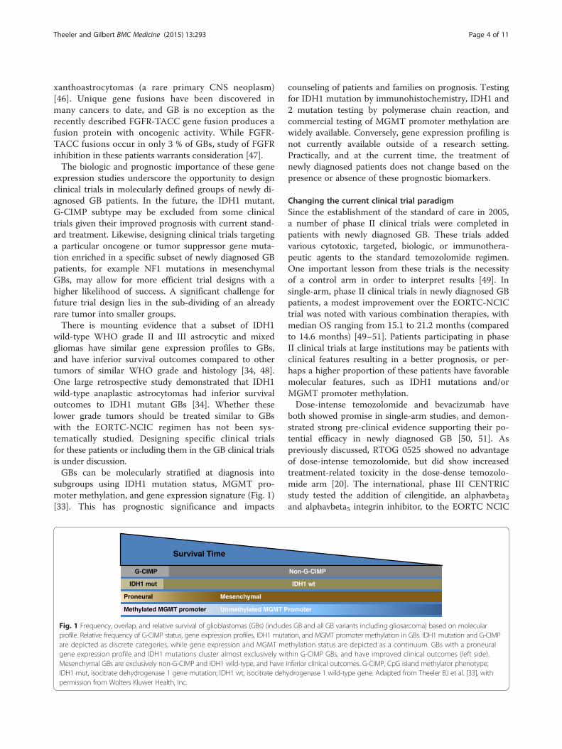
subgroups using IDH1 mutation status, MGMT pro-moter methylation, and gene expression signature (Fig. 1)[33]. This has prognostic significance and impacts
counseling of patients and families on prognosis. Testingfor IDH1 mutation by immunohistochemistry, IDH1 and2 mutation testing by polymerase chain reaction, andcommercial testing of MGMT promoter methylation arewidely available. Conversely, gene expression profiling isnot currently available outside of a research setting.Practically, and at the current time, the treatment ofnewly diagnosed patients does not change based on thepresence or absence of these prognostic biomarkers.
Changing the current clinical trial paradigmSince the establishment of the standard of care in 2005,a number of phase II clinical trials were completed inpatients with newly diagnosed GB. These trials addedvarious cytotoxic, targeted, biologic, or immunothera-peutic agents to the standard temozolomide regimen.One important lesson from these trials is the necessityof a control arm in order to interpret results [49]. Insingle-arm, phase II clinical trials in newly diagnosed GBpatients, a modest improvement over the EORTC-NCICtrial was noted with various combination therapies, withmedian OS ranging from 15.1 to 21.2 months (comparedto 14.6 months) [49–51]. Patients participating in phaseII clinical trials at large institutions may be patients withclinical features resulting in a better prognosis, or per-haps a higher proportion of these patients have favorablemolecular features, such as IDH1 mutations and/orMGMT promoter methylation.Dose-intense temozolomide and bevacizumab have
both showed promise in single-arm studies, and demon-strated strong pre-clinical evidence supporting their po-tential efficacy in newly diagnosed GB [50, 51]. Aspreviously discussed, RTOG 0525 showed no advantageof dose-intense temozolomide, but did show increasedtreatment-related toxicity in the dose-dense temozolo-mide arm [20]. The international, phase III CENTRICstudy tested the addition of cilengitide, an alphavbeta3and alphavbeta5 integrin inhibitor, to the EORTC NCIC
Fig. 1 Frequency, overlap, and relative survival of glioblastomas (GBs) (includes GB and all GB variants including gliosarcoma) based on molecularprofile. Relative frequency of G-CIMP status, gene expression profiles, IDH1 mutation, and MGMT promoter methylation in GBs. IDH1 mutation and G-CIMPare depicted as discrete categories, while gene expression and MGMT methylation status are depicted as a continuum. GBs with a proneuralgene expression profile and IDH1 mutations cluster almost exclusively within G-CIMP GBs, and have improved clinical outcomes (left side).Mesenchymal GBs are exclusively non-G-CIMP and IDH1 wild-type, and have inferior clinical outcomes. G-CIMP, CpG island methylator phenotype;IDH1 mut, isocitrate dehydrogenase 1 gene mutation; IDH1 wt, isocitrate dehydrogenase 1 wild-type gene. Adapted from Theeler BJ et al. [33], withpermission from Wolters Kluwer Health, Inc.
Theeler and Gilbert BMC Medicine (2015) 13:293 Page 4 of 11

regimen in newly diagnosed GB patients with MGMT pro-moter methylation. The parallel, randomized phase II COREstudy tested cilengitide plus standard treatment in newly di-agnosed patients with unmethylated MGMT promoters.Despite promising pre-clinical evidence, neither studyshowed an improvement in OS when adding cilengitide tostandard treatment [52, 53]. In a recent development, atreatment device delivering low-intensity, alternating elec-trical fields, or tumor treatment fields, called the Optune™system, has gained FDA approval for newly diagnosedglioblastoma based on the results of an interim analysisof the EF-14 randomized phase III clinical trial (http://www.fda.gov/NewsEvents/Newsroom/PressAnnounce-ments/ucm465744.htm). The publication of this study isawaited to evaluate the place of this treatment modal-ity in newly diagnosed GB patients.Bevacizumab gained approval for use in recurrent GB in
2009. Despite a lack of definitive evidence that combiningbevacizumab with temozolomide enhanced response, andconflicting evidence from single-arm studies combiningthese agents in the upfront treatment of GB [51, 54], beva-cizumab was being incorporated into the upfront treatmentof GB at some academic institutions and in the community.In this backdrop, two large randomized, double-blindedphase III clinical trials, RTOG 0825 and AVAglio, random-ized newly diagnosed GB patients to either chemoradiationwith temozolomide plus bevacizumab or temozolomideplus placebo [55, 56]. Neither trial demonstrated abenefit in OS. AVAglio demonstrated a progression-free survival advantage, while RTOG 0825 did notdue to differences in the predetermined statisticalmodel. In the RTOG 0825 study, prospective deter-mination of MGMT promoter methylation status anda 9-gene expression panel (previously shown to beprognostic in GB [57]) were used to stratify patientsand for subsequent subgroup analyses. However, nosubgroup of patients could be identified that specific-ally benefited from the combination treatment. RTOG0825 also demonstrated that patients treated in thebevacizumab arm had increased treatment-related tox-icity and inferior scores on symptoms, quality of life,and neurocognitive measures. In a retrospective, sub-group analysis of patients in the AVAglio study, thosepatients who had tumors which were both IDH1wild-type and had a proneural pattern of gene expres-sion may have derived survival benefit from theaddition of bevacizumab [58]. The proneural subgroupaccounts for approximately 25–30 % of newly diagnosedGBs and prospective confirmation of the benefit of up-front bevacizumab in this group of patients isneeded. Additional studies of the tumor tissues andpatient data collected from these trials are ongoing.Testing new treatments in patients with recurrent
glioblastoma then moving those with an “efficacy signal”
to the newly diagnosed setting requires re-evaluation.There have been a paucity of agents tested in the recur-rent setting that have been deemed worthy of testing asfrontline treatment. Recurrent GB is a refractory disease,and the patients often have a poor performance status andoverall health, particularly when compared to newly diag-nosed patients. The tumors are often large and unresect-able, with significant requirements for corticosteroidtreatment to control cerebral edema. Perhaps most im-portantly, overall the response rate and relative efficacy oftherapies in recurrent GB is modest, and it is often diffi-cult to demonstrate efficacy statistically even in agentswith significant promise pre-clinically. For example,cediranib, a VEGF-targeted tyrosine kinase inhibitor,had strong pre-clinical, radiographic, and biomarkerdata to support its use in GB. But cediranib failed to dem-onstrate benefit over CCNU in a well-designed phase IIItrial in recurrent GB [11], slowing the development of apotentially promising agent in newly diagnosed GB.Tumors at recurrence are different from the primary
tumor, as treatment with radiotherapy, cytotoxic chemo-therapy, contributes to genetic and biologic changes thatallow the primary tumor to overcome the host micro-environment and immune system. There is evidence thattemozolomide-induced damage to the DNA mismatchrepair system results in a hypermutated phenotype withfurther deficiency in mismatch repair [59]. In one pre-clinical study comparing primary and recurrent tumors,temozolomide-treated WHO grade II astrocytomas hadmutations in key intracellular signaling pathways, whichwere not present in the primary tumor and could mediatetreatment resistance to cytotoxic and targeted therapeuticagents [60]. Additionally, some GBs with proneural geneexpression undergo a transition to a mesenchymal patternof gene expression at recurrence, similar to that reportedfor epithelial cancers [37]. Selecting therapies effective,or ineffective, at tumor recurrence may not predict ef-fectiveness in newly diagnosed patients due to acquireddifferences between newly diagnosed and recurrent tumors.
Immune checkpoint inhibitors: a new approach to trialdesign and treatment of newly diagnosed glioblastomaThe success of immunotherapeutics in previously refrac-tory systemic solid malignancies, particularly metastaticmelanoma, has led to significant interest in testing similartherapeutic strategies in glioblastomas. A variety of ap-proaches are currently being tested in GB clinical trials,including peptide- and tumor-based vaccine strategies,oncolytic virotherapy, and adoptive immune strategies,such as autologous infusions of activated T cells [61, 62].Recurrent GB is a significant challenge for any thera-
peutic strategy, including immunotherapeutic approaches.Recurrent GB occurs after patients have undergone radio-therapy and multiple cycles of cytotoxic chemotherapy,
Theeler and Gilbert BMC Medicine (2015) 13:293 Page 5 of 11

and in addition corticosteroids are often required to treatcerebral edema resulting in relative immune suppressionin many patients. Temozolomide chemotherapy can de-crease CD4 T lymphocytes, and patients with reducedCD4 counts have worse clinical outcomes [63]. The re-lationship between temozolomide-induced lymphopeniaand its effect on OS appears to be complex in studiestesting vaccination strategies in newly diagnosed glioblast-oma patients. In a single-arm study of rindopepimut,temozolomide-induced lymphopenia was associated withimproved cellular and humoral immune responses [64],and in another single-arm phase II study testing an au-tologous formalin-fixed tumor vaccine, patients withgrade 3 lymphopenia had improved survival outcomescompared to patients with grade 4 or grade 0–2 lympho-penia [65]. Recurrent GBs may be large and unresectableand pose a significant challenge for any systemic therapy,but may be particularly challenging for immune therapieswhich need to access an already hostile tumor micro-environment. GBs have decreased expression of majorhistocompatibility complex (MHC) class I antigen andimmune-suppressive proteins, such as IL-10 and trans-forming growth factor beta, are secreted in the tumormicroenvironment [61]. Additionally, the potential riskfor immune-related inflammation (pseudoprogression,discussed subsequently) may be more problematic inthe recurrent setting.Dendritic cell (DC) vaccine strategies using autologous
tumor lysates or common tumor antigens have beentested in early-phase clinical trials in newly diagnosedglioblastoma patients [66, 67]. This methodology appearsto be feasible and safe. Changes in regulatory T cells andCTLA-4 in the systemic circulation correlates with clinicalactivity, and may provide a means of monitoring thera-peutic response [68]. Albeit in uncontrolled, single-armstudies with small numbers of patients, impressive overallmedian survival rates of 31.4–38.4 months have been ob-served in newly diagnosed GB patients. Methods to boostthe immune responses, such as tetanus toxoid, or withchemokines, such as CCL3, may increase immunogenicityand thereby improve outcomes with DC vaccines in GBpatients [69]. Controlled studies, such as the ongoing ran-domized phase III study using the autologous dendriticcell vaccine, DCVax-L (NCT00045968), are needed toclarify the efficacy of this promising therapeutic strategy.Gliomas express unique antigens, such as HER-2,
TRP-2, gp100, MAGE-1, IL-13 alpha 2, and AIM-2, andthe ICT-107, an autologous DC vaccine, has been de-veloped against these antigens [67, 70]. As discussedabove, the phase I results of this study were promising(median OS of 38.4 months) [67], and the phase II trialhas been completed and the results are forthcoming.Heat shock proteins (HSPs) are expressed during timesof cellular and environmental stress, and an autologous
HSP-96 peptide complex vaccine has been developedfor glioblastoma [70]. A single-arm phase II study hasbeen completed in newly diagnosed GB patients and theresults have not yet been published (NCT00905060). Inaddition to their prognostic and biologic significance, IDHmutations may be a tumor-specific target for immuno-therapeutics. In a recent study, an immune responsegenerated against unique epitopes expressed on IDH1mutated gliomas was successful in a mouse model using apeptide-based vaccine [71].EGFR variant III (EGFRvIII) is the most common mu-
tation of the EGFR gene in glioblastoma, is present in25–30 % of GB patients, and is absent in normal tissue[72]. EGFRvIII mutations are associated with poor long-term survival and are mutually exclusive with IDH mu-tations and G-CIMP gene expression [73, 74]. An EGFRvIIIvaccine, called rindopepimut, is a peptide-based vaccinationwhich targets the unique, tumor-specific antigen created bythe in-frame deletion of the EGFRvIII gene. Promising re-sults were reported from three phase II trials which addedrindopepimut to standard therapy in newly diagnosed GBpatients with an EGFRvIII mutation. The median OS was21.8–23.6 months in these single-arm studies [64, 73, 75].The randomized, phase III, ACT IV clinical trial test-ing the addition of rindopepimut to radiotherapy andtemozolomide in EGFRvIII mutated, newly diagnosedGB patients has completed enrollment and these re-sults are anxiously awaited.The FDA approval of the immune checkpoint inhibi-
tors, ipilimumab, pembrolizumab, and nivolumab, inmetastatic melanoma has led to significant interest inrapid development of clinical trials in GB. Ipilimumab, ahumanized IgG1 monoclonal antibody against cytotoxicT lymphocyte antigen (CTLA-4), has demonstrateddurable responses and a significant improvement in OSin metastatic melanoma [76]. Perhaps more importantlyas it pertains to GB, it has shown promising activity inpatients with melanoma brain metastases without sig-nificant central nervous system (CNS) toxicity [77].The other checkpoint inhibitors, pembrolizumab andnivolumab, are humanized monoclonal antibodies againstprogrammed cell death 1 (PD-1), and are approved for usein metastatic melanoma [78, 79]. Dacarbazine, a cytotoxicchemotherapeutic agent with a similar mechanism totemozolomide, was combined with ipilimumab andcombination treatment, and had improved outcomescompared to dacarbazine alone in metastatic melan-oma [80]. The sequencing and combination of CTLA-4 and PD-1 blockade is ongoing in clinical trials inmelanoma, with results suggesting that combinationtherapy is more effective but with more treatment-related toxicity [81].In a study using an immunohistochemical assay, 88 % of
newly diagnosed GBs had robust and diffuse expression of
Theeler and Gilbert BMC Medicine (2015) 13:293 Page 6 of 11

PDL-1, the ligand of PD-1. This rate of expression isrelatively high compared to other cancers, includingmelanoma. In this same study, PDL-1 expression wasenriched in GBs with mesenchymal gene expression,the subset of GBs which have the worse survival outcomes[82]. In a recent study analyzing PDL-1 expression usingboth immunohistochemistry and flow-cytometry, PDL-1expression was reported in 61 % of patients, but the me-dian percentage of cells expressing PDL-1 was 2.77 % witha wide range (0–86.6 %) [83]. Whether expression ofPDL-1 on a small sub-population of GB cells will correlatewith treatment efficacy will be an important determinationin early-phase clinical trials. PDL-1 expression appears tocorrelate with worsened survival outcomes [83], and theexpression of PDL-1 in the majority of GBs providesstrong rationale for ongoing clinical trials.Studies are currently being conducted in recurrent GB
using the checkpoint inhibitors, including nivolumab,pembrolizumab, and ipilimumab. The CheckMate 143trial (NCT02017717) is a randomized phase II trial testingnivolumab alone, nivolumab plus ipilimumab in two dif-ferent treatment arms versus bevacizumab as an active
comparator in recurrent glioblastoma. In another ran-domized phase II trial, pembrolizumab is being testedalone and in combination with bevacizumab in recurrentglioblastoma (NCT02337491). However, whether a lack ofefficacy in recurrent GB or even increased toxicity will besimilarly predictive of outcomes in the newly diagnosedsetting is questionable, and in our opinion should not pre-clude or delay development of trials testing checkpoint in-hibitors in newly diagnosed patients. A randomized phaseII/III trial testing combinations of temozolomide, ipilimu-mab, nivolumab, and placebo in four different treatmentarms to test whether CTLA-4 blockade alone, PD-1 block-ade alone, or a combination of both, improve outcomes inaddition to standard upfront treatment is currently openand accruing patients (NCT02311920, Fig. 2). This studyis designed to take the best experimental arm forward intoa fully-powered, phase III, placebo-controlled clinical trial.A phase I/II trial with pembrolizumab and temozolo-mide in newly diagnosed patients is open and accruingpatients (NCT02530502). Additional strategies includeadding checkpoint inhibitors to tumor vaccines to in-crease the immune response; and the upcoming AVeRT
Fig. 2 Example of a next generation phase II/III clinical trial for newly diagnosed GB. Patients are randomized after stratification by presence orabsence of MGMT promoter methylation (MGMT), clinical factors (recursive partitioning analysis or RPA), and molecular features (gene expressionprofile or MCP). All patients receive standard temozolomide (TMZ) chemotherapy on days 1 to 42 during radiotherapy and on days 1 to 5 of 28-day cyclesduring adjuvant treatment. Patients will also receive a combination of placebo, ipilimumab (Ipi), or nivolumab (Nivo) in four treatment arms. A “pick thewinner” trial design will be used during phase II to move the most efficacious treatment arm forward into a larger phase III clinical trial. A combination ofOS, treatment-related toxicity, neurocognitive function (NCF), and symptom burden will be used to pick the best treatment (arms). Figure was created forthis manuscript by the authors
Theeler and Gilbert BMC Medicine (2015) 13:293 Page 7 of 11
trial is a phase I/II study adding nivolumab to the DCvaccine in recurrent high grade gliomas testing thisstrategy (NCT02529072).This is not to suggest that upfront trials using check-
point inhibitors will not be challenging. Systemic toxicitywill need to be closely monitored and include auto-immune adverse events, including colitis, endocrinopa-thies, and dermatologic manifestations [76]; peripheralnervous system toxicity such as Guillain-Barré syndromeand myasthenia gravis have been reported [84]. CNStoxicity, including transverse myelitis, and inflammationof brain parenchyma (in the absence of brain metastasis)have also been reported in the treatment of metastaticmelanoma with checkpoint inhibitors [84, 85].Pseudoprogression, operationally defined as reversible
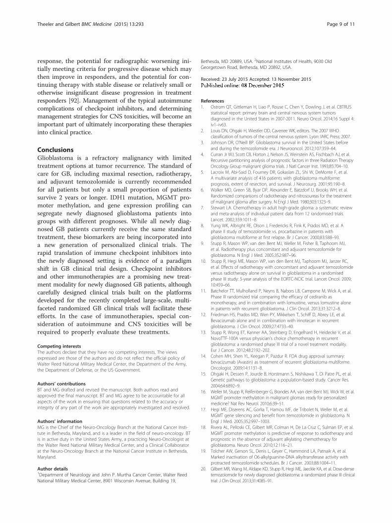
radiographic and clinical worsening due to the effects oftreatment, typically radiation therapy and temozolomide,is now well recognized. It occurs in 20–30 % of glio-blastoma patients after radiation therapy and temozolo-mide, and usually occurs within 6 months of combinedtemozolomide and radiotherapy [86]. Pseudoprogressioncan be difficult to differentiate from tumor progressionon standard MRI sequences, advanced MRI sequences,including MR spectroscopy and MR perfusion scans,
and even pathologic differentiation can be difficult[87–89]; see Fig. 3 for a typical example of pseudopro-gression in a GB patient. Pseudoprogression has beenobserved in patients with CNS metastasis from meta-static melanoma treated with ipilimumab [90], and inday-to-day practice can make the interpretation of responseto radiotherapy and checkpoint inhibitors difficult. Whencombining checkpoint inhibitors, or other immunother-apeutic agents, with standard upfront therapy, rates ofpseudoprogression may be increased, the typical inter-val during which pseudoprogression occurs may changeor be prolonged, and differentiating pseudoprogressionfrom true tumor progression may become an evenmore vexing problem. Response Assessment in Neuro-Oncology (RANO) guidelines have been developed tohelp standardize clinical and radiographic assessmentin neuro-oncology clinical trials [91]. The RANO efforthas been expanded to include immunotherapies, so-called iRANO criteria, to help with the standardizationof interpretation of clinical and imaging assessment inimmunotherapy trials [92]. Specific immune-related re-sponse criteria (iRANO) are needed for numerous rea-sons as mentioned previously, including an expectedprolonged time from therapy initiation to immunologic
Fig. 3 Example of imaging and pathologic features of pseudoprogression in a glioblastoma patient 4 months after completing chemoradiation. a Medial,right frontal lobe enhancing mass which was completely resected due to concern for tumor recurrence. Pathology revealed treatment-related necrosis,hyalinized blood vessels, and b gliosis (hematoxylin and eosin stain), and a small amount of residual tumor which was not mitotically active (not shown).Figure was created for this manuscript by the authors
Theeler and Gilbert BMC Medicine (2015) 13:293 Page 8 of 11
response, the potential for radiographic worsening ini-tially meeting criteria for progressive disease which maythen improve in responders, and the potential for con-tinuing therapy with stable disease or relatively small orotherwise insignificant disease progression in treatmentresponders [92]. Management of the typical autoimmunecomplications of checkpoint inhibitors, and determiningmanagement strategies for CNS toxicities, will become animportant part of ultimately incorporating these therapiesinto clinical practice.
ConclusionsGlioblastoma is a refractory malignancy with limitedtreatment options at tumor recurrence. The standard ofcare for GB, including maximal resection, radiotherapy,and adjuvant temozolomide is currently recommendedfor all patients but only a small proportion of patientssurvive 2 years or longer. IDH1 mutation, MGMT pro-moter methylation, and gene expression profiling cansegregate newly diagnosed glioblastoma patients intogroups with different prognoses. While all newly diag-nosed GB patients currently receive the same standardtreatment, these biomarkers are being incorporated intoa new generation of personalized clinical trials. Therapid translation of immune checkpoint inhibitors intothe newly diagnosed setting is evidence of a paradigmshift in GB clinical trial design. Checkpoint inhibitorsand other immunotherapies are a promising new treat-ment modality for newly diagnosed GB patients, althoughcarefully designed clinical trials built on the platformsdeveloped for the recently completed large-scale, multi-faceted randomized GB clinical trials will facilitate theseefforts. In the case of immunotherapies, special con-sideration of autoimmune and CNS toxicities will berequired to properly evaluate these treatments.
Competing interestsThe authors declare that they have no competing interests. The viewsexpressed are those of the authors and do not reflect the official policy ofWalter Reed National Military Medical Center, the Department of the Army,the Department of Defense, or the US Government.
Authors’ contributionsBT and MG drafted and revised the manuscript. Both authors read andapproved the final manuscript. BT and MG agree to be accountable for allaspects of the work in ensuring that questions related to the accuracy orintegrity of any part of the work are appropriately investigated and resolved.
Authors’ informationMG is the Chief of the Neuro-Oncology Branch at the National Cancer Insti-tute in Bethesda, Maryland, and is a leader in the field of neuro-oncology. BTis in active duty in the United States Army, a practicing Neuro-Oncologist atthe Walter Reed National Military Medical Center, and a Clinical Collaboratorat the Neuro-Oncology Branch at the National Cancer Institute in Bethesda,Maryland.
Author details1Department of Neurology and John P. Murtha Cancer Center, Walter ReedNational Military Medical Center, 8901 Wisconsin Avenue, Building 19,
Bethesda, MD 20889, USA. 2National Institutes of Health, 9030 OldGeorgetown Road, Bethesda, MD 20892, USA.
Received: 23 July 2015 Accepted: 13 November 2015
References1. Ostrom QT, Gittleman H, Liao P, Rouse C, Chen Y, Dowling J, et al. CBTRUS
statistical report: primary brain and central nervous system tumorsdiagnosed in the United States in 2007-2011. Neuro Oncol. 2014;16 Suppl 4:iv1–iv63.
2. Louis DN, Ohgaki H, Wiestler OD, Cavenee WK, editors. The 2007 WHOclassification of tumors of the central nervous system. Lyon: IARC Press; 2007.
3. Johnson DR, O’Neill BP. Glioblastoma survival in the United States beforeand during the temozolomide era. J Neurooncol. 2012;107:359–64.
4. Curran Jr WJ, Scott CB, Horton J, Nelson JS, Weinstein AS, Fischbach AJ, et al.Recursive partitioning analysis of prognostic factors in three Radiation TherapyOncology Group malignant glioma trials. J Natl Cancer Inst. 1993;85:704–10.
5. Lacroix M, Abi-Said D, Fourney DR, Gokaslan ZL, Shi W, DeMonte F, et al.A multivariate analysis of 416 patients with glioblastoma multiforme:prognosis, extent of resection, and survival. J Neurosurg. 2001;95:190–8.
6. Walker MD, Green SB, Byar DP, Alexander E, Batzdorf U, Brooks WH, et al.Randomized comparisons of radiotherapy and nitrosoureas for the treatmentof malignant glioma after surgery. N Engl J Med. 1980;303:1323–9.
7. Stewart LA. Chemotherapy in adult high-grade glioma: a systematic reviewand meta-analysis of individual patient data from 12 randomised trials.Lancet. 2002;359:1011–8.
8. Yung WK, Albright RE, Olson J, Fredericks R, Fink K, Prados MD, et al. Aphase II study of temozolomide vs. procarbazine in patients withglioblastoma multiforme at first relapse. Br J Cancer. 2000;83:588–93.
9. Stupp R, Mason WP, van den Bent MJ, Weller M, Fisher B, Taphoorn MJ,et al. Radiotherapy plus concomitant and adjuvant temozolomide forglioblastoma. N Engl J Med. 2005;352:987–96.
10. Stupp R, Hegi ME, Mason WP, van den Bent MJ, Taphoorn MJ, Janzer RC,et al. Effects of radiotherapy with concomitant and adjuvant temozolomideversus radiotherapy alone on survival in glioblastoma in a randomisedphase III study: 5-year analysis of the EORTC-NCIC trial. Lancet Oncol. 2009;10:459–66.
11. Batchelor TT, Mulholland P, Neyns B, Nabors LB, Campone M, Wick A, et al.Phase III randomized trial comparing the efficacy of cediranib asmonotherapy, and in combination with lomustine, versus lomustine alonein patients with recurrent glioblastoma. J Clin Oncol. 2013;31:3212–8.
12. Friedman HS, Prados MD, Wen PY, Mikkelsen T, Schiff D, Abrey LE, et al.Bevacizumab alone and in combination with irinotecan in recurrentglioblastoma. J Clin Oncol. 2009;27:4733–40.
13. Stupp R, Wong ET, Kanner AA, Steinberg D, Engelhard H, Heidecke V, et al.NovoTTF-100A versus physician’s choice chemotherapy in recurrentglioblastoma: a randomised phase III trial of a novel treatment modality.Eur J Cancer. 2012;48:2192–202.
14. Cohen MH, Shen YL, Keegan P, Pazdur R. FDA drug approval summary:bevacizumab (Avastin) as treatment of recurrent glioblastoma multiforme.Oncologist. 2009;14:1131–8.
15. Ohgaki H, Dessen P, Jourde B, Horstmann S, Nishikawa T, Di Patre PL, et al.Genetic pathways to glioblastoma: a population-based study. Cancer Res.2004;64:6892–9.
16. Weller M, Stupp R, Reifenberger G, Brandes AA, van den Bent MJ, Wick W, et al.MGMT promoter methylation in malignant gliomas: ready for personalizedmedicine? Nat Rev Neurol. 2010;6:39–51.
17. Hegi ME, Diserens AC, Gorlia T, Hamou MF, de Tribolet N, Weller M, et al.MGMT gene silencing and benefit from temozolomide in glioblastoma. NEngl J Med. 2005;352:997–1003.
18. Rivera AL, Pelloski CE, Gilbert MR, Colman H, De La Cruz C, Sulman EP, et al.MGMT promoter methylation is predictive of response to radiotherapy andprognostic in the absence of adjuvant alkylating chemotherapy forglioblastoma. Neuro Oncol. 2010;12:116–21.
19. Tolcher AW, Gerson SL, Denis L, Geyer C, Hammond LA, Patnaik A, et al.Marked inactivation of O6-alkylguanine-DNA alkyltransferase activity withprotracted temozolomide schedules. Br J Cancer. 2003;88:1004–11.
20. Gilbert MR, Wang M, Aldape KD, Stupp R, Hegi ME, Jaeckle KA, et al. Dose-densetemozolomide for newly diagnosed glioblastoma: a randomized phase III clinicaltrial. J Clin Oncol. 2013;31:4085–91.
Theeler and Gilbert BMC Medicine (2015) 13:293 Page 9 of 11

21. Barker CA, Chang M, Chou JF, Zhang Z, Beal K, Gutin PH, et al. Radiotherapyand concomitant temozolomide may improve survival of elderly patientswith glioblastoma. J Neurooncol. 2012;109:391–7.
22. Lombardi G, Pace A, Pasqualetti F, Rizzato S, Faedi M, Anghileri E, et al.Predictors of survival and effect of short (40 Gy) or standard-course (60 Gy)irradiation plus concomitant temozolomide in elderly patients withglioblastoma: a multicenter retrospective study of AINO (Italian Associationof Neuro-Oncology). J Neurooncol. 2015;125:359–67.
23. Malmström A, Grønberg BH, Marosi C, Stupp R, Frappaz D, Schultz H, et al.Temozolomide versus standard 6-week radiotherapy versus hypofractionatedradiotherapy in patients older than 60 years with glioblastoma: the Nordicrandomised, phase 3 trial. Lancet Oncol. 2012;13:916–26.
24. Gallego Perez-Larraya J, Ducray F, Chinot O, Catry-Thomas I, Taillandier L,Guillamo JS, et al. Temozolomide in elderly patients with newly diagnosedglioblastoma and poor performance status: an ANOCEF phase II trial. J ClinOncol. 2011;29:3050–5.
25. Wick W, Platten M, Meisner C, Felsberg J, Tabatabai G, Simon M, et al.Temozolomide chemotherapy alone versus radiotherapy alone for malignantastrocytoma in the elderly: the NOA-08 randomised, phase 3 trial. Lancet Oncol.2012;13:707–15.
26. Parsons DW, Jones S, Zhang X, Lin JC, Leary RJ, Angenendt P, et al. An integratedgenomic analysis of human glioblastoma multiforme. Science. 2008;321:1807–12.
27. Cancer Genome Atlas Research Network. Comprehensive genomiccharacterization defines human glioblastoma genes and core pathways.Nature. 2008;455:1061–8.
28. Yan H, Parsons DW, Jin G, McLendon R, Rasheed BA, Yuan W, et al. IDH1 andIDH2 mutations in gliomas. N Engl J Med. 2009;360:765–73.
29. Lai A, Kharbanda S, Pope WB, Tran A, Solis OE, Peale F, et al. Evidence forsequenced molecular evolution of IDH1 mutant glioblastoma from a distinctcell of origin. J Clin Oncol. 2011;29:4482–90.
30. Choi C, Ganji SK, Deberardinis RJ, Hatanpaa KJ, Rakheja D, Kovacs Z, et al.2-hydroxyglutarate detection by magnetic resonance spectroscopy inIDH-mutated patients with gliomas. Nat Med. 2012;18:624–9.
31. Elkhaled A, Jalbert LE, Phillips JJ, Yoshihara HA, Parvataneni R, Srinivasan R,et al. Magnetic resonance of 2-hydroxyglutarate in IDH1-mutated low-gradegliomas. Sci Transl Med. 2012;4:116ra5.
32. Nobusawa S, Watanabe T, Kleihues P, Ohgaki H. IDH1 mutations as molecularsignature and predictive factor of secondary glioblastomas. Clin Cancer Res.2009;15:6002–7.
33. Theeler BJ, Yung WK, Fuller GN, De Groot JF. Moving toward molecularclassification of diffuse gliomas in adults. Neurology. 2012;79:1917–26.
34. Hartmann C, Hentschel B, Wick W, Capper D, Felsberg J, Simon M, et al.Patients with IDH1 wild type anaplastic astrocytomas exhibit worseprognosis than IDH1-mutated glioblastomas, and IDH1 mutation statusaccounts for the unfavorable prognostic effect of higher age: implicationsfor classification of gliomas. Acta Neuropathol. 2010;120:707–18.
35. Wick W, Meisner C, Hentschel B, Platten M, Schilling A, Wiestler B, et al.Prognostic or predictive value of MGMT promoter methylation in gliomasdepends on IDH1 mutation. Neurology. 2013;81:1515–22.
36. Molenaar RJ, Verbaan D, Lamba S, Zanon C, Jeuken JWM, Boots-Sprenger SHE,et al. The combination of IDH1 mutations and MGMT methylation statuspredicts survival in glioblastoma better than either IDH1 or MGMT alone.Neuro Oncol. 2014;16:1263–73.
37. Phillips HS, Kharbanda S, Chen R, Forrest WF, Soriano RH, Wu TD, et al.Molecular subclasses of high-grade glioma predict prognosis, delineate apattern of disease progression, and resemble stages in neurogenesis.Cancer Cell. 2006;9:157–73.
38. Verhaak RG, Hoadley KA, Purdom E, Wang V, Qi Y, Wilkerson MD, et al.Integrated genomic analysis identifies clinically relevant subtypes of glioblastomacharacterized by abnormalities in PDGFRA, IDH1, EGFR, and NF1. Cancer Cell.2010;17:98–110.
39. Noushmehr H, Weisenberger DJ, Diefes K, Phillips HS, Pujara K, Berman BP,et al. Identification of a CpG island methylator phenotype that defines adistinct subgroup of glioma. Cancer Cell. 2010;17:510–22.
40. Aldape K, Zadeh G, Mansouri S, Reifenberger G, von Deimling A. Glioblastoma:pathology, molecular mechanisms and markers. Acta Neuropathol (Berl). 2015;129:829–48.
41. Liu XY, Gerges N, Korshunov A, Sabha N, Khuong-Quang DA, Fontebasso AM,et al. Frequent ATRX mutations and loss of expression in adult diffuse astrocytictumors carrying IDH1/IDH2 and TP53 mutations. Acta Neuropathol (Berl). 2012;124:615–25.
42. Killela PJ, Reitman ZJ, Jiao Y, Bettegowda C, Agrawal N, Diaz LA, et al. TERTpromoter mutations occur frequently in gliomas and a subset of tumorsderived from cells with low rates of self-renewal. Proc Natl Acad Sci U S A.2013;110:6021–6.
43. Liu X, Wu G, Shan Y, Hartmann C, von Deimling A, Xing M. Highly prevalentTERT promoter mutations in bladder cancer and glioblastoma. Cell Cycle.2013;12:1637–8.
44. Labussière M, Boisselier B, Mokhtari K, Di Stefano AL, Rahimian A, Rossetto M,et al. Combined analysis of TERT, EGFR, and IDH status defines distinctprognostic glioblastoma classes. Neurology. 2014;83:1200–6.
45. Schindler G, Capper D, Meyer J, Janzarik W, Omran H, Herold-Mende C, et al.Analysis of BRAF V600E mutation in 1,320 nervous system tumors revealshigh mutation frequencies in pleomorphic xanthoastrocytoma,ganglioglioma and extra-cerebellar pilocytic astrocytoma. Acta Neuropathol.2011;121:397–405.
46. Hyman DM, Puzanov I, Subbiah V, Faris JE, Chau I, Blay JY, et al. Vemurafenib inmultiple nonmelanoma cancers with BRAF V600 mutations. N Engl J Med.2015;373:726–36.
47. Singh D, Chan JM, Zoppoli P, Niola F, Sullivan R, Castano A, et al. Transformingfusions of FGFR and TACC genes in human glioblastoma. Science.2012;337:1231–5.
48. Olar A, Wani KM, Alfaro-Munoz KD, Heathcock LE, van Thuijl HF, Gilbert MR,et al. IDH mutation status and role of WHO grade and mitotic index inoverall survival in grade II-III diffuse gliomas. Acta Neuropathol (Berl). 2015;129:585–96.
49. Grossman SA, Ye X, Piantadosi S, Desideri S, Nabors LB, Rosenfeld M, et al.Survival of patients with newly diagnosed glioblastoma treated withradiation and temozolomide in research studies in the United States. ClinCancer Res. 2010;16:2443–9.
50. Clarke JL, Iwamoto FM, Sul J, Panageas K, Lassman AB, DeAngelis LM, et al.Randomized phase II trial of chemoradiotherapy followed by either dose-dense or metronomic temozolomide for newly diagnosed glioblastoma. JClin Oncol. 2009;27:3861–7.
51. Vredenburgh JJ, Desjardins A, Reardon DA, Peters KB, Herndon 2nd JE,Marcello J, et al. The addition of bevacizumab to standard radiation therapyand temozolomide followed by bevacizumab, temozolomide, andirinotecan for newly diagnosed glioblastoma. Clin Cancer Res. 2011;17:4119–24.
52. Nabors LB, Fink KL, Mikkelsen T, Grujicic D, Tarnawski R, Nam DH, et al. Twocilengitide regimens in combination with standard treatment for patientswith newly diagnosed glioblastoma and unmethylated MGMT gene promoter:results of the open-label, controlled, randomized phase II CORE study.Neuro Oncol. 2015;17:708–17.
53. Stupp R, Hegi ME, Gorlia T, Erridge SC, Perry J, Hong YK, et al. Cilengitidecombined with standard treatment for patients with newly diagnosedglioblastoma with methylated MGMT promoter (CENTRIC EORTC 26071-22072 study): a multicentre, randomised, open-label, phase 3 trial. LancetOncol. 2014;15:1100–8.
54. Lai A, Tran A, Nghiemphu PL, Pope WB, Solis OE, Selch M, et al. Phase II studyof bevacizumab plus temozolomide during and after radiation therapy forpatients with newly diagnosed glioblastoma multiforme. J Clin Oncol.2011;29:142–8.
55. Chinot OL, Wick W, Mason W, Henriksson R, Saran F, Nishikawa R, et al.Bevacizumab plus radiotherapy-temozolomide for newly diagnosedglioblastoma. N Engl J Med. 2014;370:709–22.
56. Gilbert MR, Dignam JJ, Armstrong TS, Wefel JS, Blumenthal DT, Vogelbaum MA,et al. A randomized trial of bevacizumab for newly diagnosed glioblastoma.N Engl J Med. 2014;370:699–708.
57. Colman H, Zhang L, Sulman EP, McDonald JM, Shooshtari NL, Rivera A, et al.A multigene predictor of outcome in glioblastoma. Neuro Oncol. 2010;12:49–57.
58. Sandmann T, Bourgon R, Garcia J, Li C, Cloughesy T, Chinot OL, et al.Patients with proneural glioblastoma may derive overall survival benefitfrom the addition of bevacizumab to first-line radiotherapy and temozolomide:retrospective analysis of the AVAglio Trial. J Clin Oncol. 2015;33:2735–44.
59. Cahill DP, Levine KK, Betensky RA, Codd PJ, Romany CA, Reavie LB, et al.Loss of the mismatch repair protein MSH6 in human glioblastomas isassociated with tumor progression during temozolomide treatment.Clin Cancer Res. 2007;13:2038–45.
60. Johnson BE, Mazor T, Hong C, Barnes M, Aihara K, McLean CY, et al.Mutational analysis reveals the origin and therapy-driven evolution ofrecurrent glioma. Science. 2014;343:189–93.
Theeler and Gilbert BMC Medicine (2015) 13:293 Page 10 of 11

61. Fecci PE, Heimberger AB, Sampson JH. Immunotherapy for primary braintumors: no longer a matter of privilege. Clin Cancer Res. 2014;20:5620–9.
62. Weathers SP, Gilbert MR. Current challenges in designing GBM trials forimmunotherapy. J Neurooncol. 2015;123:331–7.
63. Grossman SA, Ye X, Lesser G, Sloan A, Carraway H, Desideri S, et al.Immunosuppression in patients with high-grade gliomas treated with radiationand temozolomide. Clin Cancer Res. 2011;17:5473–80.
64. Sampson JH, Aldape KD, Archer GE, Coan A, Desjardins A, Friedman AH, et al.Greater chemotherapy-induced lymphopenia enhances tumor-specificimmune responses that eliminate EGFRvIII-expressing tumor cells in patientswith glioblastoma. Neuro Oncol. 2011;13:324–33.
65. Ishikawa E, Muragaki Y, Yamamoto T, Maruyama T, Tsuboi K, Ikuta S, et al.Phase I/IIa trial of fractionated radiotherapy, temozolomide, and autologousformalin-fixed tumor vaccine for newly diagnosed glioblastoma. J Neurosurg.2014;121:543–53.
66. Prins RM, Soto H, Konkankit V, Odesa SK, Eskin A, Yong WH, et al. Geneexpression profile correlates with T-cell infiltration and relative survival inglioblastoma patients vaccinated with dendritic cell immunotherapy. ClinCancer Res. 2011;17:1603–15.
67. Phuphanich S, Wheeler CJ, Rudnick JD, Mazer M, Wang H, Nuño MA, et al.Phase I trial of a multi-epitope-pulsed dendritic cell vaccine for patientswith newly diagnosed glioblastoma. Cancer Immunol Immunother.2013;62:125–35.
68. Fong B, Jin R, Wang X, Safaee M, Lisiero DN, Yang I, et al. Monitoring ofregulatory T cell frequencies and expression of CTLA-4 on T cells, beforeand after DC vaccination, can predict survival in GBM patients. PLoS One.2012;7:e32614.
69. Mitchell DA, Batich KA, Gunn MD, Huang MN, Sanchez-Perez L, Nair SK, et al.Tetanus toxoid and CCL3 improve dendritic cell vaccines in mice andglioblastoma patients. Nature. 2015;519:366–9.
70. Patel MA, Kim JE, Ruzevick J, Li G, Lim M. The future of glioblastomatherapy: synergism of standard of care and immunotherapy. Cancers. 2014;6:1953–85.
71. Schumacher T, Bunse L, Pusch S, Sahm F, Wiestler B, Quandt J, et al. A vaccinetargeting mutant IDH1 induces antitumour immunity. Nature. 2014;512:324–7.
72. Humphrey PA, Wong AJ, Vogelstein B, Zalutsky MR, Fuller GN, Archer GE,et al. Anti-synthetic peptide antibody reacting at the fusion junction ofdeletion-mutant epidermal growth factor receptors in human glioblastoma.Proc Natl Acad Sci U S A. 1990;87:4207–11.
73. Schuster J, Lai RK, Recht LD, Reardon DA, Paleologos NA, Groves MD, et al.A phase II, multicenter trial of rindopepimut (CDX-110) in newly diagnosedglioblastoma: the ACT III study. Neuro Oncol. 2015;17:854–61.
74. Le Rhun E, Rhun EL, Taillibert S, Chamberlain MC. The future of high-gradeglioma: Where we are and where are we going. Surg Neurol Int. 2015;6Suppl 1:S9–S44.
75. Sampson JH, Heimberger AB, Archer GE, Aldape KD, Friedman AH, Friedman HS,et al. Immunologic escape after prolonged progression-free survival withepidermal growth factor receptor variant III peptide vaccination in patients withnewly diagnosed glioblastoma. J Clin Oncol. 2010;28:4722–9.
76. Hodi FS, O’Day SJ, McDermott DF, Weber RW, Sosman JA, Haanen JB, et al.Improved survival with ipilimumab in patients with metastatic melanoma. NEngl J Med. 2010;363:711–23.
77. Margolin K, Ernstoff MS, Hamid O, Lawrence D, McDermott D, Puzanov I, et al.Ipilimumab in patients with melanoma and brain metastases: an open-label,phase 2 trial. Lancet Oncol. 2012;13:459–65.
78. Robert C, Schachter J, Long GV, Arance A, Grob JJ, Mortier L, et al.Pembrolizumab versus Ipilimumab in Advanced Melanoma. N Engl J Med.2015;372:2521–32.
79. Wolchok JD, Kluger H, Callahan MK, Postow MA, Rizvi NA, Lesokhin AM, et al.Nivolumab plus ipilimumab in advanced melanoma. N Engl J Med.2013;369:122–33.
80. Robert C, Thomas L, Bondarenko I, O’Day S, Weber J, Garbe C, et al. Ipilimumabplus dacarbazine for previously untreated metastatic melanoma. N Engl J Med.2011;364:2517–26.
81. Postow MA, Chesney J, Pavlick AC, Robert C, Grossmann K, McDermott D,et al. Nivolumab and ipilimumab versus ipilimumab in untreatedmelanoma. N Engl J Med. 2015;372:2006–17.
82. Berghoff AS, Kiesel B, Widhalm G, Rajky O, Ricken G, Wöhrer A, et al.Programmed death ligand 1 expression and tumor-infiltrating lymphocytesin glioblastoma. Neuro Oncol. 2015;17:1064–75.
83. Nduom EK, Wei J, Yaghi NK, Huang N, Kong LY, Gabrusiewicz K, et al. PD-L1expression and prognostic impact in glioblastoma. Neuro Oncol. 2015.[Epub ahead of print]
84. Liao B, Shroff S, Kamiya-Matsuoka C, Tummala S. Atypical neurologicalcomplications of ipilimumab therapy in patients with metastatic melanoma.Neuro Oncol. 2014;16:589–93.
85. Mandel JJ, Olar A, Aldape KD, Tremont-Lukats IW. Lambrolizumab inducedcentral nervous system (CNS) toxicity. J Neurol Sci. 2014;344:229–31.
86. Fink J, Born D, Chamberlain MC. Pseudoprogression: relevance with respectto treatment of high-grade gliomas. Curr Treat Options Oncol. 2011;12:240–52.
87. Young RJ, Gupta A, Shah AD, Graber JJ, Zhang Z, Shi W, et al. Potential utilityof conventional MRI signs in diagnosing pseudoprogression in glioblastoma.Neurology. 2011;76:1918–24.
88. Melguizo-Gavilanes I, Bruner JM, Guha-Thakurta N, Hess KR, Puduvalli VK.Characterization of pseudoprogression in patients with glioblastoma: ishistology the gold standard? J Neurooncol. 2015;123:141–50.
89. Ellingson BM, Wen PY, van den Bent MJ, Cloughesy TF. Pros and cons ofcurrent brain tumor imaging. Neuro Oncol. 2014;16 Suppl 7:vii2–vii11.
90. Gerber NK, Young RJ, Barker CA, Wolchok JD, Chan TA, Yamada Y, et al.Ipilimumab and whole brain radiation therapy for melanoma brain metastases.J Neurooncol. 2015;121:159–65.
91. Wen PY, Macdonald DR, Reardon DA, Cloughesy TF, Sorensen AG, Galanis E,et al. Updated response assessment criteria for high-grade gliomas:response assessment in neuro-oncology working group. J Clin Oncol. 2010;28:1963–72.
92. Reardon DA, Okada H. Re-defining response and treatment effects forneuro-oncology immunotherapy clinical trials. J Neurooncol. 2015;123:339–46.
• We accept pre-submission inquiries
• Our selector tool helps you to find the most relevant journal
• We provide round the clock customer support
• Convenient online submission
• Thorough peer review
• Inclusion in PubMed and all major indexing services
• Maximum visibility for your research
Submit your manuscript atwww.biomedcentral.com/submit
Submit your next manuscript to BioMed Central and we will help you at every step:
Theeler and Gilbert BMC Medicine (2015) 13:293 Page 11 of 11