advances in tissue regeneration 2013 conference...
TRANSCRIPT

14-15 November 2013, Lattrop, The Netherlands
ADVANCES IN TISSUE REGENERATION 2013 CONFERENCE

AdvAnces in Tissue regenerATion 2013
2 program
Table of ConTenTs
Welcome 3
organization 4
Program 6
SPeakerS ProfileS 9
PoSter PreSentation abStractS 27

AdvAnces in Tissue regenerATion 2013
program 3
WelCome
It is our great pleasure to welcome you to the advances in Tissue regeneration 2013 Conference. With this conference, we aim at providing an overview of the state-of-the-art work in the field of regenerative medicine. Success of this highly multidisciplinary field largely depends on the advances in the fields of (stem) cell biology, materials science and engineering and interactions among these fields. We are proud that world-renowned scientists from the United States, Israel, United Kingdom, Switzerland, Belgium, Portugal, Sweden and the Netherlands have agreed to give a lecture and share with us their expertise in these different fields. In addition, we have over 65 registered participants and over 30 submitted abstracts, which will be presented in the form of a poster or a short oral presentation.
Topics will range from basic sciences to clinical use, including, for example, stem cell therapies and design and fabrication of smart biomaterials for orthopedic and cardiovascular applications. In addition, novel approaches in tissue regeneration research, such as microfluidics, microfabrication technologies, high-throughput screening, imaging and computational modeling will be discussed.
We have chosen for a relatively small and informal meeting to provide plenty of opportunity for discussions with speakers and for extending scientific network.
We are convinced that we will have two days of great science. We hope you will enjoy both the conference and the beauty and hospitality of Twente!
Pamela Habibovicana barradasClemens van blitterswijk
(University of Twente, Department of Tissue Regeneration, Enschede, the Netherlands)
Dear colleagueS,
AdvAnces in Tissue regenerATion 2013
4 program
organIzaTIon
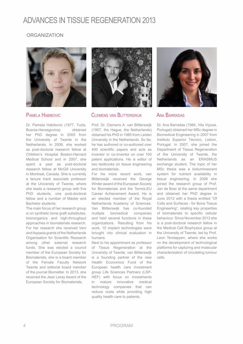
Dr. Pamela Habibovic (1977, Tuzla, Bosnia-Herzegovina) obtained her PhD degree in 2005 from the University of Twente in the netherlands. In 2006, she worked as post-doctoral research fellow at Children’s Hospital boston-Harvard Medical School and in 2007, she spent a year as post-doctoral research fellow at McGill University in montreal, Canada. she is currently a tenure track associate professor at the University of Twente, where she leads a research group with five PhD students, one post-doctoral fellow and a number of Master and bachelor students. The main focus of her research group is on synthetic bone graft substitutes, bioinorganics and high-throughput approaches in biomaterials research. for her research she received Veni and aspasia grants of the netherlands Organisation for Scientific Research among other external research funds. she was elected a council member of the European Society for Biomaterials, she is a board member of the female faculty network Twente and editorial board member of the journal biomatter. In 2013, she received the Jean leray award of the european society for biomaterials.
Pamela Habibovic clemenS van blitterSWijk ana barraDaS
Prof. Dr. Clemens A. van Blitterswijk (1957, the Hague, the Netherlands) obtained his PhD in 1985 from Leiden University in the Netherlands. So far, he has authored or co-authored over 400 scientific papers and acts as inventor or co-inventor on over 100 patent applications. He is editor of two textbooks on tissue engineering and biomaterials. for his more recent work, van blitterswijk received the george Winter award of the european society for Biomaterials and the Termis-EU Career achievement award. He is an elected member of the Royal netherlands academy of sciences. Van blitterswijk has co-founded multiple biomedical companies and held several functions in these organizations. Resulting from his work, 10 implant technologies were brought into clinical evaluation in humans. next to his appointment as professor of Tissue regeneration at the University of Twente, van Blitterswijk is a founding partner of the new Health economics fund of the european health care investment group Life Sciences Partners (LSP-HEF) with focus on investments in mature innovative medical technology companies that can reduce costs while providing high quality health care to patients.
Dr. Ana Barradas (1984, Vila Viçosa, Portugal) obtained her MSc degree in Biomedical Engineering in 2007 from Instituto Superior Técnico, Lisbon, Portugal. In 2007, she joined the Department of Tissue Regeneration of the University of Twente, the Netherlands as an ERASMUS exchange student. The topic of her MSc thesis was a bioluminescent system for nutrient availability in tissue engineering. In 2008 she joined the research group of prof. Jan de boer at the same department and obtained her PhD degree in June 2012 with a thesis entitled “of Cells and surfaces - for bone Tissue engineering”, relating key properties of biomaterials to specific cellular behaviour. Since November 2012 she is a post-doctoral research fellow in the medical Cell biophysics group at the University of Twente, led by Prof. leon Terstappen, where she works on the development of technological platforms for capturing and molecular characterization of circulating tumour cells.

AdvAnces in Tissue regenerATion 2013
program 5
aDminiStrative SuPPort
WebSite SuPPort
enDorSeD by
financial SuPPort
audrey HaarnackTom groothuis
www.tissueregeneration2013.comDavid Barata

AdvAnces in Tissue regenerATion 2013
6 program
tHurSDay, 14 november 2013
09h55 gerjo van osch | Cartilage regeneration and stem cells10h30 rf Ariane van Spreeuwel: The influence of tissue (an)isotropy on cardiomyocyte contraction in engineered cardiac microtissues10h35 marie-José goumans | stem cells for cardiac repair11h10 Coffee break
Session 1: Stem cells for tissue regeneration: a matter of communication
11h25 Melody Swartz | Immunobiology of lymphangiogenesis and implications for tissue regeneration12h00 Katarina Le Blanc | Immunomodulation by mesenchymal stem cells
Session 2: Creating cellular microenvironments
15h00 Shulamit Levenberg | engineering stem cell microenvironments for controlled induction of differentiation 15h35 Karen Hirschi | Endothelial cell differentiation and specification16h10 rf Jennifer Patterson: In vitro characterization of cell-encapsulating PEG hydrogels cultured under mineralizing conditions16h15 Christine mummery | Human pluripotent stem cells: the new patient?16h50 Coffee Break
Session 3: Stem cells for tissue regeneration: controlling and instructing
program
12h35 Walk to a nearby farm poster session & lunch Walk back
Lunch + Poster Session
09h30 Pamela HabibovicWelcome & Introduction
17h05 Carolina Wählby | extracting discoveries hidden in images17h40 séverine le gac | Microfabricated and microfluidic tools for tissue production and study18h15 rf Maciej Skolimowski: Development of a microfluidic platform for cell cultivation in narrow channels18h20 Liesbet Geris | In silico models for regenerative medicine; hype or help?18h55 Break
Session 4: Novel approaches to tissue regeneration research
19h10 Kristi Anseth | engineering hydrogel niches to promote tissue regeneration19h45 rf Xiao-Hua Qin: 3D construction of artificial ECM hydrogels by two-photon- induced polymerization20h00 Dinner
Session 5: Instructive biomaterials: cell-material interactions
Registration
AdvAnces in Tissue regenerATion 2013
program 7
friDay, 15 november 2013
08h30 Patricia Dankers | Bioinspired biomaterials - from structure to application in cardiovascular and kidney regenerative medicine 09h05 rf David Bassett: Mineralised alginate based hydrogel composites for tissue engineering09h10 Carole perry | The role of silica in composite materials for bioengineering applications including bone regeneration and cell based therapies09h45 Liz Tanner | Engineering load bearing biomaterials10h20 Coffee Break
Session 6: Instructive biomaterials: guiding tissue formation
10h35 Julie gough | nanoscale surfaces and peptide gels for tissue engineering applications 11h10 rf Rebecca Medda: Investigation of cell-surface interactions of human mesenchymal stem cells on nanopatterned β-type titanium-niobium alloy surfaces11h15 Carlijn bouten | In-situ cardiovascular tissue engineering – role of scaffold properties and functionalization11h50 manuela gomes | engineering skeletal tissues with stem cells cultured onto natural origin scaffolds in modulated environments
Session 7: Tissue regeneration: a multidisciplinary field
12h25 best poster and presentation awards 12h30 Lunch 14h00 Closure
Wrap-up
AdvAnces in Tissue regenerATion 2013
program 9
abStractengineering HyDrogel nicHeS to Promote tiSSue regeneration
Kristi S. Anseth earned her B.S. degree from Purdue University in 1992 and her Ph.D. degree from the University of Colorado in 1994. She then conducted post-doctoral research at MIT as an NIH fellow and subsequently joined the Department of Chemical and Biological Engineering at the University of Colorado at Boulder as an Assistant Professor in 1996. Dr. Anseth is presently a Howard Hughes Medical Institute Investigator and Distinguished Professor of Chemical and biological engineering. Her research interests lie at the interface between biology and engineering where she designs new biomaterials for applications in drug delivery and regenerative medicine. Dr. Anseth’s research group has published over 250 publications in peer-reviewed journals and presented over 200 invited lectures in the fields of biomaterials and tissue engineering. She was the first engineer to be named a Howard Hughes Medical Institute Investigator and received the alan T. Waterman award, the highest award of the national science foundation for demonstrated exceptional individual achievement in scientific or engineering research. In 2009, she was elected a member of the National Academy of Engineering and the Institute of Medicine. Dr. Anseth is also a dedicated teacher, who has received four University Awards related to her teaching, as well as the american society for engineering education’s Curtis W. McGraw Award. Dr. Anseth is a Fellow of the American Association for the advancement of science and the american Institute for medical and Biological Engineering. She serves on the editorial boards or as associate editor of biomacromolecules, Journal of biomedical materials research — part a, acta biomaterialia, progress in materials science, and biotechnology & bioengineering.
Kristi Anseth, Ph.D. Website: www.colorado.edu/ansethgroup/Distinguished Professor of Chemical and Biochemical Engineering, Tisone Professor, Associ-ate Professor of Surgery, Howard Hughes Medical Institute Investigator Department of Chemical and Biological Engineering, University of Colorado, Boulder, CO, USA
SPEAKERS PROFILES
Hydrogels are a unique class of polymeric materials that imbibe large amounts of water and possess a tissue-like elasticity, and when locally modified with appropriate signaling molecules, these synthetic niches can facilitate the regeneration of tissues. While the gel environment is often >90% water, the microscopic architecture and local chemistry play important roles in dictating cell function, including migration and proliferation, the secretion and distribution of extracellular matrix molecules, and ultimately the formation of tissue structures. This talk will illustrate several examples where the regeneration of tissues is highly coupled to the biophysical and biochemical properties of the gels, and demonstrate how appropriate tuning of the gel properties can create microenvironments that simply permit cells to function to those that actively promote specific cell functions. Integral to this understanding is the ability to manipulate the underlying gel chemistry and properties through the synthesis of macromolecular precursors and control of the gelation process. In this regard, photochemical reactions are increasingly used to form hydrogel biomaterials and deliver cells and biomacromolecules under physiological conditions. As an example, our recent work exploiting thiol-ene photopolymerizations to form proteolytically-degradable PEG hydrogels will be presented. Specifically, the incorporation of peptides and the role of peptide functionality on cell function and tissue regeneration will be highlighted. The overall goal of the talk will be to illustrate some of the current advances and challenges in designing gels for tissue engineering applications and place this in the broader context of potential biological applications.

AdvAnces in Tissue regenerATion 2013
10 program
abStractimmunomoDulation by meSencHymal Stem cellS
Dr. Katarina Le Blanc is a Professor of Clinical Stem Cell Research at Karolinska Institutet, Division of Clinical Immunology and Transfusion Medicine, Stockholm, Sweden. She received her MD from the Karolinska Institutet, in 1993 and her Ph.D. in 1999. In 2002 she became a certified specialist in haematology.
Her main research interest is mesenchymal stem cells, haematopoietic stem cell transplantation and immunology. She is the recipient of several awards. Dr Le Blanc is co-director of the Wallenberg Institute for Regenerative Medicine, co-director of the strategic research foundation for stem Cells and co-director for Karolinska Institutet Theme Centre for Stem Cells. She is Chair, Research and Education, Hematology Center, Karolinska University Hospital.She is a member of several international and national committees, advisory boards and scientific meetings. She has mentored many trainees, PhD students and post docs. She currently has over 100 peer-reviewed publications and review articles.
Katarina Le Blanc, MD, Ph.D.Professor of Clinical Stem Cell Research Div. of Clinical Immunology and Transfusion Medicine, Karolinska Institutet, Stockholm, SEWebsite: www.ki.se/ki/jsp/polopoly.jsp?d=7819&a=21741&l=en
Mesenchymal Stromal Cells (MSCs) are non-hematopoietic progenitor cells found in the bone marrow and many other tissues. In vitro and in vivo, the cells differentiate into adipocytes, chondrocytes and osteocytes after appropriate induction.both undifferentiated and msCs induced to differentiate, have immune-modulatory properites and promote peripheral tolerance. In vitro and in vivo in experimental animal models, msCs suppress alloreactive donor anti-host T-cell responses. MSCs also prevent the maturation of monocytes to first immature dendritic cells (DCs) and next mature myeloid DCs that support T-cell alloresponses. Instead, MSCs re-polarise pro-inflammatory DCs into tolerogenic IL-10+ DCs that together with other effects promote T-cell anergy and Treg induction. Interferon induces msC to produce indeolamine 2,3 dioxygenase, prostaglandin e2 and other factors that are believed to mediate these effects. Many questions remain to be answered before MSCs can be established as an immunomodulatory treatment. Efficacy of the cells needs to be established in clinical trials. This is particularly true since no efficacy marker has been established that predicts the clinical outcome of patients treated with MSCs. So far, data indicates low infusional toxicity. response rates in the literature indicate that msCs are a promising tool for immunomodulation.

AdvAnces in Tissue regenerATion 2013
program 11
abStractin-Situ carDiovaScular tiSSue engineering – role of ScaffolD ProPertieS anD functionalization
Carlijn bouten is professor of Cell-matrix Interactions at the department Biomedical Engineering of the Eindhoven University of Technology (TU/e). She was trained in functional anatomy and biomechanics, as well as exercise physiology, at the department of Human Movement Sciences, VU Amsterdam (MSc 1991), and obtained her PhD degree in 1995 from the TU/e.
She performed postdoctoral research at the Université Laval (Quebec), University of London, and Eindhoven University of Technology. In 1998 she became assistant professor in Cellular Biomechanics in the department of Mechanical Engineering, TU/e, and in 2002 she became associate professor of Tissue Engineering in the department of Biomedical Engineering, TU/e.
Her current research focuses on cell-matrix interactions in cardiovascular tissues, with special emphasis on regulating growth, differentiation, adaptation and remodeling. she uses ‘living’ model systems at different length scales (cell, cell-matrix, engineered tissue, native tissue) to quantify these aspects, preferably in real-time.
In 2002 she was awarded by the NWO-Aspasia program and in 2003 she received a nWo-Vici grant for her research on skeletal muscle and heart valve tissue engineering, respectively. From 2005-2010 she was member of the ‘The Young academy’ of the royal netherlands academy of arts and sciences.
Carlijn Bouten, Ph.D.Professor of Cell-Matrix interaction in Cardiovascular Regeneration Department of Biomedical Engineering, Eindhoven University of Technology, Eindhoven, NL Website: http://www.tue.nl/en/employee/ep/e/d/ep-uid/19951169/
In-situ tissue engineering is emerging as an approach to create living tissue substitutes inside the human body. The approach uses synthetic biodegradable scaffolds that gradually transform into living tissue at the site of implantation. It is built on the notion that the native immune response to a scaffold can be harnessed to induce physiological healing and guide neo-tissue formation.
This lecture discusses the role of scaffold properties and functionalization on host cell recruitment and subsequent tissue formation and remodeling with special emphasis on vessels and heart valves.
AdvAnces in Tissue regenerATion 2013
12 program
abStractbioinSPireD biomaterialS - from Structure to aPPlication in carDiovaScular anD kiDney regenerative meDicine
Patricia Y.W. Dankers, Ph.D Ph.D, studied Chemistry at the Radboud University in Nijmegen. In 2002 she started her first PhD at the Eindhoven University of Technolgoy (TU/e). Under supervision of prof. dr. Bert Meijer she investigated supramolecular biomaterials by introducing a modular approach. In a second PhD in Medical Sciences at the University of Groningen she worked on renal regenerative medicine in the group of prof. dr. Marja J.A. van Luyn (2013). additionally, she worked for the company suprapolix in eindhoven. In 2010 she moved to Chicago, USA, and performed research in the Institute for BioNanotechnology in Medicine at Northwestern University in the group of prof.dr. s.I. stupp.
Currently she is an assistant professor in the Laboratory of Chemical Biology in the biomedical engineering department, and in the Institute for Complex Molecular Systems, at TU/e. She is a Veni laureate (2008) and recently received an ERC starting grant (2013). She has been awarded various (EU) grants and awards, such as the DSM Science & Technology award, and the Pauline van Wachem award for the best thesis in biomaterials research and tissue engineering. Her particular research interests are on the design and synthesis of bioinspired functional biomaterials. Her main goal is to translate these biomaterials to applications in the field of regenerative medicine.
Patricia Dankers , Ph.D., Ph.D.Assistant ProfessorLaboratory of Chemical Biology, Eindhoven University of Technology, Eindhoven, NLWebsite: www.tue.nl/en/university/departments/biomedical-engineering/
The contact and integration of a synthetic material with(in) a living system such as cells and tissue ask for specific material requirements. We propose that these synthetic materials should be able to adapt their structure to the living system with the same dynamics as the living system can do. The interplay between synthetic and living material is a bidirectional process in which both ‘materials’ show spatiotemporal adaptation, i.e. dynamic reciprocity. Bioinspired biomaterials based on supramolecular units intrinsically show this dynamic behavior. Furthermore, in order to be able to apply these biomaterials in regenerative medicine applications these materials should also be robust. We additionally propose that both a hierarchical fiber-like structure and bioactivity are important in regulation biological processes.Here, we show the biomedical application of different supramolecular polymeric biomaterials held together via directed, non-covalent interactions. In the cardiovascular field we aim at the development of vascular grafts that in-situ can be engineered by selective capturing of progenitor cells from the blood. Additionally, we investigate the catheter-delivery of drugs, encapsulated in hydrogels, to the heart after myocardial infarction. Furthermore, our bioinspired biomaterials are also studied to intervene in processes related to kidney disease. We aim at the amelioration of hemodialysis by the development of bilayered supramolecular membranes on which kidney cells can be cultured outside the body. Secondly, we aim at the development of bioactive microcapsules that can be applied in peritoneal dialysis.
AdvAnces in Tissue regenerATion 2013
program 13
abStractmicrofabricateD anD microfluiDic toolS for tiSSue ProDuction anD StuDy
Séverine Le Gac received her diploma as engineer in Chemistry (ESPCI, Paris, France) as well as a M.Sc. diploma (MNHN, Paris, France) in 2000. Following this, she pursued a Ph.D at the University of Sciences and Technologies (Lille, France) on the development of microfluidic systems for the analysis of proteins by mass spectrometry under the supervision of Prof. Christian Rolando. She obtained her Ph.D cum laude in 2004, and her work was awarded the prize for the best Ph.D in mass spectrometry by the French Society for Mass Spectrometry (SFSM).
After as short stay of 2 months in Japan in Prof. Yoshinobu Baba’s lab, she joined BIOS, the Lab on a Chip group at the University of Twente in 2005, where she worked three years as post-doctoral researcher under the supervision of Prof. Albert van den Berg. From 2008, she became assistant professor to lead the research on Cells-on-Chip in the same group.
Currently, she is research director at the mesa+ Institute for nanotechnology, for the program Nanomedicine, and she became associate professor in 2013. Her research focuses on the development of miniaturized devices for medical and pharmaceutical applications.
Séverine Le Gac, Ph.D.Associate ProfessorBIOS Lab on a Chip Group, University of Twente, Enschede, NLWebsite: www.utwente.nl/ewi/bios/research/Cellsonchips/MicrofluidicsforNanomedicine/
Microfabricated and microfluidic devices (or Lab on a chip devices (LOC)) have become highly popular in the field of life sciences. This success can be explained by the numerous advantages miniaturized and LOC systems bring compared to lab-scale instrumentation. Microfluidic devices enable faster, more sensitive and reproducible analysis using lower amounts of reagents or less energy. Furthermore, microfluidics lends itself well to the realization of complex platforms that integrate either a series of independent but identical devices or a succession of operations. Originally, the development of LOC devices has been driven by the field of bioanalysis. Their application has however recently been diversified and extended to cellular investigations, field for which LOC present additional advantages: a better reproduction of the in vivo environment, possibly dynamic culture, as well as the possibility to combine different steps of culture, treatment and analysis on one single device. Lastly, sensors can be added in the device for monitoring cell culture conditions, cell growth, or for cell analysis. We will first briefly introduce microfabricated and microfluidic devices, and discuss their potential and advantages for the field of tissue regeneration. Next, we will present research conducted in our group on the development of microfabricated platforms for the production of uniformly-sized microtissues, which are compatible with in situ experimentation. Specifically, we will present our results on drug testing and differentiation assays using conventional (fluorescence) microscopy and non-invasive scanning probe techniques.
AdvAnces in Tissue regenerATion 2013
14 program
abStractin Silico moDelS for regenerative meDicine: HyPe or HelP?
Liesbet Geris is professor in Biomechanics and Computational Tissue Engineering at the Department of Aerospace and Mechanical Engineering at the university of Liège and associate professor at the Department of Mechanical Engineering of the KU Leuven, Belgium. From the KU Leuven, she received her MSc degree in Mechanical Engineering in 2002 and her PhD degree in Engineering in 2007, both summa cum laude. In 2007 she worked as a postdoctoral researcher at the Centre of mathematical biology of oxford University. Her research interests encompass the mathematical modeling of bone regeneration during fracture healing, implant osseointegration and tissue engineering applications. The phenomena described in the mathematical models reach from the tissue level, over the cell level, down to the molecular level. She is scientific coordinator of Prometheus, the skeletal tissue engineering division of the KU Leuven. Her research is financed by European, regional and university funding (up to date 3.5 M€ as PI and co-PI). She was recently (2011) awarded an ERC starting grant to pursue her research. Liesbet Geris is the author of 37 ISI indexed journal papers (h-index 11), 8 book chapters, 33 full conference proceedings and 48 conference abstracts. She is the editor of 2 Springer-Verlag books on computational modeling in tissue engineering and the modeling of biological processes. She has received a number of awards, including the Student Award of the European Society of Biomechanics (ESB, 2006), the Young Investigator Award of the IFMBE (2008) and the Taylor & Francis award for outstanding innovation in computational methods in biomechanics and biomedical engineering (2010). She is member of the Young Academy of Europe and the Young Academy of belgium.
Liesbet Geris, Ph.D.Professor in Biomechanics and Computational Tissue EngineeringDepartment of Aerospace and Mechanical Engineering, University of Liège, Liège, BEWebsite: www.facsa.ulg.ac.be/cms/c_285339/en/biomechanics
Recent advances in computer (in silico) modeling, simulation and imaging systems facilitate the collection, organization and integration of information from disperse data sets, providing a framework to study biological complexity. This approach enables a fully personalised and integrative investigation of human (patho)physiology and allows for the translation of the (in silico, in vitro and in vivo) observations and findings into improved understanding and therapeutic strategies. In silico models can contribute to tissue engineering (TE) by quantifying micro-environmental signals to which cells and tissues are exposed; by optimizing these signals. An example of such an integrative in silico strategy for the optimization of a specific cell-biomaterial combination in the context of bone tissue engineering will be presented in this talk. A combination of clinically relevant Calcium-Phosphate-collagen scaffolds (CaP) with human Periosteal Derived Cells (hPDCs) has been shown to lead to bone formation when ectopically implanted in nude mice. A hypothesis-driven (mechanistic) model was developed describing the effects of calcium dissolved from the scaffold on the behavior of the seeded cells and their production of growth factors and extracellular matrix. The model was able to qualitatively and quantitatively capture the experimental observations. Furthermore, the model successfully predicted specific calcium release rate windows allowing for optimal bone formation, depending on the initial cell seeding density. Patient-specific cell characteristics (e.g. young vs old, presence specific gene deficiencies) were shown to also influence the optimal scaffold characteristics. The developed model facilitates the development of cell-based clinically relevant TE products by showing robust in vivo behavior through the customization of CaP scaffold characteristics to patient-derived hPDCs.

AdvAnces in Tissue regenerATion 2013
program 15
Organizations namely, Society for Biomaterials (SFB),Tissue Engineering and Regenerative Medicine International Society (TERMIS, currently member of the Endorsement Committee), Portuguese Society for Stem Cells and Cellular Therapies (SPCE-TC, founder member/currently Secretary), and International Society for Stem Cells Research (ISSCR). M.E. Gomes was a founder researcher of 3B’s Research Group. Presently she is one of the Vice-Directors of the Group and she is also on the Board of Directors of the Portuguese Associate Laboratory. Manuela Gomes research interests focus on bone and cartilage and tendon tissue engineering strategies in the development of scaffold materials based on biodegradable natural origin polymers, stem cells sourcing, and dynamic cell culturing systems (bioreactors) for stem cells seeded onto 3D scaffolds. Recently, she has been focused on specific TE approaches for the regeneration of tendon and periodontal tissue and on the use of magnetic nanoparticles to augment Te scaffolds functionalities.
abStractengineering Skeletal tiSSueS WitH Stem cellS cultureD onto natural origin ScaffolDS in moDulateD environmentS
manuela e. gomes graduated in metallurgical and materials engineering, University of Porto, Portugal in 2007, obtained the MSc in Polymer Engineering, Univ. of Minho in 2001 and the PhD in collaboration with the Rice University (USA) in 2005. In 2005 she was awarded with a Pos-doc fellowship of the FCT (Portuguese Science Foundation). Currently, and as from July 2007, she is Invited assistant professor of the mIT-portugal program. furthermore, she is a board member of the doctoral Program on Tissue Engineering Regenerative Medicine and Stem Cells of the University of Minho. She also supports the lecturing on biomaterials, tissue engineering and stem cells to biomedical engineering students of the Univ. of Minho. Recently she was awarded with an FCT Career Development Grant from the FCT, which will enable her promotion to invited Associate Professor in the fall of 2013.Manuela E. Gomes is an active member of several International Scientific
Manuela E. Gomes, Ph.D.Principal Investigator/Invited Associate ProfessorDepartment of Polymer Engineering, University of Minho, Caldas das Taipas, Guimarães, PTWebsite: www.3bs.uminho.pt/users/megomes
Designing successful tissue engineered substitutes involves a challenging and continuous effort to balance the interplay of the scaffold with the stem cells and the culturing environment. The scaffold design requirements evolved significantly with the growing knowledge in this field that has evidenced the importance of developing stimulating scaffolds, with forms/composition tailored to specific applications, enabling to maximize interactions with cells and/or tissues. We have developed and studied several scaffolds based on natural origin polymers, in particular 3D fiber mesh scaffolds based on starch-polycaprolactone blends (SPCL), which have been used in different approaches for the regeneration of skeletal tissues, involving the culturing of stem cells from different sources, in some cases under dynamic culturing environments. The outcomes of these studies highlight the influence of the scaffold structure and of the cell-source specific behavior and differentiation stage on the resulting in vitro and in vivo functionality of tissue engineered constructs. These findings trigger our growing interest on in vitro biomechanically-stimulating culture environments that can be achieved modulating the scaffold architecture and composition and the stem cells. Thus, upgraded scaffold designs inspired in fiber meshes structures and/or incorporating additional biochemical (such as growth factors provided in platelets lysates) or physical (e.g. magnetism) features, are being developed to create skeletal tissue substitutes with enhanced functionality, addressing specific tissue requirements. This work was mainly funded through EC (FP7) and FCT projects (Portuguese Science Foundation)/ MIT-Portugal program.

AdvAnces in Tissue regenerATion 2013
16 program
abStractnanoScale SurfaceS anD PePtiDe gelS for tiSSue engineering aPPlicationS
Julie gough is a reader in biomaterials and Tissue engineering, in the School of Materials, University of Manchester. Julie has a BSc in cell and immunobiology, MSc in toxicology and PhD in osteoblast responses to biomaterials and has been at the University of Manchester as a PI for 10 years. Julie has gained over £3m funding from research councils, charities, (EPSRC, BBSRC, Leverhulme Trust), and industry, and has published over 60 research papers. Julie’s expertise is in cellular responses to biomaterials and tissue engineering scaffolds, focusing on mechanically responsive connective tissues including cartilage, bone, skin, skeletal muscle and intervertebral disc using a range of materials including hydrogels, polymers, ceramics and metal alloys. one of her main areas of research is in the development and biological characterisation of hydrogels including nanofibrous self-assembling peptide systems in collaboration with Alberto Saiani, Aline Miller, and magnetically- and thermally- responsive vesicle assemblies in collaboration with Simon Webb.
Julie Gough, Ph.D.Reader in Biomaterials and Tissue EngineeringSchool of Materials, University of Manchester, Manchester, UKWebsite: http://personalpages.manchester.ac.uk/staff/j.gough/
Cells are known to respond to their physical environment both at the micro- and nanoscale. This has been evident for decades since the first research on contact guidance. We are investigating how cells respond to their nanofibrous surroundings via two main themes:1. Cell responses to cellulose nanowhiskers (CNWs). 2. Cell responses to nanofibrous self-assembled peptide hydrogels.We have found that the CNWs (approximately 5-10nm height) are the smallest features to cause contact guidance in skeletal muscle cells and mesenchymal stem cells. Using a variety of cells types we are determining both the nanofibrous effect as well as the gel stiffness effect provided by the self-assembled hydrogels.Hopefully this will provide valuable information regarding mimiciking the extracellular matrix for tissue engineering.
AdvAnces in Tissue regenerATion 2013
program 17
abStractStem cellS for carDiac rePair
Marie-José Goumans, PhD is professor at the department of Molecular Cell Biology at the Leiden UMC. She did her PhD in cardiovascular development at the Hubrecht Laboratory, investigating the role of TGFβ in cardiovascular development, followed by postdoctoral training at the Ludwig Institute for Cancer research in Uppsala, Sweden and the Netherlands Cancer Institute where she made important contributions on how TGFβ affects endothelial cell behaviour.
In 2003, marie Jose was appointed assistant professor at the dept of cardiology, Utrecht UMC, where she initiate studies on cardiac progenitor cells. In 2004, she was awarded a prestigious NWO VIDI grant to unravel the role of cardiac progenitor cells in heart regeneration. In 2008, Dr Goumans moved to Leiden UMC and continued her studies on cardiac progenitor cell biology, in particular the role of the TGFb superfamily. In 2009, she became a member of the Young Academy of the Royal Dutch Academy of Science. From February 2012, she was appointed professor of molecular cardiovascular cell biology at the university of Leiden.
Marie-José Goumans, Ph.D.Professor of Molecular Cardiovascular Cell BiologyDepartment of Molecular Cell Biology, Leiden University Medical Centre, Leiden, NLWebsite: https://www.lumc.nl/con/1050/40835/901290026182537
Myocardial infarction (MI), blockage of a coronary artery, leads to deprivation of oxygen in a part of the heart muscle, and irreversible loss of cardiomyocytes. Since cardiomyocytes are unable to proliferate sufficiently, the damaged contractile tissue is replaced by a rigid scar, thereby diminishing the pump function of the heart. This will further attenuate cardiac contractility and ultimately result in heart failure. novel approaches to ameliorate or even reverse the progression of heart failure, include the use of progenitor or stem cells with the ability to differentiate into new cardiac tissue.
We and others have shown that multipotent cardiac stem/progenitor cells reside in the heart that can differentiate into cardiac myocytes, smooth muscle cells, and vascular endothelial cells after transplantation into the injured myocardium, but due to the low retention of cells, this strategy has thus far had limited impact on cardiac function. endogenous regeneration of the mammalian neonatal heart, and the discovery that it may still persist in adulthood sparked hope for novel cardioregenerative therapies.
In this talk, I will give an overview of the current options to restore the contractile force of the heart: the different stem cell sources as therapeutic agents in cardiac repair as well as more novel approaches like the activation of endogenous cell populations, the use of paracrine factors and their use in preclinical and clinical studies to repair the injured myocardium.

AdvAnces in Tissue regenerATion 2013
18 program
abStractenDotHelial cell Differentiation anD SPecification
Karen K. Hirschi, PhD is a tenured Professor at Yale University, and a member of the Yale Cardiovascular research Center, as well as the Yale stem Cell Center.
A primary interest of the Hirschi laboratory is to understand, at the cellular and molecular level, the events leading to blood vessel formation. They are interested in elucidating regulators of endothelial cell commitment, differentiation and specialization, as well as modulators of endothelial cell proliferation during blood vessel formation. They use the mouse model system to study vascular development in vivo (transgenesis), in situ (embryo culture) and in vitro (primary cell and co-culture systems). Information derived from the murine embryo model system is used to modulate the commitment of pluripotent human stem cells (hES and iPS cells) toward vascular cell fates, and to understand the distinct molecular mechanisms that govern the differentiation of human endothelial cells.
Insights gained from these cell and developmental studies are applied to the genesis and optimization of clinically relevant strategies to promote endogenous vascular regeneration in vivo and to produce vascular and blood cells ex vivo, as part of multi-disciplinary projects designed to translate mechanisms of tissue morphogenesis into strategies to repair damaged and diseased tissues.
Karen Hirschi, Ph.D.Professor of Medicine (Cardiology)Yale Cardiovascular Research Center, Yale University, New Haven, CT, USAWebsite: http://medicine.yale.edu/intmed/cardio/ycvrc/facultylabs/k-hirschi.aspx
The vasculature is a ubiquitously distributed organ system that nourishes almost all tissues of the body. Thus, the growth, repair and regeneration of all tissues require the formation and/or remodeling of blood vessels. similarly, the survival and optimal function of engineered tissues require vascular perfusion. Therefore, the study of blood vessel formation and its regulation remain at the forefront of critical issues to be addressed for continued advancement of tissue engineering and regenerative medicine strategies. The initial step in blood vessel formation is the generation of endothelial cells from multi-potent mesodermal progenitors. Endothelial cells constitute the luminal layer of all blood vessels and promote the recruitment of mural cell precursors that will form the surrounding vessel wall. The signal pathways that control endothelial cell development during embryogenesis can be manipulated in vitro to direct the fate of pluripotent human stem cells toward a vascular endothelial cell fate. The primordial endothelial cells that initially form in vivo, and during stem cell culture, must then be further specialized to acquire arterial and venous phenotypes and functions in order to form a functional circulatory network. A small subset of the primordial endothelium is also specialized to become blood-forming, or hemogenic, endothelium, which gives rise to multi-lineage hematopoietic stem/progenitor cells that will produce all of the blood cells in circulation. The specialization of all endothelial cell subtypes requires extrinsic signals and intrinsic regulatory events, which will be discussed at this conference.

AdvAnces in Tissue regenerATion 2013
program 19
abStractengineering Stem cell microenvironmentS for controlleD inDuction of Differentiation
Prof. Levenberg (Associate Professor, Faculty of Biomedical Engineering, Technion, Haifa) conducts interdisciplinary research on stem cells and tissue engineering.
She did her PhD at the Weizmann institute on cell adhesion and her post doctorate research at MIT on stem cells tissue engineering with Prof. Robert Langer. Her research showed that it is possible to create complex tissues including blood vessels in a laboratory and that these engineered tissue-constructs can integrate with the host when implanted. she is also developing micro bioreactors and nanoliter droplet devices for stem cell growth and manipulations.
Levenberg received the Krill Prize for excellence in scientific research by the Wolf Foundation, and was named by Scientific American as a “Research leader” in Tissue engineering. she was awarded the france-Israel foundation Prize and the Italian Excellence for Israel Prize. She won the Teva research prize and was awarded the Juludan prize. Last year she spent her sabbatical as a visiting professor at Harvard Wyss Institute for biologically Inspired engineering.
Shulamit Levenberg, Ph.D.Associate ProfessorFaculty of Biomedical Engineering, Technion, Haifa, ILWebsite: http://www.bm.technion.ac.il/~shulamit
Controlling embryonic stem (ES) cell proliferation and differentiation to form complex viable three-dimensional (3D) tissues is challenging due to their pluripotency and their potential therapeutic implications. We have previously shown that polymer scaffolds which serve as mechanical and biological supports for cell growth and functionality can promote proliferation, differentiation and organization of ESCs into 3D structures. In addition, we recently demonstrated that scaffold elasticity can influence differentiation of hESCs where high, intermediate and low elastic moduli promoted mesodermal, endodermal and ectodermal differentiation, respectively. In this manner, substrate stiffness acts as an external source of signaling between cells within a common environment. We further investigated whether other external forces applied on esCs grown on 3D matrices can mimic processes in embryogenesis and direct ESC early differentiation toward specific germ layer. For this, we applied mechanical manipulations on seeded constructs using advanced bioreactor designs and by developing new techniques for spatially defined cell stimulations.Altogether, our results show that external forces applied on embryonic stem cells in 3D through their matrix and localized stimulation of growth factors can direct their early differentiation toward a specific germ layer. Controlling the differentiation through manipulation of the microenvironment can advance our understanding of developmental mechanisms and shed light on the involvement of forces and local signals in embryogenesis. 3D scaffolds recapitulating these mechanical cues may also pave the way for generating specific cell type-enriched populations for regenerative medicine applications.
AdvAnces in Tissue regenerATion 2013
20 program
abStractHuman PluriPotent Stem cellS: tHe neW Patient?
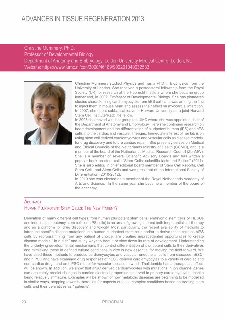
Christine Mummery studied Physics and has a PhD in Biophysics from the University of London. She received a postdoctoral fellowship from the Royal Society (UK) for research at the Hubrecht Institute where she became group leader and, in 2002, Professor of Developmental Biology. She has pioneered studies characterizing cardiomyocytes from hES cells and was among the first to inject them in mouse heart and assess their effect on myocardial infarction. In 2007, she spent sabbatical leave in Harvard University as a joint Harvard Stem Cell Institute/Radcliffe fellow. In 2008 she moved with her group to LUMC where she was appointed chair of the Department of Anatomy and Embryology. Here she continues research on heart development and the differentiation of pluripotent human (iPS) and hES cells into the cardiac and vascular lineages. Immediate interest of her lab is on using stem cell derived cardiomyocytes and vascular cells as disease models, for drug discovery and future cardiac repair. she presently serves on medical and Ethical Councils of the Netherlands Ministry of Health (CCMO), and is a member of the board of the Netherlands Medical Research Council (ZonMW). She is a member of several Scientific Advisory Boards and has written a popular book on stem cells “Stem Cells: scientific facts and Fiction” (2011). She is also editor/ in chief editorial board member of Stem Cell Reports, Cell stem Cells and stem Cells and was president of the International society of Differentiation (2010-2012). In 2010 she was elected as a member of the Royal Netherlands Academy of Arts and Science. In the same year she became a member of the board of the academy.
Christine Mummery, Ph.D.Professor of Developmental BiologyDepartment of Anatomy and Embryology, Leiden University Medical Centre, Leiden, NLWebsite: https://www.lumc.nl/con/3090/46189/902201040032533
Derivation of many different cell types from human pluripotent stem cells (embryonic stem cells or HESCs and induced pluripotency stem cells or hiPS cells) is an area of growing interest both for potential cell therapy and as a platform for drug discovery and toxicity. Most particularly, the recent availability of methods to introduce specific disease mutations into human pluripotent stem cells and/or to derive these cells as hiPS cells by reprogramming from any patient of choice, are creating unprecedented opportunities to create disease models “ in a dish” and study ways to treat it or slow down its rate of development. Understanding the underlying developmental mechanisms that control differentiation of pluripotent cells to their derivatives and mimicking these in defined culture conditions in vitro is now essential for moving the field forward. We have used these methods to produce cardiomyocytes and vascular endothelial cells from diseased hesC- and hipsC and have examined drug responses of hesC-derived cardiomyocytes to a variety of cardiac and non-cardiac drugs and an hipsC model for vascular disease in which Thalidomide has a therapeutic effect, will be shown. In addition, we show that iPSC derived cardiomyocytes with mutations in ion channel genes can accurately predict changes in cardiac electrical properties observed in primary cardiomyocytes despite being relatively immature. Examples will be shown of how metabolic diseases are beginning to be modelled in similar ways, stepping towards therapies for aspects of these complex conditions based on treating stem cells and their derivatives as “ patients”.
AdvAnces in Tissue regenerATion 2013
program 21
abStractcartilage regeneration anD Stem cellS
Gerjo van Osch (1967) studied medical biology at the University of Utrecht in the Netherlands and received her PhD in 1994 at the University of Nijmegen, the netherlands, on animal models for osteoarthritis.
In 1994 she started to work on cartilage tissue engineering which she continued doing till today. she is currently appointed as full professor at the Erasmus MC, University Medical Center in Rotterdam the Netherlands where she is leading a research line on Connective Tissue regeneration with a group of approx. 12 people that is part of the departments of orthopaedics and otorhinolaryngology.
Gerjo van Osch has been working in the field of cartilage since 1990. She is co-author on approximately 120 international peer-reviewed publications. She has been active in various committees of the International Cartilage Repair Society (ICRS), is presently vice chair of the European Science Foundation network on Regenerative Medicine (REMEDIC), associate editor of Cartilage and editorial board member of Tissue Engineering and Journal of Tissue Engineering and Regenerative medicine. She served as council member of the European chapter of the TERMIS, and chaired the TERMIS-EU meeting in Rotterdam in 2006 and the ICRS basic lab skills course in 2008.
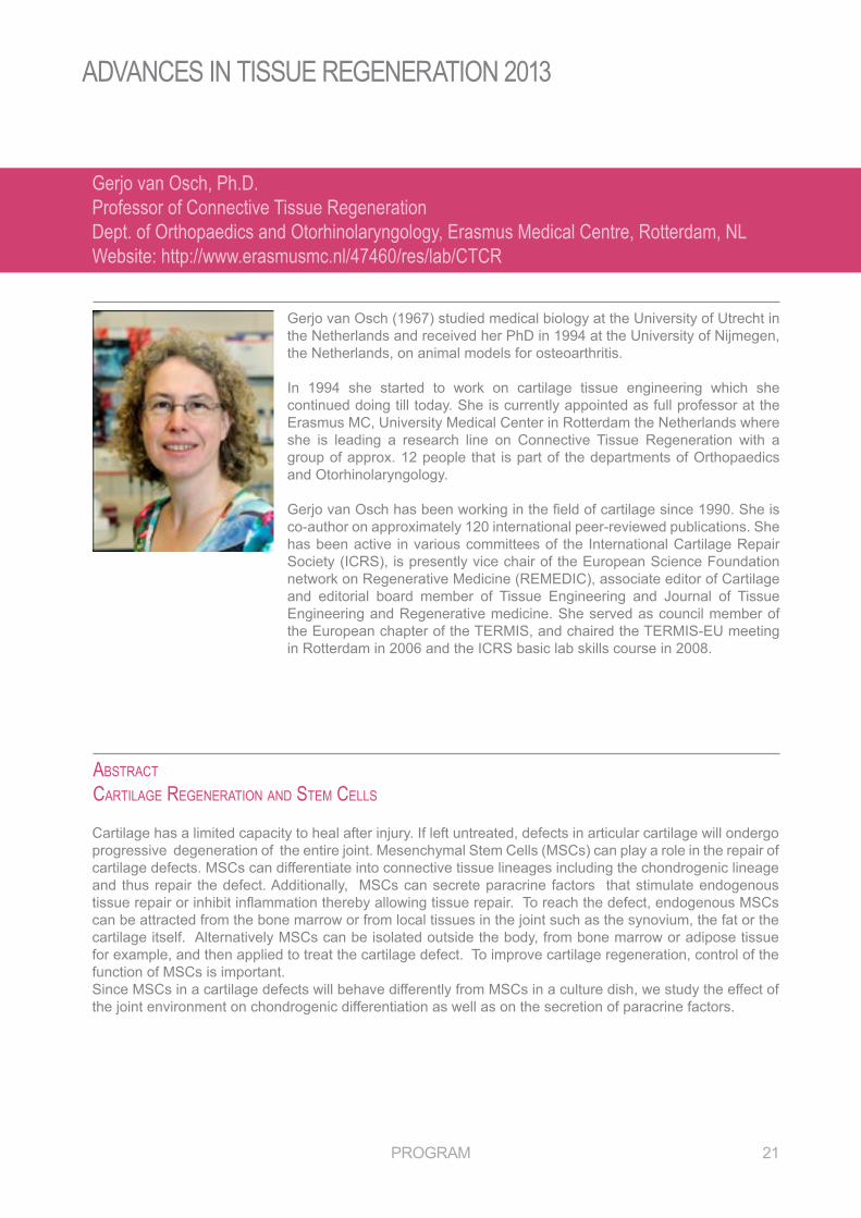
Gerjo van Osch, Ph.D.Professor of Connective Tissue RegenerationDept. of Orthopaedics and Otorhinolaryngology, Erasmus Medical Centre, Rotterdam, NLWebsite: http://www.erasmusmc.nl/47460/res/lab/CTCR
Cartilage has a limited capacity to heal after injury. If left untreated, defects in articular cartilage will ondergo progressive degeneration of the entire joint. Mesenchymal Stem Cells (MSCs) can play a role in the repair of cartilage defects. msCs can differentiate into connective tissue lineages including the chondrogenic lineage and thus repair the defect. additionally, msCs can secrete paracrine factors that stimulate endogenous tissue repair or inhibit inflammation thereby allowing tissue repair. To reach the defect, endogenous MSCs can be attracted from the bone marrow or from local tissues in the joint such as the synovium, the fat or the cartilage itself. Alternatively MSCs can be isolated outside the body, from bone marrow or adipose tissue for example, and then applied to treat the cartilage defect. To improve cartilage regeneration, control of the function of msCs is important. Since MSCs in a cartilage defects will behave differently from MSCs in a culture dish, we study the effect of the joint environment on chondrogenic differentiation as well as on the secretion of paracrine factors.
AdvAnces in Tissue regenerATion 2013
22 program
abStracttHe role of Silica in comPoSite materialS for bioengineering aPPlicationS incluDing bone regeneration anD cell baSeD tHeraPieS
Carole Perry is a professor of bioinorganic and materials chemistry at Nottingham Trent University. Her research interests lie where biology, chemistry and physics interact, and are directed in particular towards understanding how biomolecules and materials interact in aqueous media.
She has recently completed a year long sabbatical at the Radcliffe Institute for Advanced Study, Harvard University, USA as the Edward, Frances and Shirley B. Daniells Fellow and Wyss Fellow working on multidisciplinary studies of the biomolecule-mineral interface with Professor Joanna Aizenberg, Harvard University, Professor David Kaplan, Tufts University and Professor Markus Buehler, MIT. Her research at NTU has recently been recognised by the award of a Wolfson research merit award from the royal society.
Professor Perry received her BA and DPhil degrees from the University of Oxford. She has been a visiting fellow or professor at the Scripps Institute of Oceanography, Universitat Karlsruhe, Universite Pierre et Marie Curie, MIT and the Weizmann Institute of Science.
She has served as an elected trustee and council member of the Royal Society of Chemistry and chaired the UK Heads of Chemistry. From 2003-2008 she was head of Chemistry at Nottingham Trent University.
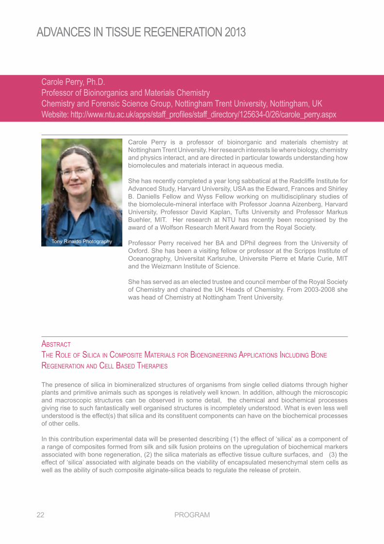
Carole Perry, Ph.D.Professor of Bioinorganics and Materials ChemistryChemistry and Forensic Science Group, Nottingham Trent University, Nottingham, UKWebsite: http://www.ntu.ac.uk/apps/staff_profiles/staff_directory/125634-0/26/carole_perry.aspx
The presence of silica in biomineralized structures of organisms from single celled diatoms through higher plants and primitive animals such as sponges is relatively well known. In addition, although the microscopic and macroscopic structures can be observed in some detail, the chemical and biochemical processes giving rise to such fantastically well organised structures is incompletely understood. What is even less well understood is the effect(s) that silica and its constituent components can have on the biochemical processes of other cells. In this contribution experimental data will be presented describing (1) the effect of ‘silica’ as a component of a range of composites formed from silk and silk fusion proteins on the upregulation of biochemical markers associated with bone regeneration, (2) the silica materials as effective tissue culture surfaces, and (3) the effect of ‘silica’ associated with alginate beads on the viability of encapsulated mesenchymal stem cells as well as the ability of such composite alginate-silica beads to regulate the release of protein.
Tony rinaldo photography

AdvAnces in Tissue regenerATion 2013
program 23
abStractimmunobiology of lymPHangiogeneSiS anD imPlicationS for tiSSue regeneration
Melody A. Swartz is Professor in the Institute of Bioengineering and the Swiss Institute for experimental Cancer research in the faculty of life sciences as well as the Institute of Chemical sciences and Technology, of the ecole Polytechnique Fédérale de Lausanne (EPFL).
previous to moving to lausanne, she was an assistant professor in biomedical Engineering and Chemical Engineering at Northwestern University. She holds a BS from the Johns Hopkins University, and a PhD from Massachusetts Institute of Technology. she undertook postdoctoral studies at Harvard medical school in boston.
Trained as a bioengineer, she uses quantitative approaches in cell biology and physiology, including biotransport and biomechanics, to investigate the role of the lymphatic system in physiology and pathophysiology. she is particularly interested in the role of the lymphatic drainage in maintaining immunological tolerance in homeostasis, and the role of lymphangiogenesis in controlling inflammation as well as inducing pathological tolerance in cancer. Her lab applies this knowledge to develop novel immunotherapeutic approaches in cancer, including lymph node-targeting vaccine approaches.
Melody Swartz, Ph.D.Professor of Bioengineering and Cancer ResearchInstitute of Bioengineering, Ecole Polytechnique Fédérale de Lausanne, Lausanne, CHWebsite: http://swartz-lab.epfl.ch/
Lymphangiogenesis occurs in nearly all cases of chronic inflammation and in the lymph node after infection or vaccination, yet the underlying reasons for lymphatic hyperplasia, or the functions that an expanded lymphatic network plays, are unknown. Furthermore, tumor-associated lymphangiogenesis has been correlated to enhanced tumor progression, indicating that it must somehow support tumor survival, invasion, or metastasis. We have recently found that tumor-associated lymphatic vessels actually help the tumor avoid host immune surveillance in multiple ways. Importantly, we found that lymphatic endothelial cells (LECs) can scavenge exogenous tumor antigens and cross-present them to naïve T cells for deletional tolerance, and that the combined effects of lymphatic expansion in the tumor microenvironment acted to impede the effects of anti-tumor immunotherapy (Lund et al, Cell Rep, 2012). Furthermore, we found that the lymphatic growth factor VEGF-C, which is often secreted by either tumor cells or tumor-associated macrophages, activates CCl21 expression, which in turn promotes changes in the tumor stromal microenvironment towards immune suppression and tolerance. When VEGF-C-driven lymphangiogenesis was blocked, antigen-specific tumor immunotherapy was more effective. In non-tumor contexts, we found that leCs could still scavenge endogenous antigens and cross-present them on mHC-I molecules for dysfunctional activation of cognate T cells. These data suggest that lymphangiogenesis serves important roles in regulating immunity to peripheral antigens, and thus could be an important process to incorporate into regenerative medicine strategies to avoid immune rejection.
AdvAnces in Tissue regenerATion 2013
24 program
abStractengineering loaD bearing biomaterialS
Professor Elizabeth (Liz) Tanner has been Professor of Biomedical Materials, University of Glasgow, since August 2007. She has been a Visiting Professor of Biomechanics and Biomaterials at the Department of Orthopaedics, Lund University Hospital, Sweden since 1998. She graduated in Engineering Science from the University of Oxford and stayed to do her DPhil in Biomedical engineering. Her thesis was measuring the movement at the fracture site in patients as their fractures healed done at the Nuffield Orthopeadic Centre. after completing her thesis, she joined Queen mary as a post doctoral research assistant working on a range of projects. Her research work continues in this area with current projects including the effect of pathology on the mechanical properties of bone and on the development of bioactive degradable composites with the stiffness and strength to be used in major load bearing applications.At Queen Mary she become Lecturer, Reader and, in 1998, Professor. She was Deputy Director of the IRC in Biomedical Materials. In 2001 she was elected a member of the Executive Council of the European Society for Biomaterials and from 2005 to 2009 she was the Secretary of the Society. In 2004 she was elected Fellow Biomaterials Science and Engineering (FBSE). In July 2006 she was elected a fellow of the royal academy of engineering. She has published over 150 papers and chapters in these fields. She as co-edited 3 books on biomaterials and biomechanics, the most recent being on “Biomaterials for Spinal Applications” with Luigi Ambrosio of the University of Naples published in 2012.
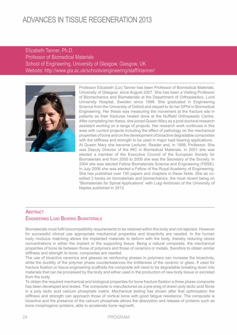
Elizabeth Tanner, Ph.D.Professor of Biomedical MaterialsSchool of Engineering, University of Glasgow, Glasgow, UKWebsite: http://www.gla.ac.uk/schools/engineering/staff/ktanner/
Biomaterials must fulfil biocompatibility requirements to be retained within the body and not rejected. However for successful clinical use appropriate mechanical properties and bioactivity are needed. In the human body modulus matching allows the implanted materials to deform with the body, thereby reducing stress concentrations in either the implant or the supporting tissue. being a natural composite, the mechanical properties of bone lie between those of polymers and those of ceramics or metals, therefore to obtain similar stiffness and strength to bone, composites are needed. The use of bioactive ceramics and glasses as reinforcing phases in polymers can increase the bioactivity, while the ductility of the polymer phase counterbalances the brittleness of the ceramic or glass. If used for fracture fixation or tissue engineering scaffolds the composite will need to be degradable breaking down into materials that can be processed by the body and either used in the production of new body tissue or excreted from the body.To obtain the required mechanical and biological properties for bone fracture fixation a three phase composite has been developed and tested. The composite is manufactured as a pre-preg of drawn poly lactic acid fibres in a poly lactic acid calcium phosphate matrix. mechanical testing has shown after that optimisation the stiffness and strength can approach those of cortical bone with good fatigue resistance. The composite is bioactive and the presence of the calcium phosphate allows the absorption and release of proteins such as bone morphogenic proteins, able to accelerate bone regrowth.
AdvAnces in Tissue regenerATion 2013
program 25
abStractextracting DiScoverieS HiDDen in imageS
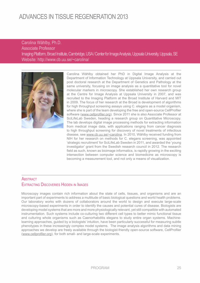
Carolina Wählby obtained her PhD in Digital Image Analysis at the Department of Information Technology at Uppsala University, and carried out post doctoral research at the Department of Genetics and Pathology at the same university, focusing on image analysis as a quantitative tool for novel molecular markers in microscopy. She established her own research group at the Centre for Image Analysis at Uppsala University in 2007, and was recruited to the Imaging platform at the broad Institute of Harvard and mIT in 2009. The focus of her research at the Broad is development of algorithms for high throughput screening assays using C. elegans as a model organism, where she is part of the team developing the free and open-source CellProfiler software (www.cellprofiler.org). Since 2011 she is also Associate Professor at SciLifeLab Sweden, heading a research group on Quantitative Microscopy. The lab develops digital image processing methods for extracting information from medical image data, with applications ranging from cancer diagnosis to high throughput screening for discovery of novel treatments of infectious disease, see www.cb.uu.se/~carolina. In 2010, Wählby received funding from nIH for her research on methods for C. elegans screening, was appointed ‘strategic recruitment’ for SciLifeLab Sweden in 2011, and awarded the ‘young investigator’ grant from the swedish research council in 2012. The research field as such, known as bioimage informatics, is rapidly growing in the exciting intersection between computer science and biomedicine as microscopy is becoming a measurement tool, and not only a means of visualization.
Carolina Wählby, Ph.D.Associate ProfessorImaging Platform, Broad Institute, Cambridge, USA / Center for Image Analysis, Uppsala University, Uppsala, SEWebsite: http://www.cb.uu.se/~carolina/
Microscopy images contain rich information about the state of cells, tissues, and organisms and are an important part of experiments to address a multitude of basic biological questions and world health problems. Our laboratory works with dozens of collaborators around the world to design and execute large-scale microscopy-based experiments in order to identify the causes and potential cures of disease. Biologists are developing model systems that are more and more physiologically relevant, yet still compatible with automated instrumentation. Such systems include co-culturing two different cell types to better mimic functional tissue and culturing whole organisms such as Caenorhabditis elegans to study entire organ systems. Machine-learning approaches, guided by a biologists’ intuition, have been particularly successful for measuring subtle phenotypes in these increasingly complex model systems. The image analysis algorithms and data mining approaches we develop are freely available through the biologist-friendly open-source software, CellProfiler (www.cellprofiler.org), for both small- and large-scale experiments.

AdvAnces in Tissue regenerATion 2013
program 27
posTer presenTaTIon absTraCTs
Sponsored by
Preparation of a biomimetic nanocomposite scaffold for bone tissue engineering
Mahmoud Azami, nafiseh baheiraei, Jafar ai
Department of Tissue Engineering, School of Advanced Technologies in medicine, Tehran University of medical sciences, Tehran, Iran.
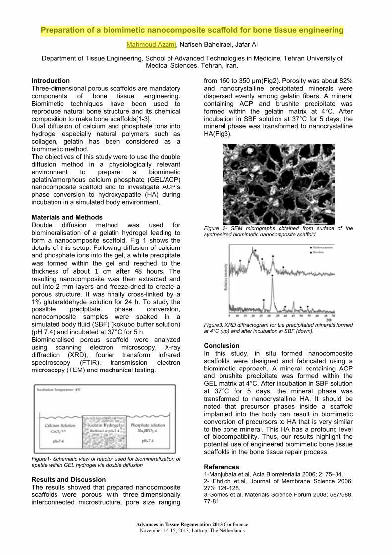
IntroductionThree-dimensional porous scaffolds are mandatory components of bone tissue engineering.Biomimetic techniques have been used to reproduce natural bone structure and its chemical composition to make bone scaffolds[1-3].Dual diffusion of calcium and phosphate ions into hydrogel especially natural polymers such as collagen, gelatin has been considered as a biomimetic method.The objectives of this study were to use the double diffusion method in a physiologically relevant environment to prepare a biomimetic gelatin/amorphous calcium phosphate (gel/aCp)nanocomposite scaffold and to investigate aCp’s phase conversion to hydroxyapatite (Ha) during incubation in a simulated body environment.
Materials and MethodsDouble diffusion method was used for biomineralisation of a gelatin hydrogel leading to form a nanocomposite scaffold. fig 1 shows the details of this setup. following diffusion of calcium and phosphate ions into the gel, a white precipitate was formed within the gel and reached to the thickness of about 1 cm after 48 hours. The resulting nanocomposite was then extracted and cut into 2 mm layers and freeze-dried to create a porous structure. It was finally cross-linked by a1% glutaraldehyde solution for 24 h. To study the possible precipitate phase conversion, nanocomposite samples were soaked in a simulated body fluid (SBF) (kokubo buffer solution)(pH 7.4) and incubated at 37°C for 5 h. biomineralised porous scaffold were analyzed using scanning electron microscopy, X-ray diffraction (XrD), fourier transform infrared spectroscopy (FTIR), transmission electron microscopy (TEM) and mechanical testing.
Figure1- Schematic view of reactor used for biomineralization of apatite within GEL hydrogel via double diffusion
Results and DiscussionThe results showed that prepared nanocomposite scaffolds were porous with three-dimensionally interconnected microstructure, pore size ranging
from 150 to 350 µm(Fig2). Porosity was about 82% and nanocrystalline precipitated minerals were dispersed evenly among gelatin fibers. A mineral containing aCp and brushite precipitate was formed within the gelatin matrix at 4°C. after incubation in SBF solution at 37°C for 5 days, the mineral phase was transformed to nanocrystalline Ha(Fig3).
Figure 2- SEM micrographs obtained from surface of the synthesized biomimetic nanocomposite scaffold.
Figure3. XRD diffractogram for the precipitated minerals formed at 4°C (up) and after incubation in SBF (down).
ConclusionIn this study, in situ formed nanocomposite scaffolds were designed and fabricated using a biomimetic approach. A mineral containing ACP and brushite precipitate was formed within the gel matrix at 4°C. After incubation in SBF solution at 37°C for 5 days, the mineral phase was transformed to nanocrystalline HA. It should be noted that precursor phases inside a scaffold implanted into the body can result in biomimetic conversion of precursors to Ha that is very similar to the bone mineral. This HA has a profound level of biocompatibility. Thus, our results highlight the potential use of engineered biomimetic bone tissue scaffolds in the bone tissue repair process.
References1-Manjubala et.al, Acta Biomaterialia 2006; 2: 75–84.2- Ehrlich et.al, Journal of Membrane Science 2006; 273: 124-128.3-gomes et.al, Materials Science Forum 2008; 587/588: 77-81.
Advances in Tissue Regeneration 2013 Conference November 14-15, 2013, Lattrop, The Netherlands

Bioactive Glass Coatings For Bone Tissue Engineering: An In Vitro Study
Nasrin Lotfibakhshaiesh1 , Jafar ai1, Mahmoud Azami1, Robert G Hill2, molly m stevens3,4
1Department of Tissue Engineering, School of Advanced Technologies in Medicine, Tehran University of medical sciences, Tehran, Iran 2Unit of Dental Physical Sciences, Barts and The London School of Medicine and Dentistry, Queen Mary University of London, London, UK3Department of Materials, Imperial College London, London, UK, 4Institute of biomedical engineering, Imperial College london, london, UK
Introductionmetallic prostheses are widely used to treat joint and skeletal injuries and disease. However, metal alloy implants can sometimes fail due to complications of fibrous encapsulation and poor stress transfer between the bone and the implant. Bioactive glass (BG) coatings may promote the formation of a strong bond with living bone tissue thus decreasing the likelihood of fibrous encapsulation and have the added benefit that their dissolution ions stimulate cell activity [1,2]. Strontium (Sr) ranelate, a drug used to treat and prevent osteoporosis, works via the action of sr ions which stimulate the formation of new bone and prevent osteoclast-mediated resorption [3]. We have previously shown that sr-substituted BGs promote osteoblast activity in vitro [4] and explored the effect of altering phosphate content on the material structure of soda-lime-phosphosilicate glasses [5]. The effect of increasing phosphate content in sr-substituted BG on cultured osteoblasts, however, remains unexplored. Here, we created sr-substituted BG coatings with a range of phosphate contents and thermal expansion coefficients that matched that of Ti alloy, producing materials that combine the bone remodelling benefits of sr and bg with phosphate to mediate pH changes which can affect cell viability. In the study presented here we report the characterisation of these multicomponent bg coatings in terms of their bioactivity and interaction with cells.
Materials and Methodsbioactive glasses in the system sio2-mgo-na2o-K2o-zno-p2o5-CaO in which 10% of the Ca was replaced by Sr and the P2o5 content was increased from 1.07 to 6.42 mol% were produced by a melt quench route. Sufficient cations were added to ensure charge neutrality in the po4
3-
complex formed. Simulated body fluid (SBF) was prepared according to Kokubo [6]. Glass particles (<38 micrometer) were immersed for up to 28 days and agitated at 60 rpm at 37°C. At indicated time points samples were filtered and dried for X-ray Diffraction (XRD) analysis. Culture media containing ions from glasses were created by incubating 1.5g/L of glass powder (<38 micrometer) in RPMI 1640 on a roller for 4 hours at 37°C and then passed through a 0.2 micrometer filter. This media was then supplemented with 10% (v/v) foetal bovine serum (FBS), 2mM L-glutamine and 1% (v/v) penicillin/streptomycin. The human osteosarcoma cell line, saos-2, was seeded at
30,000 cells/cm2 in conditioned medium and cultured for up to 28 days. On days 1, 14, 21 and 28 cell metabolic activity was measured using the tetrazole MTT as an indicator of cell proliferation. glasses were coated on the surface of Ti6al4V coupons with an enameling technique. 10,000 saos-2/cm2 were seeded on bg coatings and viability was assessed after 1, 7 and 14 days with a LIVE/DEAD stain. Some glass coatings cultures were also fixed, gold coated and viewed on a leo 1525 gemini sem.
Results and Discussionbg with high p2o5 content forms more apatite after immersion in sbf for 4 weeks than bg with low p2o5 content, as examined by XRD. MTT activity in saos-2 cells treated with dissolution ions from bg increased in all samples with time in culture. MTT activity was also significantly greater (p<0.01) in cells treated with dissolution ions from 4.28 and 6.24 mol% P2o5 bgs as compared to controls at day 28. LIVE/DEAD staining indicated that all coating materials were not cytotoxic. sem imaging demonstrated that the bg coating encouraged cell attachment and that cells spread well over thesurface.
ConclusionWith increasing p2o5 content in the series of sr-substituted BG, Bragg peaks in XRD traces associated with apatite crystallisation increase suggesting the glass becomes more bioactive. apatite formation on the coating surface is an essential factor for bone bonding as the more apatite that forms on the glass coating the more bone bonding will be expected. Adding P2o5 to the glass composition in a controlled way prevents extreme pH rises, which can affect cell viability and proliferation.
References[1] Hench LL et al. J. Inorg Mater 2002;17:897–909[2] Jell, g, stevens mm, J mater sci mater med, 2006. 17(11): 997 [3] Hamdy NA, Rheumatology, 2009; 48(4): 9 [4] gentleman e, fredholm YC, Jell g, Lotfibakhshaiesh N, O'Donnell MD, Hill rg, stevens mm. , Biomaterials, 2010; 31(14): 3949 [5] O’Donnell MD et al., J Mater Sci Mater Med, 2009. 20:1611 [6] Kokubo T et al., Biomaterials, 2006; 27:2907–15
Advances in Tissue Regeneration 2013 Conference November 14-15, 2013, Lattrop, The Netherlands

Microfluidic strategies to study interactions between cells and biomaterials for bone regeneration
David barata*, C. Correia, björn Harink, roman Truckenmüller, Clemens van blitterswijk, Pamela HabibovicDepartment of Tissue Regeneration, University of Twente, The Netherlands
*e-mail: [email protected]
IntroductionCombining tools from micro-engineering and tissue regeneration fields offers new possibilities to simulate biomaterial/cell and biomaterial/tissue interactions in vitro.One of the objectives of our research group is to develop synthetic alternatives to autologous bone grafts, that suffer from a number of disadvantages, limited availability being the most critical one. In order to be considered a comprehensive alternative to natural bone grafts, synthetic biomaterials need to meet various requirements, including mechanical stability, and bioactivity in terms of osteoconduction and osteoinduction. While in the past decades a great number of synthetic bone graft substitutes, including calcium-phosphate ceramic-based oneshas been developed [1, 2], the majority still needs further improvement to be accepted as a true alternative to natural bone grafts. Fundamental understanding of interactions between materials and cells and/or tissues is of great value when it comes to improvement of synthetic bone graft substitutes. Platforms based on microfluidics offer possibilities to increase the throughput of testing of cell/material interactions. In addition, they allow for recreating the biological microenvironment surrounding an implanted bone graft substitute in vitro [3].
Materials and methodsIn order to mimic the microenvironment of biomaterials relevant for bone repair and regeneration, two microfabricated systems have been developed:A) A wet etched glass microfluidic cell culture chamber, assembled with a glass cover, and fed by 4 independent diffusive side-channels for nutrient and oxygen supply;B) polydimethylsiloxane (PDMS) multi-chamber microfluidic device, assembled over glass/polymeric layers, and fed by 2 independent diffusive side-channel for nutrient and oxygen supply.Functional studies are performed by culturing of mg-63 human osteosarcoma cells.
Results and discussionTo make the developed microfluidic systems suitable for studies of interactions between cells and biomaterials for bone regeneration, surface properties of the cell culture chambers were modified following two different strategies.In strategy A, the microfluidic culture chamber wascoated by using a sputtering technique. This allowed for deposition of a titanium film with a thickness of few nanometers, that was oxidized in
reactive atmosphere (Figure 1). This technique is now being employed to deposit a range of different biomaterials, including bioactive calcium-phosphates.
figure 1: left, scanning electron microscopy image of glass chip; right,X-ray map analysis of sputtered titanium inside the culture chamber of glass chip.
In strategy b, the surface of the microfluidic cellculture chamber was covered by a layer of polymer or polymer/ceramic composite by spin-coating before assembling the system with the support glass and the fluidics in PDMS (Figure 2).
figure 2: left, image of PDMS multi-chamber device; right, mg-63osteosarcoma cells on the glass surface of the chip (control).
future experiments will focus on functionalizing the systems with thin films of other materials relevant tobone repair and regeneration, and performing studies on cell-material interactions under flow regimes which more closely resemble the in vivocell microenvironment.
References[1] Dinopoulos H et al. (2012) Bone graft substitutes – what are the options?. Surgeon 10(4): 230-239.[2] barradas mC et al. (2011) osteoinductivebiomaterials: current knowledge of properties, experimental models and biological mechanisms.European Cells and Materials, 21: 407-429[3] Whitesides gm (2006) The origins and the future of microfluidics. Nature 442: 368-373.
Acknowledgements: The netherlands Institute for regenerative medicine is acknowledged for financial support.
Advances in Tissue Regeneration 2013 Conference November 14-15, 2013, Lattrop, The Netherlands
Advances in Tissue Regeneration 2013 Conference November 14-15, 2013, Lattrop, The Netherlands
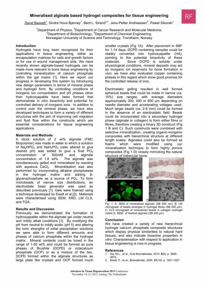
Mineralised alginate based hydrogel composites for tissue engineering
David Bassett1, sindre Hove bjørnøy1, berit l. strand2,3, Jens-petter andreassen4, pawel sikorski1
1Department of Physics; 2Department of Cancer Research and Molecular Medicine; 3Department of Biotechnology, 4Department of Chemical engineering, Norwegian University of Science and Technology, Trondheim, norway
Introduction Hydrogels have long been recognized for their applications in tissue engineering, either as encapsulation matrices for cells and growth factors or for use in wound management aids. We have recently shown alginate-based hydrogels can be made more relevant to bone tissue engineering by controlling mineralization of calcium phosphate within the gel matrix [1]. Here we report our progress in developing this system by introducing new design parameters in terms of mineral phase and hydrogel form. by controlling conditions of inorganic ion concentration and pH phases other than hydroxyapatite have been formed that demonstrate in vitro bioactivity and potential for controlled delivery of inorganic ions. In addition to control over the mineral phase, we have also developed techniques to form a variety of different structures with the aim of improving cell migration and fluid flow within the constructs which are essential considerations for tissue engineering applications. Materials and Methods a stock solution of 2 wt% alginate (fmC biopolymer) was made in water to which a solution of na2Hpo4 and naH2po4 (ratio altered to give desired pH) was added to give a final po4 concentration of 300mm and alginate concentration of 1.8 wt%. The alginate was simultaneously gelled and mineralised by reacting with aqueous CaCl2. mineralisation was also performed by incorporating alkaline phosphatase in the hydrogel matrix and adding β-glycerophosphate as a source of po4. To form microbeads of narrow size distributions, an electrostatic bead generator was used as described previously [1]. gels were foamed using a technique developed by Eiselt et al.[2]. materials were characterised using SEM, XRD, LM CLS, and Tga. Results and Discussion previously we demonstrated the formation of hydroxyapatite within the alginate gel under neutral and mildly alkali conditions [1]. by modifying the pH from neutral to mildly acidic (pH 5) and altering the ionic strengths of initial precipitation solutions we were able to form different amounts and phases of calcium phosphate within the hydrogel matrix. Mineral contents could be tuned in the range of 1-55 wt% and could be formed as pure phases of Brushite (DCPD) or octacalcium phosphate (OCP) or as a mixture of the two. DCPD formed within the alginate structures as large plate like crystals and oCp formed much
smaller crystals (Fig 1A). After placement in SBF for 1-14 days, DCPD containing samples could be readily converted into hydroxyapatite (Ha), pointing to the potential bioactivity of these materials. Since DCPD is soluble under physiological conditions, mineral deposits may act as inorganic ion reservoirs for bone formation in vivo; we have also evaluated copper containing phases in this regard which show good promise for the controlled release of ions. electrostatic gelling resulted in well formed spherical beads that could be made in narrow (ca. 10%) size ranges, with average diameters approximately 200, 400 or 600 µm depending on needle diameter and accelerating voltages used. Much larger beads (ca. 2-6 mm) could be formed in the absence of an electrostatic field. Beads could be incorporated into a secondary hydrogel phase (alginate or collagen) to form either films or fibres, therefore creating a binary 3D construct (Fig 1 b and C). such constructs were combined with selective mineralization, creating organic-inorganic composites with hierarchical structure at different length scales. alginates could also be formed as foams which were modified using our mineralization technique to form highly porous composites (Fig 1 D) closely mimicking the natural structure of trabecular bone.
a b
C D fig. 1: a: sem of mineralized alginate (sb 500 nm), b: lm micrograph of beads arranged in hydrogel fibres (sb 200 µm), C: Cls micrograph of mineralized beads in collagen hydrogel matrix D: sem– of foamed alginate (sb 200 µm). Conclusion We have created a variety of new hierarchical hydrogel calcium phosphate composite structures which display physical similarities to natural hard tissues, and also indicate bioactive properties in vitro. Characterisation with respect to application in tissue engineering is now in progress. References 1. Xie, m.l., et al., acta biomaterialia, 2010. 6(9): p. 3665-
3675. 2. eiselt, p., et al., biomaterials, 2000. 21(19): p. 1921-1927.
RF

Liposome-mediated stabilization of Wnt ligands enables defined culture of human organ stem cells
Nesrin Tüysüz1, louis van bloois2, Enrico Mastrobattista2, stieneke van den brink3, Robert Vries3, Hans Clevers3, Derk ten Berge1 .
1erasmus mC stem Cell Institute, erasmus medical Center, rotterdam. 2Utrecht Institute for Pharmaceutical Sciences (UIPS), Utrecht University, Utrecht. 3Hubrecht Institute for Developmental Biology and Stem Cell
Research, Utrecht.
Introductionone of the promises of regenerative medicine is to create or repair tissues and organs using stem cells. Over the past 15 years it has been shown that the self-renewal and cell fate choices of a variety of stem cells, such as those from the gastrointestinal system, skin, hair, and nervous system, are regulated by the Wnt signaling pathway. Wnt proteins may therefore support the self-renewal of these stem cells in culture, allowing their in vitro manipulation and expansion for therapeutic purposes. Culture systems dependent on Wnt agonists haverecently been defined for several human and mouse adult stem cells. mouse intestinal stem cells can be expanded in organoids that contain niche cells that produce the required Wnt signals. However, for stem cells from other organs, such as colon, stomach, and liver, and for human stem cells, these endogenous Wnt signals are insufficient and exogenous Wnt3a protein is required to support stem cell expansion. We noticed however that this exogenous Wnt3a protein is only effective when provided as an unpurified conditioned medium, which contains many undefined factors secreted by the Wnt-producing cells or originating from the serum added to support those cells. not only do these factors promote differentiation of the stem cells, their presence is highly undesirable for clinical applications. We therefore investigated why purified Wnt3a protein failed to support derivation or maintenance of human adult stem cells.
Results and DiscussionHere, we demonstrate that purified Wnt3a protein rapidly loses its activity in serum-free culture media, displaying a half-life of less than 2 hours. In contrast, in the presence of serum or conditioned medium, Wnt3a activity is maintained for several days, suggesting that serum is required for the maintenance of human adult stem cells by stabilizing Wnt3a protein. Based on our previous observation that Wnt proteins associate with lipid vesicles1, we explored whether liposomes stabilize Wnt proteins in defined stem cell cultures. We show that association with lipid vesicles considerably increases the half-life of purified Wnt3a protein in the absence of serum. Moreover, our Wnt3a/liposome complexes support the highly efficient derivation and expansion of human duodenum and ileum stem cells in serum free conditions. Derivation in the presence of Wnt3a liposomes was more efficient than in the presence
of Wnt3a conditioned medium, and the organoids displayed less evidence of differentiation.
ConclusionThe short half-life of purified Wnt proteins in serum-free culture systems prevents the derivation and efficient expansion of human adult stem cells.However, by complexing purified Wnt3a protein with lipid vesicles, its half-life is considerably increased. We show that Wnt3a liposomes not only enable the establishment of adult human stem cell cultures in defined conditions, but also increase the efficiency of derivation and culture by eliminating differentiation-inducing factors from the culture system. Our Wnt/liposome technology enables the derivation and culture of human adult stem cells under defined conditions, and has now made possible their use for clinical applications.
Reference1 - Morrell, N.T., Leucht, P., Zhao, L., Kim, J.-b., ten Berge, D., Ponnusamy, K., Carre, A.L., Dudek, H., zachlederova, m., mcelhaney, m., brunton, S., Gunzner, J., Callow, m., polakis, p., Costa, m., zhang, X.m., Helms, J.A., and Nusse, R. (2008) PLoS ONE 3, e2930. liposomal packaging generates Wnt protein with in vivo biological activity.
Advances in Tissue Regeneration 2013 Conference November 14-15, 2013, Lattrop, The Netherlands

Anterior Cruciate Ligament- versus Hamstring-derived cells
C.A.Ghebes1, C. Kelder1, T. schot1, a.J. renard2, D.F.M Pakvis3, H. fernandes1, D. Saris1.1University of Twente, Department of Tissue Regeneration, Zuidhorst 130, Postbus 217, 7500AE, Enschede,The netherland; 2Department of Orthopaedic Surgery, Medisch Spectrum Twente Hospital, Enschede, The
netherland; 3Department of Orthopaedic and Traumatology, Orthopedic Center OCON, Hengelo, The netherland
IntroductionAnterior Cruciate Ligament (ACL) injuries are common clinical problems that impair the functionality of the knee joint resulting in improper gait. Due to the poor healing potential of the ACL, ACL reconstruction is the treatment of choice. The torn ACL is replaced by a new graft, often a Hamstring (HT) and the functionality of the knee joint is regained. Clinical outcome and functional follow-up assessment after the surgical treatment proof that the HT graft is suitable for replacing the torn ACL. Even so, although it is described that both ACl and HT are similar in structure, biology and mechanical properties [1], cells derived from these two tissues might be intrinsically different as result of variation in anatomical location and function. Both ACL and HT are described by closely packed collagen fibers and low cellular content, with cells aligned with the fibers. on the opposite, the aCl is located between the femur and the tibia, transmitting force from one bone to the other, while the HT is located on the back of the knee joint attaching the hamstring muscle group to the tibia and thereby transmitting force from the muscle to the bone. Understanding the differences and similarities between the two cell types could have a great impact on refining aCl reconstruction surgeries, such as accelerating the process of healing.
AimThe aim of this study was to analyze the phenotypic differences between ACL- and HT-derived cells.
MethodsaCl- and HT-derived cells were isolated from tissue harvest from patients undergoing total knee arthroplasty or aCl reconstruction upon informed consent. The self-renewal and multilineage potential, as well as the expression of surface markers and gene expression profile of both cell types were analyzed.
Resultsboth aCl- and HT-derived cells showed limited self-renewal potential and had a surface marker profile distinct from mesenchymal stromal cells (MSCs). The colony forming units (CFUs) from aCl- and HT-derived cells did not show the circular shape that is characteristic of self-renewing colonies obtained from MSCs. Surface marker expression showed high expression of CD90 but limited expression of other established MSCs markers, such as CD73, CD105 and CD146.Differences in ACL- and HT-derived cells were observed on multilineage potential. While ACL-derived cells showed high potential to differentiate into chondrocytes and adipocytes but not osteoblast, HT-derived cells showed limited multilineage potential. The expression of tendon/ligament related genes (COL1A1, COL3A1 and TNC) was similar on both cell types.
Conclusionbased on our findings, we support the use of HT graft as a replacement for a torn aCl, as the intrinsic properties of the aCl-derived cells can be to a certain extent replaced by HT-derived cells. nevertheless, our findings also indicate that HT-derived cells would need some modulating factors to reduce the differences between the two cell types and consequently improve the clinical outcome.
References
1.Zhu, J., et al., Ultrastructural and Morphological Characteristics of Human Anterior Cruciate Ligament and Hamstring Tendons. The Anatomical Record: Advances in Integrative Anatomy and Evolutionary Biology, 2012. 295(9): p. 1430-1436.
Advances in Tissue Regeneration 2013 Conference November 14-15, 2013, Lattrop, The Netherlands

Bioinspired Supramolecular Biomaterials for in-situ Cardiovascular Tissue Engineering
o.J.g.m. goor1, g.C.v. almen1 and P.Y.W. Dankers1.
1Department of Biomedical Engineering, Laboratory of Chemical Biology, and Institute for Complex Molecular systems, eindhoven University of Technology, PO Box 513, 5600 MB Eindhoven, the Netherlands
Cardiovascular and valvular diseases are responsible for a significant cause of death worldwide. although surgical interventions to replace heart valves or arteries provide asignificant improve in quality of life, long-term failure of the implants often leads to a decreased life expectancy. To this extend, in-situcardiovascular tissue engineering has evolved as a potential alternative in order to create an instructive, biodegradable scaffold. This scaffold aims at using the natural regenerative potential of the human body to engineer a replacement tissue in vivo. In our group, we aim at the synthesis and development of cell-free vascular graft materials using supramolecular chemistry. These graft materials should be indistinguishable from nature, requiring control over both their mechanical properties and their bioactive adaptation capacity. Tailor-made materials are developed based on supramolecular polymers end-capped with ureido-pyrimidinone (UPy) moieties, able to dimerize upon quadruple hydrogen-bond formation giving rise to a dynamic character, that are spaced with
polycaprolactone blocks to meet mechanical requirements of the material. Bioactive UPy-modified peptides are incorporated via a modular approach in order to provide necessary biological signals to attract and stimulate cells inside the scaffold. In the design presented here, the supramolecular scaffold consists of two layers. The first, luminal layer targets endothelial progenitor cells (EPC) that will be recruited from the blood stream by UPy-SDF-1 α (stromal cell-derived factor 1 alpha) peptides incorporated in the scaffold. These EPCs subsequently are proposed to differentiate into endothelial cells. In the second layer UPy-TGFβ (transforming growth factor beta) peptides are incorporated. It is proposed that these TGFβ-derived peptides are able to induce endothelial-to-mesenchymal transition (EndMT) that should result in the formation of a layer of smooth muscle cells. This approach is a step forward in the design of a cell-free instructive synthetic biomaterial for future in-situcardiovascular tissue engineering applications.
Advances in Tissue Regeneration 2013 Conference November 14-15, 2013, Lattrop, The Netherlands

Development of a microfluidic platform for cell cultivation in narrow channelsmaciej grajewski1, patty p.f.m.a. mulder1, grietje molema 2, Elisabeth Verpoorte1
1pharmaceutical analysis, groningen research Institute of pharmacy, University of Groningen2Endothelial Biomedicine and Vascular Drug Targeting, Medical Biology, University Medical Center Groningen
The netherlands
Introduction
The current report focuses on the development of microfluidic devices for endothelial cell (EC) culture. microchannels are a good mimic for the in vivo environment of endothelial cells. However, reported channels have large dimensions [1],whereas our interest lies in the investigation of microvasculature. seeding eC into channels as small as 25 µm is difficult, with cells tending to distribute unevenly along channels; cell layerscontaining gaps are often the result. This report describes optimization of previously reported microfluidic device designs [2] for improved seeding and cultivation of eC in narrow channels.
Materials and MethodsDevices poly(dimethyl)siloxane (PDMS) - glass hybrid devices were used, with microchannels replicated in PDMS and sealed with a glass microchip. The bottom glass surface of the channels was coated with gelatin (porcine, type A, Sigma Aldrich) at 1% (W/V), a substitute extracellular matrix (ECM) which encourages eC to adhere to this surface. gelatin strips were patterned on the glass substrate using a stencilling technique before final device assembly. 0.5% glutaraldehyde solution (V/V) in phosphate-buffered saline (PBS) solution was employed as a cross-linker to improve adhesion of the gelatin to the glass. Irreversible bonding of the pDMS and glass chips was achieved using oxygen plasma treatment to activate both PDMS and glass surfaces. The PDMS channels were then precisely aligned (± 2 µm) with the gelatin strips [3], and the two chips brought together to bond.Cell culture: Human umbilical vein endothelial cells (HUVEC) obtained from the UMCG endothelial Cell facility were seeded at concentrations of 5,000 cells/µL. endothelial cell medium (RPMI medium supplemented with 10% fetal calf serum) was used for seeding and subsequent cell culture. Cell culture was carried out in an incubator at 37 °C in an atmosphere of 5% CO2.
Results and Discussion: successful cultivation of HUVEC was achieved for up to 72h without cell death in microchannels in irreversibly bonded chips with different dimensions. Three different designs were tested and progressive improvement of EC distribution along the microchannels was observed (Fig.1). The difference between the first and second design was a decreased length of the microchannel from 25 mm to 10 mm (Fig.1 A1 and B1). However, cell distribution was not improved sufficiently to reproducibly obtain confluent layersof eCs. Therefore, a third design was introduced
which included a narrow cell-introduction channel before the cultivation channel as well as a narrow exit channel (Fig.1 C1). This design yielded improved cell distribution along the microchannels.This significant improvement in cell distribution is thought to be due to the back pressure introduced to the system by the presence of the narrow exit channel. This results in better cell retention in the cultivation channel itself, and a more even distribution of cells as they settle and adhere to the gelatin layer. The confluent layers formed have far fewer gaps in them, making them more suitable for cell-based studies.
Channel length:25 mm; reservoir diameter: 2.5 mm
Channel length: 10 mm; reservoir diameter: 1 mm
Channel length: 10mm; reservoir diameter: 1 mm
Fig.1 Three microfluidic device designs are presented, withprogressive improvement of EC distribution observed from left to right (A2<B2<C2). Each photo was taken 2h after cell seeding, before confluency. Magnification: 20x Conclusion: Cultivation channels having widths less than 120 µm will be tested first with HUVEC. once successful cell cultures are demonstrated, we will commence experiments with murine microvascular cells.References:[1] Young EWK, Simmons CA, Macro- and microscale fluid flow systems for endothelial cell biology, Lab Chip 2010;10:143-160.[2] mulder ppmfa et al., Characterization of HUVEC cultivated in microfluidic channels, proc. micro Total analysis systems 2005, Transducers Research Foundation, San Diego, U.S.A., 1383-1385 (2005).[3] Kim J.Y. et al., Automatic aligning and bonding system of PDMS layer for the fabrication of 3D microfluidic channels; sens. and act. a 2005, 119: 593-598.
A1 B1 C1
120µm
A2
120µm
B2
120µm
C2
Advances in Tissue Regeneration 2013 Conference November 14-15, 2013, Lattrop, The Netherlands
From high-throughput gene expression profiling to high-throughput materials screening
nathalie groen1, Gülistan Koçer1, Huipin Yuan1, Clemens a van blitterswijk1 and Jan de boer1.1 Department of Tissue Regeneration, MIRA Institute for Biomedical Technology and Technical medicine,
University of Twente, Enschede 7500 AE, The Netherlands.
Introduction
In improving the biological response of bone graft substitute materials, not only the biological performance but also the handling and mechanical properties are still part of current research activities. This appears challenging because in vitro models which truthfully reconcile the behavior of these materials in vivo are largely lacking. our approach in this biomaterial research focusses on discovering biological differential responses induced by virtue of specific biomaterial properties and henceforth applying this biological knowledge for improving biomaterial properties.as a model system we use well-studied calcium phosphate based ceramic materials together with the human osteoblastic cell line MG-63 [1, 2].
Materials and Methods
In the work described here, we aim at characterizing the (differential) response of osteoblasts to biomaterials with varying properties of interest for bone regeneration applications.Therefore, a set of calcium phosphate based biomaterials was characterized both in vitro and in vivo (i.e. their bone inducing capacity at ectopic sites). besides confirming their related physico-chemical properties by FTIR and XRD, we specifically compared the structural surface properties that are hypothesized to play a role in in vivo bone formation capacity.Then, we determined the gene expression profiles these materials elicited in mg-63 using DNA microarray technology. Using bioinformatics analysis tools we compared the transcriptional profiles on the different materials in relation to their structural properties. These differences and similarities were further confirmed and analyzed in different studies and model systems.
Results and Discussion
Correlating the material properties with their bone forming capacity in vivo and the expression of
genes in vitro revealed differences in gene expression which may be important for the biological response to biomaterials. We next validated the expression of the identified genes in a different but defined set of calcium phosphatebased materials with corresponding differences in the studied property.
ConclusionWe identified and confirmed the expression of specific genes in correlation to material properties of interest for improving the performance of bone graft substitute materials. Thereafter, we aim at further validating the expression and exploring the biological functionality of these found transcriptional differences. for this purpose we designed artificial materials with the specific properties of interest. eventually these discovered hits may be used to further develop, screen and improve materials for bone regeneration purposes.
References
[1] Yuan H, Fernandes H, Habibovic P, de Boer J, barradas amC, de ruiter a, et al. osteoinductive ceramics as a synthetic alternative to autologous bone grafting. proceedings of the national academy of sciences 2010; 107 (31): 13614-9.
[2] groen n, van de peppel J, Yuan H, van leeuwen Jp, van blitterswijk Ca, de boer J. bioinformatics-based selection of a model cell type for in vitro biomaterialtesting. biomaterials 2013; 34 (22): 5552-61.
This research forms part of the Project P2.04 BONE-IP of the research program of the BioMedical Materials institute, co-funded by the Dutch Ministry of Economic Affairs, Agriculture and Innovation.
Advances in Tissue Regeneration 2013 Conference November 14-15, 2013, Lattrop, The Netherlands
Hn
Hn
o
Hn
Hn
on
Introduction of fluorescent guest into segmented bisurea based supramolecular polymers for tissue engineering
Samaneh Kheyrrooz 1,3, Patricia Y.W. Dankers 2,3, Rint P.Sijbesma 1,3
1. Laboratory for Macromolecular and Organic Chemistry,2. Laboratory of Chemical biology, 3.Institute for Complex molecular systems.
Eindhoven University of Technology, Eindhoven, The [email protected]
Introductionmonomeric units in supramolecular polymers are held together by non-covalent interactions. Due to these reversible non-covalent interactions, supramolecular materials are dynamic in nature. This adaptive behaviour makes supramolecular polymers promising materials for biomedical applications.Urea groups are known to form strong bifurcated hydrogen bonds. Self-assembly of bisurea based bolaamphiphiles led to formation of rod-like micelles in water.1 Inspired by the bisurea based bolaamphiphiles, we have designed segmented polymers with bisurea units in a hydrophobic alkyl motif, and poly(ethylene glycol) (PEG) as hydrophilic domain (Figure 1).2
Figure 1: Molecular structure of the segmented bisurea based polymers PUnU.
These bisurea-PEG polymers (PUnU) form hydrogels in aqueous environment. bioactive molecules functionalized with the same bisurea motif are proposed to co-assemble as guest molecules with the host bisurea-peg polymers. In this way bioactivity can be introduced into the bisurea-peg hydrogels by simply mixing-and-matching. To study the possibility of bioactivation a model system based on bisurea fluorescent guest molecules was designed.
MethodsFluorescent probe molecules are composed of two urea groups in the central part and the fluorescent pyrenes were coupled to one end (PyUnU) (Figure 2). These probe molecules can be incorporated into the supramolecular network by making intramolecular H-bonds with the urea of the host polymers. at high concentration pyrene molecules form intermolecular excimers. Dissociation of excimers was used to probe the dynamic nature of the bisurea polymers.
Figure 2: Molecular structure PyUnU.
Results and discussionDilution of a solution of PU4U with incorporated PyU4U molecules with a solution of PU4U (without PyU4U) led to higher ratio of monomer to excimer.Due to the reversibility of H-bonds between the guest molecules and host polymers, bisurea pyrene guest molecules are incorporated into the newly added hosts. This led to a decrease in the local concentration of guest, and subsequently excimer species dissociated into monomeric pyrenes. (Figure 3).
Figure 3: The ratio of monomer to excimer emission intensity as a function of pyrene concentration.
Conclusion and outlookThe dynamic nature of the bisurea based polymers was confirmed. These results give us more insight into the host-guest interactions in our bisurea-peg systems. This will help us to design bioactive guest molecules that in future can be used in these bisurea-PEG hydrogels to be applied for tissue engineering purposes.
References(1) a. pal, S. Karthikeyan, R. P. Sijbesma, J.
Am. Chem. Soc. 2010, 132, 7842-7843.(2) G. M. Pawar, M. Koenigs, Z. Fahimi, M.
Cox, I. K. Voets, H. M. Wyss, R. P. Sijbesma, Biomacromolecules. 2012, 13, 3966−3976.
Acknowledgment. The authors would like to thank nWo for financial support of the project.
OHN
PEG O10
HN
O
HN
HN
OO
O
n 10p(8k/20k)
0.0 1.0x10⁻⁶ 2.0x10-60.2
0.4
0.6
0.8
1.0
Dilution with polymer solutionDilution with water
Mon
omer
/Exc
imer
Pyrene concentration (M)
Advances in Tissue Regeneration 2013 Conference November 14-15, 2013, Lattrop, The Netherlands

Bottom-up strategy to build up functional 3D Dermis equivalent in vitro with different shapes
francesca martorina1, giorgia Imparato1, francesco Urciuolo1, Costantino Casale1, paolo a. netti1
1 Center for advanced biomaterials for HealthCare@CrIb Istituto Italiano di Tecnologia (IIT), largo barsanti e Matteucci n. 53, 80125 naples, Italy
Introductionmimicking the natural tissue organization, several strategies were implemented to successfully create large and viable constructs in vitro using a bottom-up approach [1,2]. an emerging tissue engineering field known as “modular tissueengineering” focuses on fabricating tissue building blocks with specific microarchitectural features and using these modular units to engineer biological tissues from the bottom up [3,4]. according to these strategies, our tissue fabrication process consisted of gelatin porous micro beads culturedwith Human Dermal Fibroblasts (HDFs) which were used for the construction of millimeter-thick tissues with different shapes and complex microstructures. The versatile strategy allowed to obtain 3D dermis tissue constructs of defined size and geometry by means of the biological sintering of cell seeded microscaffold so-called microtissue precursors (μTPs). 3D dermis tissues of different shapes were obtained and, exploiting μTPs assembling strategy, the role of microscaffold properties in guiding extracellular matrix (eCm)organization was investigated histologically and morphologically.
Materials and MethodsDegradable gelatin porous microcarriers (GPM) were prepared according to a modified double emulsion technique (O/W/O) and then chemically crosslinked by means of glyceraldeide. Using aspinner flask bioreactor, building blocks for bottom-up tissue applications such as µTPs were obtained by dynamic cell seeding of HDFs on the realized gpms. after nine days of dynamic culture, µTps samples were withdrawn for morphological and histological analyses. at the end of spinner culture,µTps were transferred into maturation chamber with different geometry. Under optimized culture conditions µTps were induced to assemblegenerating a 3D millimeter-thick dermal equivalent tissue able to maintain the shape of the maturation chamber. The 3D dermis equivalent constructs were detached from the molds and investigated by means of histological analyses (picro sirius red,hematoxylin and eosin stainings) and second harmonic generation imaging (SHG) ultra-structure analyses.
Results and Discussionresults showed that 3D millimeter-thick macroscopic dermis equivalent with different shapes and complex microstructures were obtained. The presence of the microbeads in the initial stage of the maturation process supplied
mechanical support to the neo tissue. Despite to other bottom-up approaches, in this case just after one week of maturation, the 3D biohybrid constructs, which were characterized by uniform cellular colonization and functional tissue formation throughout their thickness and shape, wereobtained and handled without any mechanical breakage. moreover, results showed the presence of endogenous eCm in the 3D dermis equivalent with organized collagen fibers as confirmed by histological and ultra-structural analyses.moreover, the shapes of the 3D dermal tissues strongly depended on the geometry of the maturation chambers and on dynamic culture conditions inside the chambers where µTps were assembled, as demonstrated by different kinds of maturation chambers developed for the aim.
ConclusionThe proposed bottom-up strategy demonstratedthat μTP precursor assembly approach can be exploited to build-up 3D human dermal tissue equivalent in vitro of different shapes overcoming any shape and size limitations. Depending on the structure of the maturation chambers, 3D human dermis equivalent can assembly with different shapes and microstructures allowing to build complex tissue composed by several cell layers.future studies will focus on the application of the 3D dermis equivalent for human dermal tissue regeneration and repair.
References1. Y.T. matsunaga, Y. morimoto, and s. Takeuchi.Adv. Mater. 2011, 23, H90–H942. n.C. rivron, J. rouwkema, r. Truckenmu¨ ller, m. Karperien, J. De Boer, C.a. Van blitterswijk;Biomaterials 30 (2009) 4851–48583. f. Urciuolo, g. Imparato, C. palmiero, a. Trilli, p.a. netti. Tissue Eng Part C Methods. 2011 feb;17(2):155-644. g. Imparato, f. Urciuolo, C. Casale, p.a. netti.Biomaterials 34 (2013) 7851–7861.
Advances in Tissue Regeneration 2013 Conference November 14-15, 2013, Lattrop, The Netherlands

Investigation of cell-surface interactions of human mesenchymal stem cells onnanopatterned β-type titanium-niobium alloy surfaces
Rebecca Medda1, arne Helth2, patrick Herre3, Darius Pohl3, bernd rellinghaus3, nadine perschmann1,Stefanie Neubauer4, Horst Kessler4, Jürgen eckert2, Joachim P. Spatz1, Annett Gebert2, Elisabetta A.
Cavalcanti-adam1.1Institute for physical Chemistry, ruprecht-Karl-University of Heidelberg, 69120 Heidelberg, Germany and
max-planck-Institute for Intelligent Systems, 70569 Stuttgart, germany. 2Institute for Complex materials, IfW Dresden, 01171 Dresden, Germany. 3Institute for Metallic Materials, IFW Dresden, 01171 Dresden, Germany. 4Institute for Advanced Study (IAS) and Center of Integrated Protein Science (CIPSM), Technische Universität
München, 85747 Garching, Germany.
IntroductionTitanium alloys are widely used for orthopedic and dental implants. especially β-type titanium alloys, such as Ti-Nb with satisfactory mechanical features are the preferred material for medical applications[1]. The ever-growing interest in using human adult mesenchymal stem cells (hMSC) for regenerative medicine approaches is due to their potential to differentiate into a variety of lineages[2].However, the presence of non-proliferative senescent cells is problematic; therefore, it is of upmost significance to control initial cell adhesion on implant materials.Here, we present Ti-40Nb alloys and the effects of nanotopography (deposited gold nanoparticles) and biofunctionalization (integrin ligand peptides)on hmsC early adhesion.
Materials and MethodsTi-40Nb alloys were prepared as described elsewhere[3]. gold nanoparticles were deposited on Ti-40Nb alloys by block copolymer micelle nanolithography (BCMN)[4] and characterized by scanning electron microscopy (SEM) and high-resolution transmission electron microscopy (HR-TEM). Ligands were immobilized on the gold nanoparticles via a thiolated linker. analysis of cell size, cell number and focal adhesions was based on images of immunofluorescently labeled hMSCs.
Results and DiscussionWe modified Ti-40Nb alloy surfaces by deposition of gold nanoparticles via bCmn. The particles served as topographical cues on the one hand and as anchorage sites for ligands on the other hand.Here, hmsCs were seeded on these disks for 24 h and heterogeneity of the population was analyzed at first as total number and fraction of large-sized cells. as shown in figure 1, cell heterogeneity is reduced compared to control. restricting cell adhesion to the functionalized nanoparticles (cRADfK and cRGDfK) by passivation of the interparticle space with peg promotes the non-committed phenotype and significantly reduces the giant cell phenotype, associated with committed or senescent cells.The ability to establish mature focal adhesions(FA) is a crucial determinant for cell fate. Thus, we analyzed number and size of fa clusters shown in figure 2.
Figure 1. Cell adhesion and cell size distribution of hMSCs adhering to different surfaces after 24 h.
The decrease in FA number seen on nanopatterned (NP) and cRADfK functionalized NP is partly rescued by the adhesion promoting ligand cRGDfK.
Figure 2. FA analysis of hMSCs on different Ti-40Nb surfaces.(a) Non-patterned, (b) nanopatterned, not passivated, (c) cRADfK and (d) cRGDfK functionalized nanopatterned,passivated Ti-40Nb surfaces. (e) Number of FAs per cell, (f) Box plot of FA cluster sizes.
ConclusionWe showed that by the combination of spatial cues with specific adhesive cues, hmsC adhesion is optimized while the typical phenotype of non-committed stem cells is maintained.
References[1] Hon Y-H et al. Materials Transactions. 2003[2] Wang XX et al. Stem Cells. 2006[3] Helth a et al. J Biomed Mater Res Part B. 2013[4] glass r et al. Nanotechnology. 2003
Advances in Tissue Regeneration 2013 Conference November 14-15, 2013, Lattrop, The Netherlands
RF
Advances in Tissue Regeneration 2013 Conference November 14-15, 2013, Lattrop, The Netherlands
Measuring oxygen using electrochemical microsensors for improved control and understanding of microfluidic incubation of precision-cut liver slices
p.e. oomen1, g.m.m. groothuis2, e. Verpoorte1.
1pharmaceutical analysis, 2pharmacokinetics, Toxicology and Targeting
groningen research Institute of pharmacy, University of Groningen, The Netherlands
Introduction precision-cut liver slices (PCLS) are a well-established model for the in vitro study of toxicology and pharmacokinetics, as they closely resemble the complete organ. A poly(dimethylsiloxane) (PDMS)-based microfluidic tissue perfusion system (biochip) was developed to introduce controlled flow to the incubation and expand the (on-line) analysis of drug metabolites. During 24 hours of incubation in the biochip, PCLS showed viability and metabolic functionality comparable to the traditional well-plate technique [1]. However, during longer incubation times, phase I metabolite formation in PCLS incubated in both well plates and biochip declines rapidly – down to 25% of the original metabolite formation after 48 hours [2]. Embedding liver slices in Matrigel improved phase II metabolic stability through the support to the physical environment of the slice, and the compensation of the possible loss of inductive stimuli with extracellular matrix proteins from the hydrogel [3]. another factor whose possible influence on slice function is under investigation is oxygenation of the slice. To gain a better understanding of, and control over, the incubation conditions, accurate on-line oxygen measuring techniques are being developed. optical measurements using fibers coated with oxygen-sensitive dye inside the culture chambers of the biochip proved impossible as fiber diameters (500 µm) were large compared to the chamber. Inserted fibers thus tended to physically damage the slices. electrochemical techniques, which are based on the generation of cathodic current as a result of oxygen reducing on an electrode, are miniaturized more easily. Due to the nature of the reaction that is observed, oxygen is consumed. The extent of this effect is studied, as oxygen overconsumption during analysis could actively change the o2 gradient in the chamber, an undesired effect. Materials and Methods
Figure 1: Cross-section of a biochip microchamber.
a cross-section of one of the six 25 µl culture chambers of the biochip is displayed in figure 1. oxygenated medium is continuously pumped through the chambers at a rate of 10 µL/min. Polycarbonate membranes ensure an equal distribution of medium flow from the bottom to the top of the chamber. PDMS membranes (250 µm thick) enable further oxygen diffusion from the incubator environment (95% O2; 5% CO2) of the biochip to the chamber. a needle-type microelectrode based on a 50-µm-diameter pt wire was made. In an amperometric set-up with Pt counter and miniaturized Ag/AgCl pseudo-reference electrode, the current generated in PBS equilibrated with 0, 21 and 95% O2 was measured. The limiting current found in these experiments was used to calculate an estimation of the diffusion layer thickness. Results and Discussion The microelectrodes showed good sensitivity (-12 ± 2 nA/%O2 and a LOD for oxygen of 0.1 ± 0.05 %O2). assuming planar dimensions and using an observed limiting current of 1 µA during normal incubation conditions (aqueous solution, 37o C, 1 mm o2), the extent of the diffusional layer over which the depletion of oxygen affects the bulk concentration in our biochip was calculated to be 100 µm. placing two of these microelectrodes in the 4-mm-diameter culture chamber will hence not have a significant negative effect on the delivery of oxygen to the slice. This set-up could therefore be used to measure consumption of oxygen by the slice. Conclusion needle-type microelectrodes for oxygen measurements were developed. Using preliminary data the extent of oxygen depletion at the electrode surface was calculated, and found to be small enough to make measurement of oxygen consumption of incubated liver slices in the biochip possible. We will now focus on integrating the sensors in the biochip’s culture chambers. References [1] Van midwoud pm et al. Microfluidic biochip for the perifusion of precision-cut liver slices for metabolism and toxicology studies. Biotechnol Bioeng 2010; 105:184-194 [2] Van midwoud pm et al. on-line HplC analysis system for metabolism and inhibition studies in precision-cut liver slices. Anal Chem 2011; 83:84-91 [3] Van midwoud pm et al. Hydrogel embedding of precision-cut liver slices in a microfluidic device improves drug metabolic activity. biotechnol bioeng 2011;108:1404-1412
Biomaterials for tissue regeneration: study of cell-material and cell-tissue interaction using protein analyses
ziryan othman1, Theo luider2, Clemens van blitterswijk1 and Pamela Habibovic1
1University of Twente, Department of Tissue regeneration, enschede, the netherlands2erasmus medical Center, neuro-Oncology/ Clinical and Cancer Proteomics, Rotterdam, the Netherlands
IntroductionSynthetic biomaterials are becoming increasingly important in the field of orthopedics and cranio-maxillo-facial surgery, since application of natural bone grafts (i.e patient’s own bone, bone from a donor) is associated with important drawbacks of donor site morbidity, infections and limited availability. Synthetic bone graft substitutes, whichare relatively inexpensive, and available in large quantities and off-the-shelf, present an interesting alternative to natural bone grafts, but their biological performance, in terms of osteoconductivity and osteoinductivity is generally considered inferior to that of their natural counterparts1. In order to be able to improve bioactivity of synthetic bone graft substitutes, it is imperative to understand mechanisms of their interaction with the biological environment. Here we aim to use advanced proteomics techniques to study interactions between synthetic biomaterials and cells/tissues.
Materials and MethodsHuman mesenchymal stromal cells (hMSCs) were cultured for 8 hours, 2 days and 7 days respectively on four different types of calcium phosphate ceramics: hydroxyhapatite (HA), two biphasic calcium phosphate ceramics sintered at different temperatures (BCP1150 and BCP 1300) and β-tricalcium phosphate (TCP). These materials have previously been demonstrated to possess distinct levels of bioactivity in terms of osteoinductivity and bone regenerative potential3.Mass spectrometry (MS) was applied to determine expression of protein profiles of cells on different materials. Validation of protein expression was performed by using Western blot. In addition, qpCr was used to study expression of relevant markers on mRNA level (figure 1).
Figure 1. Experimental set-up
Resultsms analysis resulted in identification of 661proteins. The identified proteins were categorized based on their function (figure 2a). The group with proteins categorized as “others”, i.e. with poorly
identified functions was the largest (53%), followed by enzymes (24%). based on their role in process related to bone regeneration, 6 proteins were selected for further analyses. expression of theseselected proteins was shown to be influenced, to a great extent, by the type of calcium-phosphate ceramic used. The level of their expression was also dependent on culture time. as an example, figure 2 shows the spectrum counts (2B) and protein expression (2C) of ectonucleotidepyrophosphatase /phosphodiesterase-1 (ennp1)obtained by ms and Western blot respectively.The expression of enpp1 was higher by cells cultured on BCP1150 and TCP after 7 days of culture, as compared to the other two ceramics used.Figure 2. (A) Pie chart of the identified proteins categorized based on functionality. (B) spectrum count of Enpp1 of MSCs. The cells (n=2) were cultured on HA, BCP1500, BCP1300 and TCP for 8 hours, 2 days and 7 days, respectively. (C) Black bar shows Western blot for Enpp1 and GAPDH (loading control). 1,5,9=HA, 2,6,10=BCP1500, 3,4,11=BCP1300 and 4,8,12
=TCP.
Discussion Enpp1 is an establish mediator for mineralization initiation in tissue and has an important role in osteoblast differentiation2. In this study enpp1 was differentially expressed on the four materials, as analyzed by MS and confirmed by Western blot. The trend of its expression was in accordance with the trend of in vivo bioactivity of the four materials tested3.
Acknowledgement This project is supported by nWo Ta-CoasT grant # 05321104.
References 1. p Habibovic and K de groot, J Tissue Eng
Reg Med 20072. HK nam et al. J biol Chem 2011.3. H Yuan et al. Proc Natl Acad Sci USA 2010
C
Bs
pect
rum
cou
nt
GAPDH
A
Advances in Tissue Regeneration 2013 Conference November 14-15, 2013, Lattrop, The Netherlands
In Vitro Characterization of Cell-Encapsulating PEG Hydrogels Cultured under Mineralizing Conditions
Çiğdem Demirkaya1, Yoke Chin Chai2,3, Abhijith Kundadka Kudva3,4, Jennifer patterson3,4
1Department of Bioengineering, Ege University, TURKEY; and 2skeletal biology and engineering research Center, 3Research Division Prometheus, and 4Department of Metallurgy and Materials Engineering, KU
Leuven, BELGIUM
Introductionresearchers are investigating methods through which biomaterials, from ceramics to polymers, can be better integrated with natural bone as well as help to promote bone regeneration. These approaches include the creation of functional polymer scaffolds that mimic the bone extracellular matrix (ECM); direct biomineralization; and stimulate cell adhesion, proliferation, migration, and differentiation.
Hydrogels are polymer-based materials with a high water content and thus suitable for use as scaffolds for tissue regeneration. They are capable of encapsulating cells in 3D and supporting nutrient-waste exchange. Hydrogels made from poly(ethylene glycol) (PEG) are one of the most known and studied hydrogel systems because they offer good biocompatibility, non-immunogenicity, and resistance to protein adsorption. end-functionalized multi-arm peg precursors can be crosslinked into an enzymatically degradable hydrogel network using di-thiol containing protease substrates in amichael-type addition reaction [1]. Using the same chemistry, various bioactive peptides, such as arg-gly-Asp (RGD), can be incorporated into the peg hydrogels to enhance the adhesion of cells and promote a biomimetic environment for cells encapsulated in the hydrogel.
In this study, we aimed to determine the mineralization potential of cell-encapsulating 3D peg scaffolds under different culture conditions as well as to examine the effect of the treatment on cell behavior. This work builds upon observationsthat human periosteum-derived cells (hPDCs), a type of skeletal precursor cell, can respond in vitroto supplementation with calcium (Ca2+) and phosphate (Pi) in the growth medium, leading to increased cell proliferation, upregulation of osteogenic gene expression, and mineralization. This effect has been shown in 2D culture [2] as well as in 3D culture on solid (non-degradable) scaffolds [3]. Here, we focus on degradable scaffolds.
Materials and MethodsHuman periosteum-derived cells (hPDCs) were encapsulated in degradable PEG hydrogels that were modified with an RGD-containing peptide. The PEG scaffolds (with and without cells) were incubated in three different culture conditions of growth medium supplemented with Ca2+ and pi.
PEG scaffolds were incubated in growth medium or in growth medium containing Ca2+ and pi for 21 days. The third set of peg scaffolds wasincubated in medium containing Ca2+ and dexamethasone for 21 days before being switched to medium containing Ca2+ and pi for an additional 21 days. Constructs were analyzed weekly to measure cell proliferation (Presto Blue metabolic activity assay), cell attachment and morphology(staining with phalloidin), cell viability (Live/Dead assay), and cell differentiation (alkaline phosphatase activity). staining of cryosections with H&E, picrosirius red, Von Kossa, and alizarin red was also performed. nanoCT was used to characterize the distribution and volume of mineral deposited with the scaffold.
Results and DiscussionThrough 42 days, hPDCs encapsulated in the PEG hydrogels and cultured in the two phase procedure (medium with dexamethasone followed by medium with Ca2+ and pi) continued to proliferate,according to the Presto Blue assay. Metabolic activity of encapsulated cells cultured in growth medium as well as in growth medium with Ca2+
and pi peaked at 14 days and then decreased. From images of phalloidin and DAPI stainedconstructs, hPDCs in PEG hydrogels cultured in medium containing Ca2+ and pi exhibited a more spread morphology than the other conditions. Interestingly, nanoCT analysis showed that incubation of cell-free hydrogels in medium containing Ca2+ and pi led to mineral deposition primarily on the surface of the hydrogels whereas cell encapsulation in the hydrogels led to mineral being deposited also inside the scaffolds.
ConclusionIn this study, we have identified in vitro culture conditions that support the growth of hPDCs in a 3D PEG hydrogel scaffold while promoting construct mineralization.
References1. Patterson J, Hubbell JA. Biomaterials. 2010; 31(30):7836-7845.2. Chai YC, Roberts SJ, Schrooten J, Luyten FP. Tissue Engineering: Part A. 2011; 17(7-8):1083-1097.3. Chai YC, Roberts SJ, Van Bael S, Chen Y, luyten fp, schrooten J. Tissue Engineering: Part C. 2012; 18(2):90-103.
Advances in Tissue Regeneration 2013 Conference November 14-15, 2013, Lattrop, The Netherlands
RF

3D Construction of Artificial ECM Hydrogels by Two-Photon-Induced PolymerizationX.-H. Qin,1,2 s. mühleder,2 J. Torgersen,1 a. ovsianikov,1 J. stampfl,1 H. redl,2 r. liska1
1 TU Vienna, 2 Ludwig Boltzmann Institute for Traumatology; 1,2 austrian Cluster for Tissue regeneration
Introduction
Development of 3D hydrogels mimicking in vivo-like cell culturing conditions and free transport properties has become increasingly important intissue engineering. However, the lack of a general approach to rapidly construct 3D hydrogels with user-defined architectures remains a major challenge.1 Two-photon-induced polymerization (2PP) strategy presents the most promise to createeCm-mimetic hydrogels because it enables the true 3D-printing of user-dictated shapes with μm-scale accuracy. We previously have proved: 1) vinyl esters are much less cytotoxic than acrylates; 2) reactivity-limitation of vinyl esters can be circumvented by using thiol-ene chemistry.2 These efforts motivated us to exploit thiol-ene chemistry for 2pp microfabrication of artificial eCm hydrogels. presented is the synthesis of naturally-derived macromers that are thiol-ene photopolymerizable and 2PP microfabrication of hydrogels with user-dictated microstructures.3
Materials & Methods
gelatin hydrolysate vinyl ester (GH-VE) was prepared through aminolysis reaction between lysine units of gH and excessive divinyl adipate(DVA). products were purified by dialysis and lyophilization and further analyzed via 1H-nmr and MALDI-Tof-ms. Cytotoxicities of gH-Ve on mg63 cells were measured via mTT assay. reduced bsa (BSA-SH) was used as model macrothiols to donate varying amount of cysteines for thiol-ene polymerization where cysteine concentration was quantified via ellman’s test. one-photon photoreactivity of gH-VE/BSA-sH formulations were evaluated via in-situ photo-rheometry. a water-soluble two-photon initiator (WSPI) was synthesized according to literature.4 aTi:sapphire laser (800 nm) was used for 2pp.
Results & Discussion
Synthesis & Characterization
schematic design and synthesis of thiol-ene photopolymerized hydrogels are shown in fig. 1.specifically, nmr analysis quantitatively showed that the vinyl group concentration in gH-Ve was 0.57 mmol/g. further calculation suggested that 2.7 DVA moieties on average were present pergH-Ve peptide. Quantification of free cysteines in bsa-sH showed that around 2-12 cysteines were accessible after reduction. photo-rheometry measurements indicated that the cysteine concentration had a major influence on the one-photon curing kinetics. negligible cytotoxicities ofgH-Ve macromer and hydrogel pellets were
verified via mTT assay. Interestingly, it was found that cell adhesion was dependent on the relative ratio of gelatin (adhesive) to BSA (repellent).
Fig. 1: Hydrogel formation by thiol-ene photopolymerization.
2PP microfabrication: We firstly designed a 3D CAD model with free-transport properties. It consists of three layers of packed cylinders with a hexagonal arrangement. by using the 2pptechnique, well-defined hydrogel structures were written within the gH-VE/BSA-sH matrix in a high writing speed (50 mm/s). laser scanning microscope (LSM) images thereof are shown in Fig. 2. The structures’ feature size was around 10 μm that is relevant to the size scale of most mammalian cells.
Fig. 2: 2PP setup and LSM images of microfabricated 3D hydrogels (scale bar:100 μm)
Conclusion
In all, a new approach to directly assemble 3D artificial eCm hydrogels is presented. We envisagethat the fusion of rationally-designed hydrogels, robust thiol-ene chemistry and 2pp technique will enable researchers to engineer customized cell microenvironments with highly controlled matrix features (chemical, physical, topographic…).
References1 J. Torgersen, X.-H. Qin, et al. Adv. Funct. Mater.(doi:10.1002/adfm.201203880).2 a. mautner, X.-H. Qin, et al. (2013) J Polym. Sci.Chem. 51: 203-12.3 X.-H. Qin, J. Torgersen, et al. J Polym. Sci. Chem., (doi:10.1002/pola.26903).4 H. Woo, J. Hong, et al. (2005) JACS 127:14721-29.
Advances in Tissue Regeneration 2013 ConferenceNovember 14-15, 2013, Lattrop, The Netherlands
RF

Design and Synthesis of a Novel Hydrogel Precursor: Hyaluronate Vinyl EstersXiao-Hua Qin,1,2 aleksandr ovsianikov,1 Heinz Redl,2 robert liska1
1TU Vienna, 2ludwig Boltzmann Institute for Traumatology; 1,2austrian Cluster for Tissue regeneration
Introduction
Cytocompatible and photocurable hydrogelprecursors are important materials for tissue engineers to repair cartilage defects in a non-invasive manner.1 Up till now, peg diacrylates(PEGDA) have been widely used in the biomaterials community due to high reactivity. However, irritancy and potential cytotoxicity of unreacted acrylate groups might preclude PEGDA from further clinical use.2 We previously have proved that vinyl esters are much less cytotoxic monomers when compared with acrylateanalogues.3 We here report the design and synthesis of a novel hydrogel precursor(hyaluronate vinyl esters, HAVE) which is cytocompatible and photocrosslinkable.4Importantly, the unique molecular design of HaVe enables that major degradation products out of HaVe hydrogels are pVa and adipic acid (both FDA-approved).
Materials & Methods
HaVe with varying degree of substitution (DS)were prepared by lipase-catalyzed transesterification reaction between hyaluronate(10 K) and divinyl adipate. The products were purified by ion-exchange dialysis and subsequent lyophilization. Chemical structure and DS of HaVe were confirmed via 1H-nmr. The photoreactivity and degradation behaviour of HAVE were evaluated via in-situ photo-rheometry. mTT assay was applied to measure the cytotoxicities of the macromer solutions and extractions of hydrogelpellets against mg63 cells.Results & Discussion
NMR results proved that a wide range of DS (0.05-0.68) was accessible by tuning the reaction time and stoichiometry. photo-rheometry measurements showed that both the photoreacitivy and crosslinking degree of HaVe depends on the DS degree. Fig. 1 shows that HAVE could be photopolymerized with temporal control. although photoreactivity of vinyl esters is generally lower than acrylates, we were able to improve HAVE’s reactivity by using the robust thiol-ene chemistry.In addition, in-situ rheometry was applied to monitor the degradation of HaVe hydrogels upon exposure to hyaluronidase (Fig. 2). Cleavage of linkages between HA repeating units leads to an enzyme-dose dependent decrease in the elastic modulus (G’) over time. MTT assay proved that HAVE macromers and pellets presented negligible cytotoxicities on mg63 cells.
Fig. 1: chemical structure of HAVE and in situ photo-rheometry measurements for elastic modulus of network formed at 10% HAVE(DS-0.15), 0.5% I2959, 10 mW cm-2, interrupted for 80 s at 100s and again at 260s, 25 °C.
Fig. 2: elastic modulus evolution due to enzymatic degradationof hydrogel networks at 37°C with 5, 30, or 60 mg/mL hyaluronidase.
Conclusion
molecular design and synthesis of a novel hydrogel precursor is presented. In all, HaVe proves to be a promising material for the design of cytocompatible hydrogels with tunable properties. further exploration of HaVe as artificial eCm hydrogels is under way.
References1J. elisseeff, K. Anseth, et al. (1999) PNAS 96:3104-07. 2 J. Torgersen, X.-H. Qin, et al (2013)Adv. Func. Mater., in press. 3 a. mautner, X.-H.Qin, et al (2013) J Polym. Sci. Pol. Chem. 51: 203-12. 4 X.-H. Qin, manuscript in preparation.
Acknowledgement
The authors acknowledge the financial support from China scholarship Council (No. 2009688014), austrian research agency and erC starting grant (No. 307701).
Advances in Tissue Regeneration 2013 Conference November 14-15, 2013, Lattrop, The Netherlands
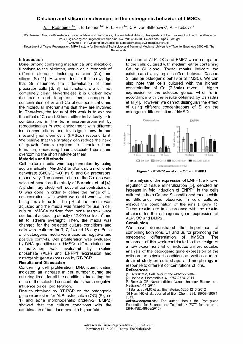
Calcium and silicon involvement in the osteogenic behavior of hMSCs
a. I. rodrigues 1,2, I. b. leonor 1,2, r. l. reis1,2, C.a. van blitterswijk3, P. Habibovic3
13b’s research group – Biomaterials, Biodegradables and Biomimetics, Universidade do Minho, Headquarters of the european Institute of excellence on Tissue Engineering and Regenerative Medicine, AvePark, 4806-909 Caldas das Taipas, Portugal
2ICVS/3B's – PT Government Associated Laboratory, Braga/Guimarães, Portugal3Department of Tissue Regeneration, MIRA Institute for Biomedical Technology and Technical Medicine, University of Twente, Enschede 7500 AE, The
netherlands
IntroductionBone, among conferring mechanical and metabolic functions to the skeleton, works as a reservoir of different elements including calcium (Ca) and silicon (Si) [1]. However, despite the knowledge that Si influences the differentiation of bone precursor cells [2, 3], its functions are still not completely clear. nevertheless it is unclear how the acute and long-term local changes in concentration of Si and Ca affect bone cells and the molecular mechanisms that they are involved in. Therefore, the focus of this work is to explore the effect of Ca and si ions, either individually or in combination, in the bone microenvironment by reproducing an in vitro environment with different ion concentrations and investigate how human mesenchymal stem cells (hMSCs) respond to it.We believe that this strategy can reduce the need of growth factors required to stimulate bone formation, decreasing their associated costs and overcoming the short half-life of them.Materials and MethodsCell culture media was supplemented by using sodium silicate (Na2sio3) and/or calcium chloride dehydrate (CaCl2*2H2O) as Si and Ca precursors, respectively. The concentration of the Ca ions was selected based on the study of Barradas et. al [4].a preliminary study with several concentrations of si was done in order to define the range of siconcentrations with which we could work without being toxic to cells. The pH of the media was adjusted and the media was filtered for use in cell culture. hmsCs derived from bone marrow were seeded at a seeding density of 2.000 cells/cm2 andlet to adhere overnight. Then, the media was changed for the selected culture conditions and cells were cultured for 3, 7, 14 and 18 days. Basic and osteogenic media were used as negative and positive controls. Cell proliferation was evaluatedby DNA quantification. hmsCs differentiation and mineralization was evaluated by alkaline phosphate (ALP) and enpp1 expression and osteogenic gene expression by RT-pCr. Results and DiscussionConcerning cell proliferation, DNA quantification indicated an increase in cell number during the culturing times for all the conditions, indicating that none of the selected concentrations has a negative influence on cell proliferation.results obtained by RT-pCr on the osteogenic gene expression for ALP, osteocalcin (OC) (Figure 1) and bone morphogenetic protein-2 (BMP2) showed that the culture conditions with the combination of both ions reveal a higher fold
induction of alp, oC and bmp2 when compared to the cells cultured with medium either containingCa or si alone. These results indicate the existence of a synergistic effect between Ca and si ions on osteogenic behavior of hMSCs. We can also note that cells cultured with the highest concentration of Ca (7.8mM) reveal a higher expression of the selected genes, which is in accordance with the results obtained by barradas et al [4]. However, we cannot distinguish the effect of using different concentrations of si on the osteogenic differentiation of hmsCs.
The analysis of the expression of enpp1, a known regulator of tissue mineralization [5], denoted an increase in fold induction of enpp1 in the cells cultured in both Ca and si conditioned media while no difference was observed in cells cultured without the combination of the ions (Figure 1).These results are in accordance with the results obtained for the osteogenic gene expression of alp, oC and bmp2.ConclusionWe have demonstrated the importance of combining both ions, Ca and si, for promoting the osteogenic differentiation of hmsCs. The outcomes of this work contributed to the design of a new experiment, which includes a more detailed analysis of the osteogenic gene expression of the cells on the selected conditions as well as a more detailed study on cells shape and morphology in response to different concentrations of ions. References[1] Dvorak MM, Cell Calcium 35: 249-255, 2004.[2] Hoppe a, Biomaterials 32: 2757-2774, 2011.[3] beck Jr gr, nanomedicine: nanotechnology, biology, and medicine,1-11, 2011.[4] barradas amC et al., biomaterials 3205-3215, 2012.[5] Nam HK et al., Journal of Biol. Chem. 286; 39059–39071, 2011.Acknowledgements: The author thanks the portuguese Foundation for Science and Technology (FCT) for the grant (SFRH/BD/69962/2010).
Figure 1 - RT-PCR results for OC and ENPP1
Advances in Tissue Regeneration 2013 Conference November 14-15, 2013, Lattrop, The Netherlands
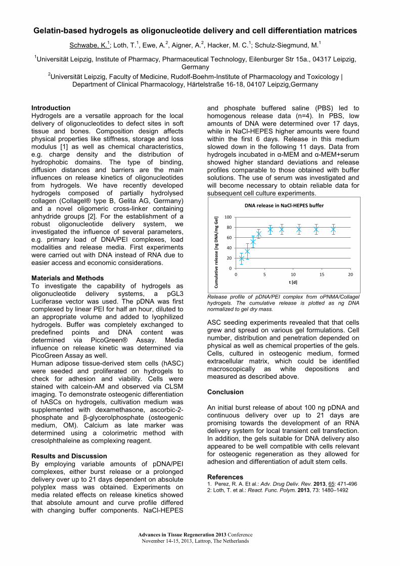
Gelatin-based hydrogels as oligonucleotide delivery and cell differentiation matrices
Schwabe, K.1; loth, T.1, ewe, a.2, aigner, a.2, Hacker, m. C.1; Schulz-siegmund, m.1
1Universität Leipzig, Institute of Pharmacy, Pharmaceutical Technology, Eilenburger Str 15a., 04317 Leipzig, germany
2Universität Leipzig, Faculty of Medicine, Rudolf-boehm-Institute of pharmacology and Toxicology | Department of Clinical Pharmacology, Härtelstraße 16-18, 04107 Leipzig,Germany
IntroductionHydrogels are a versatile approach for the local delivery of oligonucleotides to defect sites in soft tissue and bones. Composition design affects physical properties like stiffness, storage and loss modulus [1] as well as chemical characteristics, e.g. charge density and the distribution of hydrophobic domains. The type of binding, diffusion distances and barriers are the main influences on release kinetics of oligonucleotides from hydrogels. We have recently developed hydrogels composed of partially hydrolysed collagen (Collagel® type B, Gelita AG, Germany) and a novel oligomeric cross-linker containing anhydride groups [2]. For the establishment of a robust oligonucleotide delivery system, we investigated the influence of several parameters,e.g. primary load of DNA/PEI complexes, load modalities and release media. first experimentswere carried out with DNA instead of RNA due to easier access and economic considerations.
Materials and MethodsTo investigate the capability of hydrogels as oligonucleotide delivery systems, a pgl3 Luciferase vector was used. The pDNA was first complexed by linear PEI for half an hour, diluted to an appropriate volume and added to lyophilized hydrogels. buffer was completely exchanged to predefined points and DNA content was determined via PicoGreen® Assay. media influence on release kinetic was determined via picogreen assay as well.Human adipose tissue-derived stem cells (hASC) were seeded and proliferated on hydrogels to check for adhesion and viability. Cells were stained with calcein-AM and observed via CLSM imaging. To demonstrate osteogenic differentiation of hasCs on hydrogels, cultivation medium was supplemented with dexamethasone, ascorbic-2-phosphate and β-glycerolphosphate (osteogenic medium, OM). Calcium as late marker was determined using a colorimetric method with cresolphthaleine as complexing reagent.
Results and DiscussionBy employing variable amounts of pDNA/PEI complexes, either burst release or a prolonged delivery over up to 21 days dependent on absolute polyplex mass was obtained. experiments on media related effects on release kinetics showed that absolute amount and curve profile differed with changing buffer components. naCl-Hepes
and phosphate buffered saline (PBS) led to homogenous release data (n=4). In pbs, low amounts of DNA were determined over 17 days, while in naCl-Hepes higher amounts were found within the first 6 days. release in this medium slowed down in the following 11 days. Data from hydrogels incubated in α-MEM and α-mem+serum showed higher standard deviations and release profiles comparable to those obtained with buffer solutions. The use of serum was investigated and will become necessary to obtain reliable data for subsequent cell culture experiments.
Release profile of pDNA/PEI complex from oPNMA/Collagel hydrogels. The cumulative release is plotted as ng DNA normalized to gel dry mass.
asC seeding experiments revealed that that cells grew and spread on various gel formulations. Cell number, distribution and penetration depended on physical as well as chemical properties of the gels. Cells, cultured in osteogenic medium, formed extracellular matrix, which could be identified macroscopically as white depositions and measured as described above.
Conclusion
An initial burst release of about 100 ng pDNA and continuous delivery over up to 21 days are promising towards the development of an rna delivery system for local transient cell transfection. In addition, the gels suitable for DNA delivery also appeared to be well compatible with cells relevant for osteogenic regeneration as they allowed for adhesion and differentiation of adult stem cells.
References1. Perez, R. A. Et al.: Adv. Drug Deliv. Rev. 2013, 65: 471-4962: loth, T. et al.: React. Func. Polym. 2013, 73: 1480–1492
0
20
40
60
80
100
0 5 10 15 20
Cum
ulat
ive
rele
ase
[ng
DN
A/m
g G
el]
t [d]
DNA release in NaCl-HEPES buffer
Advances in Tissue Regeneration 2013 Conference November 14-15, 2013, Lattrop, The Netherlands

Advances in Tissue Regeneration 2013 Conference November 14-15, 2013, Lattrop, The Netherlands
Silencing of BMP-2 antagonist chordin increases the osteogenic potential of adipose tissue derived stromal cells (ASCs) - Concept of a controlled release system for
siRNA
bita sedaghati1, Hellen schneider1, michael C. Hacker1, Michaela Schulz-siegmund1
1Pharmaceutical Technology, Institute of Pharmacy, University of Leipzig, Leipzig, Germany.
Introduction adipose derived stromal cells (ASCs) have been widely applied in bone regeneration due to the ease of isolation and availability. bone morphogenic proteins (BMPs) are of high interest in enhancing bone regeneration approaches. bmp-2 antagonists such as chordin, noggin and twisted gastrulation complex bmp-2 and modulate its action. suppression of bmp-2 antagonists, using sirna, may provide a platform to locally augment bmp induced bone formation. We could recently show that silencing of chordin in an in vitro study enhanced osteogenic differentiation of human asCs [1]. However, the sirna effects were transient. Hence, to silence chordin or other bmp modulators in a 3-D bone regeneration approach and to maintain the effect over a longer period of time, a controlled release system is required. This study aims to examine the effects on suppression of the bmp-2 antagonist chordin on osteogenic differentiation of asCs. To overcome the limitations of a single application of sirna, we started to set up a controlled release system for sirna with polycaprolactone (pCl) particles in combination with low concentrations of gelatin and peg carrying DNA. In order to develop an applicable strategy for a long time release, this mixture was compressed into thin polymer discs. release of DNA into pbs was investigated. The disc system serves as model for an envisioned pCl scaffold as drug delivery system. future experiments will also transfer the system to the delivery of sirna against chordin in combination with 3-D cultivation of ASCs as an approach to improved osteogenic regeneration. Materials and Methods -asCs silencing and differentiation: asCs were isolated from male and female donors applying collagenase digestion. on fifth passage, cells were seeded nearly confluent and were treated with transfection medium against noggin and chordin. next day, the transfection medium was replaced by osteogenic medium with and w/o 100 ng/ml BMP-2 (BOM and OM). Gene expression of chordin, noggin and bmp-2was determined by rt-pCr on day 4. ALP activity normalized to cell number and matrix mineralization as early and late osteogenic markers, respectively, were quantified.
-biomaterials: pCl particles were generated from large pCl pellets using a precipitation method. The particles were combined with different amounts of gelatin (type A, 140 bloom), peg4000 and DNA. These were finally compressed to thin discs. release of DNA from these discs was determined with the pico green assay over a period of one month. Results and Discussion gene expression assay on asCs treated with sirna against chordin showed significant silencing of chordin on day 4. asCs silenced against chordin showed enhanced ALP activities on day 8 over non-treated controls. This effect was more pronounced in groups treated with bom rather than om. matrix mineralization took place as early as day 12 in the silenced group treated with bmp-2. Mineralization quantified on day 14 was also improved in silenced groups treated with bmp-2 compared to the non-silenced groups. furthermore, donor dependency has been evident from the results. first results of DNA release from PCL discs showed positive effects of gelatin in the disc composition on the total amount of released DNA and the release profile over time. When compared to pure pCl discs and discs containing 5% PEG4000, a content of 5-10% gelatin in the composition prolonged the DNA release. In addition to the gelatin content, the amount of DNA loading has an influence on the release. further investigations towards the release of sirna are ongoing. Conclusion silencing of chordin improved osteogenic differentiation of asCs. Concept experiments towards a pCl-based scaffold-type controlled nucleotide delivery system have been presented.
References [1] schneider H, sedaghati b, naumann a, Hacker
mC, siegmund-Schulz M. Gene silencing of chordin improves bmp-2 effects on osteogenic differentiation of human adipose tissue derived stromal cells. Tissue engineering part a 2013:130809200247003.

Lab-on-a-chip device to rapidly search for optimal combinations of surface strains and fluid flow shears for stimulating cells in tissue regeneration strategies
r. sinha1, s. le gac2, a. van den berg2, H.f.J.m. Koopman1, J. rouwkema1.1Dept. of Biomechanical Engineering, University of Twente, Netherlands. 2bIos – lab on a chip group,
University of Twente, Netherlands.
IntroductionOne of the major bottlenecks in tissue engineering is the lack of a vascular network shortly after implantation, resulting in a lack of nutrients and oxygen for the cells in the engineered tissue (1). We have pioneered a promising strategy to overcome this problem, by including endothelial cells that form a (pre)vascular network (2) that can connect to the host vasculature shortly after implantation (3). Since mechanical signals are important for the development of both bone and vascular networks, we hypothesize that prevascularized bone tissue engineering can be further optimized by the application of mechanical signals. The role of mechanical signals in affecting cell behavior is well established. There is however limited quantitative data on this correlation, making it hard to be effectively utilized in tissue engineering approaches. To obtain the required mechanical information we have designed a semi high throughput screening system. With this system, we will study the effect of combinations of (i) strains in the surface to which they are attached and (ii) fluid shear on their free surfaces on the cells that are used in prevascularized bone.
Materials and MethodsThe device design consists of an array of units generating different strains overlaid with areas experiencing varying fluid shear. strains are generated by deforming a PDMS membrane over noa-81 pillars using a pressure drop and varied between units by varying the area to which thepressure drop is applied. Fluid shear is applied by flowing fluid in chambers over the strain units and varied by varying the flow channel width along the way. Finite element (FEA) and computational fluid dynamics (CFD) modeling have been done using ANSYS to assess the feasibility of the design. A prototype has been built to look at 5 strain x 5 shear combinations each in 4 replicates (so 100 units on a chip). The chip has the dimensions of regular 96 well plates making possible automated imaging by a bioimager like the BD Pathway. strains and fluid flows have been determined empirically by tracking beads. Next, the cells (hMSCs, HUVECs and co-cultures of these cell types) will be included in the experiments.
Results and DiscussionFEA modeling showed that by varying (i) pressure or (ii) the area to which the pressure is applied, different strains can be achieved in the membrane. Fluid dynamics modeling showed that by using a varying width fluid flow channel, different shear
domains can be achieved. Fluid structure interaction (FSI) modeling showed that design parameters can be adjusted so that the membrane deformation has minimal effect on the fluid flow. empirical strain and fluid flow determination have shown proof of principle and good correlation with computational modeling.
ConclusionA device to expose cells to combinations of surface strains and fluid flow shears has been designed. A working prototype is currently being validated.
References1. J. rouwkema et al, Trends in Biotechnology,2008, 26, 434.2. J.rouwkema et al, Tissue Engineering, 2006, 12, 2685.3. S. Levenberg, J.Rouwkema et al, Nature Biotechnology, 2005, 23, 879.
Figure 1: Device schematic and device prototype
Figure 2: Strains and fluid flows(computational modeling and empirically determined values)
Advances in Tissue Regeneration 2013 Conference November 14-15, 2013, Lattrop, The Netherlands
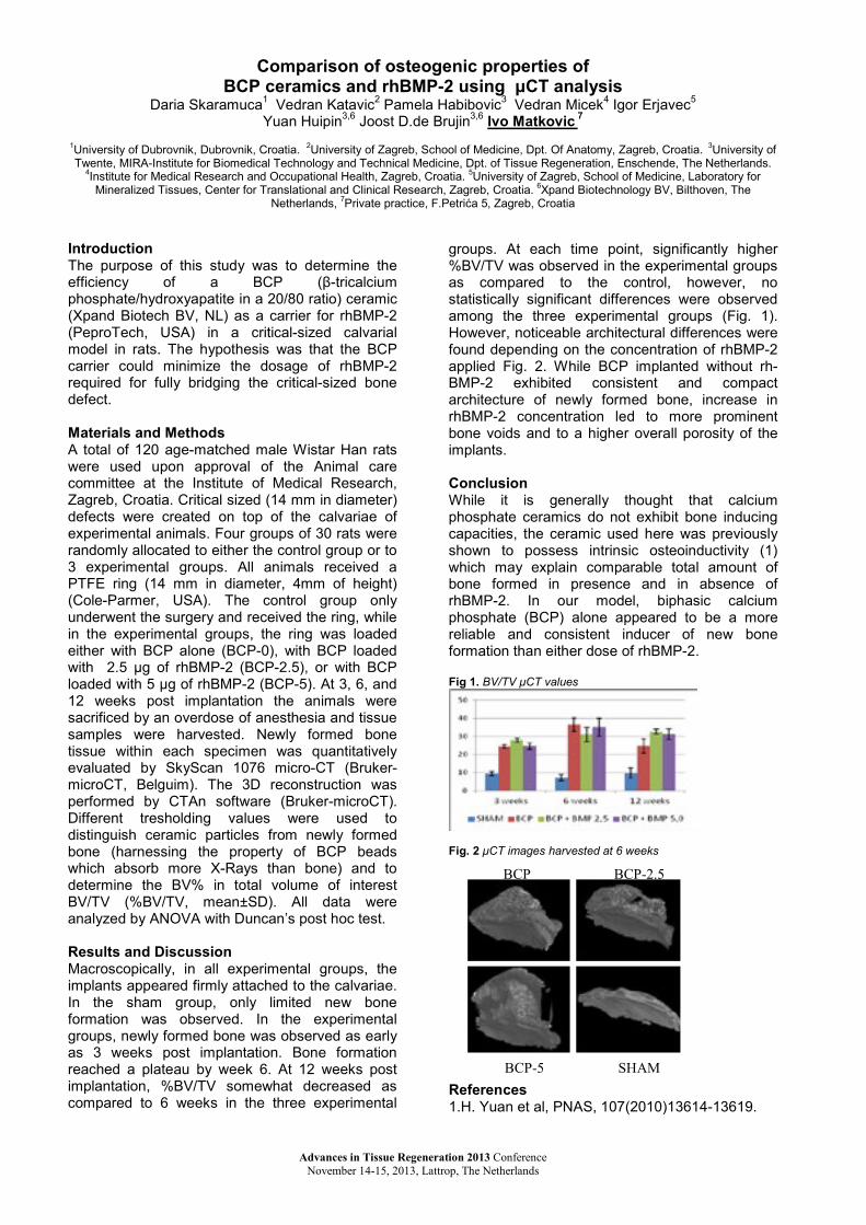
Comparison of osteogenic properties of BCP ceramics and rhBMP-2 using μCT analysis
Daria Skaramuca1 Vedran Katavic2 Pamela Habibovic3 Vedran micek4 Igor erjavec5
Yuan Huipin3,6 Joost D.de Brujin3,6 Ivo Matkovic 7
1University of Dubrovnik, Dubrovnik, Croatia. 2University of Zagreb, School of Medicine, Dpt. Of Anatomy, Zagreb, Croatia. 3University of Twente, mIra-Institute for Biomedical Technology and Technical Medicine, Dpt. of Tissue Regeneration, Enschende, The netherlands.
4Institute for medical research and occupational Health, Zagreb, Croatia. 5University of Zagreb, School of Medicine, Laboratory for Mineralized Tissues, Center for Translational and Clinical Research, Zagreb, Croatia. 6Xpand biotechnology bV, bilthoven, The
netherlands, 7private practice, F.Petrića 5, Zagreb, Croatia
IntroductionThe purpose of this study was to determine the efficiency of a bCp (β-tricalcium phosphate/hydroxyapatite in a 20/80 ratio) ceramic (Xpand Biotech BV, NL) as a carrier for rhBMP-2(PeproTech, USA) in a critical-sized calvarial model in rats. The hypothesis was that the bCp carrier could minimize the dosage of rhBMP-2required for fully bridging the critical-sized bone defect.
Materials and Methodsa total of 120 age-matched male Wistar Han rats were used upon approval of the animal care committee at the Institute of medical research, Zagreb, Croatia. Critical sized (14 mm in diameter)defects were created on top of the calvariae of experimental animals. four groups of 30 rats were randomly allocated to either the control group or to 3 experimental groups. all animals received a pTfe ring (14 mm in diameter, 4mm of height)(Cole-Parmer, USA). The control group only underwent the surgery and received the ring, while in the experimental groups, the ring was loaded either with BCP alone (BCP-0), with BCP loaded with 2.5 μg of rhBMP-2 (BCP-2.5), or with bCploaded with 5 μg of rhBMP-2 (BCP-5). At 3, 6, and 12 weeks post implantation the animals were sacrificed by an overdose of anesthesia and tissue samples were harvested. Newly formed bone tissue within each specimen was quantitatively evaluated by SkyScan 1076 micro-CT (Bruker-microCT, Belguim). The 3D reconstruction was performed by CTAn software (Bruker-microCT). Different tresholding values were used to distinguish ceramic particles from newly formed bone (harnessing the property of BCP beads which absorb more X-Rays than bone) and to determine the BV% in total volume of interest BV/TV (%BV/TV, mean±SD). all data were analyzed by ANOVA with Duncan’s post hoc test.
Results and Discussionmacroscopically, in all experimental groups, the implants appeared firmly attached to the calvariae. In the sham group, only limited new bone formation was observed. In the experimental groups, newly formed bone was observed as early as 3 weeks post implantation. bone formation reached a plateau by week 6. at 12 weeks post implantation, %BV/TV somewhat decreased as compared to 6 weeks in the three experimental
groups. at each time point, significantly higher %BV/TV was observed in the experimental groups as compared to the control, however, no statistically significant differences were observed among the three experimental groups (fig. 1). However, noticeable architectural differences were found depending on the concentration of rhbmp-2applied fig. 2. While bCp implanted without rh-bmp-2 exhibited consistent and compact architecture of newly formed bone, increase in rhbmp-2 concentration led to more prominent bone voids and to a higher overall porosity of the implants.
ConclusionWhile it is generally thought that calcium phosphate ceramics do not exhibit bone inducing capacities, the ceramic used here was previously shown to possess intrinsic osteoinductivity (1) which may explain comparable total amount of bone formed in presence and in absence of rhbmp-2. In our model, biphasic calcium phosphate (BCP) alone appeared to be a more reliable and consistent inducer of new bone formation than either dose of rhbmp-2.
Fig 1. BV/TV μCT values
Fig. 2 μCT images harvested at 6 weeks
References1.H. Yuan et al, PNAS, 107(2010)13614-13619.
BCP BCP-2.5
BCP-5 SHAM
Advances in Tissue Regeneration 2013 Conference November 14-15, 2013, Lattrop, The Netherlands

Advances in Tissue Regeneration 2013 Conference November 14-15, 2013, Lattrop, The Netherlands
A new nanotechnology-based paradigm for engineering vascularised liver tissue for transplantation
m. skolimowski1, p.e. oomen1, V. Starokozhko2 H. b. muhammad3 , C. Canali3, s. mohanty3,
m. Hemmingsen3, a. Wolff3, M. Dufva3, J. Emnéus3, g. m. m. groothuis2, e. Verpoorte1
1pharmaceutical analysis, 2pharmacokinetics, Toxicology and Targeting
Groningen Research Institute of Pharmacy, University of Groningen, The Netherlands 3DTU Nanotech, Technical University of Denmark, Kgs. Lyngby, Denmark
Introduction The liver is the largest internal organ in the body, responsible for over 500 metabolic, regulatory and immune functions [1]. loss of liver function leads to liver failure, for which organ transplantation is the primary medical treatment approach. limitations in this procedure, such as shortage of donor organs and tissue rejection, have spurred research in the area of liver tissue engineering. There have been significant advances in the 3D (co)culture of hepatocytes. However, the tissues generated have dimensions limited to a few hundred µm, since the absence of a vascular system means nutrient delivery to and waste removal from inner cell layers by diffusion only. We are exploring the use of micro- and nanotechnological approaches to provide vascularization to 3D cultures as a means of scaling up tissue production to larger tissue amounts, as ultimately required by the transplantation application. Materials and Methods The goal of this project is to develop a highly vascularised, extracorporeal bioartificial liver (EBAL) support system. This will be done by integrating stem cell and novel 3D polymer scaffolds for cell culture, and sensor and microfluidic technologies for control of culture conditions (Fig. 1).
3D scaffolds and microfluidic support systems are being developed in polylactic acid (PLA) and
polydimethylsiloxane (PDMS) by exploiting such technologies as fused-filament deposition-based 3D-printing technology, micromilling and soft photolithography. Results and Discussion To investigate the compatibility of cells in the scaffolds, Hepg2 liver cells have been cultured in the porous polymer scaffolds (Fig. 2).
Fig. 2 (A) 3D polymeric scaffold. (B) Immuno-stained confocal imaging of liver cells after 10 days of culturing (red-nucleus, green-cytoskeleton). an integrated modular system has been developed (Fig. 3a) incorporating miniaturised peristaltic pumps, a biochip containing a scaffold with liver cells, and needle electrodes for bioimpedance tomography. The EBAL will be characterised with respect to its metabolic capability and other essential functions (e.g. clearance of ammonia, bilirubin, toxins and bile acids) to assure its suitability and safe use in extracorporeal applications and in potential future transplantation. precision-cut liver slices (PCLS) incubated in liver-on-a-Chip systems (Fig. 3b) will be used.
Future perspective The ebal being developed in this project will be used for supporting liver function in patients while they wait for an appropriate donor organ, or until their own liver has recovered. We expect the results will be the first step in the development of a transplantable bioartificial liver in the future. References [1] Y. nahmias et al. Adv Biochem Eng/Biotechnology, 2006. 103: 309-329 [2] p. m. van midwoud et al. Biotechnol Bioeng, 2010:105(1) 184-194
Fig. 1 Bioartificial liver support system in a microfluidic chip process: (A) 3D scaffold with a highly branched, perfusable channel network that allows flow through the material from one inlet to one outlet, (B) Stem cell derived endothelial and hepatocyte cells seeded and grown inside the
primary capillary channel network, mimicking the in vivo situation of blood vessels lined with endothelial cells on top of which hepatocytes are formed, (C) A fully developed BAL support system, with integrated sensing and imaging tools for control of growth and viability, with a final releasable bioartificial liver for future transplantation.
a
b
a
b
Fig. 3 (A): Integrated EBAL on a chip system consisting of an array of 3D biochips, (B) Liver-on-a-Chip system for PCLS culture [2].
This work has been supported by the EC within the 7th framework programme under grant agreement no. 304842
RF

The influence of tissue (an)isotropy on cardiomyocyte contraction in engineered cardiac microtissues
ariane van spreeuwel1, noortje bax1, alex bastiaens1, Jasper foolen1, sandra loerakker1, michael borochin2, Daisy van der Schaft1, frank baaijens1, Christopher Chen2, Carlijn bouten1
1Eindhoven University of Technology, 2University of Pennsylvania
IntroductionThe myocardial microenvironment is highly organized, with cardiomyocytes (CMs) residing in a structured and aligned matrix to maintain the coordinated contractile function of the heart.Cardiac fibroblasts (FBs) are responsible forproducing and remodeling this matrix. In cardiac diseases, however, ongoing remodeling may lead to adverse changes in cell and matrix distribution, often resulting in a disturbed, chaotic tissueorganization. In this study, we present an in vitrocardiac model system with biaxial or uniaxial constraints to induce (an)isotropy in 3D microtissues, thereby mimicking ‘healthy’ alignedand ‘diseased’ chaotic cardiac matrices. The effectof matrix (an)isotropy on cellular composition,structure, and contractile properties of the tissuewas quantified. We hypothesized that matrix anisotropy will to lead to improved contractile properties.
Materials and Methodsmicrofabricated tissue gauges (μTUGs [1]) withflexible microposts as biaxial or uniaxial constraints were fabricated using soft lithography.neonatal mouse Cms were seeded in collagen/matrigel in µTUGs and cultured for up to7 days. Displacement of the posts was monitored to calculate contractile properties. Viable fluorescent probes were used to visualize matrix over time. CM/FB ratios and Cm maturation and organization were checked using fluorescent stainings and confocal microscopy.
Results and DiscussionUsing the μTUG system we successfully obtained isotropically and anisotropically organized tissues(Fig 1). although results confirmed that Cms in anisotropic microtissues were more aligned, these tissues did not generate higher contractile forces (Fig 2A). However, strain analysis showed that contraction was much more homogeneous
Figure 1: Microtissues were biaxially (A) or uniaxially (B) constrained to manipulate tissue organization. Collagen had a random (black graph) and aligned (grey graph) orientation respectively, as was confirmed by image analysis (C).
in anisotropic tissues than in isotropic tissues (Fig 2E+F). Contraction frequency was not affected by matrix organization, but drastically decreased at day 7 in both designs (Fig 2B). We suggest that this is caused by the high percentage of fbs at day 7 (Fig 2C). A threshold value of 55% FBs wasfound to correlate with a reduction of the frequency from 4 Hz to almost 0 Hz (Fig 2D), suggesting that this percentage of fbs and concomitant distance between CMs interferes with signal conduction between CMs.
Figure 2: Microtissue contraction force (A), frequency (B) andpercentage of fibroblasts (C) was determined at day 2, 4, and 7.Analysis of all time points together showed that beating frequency rapidly decreased when the percentage of FBs reached 55% (D). A representative example shows the inhomogenous distribution of Exx in isotropic tissues when compared to anisotropic tissues (E), which is confirmed by the difference in standard deviation for all time points (F).
ConclusionIn contrast to our hypothesis, our data suggest that Cms can generate similar contractile forces inchaotic and aligned matrices. However, the disorganized contraction that was seen in isotropic tissues may lead to arrhythmia. furthermore, our data indicate that increased numbers of FBs can further disturb the frequency of contraction.Together, these results show that this in vitromodel system is suitable to study cellular and pathophysiological processes that occur during heart disease and may hence serve to screen for possible therapeutic targets.
References[1] legant W.r., et al., PNAS, 2009
Advances in Tissue Regeneration 2013 Conference November 14-15, 2013, Lattrop, The Netherlands
RF

Assessment of organ interactions in a multichamber microfluidic perfusion device using rat precision-cut liver and intestinal slices
Starokozhko V.1, van midwoud p.2, de Ruiter K.1, Verpoorte e.2, groothuis g.m.m.1
1pharmacokinetics, Toxicology and Targeting, 2pharmaceutical analysis, groningen research Institute of pharmacy, University of Groningen, The Netherlands
IntroductionHistorically, animal models have been used in many pharmacological and toxicological studies. However, in vivo methods mean the use of large numbers of animals. precision-cut liver slices(PCLS) can be used as an alternative ex vivomodel, one which has been shown to be functional and efficient in numerous metabolism and toxicity studies [1]. Though the liver is known to be the main organ involved in the metabolism of xenobiotics, increased insight into intestinal drug transport and metabolism has shown the importance of the intestine in drug biotransformation and toxicity. However, stand-alone pCls or precision-cut intestinal slice (PCIS) models cannot fully cover and predict drug biotransformation in vivo, as interplay of the liver and intestine plays an important role in many physiological processes, e.g. expression and activity of different enzymes and transporters [2].Therefore, development of an in vitro model that includes both organs would considerably broadenthe scope of toxicological and pharmacological studies. The conventional incubation of PCLS and pCIs in well plates has its limitations, such asdepletion of nutrients and accumulation of waste products in the media. Moreover, it would not be possible to study organ interactions, since mutual interaction can take place between pCls and pCIs placed in one well. However, a recently developed microfluidic perfused incubation system (biochip) has made it possible to overcome theselimitations by enabling continuous perfusion of slices with medium to deliver oxygen and nutrients and remove waste products [3]. moreover, by using this system it becomes possible to sequentially perfuse intestinal and liver slices, mimicking in vivo first-pass metabolism. The aim of this study was then to investigate the applicability of the biochip for the culture of pCls and pCIs, and their sequential perfusion for organ interaction studies.
Materials and MethodsRat PCLS and PCIS were incubated for 24 h in thebiochip and well plates as control. The viability of pCls and pCIs was assessed by means of lactate dehydrogenase (LDH) leakage and aTp content, respectively. furthermore, to compare the metabolic rates of slices in the biochip and in the well plates, metabolite formation for the substrates 7-ethoxycoumarin (7-EC) and 7-hydroxycoumarin (7-HC) was monitored following 3 h exposure. The interplay between the liver and intestine was demonstrated by sequentially exposing intestinal
and liver slices in connected microchambers to the bile acid, chenodeoxycholic acid (CDCA). The gene expression for fibroblast growth factor 15 (fgf15) in the intestinal slices and the enzyme,cytochrome P450 7A1 (CYp7a1), in the liver sliceswas determined after the incubation experiment.
Results and Discussionno significant differences in LDH leakage from pCls or in aTp content of pCIs were found between the biochip and the well plates following 24 h incubation. Furthermore, metabolite formation of 7-EC and 7-HC by pCls and pCIs was shownto be similar compared to the well plates for periods of up to 3 h. significant up-regulation of FGF15 in the intestinal slices after CDCA treatment was demonstrated, which resulted in stronger down-regulation of CYP7A1 in liver slicesin the second compartment, compared to liver slices that were exposed separately to CDCA [4].These results are in agreement with the in vivosituation, where fgf15 is formed in rat intestine following CDCA exposure and is transported via the portal vein to the liver, resulting in an additional down-regulation of CYP7A1 [5].
ConclusionThe present study demonstrated that the biochip can be used as an alternative system to study drug metabolism and toxicity, since no differences in viability and metabolic rate of the liver and intestinal slices were shown for incubation in well plates versus biochip. additionally, sequential perfusion of slices from the intestine to the liver was shown to be successful and promising for the study of interorgan effects and first-pass metabolism of xenobiotics.
References1. De Graaf I.A.M., Groothuis G.M.M., Olinga p. precision-
cut tissue slices as a tool to predict metabolism of novel drugs. Expert Opin. Drug Metab. Toxicol, 2007; 3(6): 879-98.
2. siissalo s., Heikkinen a.T. In vitro methods to study the interplay of drug metabolism and efflux in the intestine. Current Drug Metabolism, 2013; 14: 102-111.
3. Van midwoud p.m., groothuis g.m.m., merema m.T., Verpoorte E. Microfluidic biochip for the perifusion of precision-cut liver slices for metabolism and toxicology studies. Biotech. Bioeng., 2010; 105(1): 184-94.
4. Van midwoud p.m., merema m.T., Verpoorte e., groothuis g.m. a microfluidic approach for in vitro assessment of interorgan interactions in drug metabolism using intestinal and liver slices. Lab on a Chip, 2010; 10(20): 2778-86.
5. Inagaki T., Choi m., moschetta a., peng l., Cummins C.l., McDonald J.G., Luo G., Jones S.A., Goodwin B., Richardson J.A., Gerard R.D., Repa J.J., Mangelsdorf D.J., Kliewer S.A. Fibroblast growth factor 15 functions as an enterohepatic signal to regulate bile acid homeostasis. Cell Metabolism, 2005; 2(4): 217-25.
Advances in Tissue Regeneration 2013 Conference November 14-15, 2013, Lattrop, The Netherlands

Effects of cobalt ions on proliferation and differentiation of human mesenchymal stromal cells
Z. Tahmasebi Birgani, C. A. van Blitterswijk, P. HabibovicDepartment of Tissue Regeneration, University of Twente, Enschede, the Netherlands.
IntroductionIncorporating bioinorganics, which are either present in bone or known to affect bone formation and remodeling mechanisms, into bone graft substitutes is an interesting approach to improve their biological performance while retaining their synthetic character [1]. earlier studies have shown that short-term exposure to cobalt ions (Co2+) may affect osteoclast and osteoblast proliferation and function [2-3]. moreover, Co2+ is known to be a hypoxia-mimicking factor, which in turn may be a way of enhancing angiogenesis inside bone graft substitutes [4]. In the present study, the effects of Co2+ on proliferation and differentiation of human mesenchymal stromal cells (hMSCs) were evaluated.
Materials and MethodshmsCs were cultured at a density of 10000 cells/cm2 in treated tissue culture plates in eitherbasic or osteogenic medium. appropriate volumes of a Tris buffer solution containing 10 µM CoCl2were added to the cell medium to reach the concentrations of 0, 0.1 or 20 µm of Co2+ in the wells. proliferation and osteogenic differentiation of cells were studied at different time points by quantifying DNA and ALP activity, respectively.
Results and DiscussionDNA content of hMSCs treated with varying concentration of Co2+ after 1, 3, 7 and 14 days of culture in basic and osteogenic medium are shown in figure 1. at all time points, the DNA content of hmsCs treated with 0,1 µm of Co2+ was higher than in conditions without Co2+ or with 20 µm Co2+,particularly when cells were cultured in basic medium. The differences were, however, notstatistically significant.
Figure 1. DNA content of hMSCs cultured with cobalt ions in basic and osteogenic medium after 1, 3, 7 and 14 days.
results of alp staining showed a decrease in the number of positively stained cells in 20 µm Co2+
condition (data not shown). Normalized alp activity results (figure 2) also showed adescending trend in ALP activity of hMSCs by adding Co2+ to the cell medium after 14 days of culture, however, these differences were not statistically significant.
Figure 2. ALP activity of hMSCs cultured with cobalt ions in basic and osteogenic medium after 7 and 14 days.
our data obtained so far suggests that cobalt ionsdo not have a major effect on viability, proliferation and alp expression of hmsCs in the examined range of concentrations. These findings are consistent with the data presented by Andrews et. al. [3], who at lower concentrations, showed no effect of Co2+ ions on proliferation and osteogenic differentiation of human osteoblasts, whereas at aCo2+ concentration of 100 µm a significant decrease in proliferation, alp activity and mineralization was observed.andrews et al. [3] observed a significant effect of Co2+ on proliferation and differentiation of human osteoclasts. They showed that Co2+ ions at concentrations higher than 0,1 µm significantly decreased the number of forming and mature osteoclasts, suggesting that the effect of Co2+ is more pronounced on osteoclasts than on osteoblasts.
ConclusionThe results presented here showed a minor dose-dependent effect of Co2+ on proliferation and osteogenic differentiation of hmsCs. further studies will focus on analysis of the expression of other osteogenic markers and cell mineralization.
References1- l. Yang, b. Harink, p. Habibovic, Comprehensive
Biomaterials V. 1, Elsevier, 299-312, 2011.2- s. patntirapong, P. Habibovic, P.V. Hauschka,
biomaterials 30, 548–555, 2009.3- R.E. Andrews, K.M. Shah, J.M. Wilkinson, A.
Gartland, Bone 49, 717–723, 2011.4- W. fan, r. Crawford, Y. Xiao, biomaterials 31,
3580–3589, 2010.
Advances in Tissue Regeneration 2013 Conference November 14-15, 2013, Lattrop, The Netherlands

Melatonin, Telomerase and The Regeneration Liver Tissue of the Rats
Alexandre Tavartkiladze
Introductionfrom the songs of my childhood I can
remember one such song: " The cranes are flying far and they will never come back, the rumour is spread ... . " The author of the song text assumes that the cranes can not come back , but he/she can't bear the thought of losing lovely birds and adds: " it's a rumour !" However, I am saying once more he lets the thought that maybe the cranes won't be back any more .... I loved this song and while listening sorrow used to enter my soul quietly. Although I knew that birds return from warm countries ... and still there was something ominous in those " never come back." Then , after so long I read the following: "Each species has it's own quantity of wing waving , after which the bird dies ." ..That's where that subconscious sorrow came from ... mine or of that song text author, as we mentioned above... Yes, it's so, the organism tissues , cells "get worn" while acting and get restored too; The only bad thing is that with age , damage of cells exceeds their reparation i. e. the mentioned fact becomes expressed with aging! every creature turned to have its own strictly defined potential, after which the life expires. Then where is immortality? Only in fairy tales ? "The truth used to be written in fairy tails , the truth , written in a creative and "fairy" way " ,-"In fairy tales , many secrets of nature are explained in "fairy" language ,many things that are unsolved and not clear at all! "... Then where's immortality? The water or spring of immortality ? Maybe it is nearby , here next to us...
The primary physiological function of melatonin, whose secretion adjusts to night length, is to convey information concerning the daily cycle of light and darkness to body physiology. Althoughfunctions of this hormone in humans are mainly based on correlative observations, there is some evidence that melatonin stabilises and strengthens coupling of circadian rhythms, especially of coretemperature and sleep-wake rhythms. The circadian organisation of other physiological functions could depend on the melatonin signal, for instance immune, antioxidative defences, hemostasis and glucose regulation. since the regulating system of melatonin secretion is complex, following central and autonomic pathways, there are many pathophysiological situations where the melatonin secretion can be disturbed. The resulting alteration could increasepredisposition to disease, add to the severity of symptoms or modify the course and outcome of the disorder.
The human liver has a remarkable capacity to regenerate, as demonstrated by its growth after partial hepatectomy, which may be performed for tumor resection or for living-donor hepatic transplantation. The popular image of liver
regeneration is the daily regrowth of the liver of Prometheus, which was eaten every day by an eagle sent by Zeus (Zeus was angry at Prometheus for stealing the secret of fire, but did he know that prometheus’s liver would regenerate?). The reality, although less dramatic, is still quite impressive. In humans, resection of approximately 60% of the liver in living donors results in the doubling of the liver remnant in about one month.
The essence of genes chromosome telomere shortening is interesting. The limitedreplication ability of the cell can be explained by the following: During each division, thechromosomal endings go through the unfinished replication (telomere shortening). aftereach division telomeres shorten, that ultimately causes stopping the cell division. Telomeres are important in stabilization of terminal parts of the chromosomes and also their fixing atthe nucleus matrix. Telomeres gradually get shorter in later passages of the culture and also in older people's cell cultures. Telomeres are the longest in spermatozoids, they are longer infetus than in an adult.
Materials, Methods, Results, Discussion and ConclusionWe have studied the influence of melatonin on the regeneration liver tissue of the rats in experiment. for this experiment we studied 30 male rats (+ 30 Rats for the control group). The both group (Research Group and Control Group) with surgery intervention amputated the liver tissue approximately by 40%. During 4 weeks we administrated each rat 25 mg/kg melatonin (per os) only Research Group’s Rats. After 4 weeks by liver biopsy we studied the telomerase activity in the research group and control group also by Immunohistochemisrty method. The results was following: activity Telomerase was increased in research Group by 74% comparative Control group. The regeneration of the liver in the research group was noticeably better by function activity (ALT, AST, GGT, T.Bill, D.Bill, TP and Alb) comparative Control group. This experiment clearly testified a fact about influence of melatonin in the regeneration process. This fact should be used in the more the experimental (in the animal’s model) and clinical research of Liver Cirrhosis and in Cancer’s pathogenesis also.
ReferencesThe basic physiology and pathophysiology of melatonin. Sleep Medicine Reviews (2005) 9, 11–24;programming and Implementation of age-related Changes. Scenecence (2011) Chapter 12 InTech.
Advances in Tissue Regeneration 2013 Conference November 14-15, 2013, Lattrop, The Netherlands
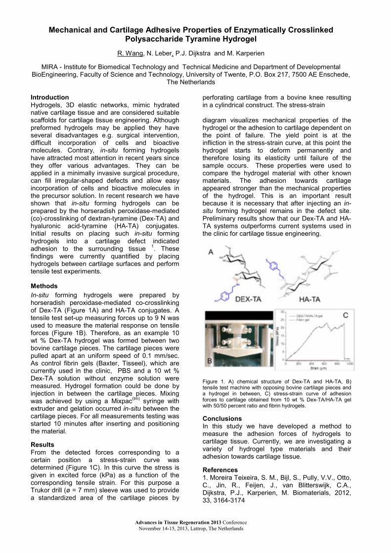
Mechanical and Cartilage Adhesive Properties of Enzymatically Crosslinked Polysaccharide Tyramine Hydrogel
r. Wang, N. Leber, p.J. Dijkstra and M. Karperien
mIra - Institute for Biomedical Technology and Technical Medicine and Department of Developmental BioEngineering, Faculty of Science and Technology, University of Twente, P.O. Box 217, 7500 AE Enschede,
The netherlands
IntroductionHydrogels, 3D elastic networks, mimic hydrated native cartilage tissue and are considered suitable scaffolds for cartilage tissue engineering. although preformed hydrogels may be applied they have several disadvantages e.g. surgical intervention, difficult incorporation of cells and bioactive molecules. Contrary, in-situ forming hydrogels have attracted most attention in recent years since they offer various advantages. They can be applied in a minimally invasive surgical procedure, can fill irregular-shaped defects and allow easy incorporation of cells and bioactive molecules in the precursor solution. In recent research we have shown that in-situ forming hydrogels can be prepared by the horseradish peroxidase-mediated (co)-crosslinking of dextran-tyramine (Dex-TA) and hyaluronic acid-tyramine (HA-TA) conjugates.Initial results on placing such in-situ forming hydrogels into a cartilage defect indicated adhesion to the surrounding tissue 1. These findings were currently quantified by placing hydrogels between cartilage surfaces and perform tensile test experiments.
MethodsIn-situ forming hydrogels were prepared by horseradish peroxidase-mediated co-crosslinking of Dex-TA (Figure 1A) and Ha-Ta conjugates. atensile test set-up measuring forces up to 9 N was used to measure the material response on tensile forces (Figure 1B). Therefore, as an example 10wt % Dex-TA hydrogel was formed between two bovine cartilage pieces. The cartilage pieces were pulled apart at an uniform speed of 0.1 mm/sec. as control fibrin gels (Baxter, Tisseel), which arecurrently used in the clinic, pbs and a 10 wt % Dex-TA solution without enzyme solution were measured. Hydrogel formation could be done by injection in between the cartilage pieces. Mixing was achieved by using a mixpac(tm) syringe with extruder and gelation occurred in-situ between the cartilage pieces. for all measurements testing was started 10 minutes after inserting and positioning the material.
Resultsfrom the detected forces corresponding to a certain position a stress-strain curve was determined (Figure 1C). In this curve the stress is given in excited force (kPa) as a function of the corresponding tensile strain. for this purpose a Trukor drill (ø = 7 mm) sleeve was used to provide a standardized area of the cartilage pieces by
perforating cartilage from a bovine knee resulting in a cylindrical construct. The stress-strain
diagram visualizes mechanical properties of the hydrogel or the adhesion to cartilage dependent on the point of failure. The yield point is at the infliction in the stress-strain curve, at this point the hydrogel starts to deform permanently and therefore losing its elasticity until failure of thesample occurs. These properties were used to compare the hydrogel material with other known materials. The adhesion towards cartilage appeared stronger than the mechanical properties of the hydrogel. This is an important result because it is necessary that after injecting an in-situ forming hydrogel remains in the defect site. Preliminary results show that our Dex-Ta and Ha-Ta systems outperforms current systems used in the clinic for cartilage tissue engineering.
Figure 1. A) chemical structure of Dex-Ta and Ha-Ta, B) tensile test machine with opposing bovine cartilage pieces and a hydrogel in between, C) stress-strain curve of adhesion forces to cartilage obtained from 10 wt % Dex-Ta/HA-Ta gel with 50/50 percent ratio and fibrin hydrogels.
ConclusionsIn this study we have developed a method to measure the adhesion forces of hydrogels to cartilage tissue. Currently, we are investigating a variety of hydrogel type materials and their adhesion towards cartilage tissue.
References1. moreira Teixeira, s. m., bijl, s., pully, V.V., otto, C., Jin, r., feijen, J., van blitterswijk, C.a., Dijkstra, P.J., Karperien, M. biomaterials, 2012,33, 3164-3174
Advances in Tissue Regeneration 2013 Conference November 14-15, 2013, Lattrop, The Netherlands

Polytrimethylene Carbonate Barrier Membranes in Bone Augmentation for Dental Implants
ni zeng1, anne van leeuwen2, ruud r.m. bos2, Dirk W. Grijpma1,3, Roel Kuijer1
1Department of Biomedical Engineering, University Medical Center Groningen and University of Groningen, groningen, The netherlands
2Department of Oral and Maxillofacial Surgery, University Medical Center Groningen and University of groningen, groningen, The netherlands
3Department of Biomaterials Science and Technology, University of Twente, enschede, The netherlands
IntroductionThe success rate of inserting a dental implant highly depends on the quantity and quality of the jaw bone in which it is placed. Autologous bone grafts are often used to enhance bone stock where needed. In clinical practice, barrier membranes are used to cover bone grafts in order to inhibit/prevent graft resorption and both resorbable (collagen) and non-resorbable (Gore-tex) membranes are used.(1) Both types of membranes have significant disadvantages: collagen membranes lack mechanical stability and Gore-tex membranes have to be removed before placing the implant. In this study we evaluated biodegradable membranes prepared from poly(trimethylene carbonate) (PTMC) with sufficient mechanical stability to help in bone augmentation for dental implants. Bone grafting using PTMC membranes was compared to using collagen membranes or ePTFE membranes in an established rat model. Controls using no membranes were also evaluated.(2)Materials and MethodsDisk-shaped autologous bone grafts of 5 mm in diameter were harvested from rat mandibular angle and transplanted onto the surface of the contralateral mandibular angle. The grafts were fixed with suture to the recipient bone and covered either with membranes or not. The membranes used in the study included collagen membranes (Geistlich Bio-Gide) of 0.3-0.4 mm in thickness, expanded poly(tetrafluoroethylene) (e-PTFE) membranes (Gore-tex) of 0.15 mm in thickness and PTMC membranes of 0.3 mm in thickness. All used membranes were 8 mm in diameter. after 2, 4 and 12 weeks the rat mandibles were retrieved and analyzed using histology, histomorphometry and µCT quantification.Results and discussionThe histological study revealed that in time the grafted autologous bone was well integrated in the recipient bone and did not show signs of resorption, independent of membrane coverage of the graft. The type of barrier membrane did not affect the result. at the time point of 12-week, the contour of the grafts were no longer recognizable and the mandibles appeared to be intact and continuous. The histomorphometric analysis showed that it was the recipient beds and not the autologous bone grafts that underwent resorption. The results of µCT quantification confirmed this finding: the volume of the bone graft and recipient bone was maintained by new bone formation and
resorption of recipient bone. The dynamic change is likely caused by changes in the transduction of forces through the bone after graft transplantation.Conclusion The use of novel PTMC membranes results in similar bone remodeling as clinically used resorbable and non-resorbable barrier membranes. The used animal model does not seem to be sensitive enough to detect differences. In this model the envisaged benefits of barrier membranes in preventing bone graft resorptioncould not be illustrated.
References
1. Bone grafting the mandible. Oral and maxillofacial surgery clinics of north america. (23): 209.
2. gielkens pf, schortinghuis J, de Jong Jr, Paans AM, Ruben JL, Raghoebar GM, et al. The influence of barrier membranes on autologous bone grafts. J Dent Res. 2008 Nov;87(11):1048-52.
Advances in Tissue Regeneration 2013 Conference November 14-15, 2013, Lattrop, The Netherlands

DKK1, FRZB, GREM1 as gatekeepers of articular cartilage homeostasis
leilei zhong, Marcel Karperien, Janine N. Post
Developmental BioEngineering, MIRA Institute for Biomedical Technology and Technical Medicine, University of Twente, enschede, The netherlands
Introductionosteoarthritis (OA) is a multifactorial disease characterized by progressive degradation of joint cartilage. It affects several million people in the world. Currently there is no cure for oa. In a subset of patients oa is associated with hypertrophic differentiation of chondrocytes. This process normally occurs in growth plate. Healthy articular cartilage is protected against hypertrophy[1] and chondrocytes in coculture with msCs prevent hypertrophic differentiation of msCs [2].The underlying mechanism of this protection is not yet elucidated. Our group identified DKK1, FRZB (WNT antagonists) and GREM1 (BMP antagonist) as the natural brakes on hypertrophic differentiation and regulation of the maintenance of the articular phenotype [3]. In addition, we found that when mesenchymal stem cells undergo chondrogenic differentiation in hypoxia, the expression of DKK1, FRZB and GREM1 is increased as compared to normoxia, and that hypoxia inhibits endochondral ossification in explanted tibiae [4]. In this project, we hypothesizethat DKK1, FRZB, GREM1 are the gatekeepers for the maintenance of homeostasis in articular cartilage. To prove this, we will investigate whether i) knock-down of DKK1, FRZB and GREM1 is sufficient to cause hypertrophic differentiation in healthy articular chondrocytes; and ii) whether DKK1, FRZB and GREM1 are sufficient for preventing chondrocyte hypertrophy in oa or differentiating msCs.
Materials and Methodsarticular cartilage samples were isolated from 6 donors with primary oa undergoing total joint replacement surgery. Cartilage was cut into smallpieces and processed for rna isolation and histological analysis. Cartilage sections were scored according to ICrs guidelines. low grade(0-1) and high grade (2-4) cartilage samples were combined for further analysis. Human primary chondrocytes (hPC) were isolated from cartilageas described [2]. Human mesenchymal stem cells(MSC) were isolated from fresh bone marrow samples. Coculture pellets were formed as published [2]. shRNA and neutralizing antibodieswere tested for knockdown of DKK1, FRZB and grem1. Cells were cultured under hypoxia (2.5% o2) and normoxia (21% O2).
Results and DiscussionIn low grade cartilage, DKK1, FRZB, GREM1 mrna expression was significantly higher as compared to high grade OA cartilage (Fig 1). Therefore, deregulation of their expression is likely to be associated with the development of oa.
Fig 1. Gene expression of DKK1, FRZB, GREM1 was measured by RT-PCR in low grade cartilage and high grade cartilage. All three antagonists are significantly lower expressed in high grade OA .
To test the expression of DKK1, FRZB and grem1 in hpC and msC under hypoxia, we cultured the cells in monolayer for 1 and 3 weeks and tested the relative gene expression of the antagonists by rT-qpCr. after 1 week, the expression of all 3 antagonists was significantly reduced in hypoxic conditions in both cell types. at 3 weeks we observed a significant increase in DKK1 expression in hPC in hypoxia, and of FRZB in msC in hypoxia as compared to the normoxic condition. Taken together, these data suggest that the effects of oxygen level on mrna expression of DKK1 and FRZB are dependent on culture time and cell type.We further tested whether pellet coculture of msC with hPC influences the expression of DKK1, frzb and grem1. We found that after 3 weeks, the expression of grem1 in coculture equaled that of the hPC monoculture, and that DKK1 was 10-fold reduced compared to the hpC monoculture.
ConclusionThe antagonists DKK1, FRZB, GREM1 areinvolved in the regulation of articular cartilage homeostasis. The expression is changed in osteoarthritic chondrocytes and is regulated by oxygen concentration.
References1. Blanke M, Carl HD, Klinger P, et al., Calcif Tissue Int. 2009. 85(5): p.421-33. 2. Wu l, leijten JC, georgi n, post Jn, van Blitterswijk CA, Karperien M.Tissue eng part a. 2011 .17(9-10):1425-363. leijten JC, emons J, sticht C, et al., arthritis rheum. 2012. 64(10): p.3302-12. 4. leijten JC, moreira Teixeira ls, landman eb, van Blitterswijk CA, Karperien M . plos one.2012. 7(11): e49896.
Advances in Tissue Regeneration 2013 Conference November 14-15, 2013, Lattrop, The Netherlands