advantages of supplementary ct in myelography of ... · advantages of supplementary ct in...
TRANSCRIPT
618
Advantages of Supplementary CT in Myelography of Intraspinal Masses Rina Tadmor, 1 Edwin D. Cacayorin,2 and Stephen A. Kieffer1, 2
Computed tomographic (CT) myelography subsequent to nonionic water-soluble contrast medium myelography has provided additional diagnostic information for evaluating intraspinal mass lesions. In 20 patients thus studied, there were eight intramedullary tumors, eight intradural extramedullary masses, and four extradural neoplasms. In intramedullary tumors CT enabled more precise delineation of the extent and location of expansion of
the spinal cord . In intradural and extradural tumors, rotation and compression of the spinal cord as well as bony and paraspinal soft-tissue changes were more accurately demonstrated in the axial plane. When a complete block was present, the greater contrast sensitivity of CT permitted visualization beyond the level of the block. Histologic prediction is not feasible by CT myelography except for hyperlucent lipoma. CT can provide
useful supplemental information to conventional metrizamide myelography.
Computed tomographic (CT) evaluat ion of the spine in spinal stenosis, herniated lumbar intervertebral disk , trauma to the vertebral co lumn , and oth er bony pathology has been well established
[1 -3]. CT also permits accurate cross-sectional assessment of the spinal cord and nerve roots within the opacified thecal sac as well as th e extradural soft ti ssues surrounding the sac [4]. CT myelography for the investigation of syringomyelia and of spinal dysraphism has added a further dimension to our understanding of these anomalies of the spine [5-7]. Application of CT for the detection and localization of intraspinal mass lesions has also been described [8-10]. This report analyzes our accumulated experience in the application of CT myelography to the evaluation of intraspinal mass lesions and emphasizes th e supplementary information it may provide after conventional myelography with metrizamide.
Materials and Methods
Twenty patients ranging in age from 5 months to 65 years with clinica l suspic ion of an intraspin al mass lesion had plain radiographic studies of the spine and subsequently underwent conventional metrizamide myelog raphy of that region. Volumes of metrizamide injected into the spinal subarachnoid space were 8-15 ml at concentrations of 170-200 mg 11 m!. Within 1.5-2 hr after the
intrathecal injec tion of contrast medium, CT examination of the area of interest was obtained . Patients with spinal dysraphism . syringomyelia, spinal stenosis. or herniated disk are not included in this
study. All masses were pathologically proven by surgical exploration and histologic verification.
The CT examinations were performed on Elscint Exel-905, Technicare 2020. and General Electric 8800 scanners. In most
cases. a scan projection digital radiograph was first obtained which permitted rapid and accurate delineation of the area of interest; subsequent axial CT sections cou ld then be made above, through . and below the lesion. Each study was tailored according to the
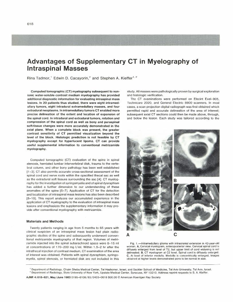
A c Fig . 1.-lntramedullary glioma with intracranial extension in 42-year-old
woman. A, Cervical mye logram, anteroposterior view. Cervical spinal cord is diffusely en larged from level of T2, but upper limit of cord widening is not delineated. B, CT mye logram at C2 level. Spinal cord is diffusely enlarg ed. C, At level of inferior medulla. Medulla is concentrically enlarged. tmages obtained at higher levels demonstrated pons to be normal in size.
, Departmen t of Radiology , Chaim Sheba Medical Center, Tel Hashomer, Israel, and Sack ler School of Medic ine, Tel Aviv University, Tel Aviv , Israel. 2 Department of Radiology, State University of New York, Upstate Medical Center, Syracuse, NY 13210. Address reprint requests to S. A. Kieffer.
AJNR 4 :618-621. May/ June 1983 0195-6108 / 83 / 0403-0618 $00.00 © American Roentgen Ray Society

AJNR:4, May/ June 1983 SPINE AND INTERVERTEBRAL DISK 619
A
extent of the tumor. The scanning techniques employed either a small scan c irc le (1 25 or 140 mm) or a large scan ci rc le (420 mm) with image postprocessing confined to a smaller (125 mm d iameter) region of the c irc le . Sl ice thic kness was 5- 8 mm. In many cases, overlapping of consecutive ax ial sections permitted subsequent image reconstructions in sagittal or coronal planes .
Results
In all 20 patients, the diagnosis of intraspinal mass was initially made by myelography, and CT myelog raphy was performed subsequently. There were eight intramedullary tumors, eight intrad ural extramedullary masses, and four extradural neoplasms.
Four of the eight intramedullary lesions were located in the cervical area; CT myelography demonstrated intracranial extension along the neuraxis into the medulla and pons in all four patients. These included one glioma, one hemangioblastoma with a large cystic component , and two lipomas. Evaluation of intrac ranial extension by conventional metri zamide myelog raphy was often not possible (fig . 1), either because of our reluc tance to carry the contrast medium intracranially or because of apparent block to upward flow of the contrast bolus in the cervical region. In the region of the conus medullari s, axial CT myelography allowed definite diagnosis of spinal cord enlargement when conventional myelographic evaluation was equ ivocal. Histological pred iction was possible only in the two cases of lipoma, which demonstrated negative attenuation coeffi c ients.
The eight intradural extramedullary masses inc luded two thorac ic meningiomas, three neurofibromas, two patients with seed metas
tases, and one case of an intradural disk hern iation presenting as a tumor. Differentiation between exophytic ex tension of intramedullary tumor and intradural extramedullary neoplasm was accomplished with CT myelography in one instance where conventional
A
Fig. 2.-lntramedullary astrocytoma o f conus medullari s in 13-year-old boy. A, Lumbar metrizamide myelogram, lateral view. Complete block with meniscuslike configurati on o f contrast column suggesting that mass may be in tradural extramedullary in locati on . Note displacement o f roots of cauda equina by mass. B, CT myelography at level of upper marg in of L 1. Inferior aspect of mass is eccentric in location and irregular and exophytic in configurat ion. Nerve roots lie ipsilateral and c lose to mass, whi ch suggests that mass actuall y represents enlarged conus meduliaris- extramedullary tumor wou ld displace adjacent nerve roots to contralateral side of thecal sac. C , CT myelography at level o f lower margin of T1 2. En larged conus medullaris virtuall y fills theca l sac.
B Fig. 3. - Thorac ic meningioma in 6g-year-o ld man. CT myelog raphy. A, At
T1 0 level. Spinal cord is located eccentrically on posterior aspec t of thecal sac to left of midline. B , At T9 level. Lobu lated int radural extramedullary mass (arrows) on right anteri orly d isplaces and compresses cord posteriorly and to lefl.
myelographic evaluation was equivocal (fig. 2). The effectr, of intradural extramedullary tumors on th e adjacent spinal cord (rotation, deformity, displacement) were more c learl y delineated on the sequential axial CT myelog rams than on the conventional myelograms (fig . 3).
The four extradural lesions inc luded two lymphomas and two

620 SPINE AND INTERVERTEBRAL DISK AJNR:4, May / June 1983
A B
c o Fig. 4. - Extradural thoraco lumbar lymphoma in 60-year-old man. A, Lat
eral radiograph showing severe compression and destruction of body of L 1 . B, CT myelography at L2. Opac ified thecal sac is concentricall y compressed by thi ckening of ex tradural soft tissues. C, At L 1 . Destruction with pathological fractu re involving body of L 1. Disp laced posterior portion of vertebral body encroaches upon spinal canal. Thecal sac is severely compressed and barely recognizable (arrow). Conventional myelographic images demonstrated ex tradural block at this level. D, At T1 2. Extradural soft-tissue mass impinges upon an terior aspect of thecal sac wh ich is opacified above level o f apparent block. Also extensive soft-tissue mass surrounds external margins of vertebral body, bu lges diaphragmatic c rura (arrows ), and obscures margins of aorta.
cases of carcinomatous metastasis. CT myelography delineated the extent and location of th e thickened extradural space due to tumor infiltration more precisely than th e conventional myelog raphic images. Exten.sion of extradural neoplasm into adjacent osseous structures and pre vertebral and paraspinal soft ti ssues was also demonstrated (fig. 4).
In instances of apparent complete block on conventional myelography, it was occasionally possible to establish the distal limit of intraspinal tumor extension by visualization of contrast opacification beyond the " block " on CT myelograms (fig . 40). This was achieved in two patients in this series (one intramedullary metastasis, one extradural lymphoma), thu s obviating a second myelogram with puncture above the level of the block .
Discussion
Myelog raphy with non ion ic water-soluble contrast medium is currently the accepted initial technique for detection and localization of intraspinal mass lesions. When properly performed, such examinations provide superior radiographic demonstration of the presence, location , and extent of space-occupying masses. However , post myelography CT can provide additional diagnostic information which may enhance accuracy and obviate th e need for further invasive examinations.
Th e add itional information obtained from display in th e axial plane regarding presence and degree of deformity, displacement, and rotation of the spinal cord and thecal sac may aid in characterization of the mass as intramedullary, intradural extramedullary, or extradural when conventional myelographic findings are equivocal. Axial CT myelog raphic images also provide improved appreciation of cord / tumor relations, which may aid in planning the surgical approach, biopsy, or resection.
Demonstration of cord enlargement or displacement by conventional myelography may prove difficult in two regions, the cervicomedullary junction and the conus medullari s, in whic h overlying bony structures obscure anatomic detail. Information provided by CT myelography in the axial plane is often sufficient to eliminate diagnostic uncertainty reg arding presence and / or extent of a mass
lesion in these areas . CT myelography offers two further advantages as a supplemental
and complementary examination to myelography. The superior contrast resolution may permit recog nition of subarachnoid space opacification too faint to be recognized by conventional film-screen techniques. This has proven valuable in demonstrating th at some apparent "complete " blocks to the flow of contrast medium are indeed incomplete, thus allowing delineation of tumor margins beyond the level of obstruction and obviating a second spinal puncture . Also , the demonstration by CT of involvement of adjacent osseous structures and soft tissues not only increases the understanding of degree of tumor extension but also may permit more accurate preoperative characterization of tumor type.
At the present stage of technologic development, state-of-the-art CT units lack suffic ient con trast and spatial resolution to provide c lear and reliable definition of the contents of most of the spinal canal without intrathecal introduction of contrast material [1, 11]. The spinal cord can be reliably demarcated on plain CT images only in the high cervical reg ion (C1 and C2 levels) where the subarachnoid space is considerably larger than in the remainder of th e spine. In the upper thoracic region , identification of the margins of the
spinal cord on plain CT images was possible in only one-third of the cases in a consecutive nonselected series [12] . While intravenous injection of iod inated contrast medium results in enhancement of the normal extradural soft ti ssues and spinal dura as well as some large arteriovenous malformations and tumors [8, 13, 14], this opacification is by no means universal or clearly detectable within the resolution limits of current CT apparatus , Thus, it appears that CT myelography will continue to be the procedure of choice to supplement conventional myelography in providing accurate definition of the contents of the spinal canal in patients with suspected intraspinal mass lesions.
REFERENCES
1. Cacayorin ED, Kieffer SA. Applications and limitations of computed tomography of th e spine. Radial Clin North Am 1982;20: 185-206
2. Harwood-Nash DC. Computed tomography of the pediatric spine: a protocol for the 1980s. Radial Clin North Am 1981 ;19 : 479-494

AJNR :4, May / June 1983 SPINE AND INTERVERTEBRAL DISK 621
3. Tadmor R, Davis KR , Roberson GH, New PFJ , Taveras JM . Computed tomographic evaluat ion of traumatic spinal injuries. Radiology 1978; 127 : 825-827
4. Di Ch iro G, Schellinger D. Computed tomography of spinal cord after lumbar intrathecal introduction of metrizamide (computer assisted myelography) . Radiology 1976; 120: 1 01 - 1 04
5. Aubin ML, Vignaud J, Jardin C, Bar D. Computed tomography in 75 c linical cases of syringomyelia. AJNR 1981 ;2: 199-204
6. Resjo 1M , Harwood-Nash DC, Fitz CR, Chuang SH . Computed tomographic metrizamide myelography in spinal dysraphism in
infants and children. J Comput Assist Tomogr 1978;2: 549-558
7. Scotti G, Musgrave MA, Harwood-Nash DC, Fitz CR , Chuang SH. Diastematomyelia in chi ldren : metrizamide and CT metrizamide myelography. AJNR 1980;1 :403 -410, AJR 1980; 135: 1225-1 232
8 Aubin ML, Jardin C, Bar D, Vignaud J. Computerized tomography in 32 cases of intraspinal tumor. J Neuroradiol 1979;6:81-92
9. Resjo 1M , Harwo'Jd-Nash DC, Fitz CR, Chuang SH. Computed tomographic metri zamide myelography (CTMM) in intraspinal and paraspinal neoplasms in infants and children. AJR
1979;132: 367 -372 10. LaM asters DL, Watanabe T J, Chambers EF, Norman D, Newton
TH. Multiplanar metri zamide-enhanced CT imag ing of the foramen magnum. AJNR 1982;3: 485-494
11 . Haughton VM , Syvertsen A, Williams AL. Soft-ti ssue anatomy within th e spinal canal as seen on computed tomography . Radiology 1980; 134: 649-655
12. Taylor AJ, Haughton VM , Doust BD . CT imag ing of the thoracic spinal cord without intrathecal contrast medium . J Comput Assist Tomogr 1980;4: 223- 224
13. Di Chiro G. Doppman JL, Werner L. Compu ted tomography of spinal cord arteriovenous malformations. Radiology
1977; 123: 351-354 14. Handel S, Grossman R, Sarwar M . Computed tomography in
the diagnosis of spinal cord astrocy toma. J Comput Assist Tomogr 1978; 2: 226-228