advertiment. lʼaccés als continguts dʼaquesta tesi queda ...€¦ · sobre la rellevÀncia del...
TRANSCRIPT

ADVERTIMENT. Lʼaccés als continguts dʼaquesta tesi queda condicionat a lʼacceptació de les condicions dʼúsestablertes per la següent llicència Creative Commons: http://cat.creativecommons.org/?page_id=184
ADVERTENCIA. El acceso a los contenidos de esta tesis queda condicionado a la aceptación de las condiciones de usoestablecidas por la siguiente licencia Creative Commons: http://es.creativecommons.org/blog/licencias/
WARNING. The access to the contents of this doctoral thesis it is limited to the acceptance of the use conditions setby the following Creative Commons license: https://creativecommons.org/licenses/?lang=en

Doctoral program in Medicine
Doctoral Thesis
AN IN VIVO STUDY OF THE IMPORTANCE OF THE INNATE
IMMUNE SYSTEM IN THE PATHOGENESIS OF EPIDERMOLYSIS
BULLOSA ACQUISITA

Programa de doctorat en Medicina. Departament de Medicina
ESTUDI IN VIVO SOBRE LA RELLEVÀNCIA DEL SISTEMA IMMUNE
INNAT EN LA PATOGÈNIA DE LA EPIDERMÒLISI AMPUL·LAR
ADQUIRIDA
Tesi doctoral presentada per:
Maria Estela Martinez Escala
Directors:
Dr. Josep E. Herrero González Dr. Ramon M Pujol Vallverdú
Barcelona, 2016


“Once we accept our limits, we go beyond them.”
Albert Einstein


ACKNOWLEDGMENT


Acknowledgements
This study has been made possible by the financial support from the Fondo de Investigación
Sanitaria, Instituto de Salud Carlos III/FEDER (Spanish Ministry of Science and Innovation) granted
to Dr. Josep E. Herrero González (PS09/01410).
The author is greatly indebted with the Molecular Laboratory of Universitätsklinikum Freiburg
(Germany) for all of their contributions to this project, including the supply of the protein mCVIICr
by Jessica Kleindienst, and the training on the experimental animal model technique by Kinga
Melinda Csorba and Florina Florea. I would like to express my sincere gratitude to Dr. Cassian
Sitaru for his teaching and guidance throughout the project development.
A specially thank you to Boston Children’s Cancer and Blood Disorders Center, which provided us
with the Rac2 knock-out mice.
The author also gives thanks to the veterinarians and technicians at the animal facilities of the
Parc de Recerca Biomedica de Barcelona, for all their assistance in teaching some of the animal
procedures, and to the Department of Pathology of Parc de Salut Mar for the hematoxilin-eosin
staining of the murine skin biopsies.
In addition, the author wants to thank the laboratory of Dr. Gabriel Gil, Apoptotic signaling at Parc
de Recerca Biomedica de Barcelona, who performed the genotyping of the inbred Rac2 knock-out
mice.
The author wants to thank her fellow labmates that helped through the long struggles during the
thesis, including Xavier Joya for his assistance in some laboratory techniques, and Dr. Irene Garcia
Diez, who assisted me to finish some experiments of this thesis.
The author gives special thanks to Kimberly A. Sable for editing and reviewing the manuscript.
The author wants to give her gratitude to her thesis advisors, Dr. Josep E. Herrero and Dr. Ramon
M. Pujol, who both assisted providing their critical opinion and suggestions to improve this work.
Least but not last, the author cannot thank enough to her family and friends for all the support
given, not only in this thesis but also in my life project.


Index
1. Abbreviations 13
2. Introduction 17
2.a. Basement membrane zone 19
2.b. Autoimmune blistering disorders 22
2.c. Epidermolysis bullosa acquisita 24
2.d. Experimental models of EBA 35
3. Background 39
3.a. Type VII collagen 41
3.b. Loss of immunological tolerance 45
3.c. Tissue injury 46
3.c.i. Role of complement 47
3.c.ii. Neutrophils 47
3.c.iii. Fc receptors 48
3.c.iv. Reactive oxygen species 49
3.d. Rac2 protein 53
4. Objectives 59
5. Materials and methods 63
5.a. Mice 65
5.b. Mice genotyping 65
5.c. Recombinant protein CVII 66
5.d. Rabbit immunization 66
5.e. Affinity purification of IgG 67
5.f. Induction of experimental EBA 67
5.g. Drug administration 68
5.h. Histology 68
5.i. Immunefluorescence microscopy 69
5.j. Enzyme linked immunosorbent assay 69
5.h. Statistical analysis 70
6. Results 71
7. Discussion 99
8. Conclusions 107
9. References 111

13
1. ABBREVIATIONS

14
1. ABBREVIATIONS
A/J Mouse substrain
ALI Acute lung injuryBALB/C
BMZ Basement membrane zone
BP Bullous pemphigoid
BPAG1 Bullous pemphigoid antigen 1
BPAG2 Bullous pemphigoid antigen 2
BSA Body surface area
C Complement (C3, C1q, C5a)
C57BL/6 Mouse substrain
CaaL C-terminal sequence, aliphatic aminoacid, Leucine
CCR4 C-C motif chemokine receptors
CD Cluster of differentiation (CD151, CD4)
CGD Chronic granulomatous disease
CMP Cartilage matrix protein
COL7A1 Type collagen VII gene
CVII Type VII collagen
CXCR C-X-C motif chemokine receptors
CXCL C-X-C motif ligand
DC Dendritic cells
DEX Dexamethasone
DIF Direct immunofluorescence
EBA Epidermolysis bullosa acquisita
ELISA Enzyme-linked immunesorbent assay
F(ab) Variable portion of antibody

15
Fc Fragment crystallizable, constant fragment of the antibody
FcR Receptor of the constant fragment of antibodies
FITC Fluorescein isothiocyanate
Fn Fibronectin
FOAM Fluorescent overlay antigen mapping
GAP GTPases activating protein
GDP Guanosine diphosphate
GDI GDP dissociation inhibitors
GEF guanine nucleotide exchange factor
GM-CSF Granulocyte-macrophage colony-stimulating factor
Gr-1 Myeloid differentiation antigen Gr-1
GTP Guanosine triphosphatase
GST Glutathione-S-transferase
HLA Human leukocyte antigen
Hsp Heat shock protein
IBD Inflammatory bowel disease
IEM Immunoelectron microscopy
IIF Indirect immunofluorescence
Ig Immunoglobulin
IL Interleukin
ITAM Immunereceptor tyrosine-based activation motif
ITIM Immunereceptor tyrosine-based inhibition motif
IV Intravenous
KO Knock-out
LAD Leukocyte adhesion deficiency
LPS lipopolysaccharide

16
mCVIICr Murine CVII corresponding to fragment Cr
MHC Major histocompatibility complex
mg milligrams
MMP Mucous-membrane pemphigoid
MPO Myeloperoxidase
NADPH Nicotinamide adenine dinucleotide phosphate
NBT Nitroblue-tetrazolium
NC Non-collagen
NSC23766 Rac1/Rac2 inhibitor
Phox Phagocytic oxidases
Rac2 Ras-related C3 botulinum toxin substrate 2
R-IgG Rabbit IgG
ROS Reactive oxygen species
SJL-1 Mouse substrain
SKH-1 Mouse substrain
TEM Transmission electron microscopy
vWF von Willebrand factor

17
2. INTRODUCTION

18

19
2. INTRODUCTION
2.a. Basement membrane zone
The basement membrane zone (BMZ) is a complex structure with different functions
including cell adhesion, differentiation, motility, transmission of extracellular signals and
growth factors, and formation of permeability barriers (1, 2). Broadly, the main structure
is made of intracellular and extracellular proteins, in which some of them are organ-
specific (e.g. kidney, lung, skin) proteins, yet others are ubiquitous in all basement
membranes of the body, making up the basic scaffolding of the BMZ. Dermal-epidermal
BMZ structure is one of the best studied. Structural proteins are interconnected forming
different layers from the basal keratinocytes of the epidermis to the dermis, which
includes lamina lucida, lamina densa and sublamina densa. Main proteins involved in
dermal-epidermal adhesion are represented in figure 1(2). There are two main structures
of the dermal-epidermal BMZ that strengthen the attachment among the different layers,
the hemidesmosome plaque and the focal adhesions. The hemidesmosome plaque is the
main adhesion structure of the dermal-epidermal BMZ that specifically connects the
keratin intermediate filaments to lamina lucida. In addition to hemidesmosome plaque,
focal adhesions, also known as focal contacts or integrin adhesomes, are located between
two different hemidesmosomes and connect actin to the lamina lucida.
The hemidesmosome plaque is formed by plectin, bullous pemphigoid antigen 1 (BPAG1,
also known as BP230), integrins (α6β4) and type XVII collagen (also known as BPAG2 or
BP180). Integrins and BPAG2 connect to extracellular proteins such as laminins (-332, -
311) forming the anchoring filaments. Lamina densa is formed by the interaction of the
laminins (-332, -311) plus three additional proteins: nidogen, laminin-511, and type IV
collagen. These three additional proteins are found ubiquitously in the BMZ of different
organs (non-organ-specific). Finally, structures that extend down from the lamina densa
are known as anchoring fibrils and their most abundant component is collagen VII (CVII)
(1, 2).

20
Focal adhesion or integrin adhesomes are formed by integrin α3β1 and adhere the actin
cytoskeleton to the lamina lucida. It is stated that integrin adhesome has a higher
complexity compared with the hemidesmosome structure, which suggests extensive
functional diversity. Finally, CD151 is a member of the tetraspan superfamily, also
expressed ubiquitously, and is co-distributed with both integrins, α3β1and α6β4 (2).

21
Figure 1. Dermal-epidermal BMZ. Structure, main proteins and their distribution forming the BMZ are shown (2).

22
2.b. Autoimmune blistering diseases of the skin
Autoimmune mucocutaneous blistering diseases are a wide group of rare disorders
characterized by the presence of circulating antibodies against different proteins of either
cell-to-cell adhesion structures (desmosomes) or BMZ of the skin and mucous
membranes. These antibodies induce intraepidermal or dermal-epidermal separation
depending on the target protein, which is clinically manifested as blisters, erosions or
crusts. The level of blister formation allows classifying these conditions in two distinct
groups. First group are the intraepidermal bullous disorders where the proteins involved
are desmosomes, plakin family or armadillo family proteins, and they manifest as fragile
blisters and erosions. The second group corresponds to the subepidermal bullous
disorders, where hemidesmosomes, proteins from the lamina densa and lucida (e.g.
laminin and integrins), and anchoring fibrils are the main targets, and typically present
with tense blisters. Table 1 shows the different autoimmune bullous diseases with their
main associated antigens. This thesis is focused on epidermolysis bullosa acquisita (EBA),
which belongs to the second group and where anchoring fibrils are the main target
protein of autoantibodies.

23
Table 1. Main target proteins and type of antibodies of each autoimmune bullous disease
(3-6).
Main classification
Disease Protein Type of antibody
Epidermal Pemphigus vulgaris Dsg 3 and 1 IgG
Pemphigus foliaceus Dsg 1 IgG
Pemphigus vegetans Dsg 3 and 1, Dsc 1 -3
Pemphigus erythematosus Dsg 1
Paraneoplastic pemphigus Plectin, Epiplakin, Desmoplakin I/II, Envoplakin, Periplakin, A2ML1, Dsg 1, Dsg 3, Dsc 1-3, BP230
IgG
IgA pemphigus Dsc 1, Dsg 1 and 3 IgA
Subepidermal Bullous pemphigoid BP180, BP230 IgG/IgE
Pemphigoid gestationis BP180 (NC 16a domain) IgG
Linear IgA dermatosis LAD-1, 97kDa/120kDa IgA
Mucous membrane pemphigoid
BP180, Laminin 332, α6 integrin, β4 integrin, 168kDa protein,
IgG/IgA
Epidermolysis bullosa acquisita
CVII IgG/IgA
Bullous systemic lupus erythematosus
CVII IgG, IgA, C3, C1q
Anti-p200 pemphigoid Laminin γ1 (subunit of the integrin)
IgG/IgA
Dermatitis herpetiformis Epidermal transglutaminases IgA
A2ML1: Alpha-2-Macroglobulin Like; Dsg: Desmoglein; Dsc: Desmocollins; NC: Non-collagenous; LAD-1: fragment of the extracellular domain of the BP180, linear IgA dermatosis

24
2.c. Epidermolysis bullosa acquisita
Epidermolysis bullosa is term defining a group of disorders characterized by the presence
of blisters, erosions and crust on the skin and mucous membrane. There are two main
groups that can be differentiated, inherited and acquisita. The inherited forms are
characterized by the deficiency of the specific skin proteins and most of them are present
since born. The acquisita form is an acquired and chronic autoimmune disease
characterized by the presence of circulating autoantibodies against CVII that bind to the
BMZ inducing dermal-epidermal separation. It was first described by Elliot and named by
Kablitz in 1985, in order to differentiate it from the inherited forms (7-9).
2.c.i. Epidemiology
EBA is a rare condition with an incidence of 0.2 to 0.5 new cases per million per year (9-
11). Even though EBA has been reported in all ages, it commonly presents in adults with a
median age of 47 years (75th percentile: 30 – 66 years of age). Initially, no predominant
gender or ethnic group was noted in EBA patients. However, more recently, three large
studies with a total of 83 patients have shown a female to male predominance (61%
female versus 39% male) (11-13). Moreover, a genetic association has been established
with the major histocompatibility complex (MHC) class II haplotype, in particular human
leukocyte antigen (HLA)-DR2. It has been found that EBA is more common among the
African American population with DRB1*15, and in Koreans with DRB1*13 (14, 15).
2.c.ii. Clinical features
Tense blisters and erosions in mucocutaneous areas are the main clinical features;
however, EBA can present in diverse manners. In broad terms, two clinical forms are
differentiated: mechanobullous (or non-inflammatory) and inflammatory (the most
25
frequent presentation in humans). The first type, also known as the classical form, is seen
in only one third of the patients, and usually manifests with blisters, erosions and scars
appearing in trauma prone locations, such as fingers and toes, where the loss of nails is
characteristic. The inflammatory form is classified in four different subtypes that
resemble other types of autoimmune mucocutaneous blistering conditions: bullous
pemphigoid (BP)-like EBA, mucous-membrane pemphigoid (MMP)-like EBA, Brunsting-
Perry pemphigoid-like EBA and linear IgA dermatitis-like EBA.
BP-like EBA presents with the clinical features of BP or a mixture of BP and the classical
form of EBA. Usually lesions are widespread along the trunk and extremities with
pruritus. Tense blisters are surrounded by inflamed or urticarial skin, and no scarring or
millia formation is seen in contrast to the classical form. In MMP-like EBA, blisters and
scars involve the mouth, upper esophagus, conjunctiva, anus and genital mucosa.
Involvement of the trachea has been reported, as well as, resolution without scarring of
the involved mucosa. Brunsting-Perry pemphigoid-like EBA presents with lesions mainly
involving the head and neck and usually leaves residual scars. Mucous membranes are
not afflicted in contrast to MMP-like EBA. Linear IgA bullous dermatosis-like EBA
presentation is characterized by annular pattern of the tense blisters and mucous
membranes are frequently involved.
Although EBA patients are predominantly adults, pediatric cases have also been described
with similar clinical manifestations. A study consisting of 14 children showed 5 with IgA
linear dermatosis-like EBA, 5 with BP-like EBA, and the remaining 4 presenting as the
classical form of EBA (16). Mucosal involvement was observed in 11 of the 14 children,
which seemed more severe and frequent when compared to adults. However, the overall
prognosis and treatment was more favorable in pediatric than in adult EBA.
Mucocutaneous involvement and a higher treatment response were also seen in another
study with 33 pediatric patients, which confirmed the data presented previously (11, 12,
17). Finally, congenital EBA has also been described due to the transference of IgG from
the mother to the fetus (18).

26
Overall, the clinical presentation of EBA may change during the course of the disease or
may show overlap between the different forms within the same patient. Moreover, the
disease will eventually become non-inflammatory over time. Despite of that, disease
severity varies from one patient to another and usually correlates with antibody levels.
Also, it has been suggested that clinical presentation may correlate with the epitope
profile, but this has not yet been fully demonstrated.
With disease progression, these patients develop dramatic consequences, such as
dystrophic changes, including atrophic scars that may also involve the scalp, nail
deformities and digital contractures (9, 19, 20). Yet mucous membrane involvement can
trigger severe sequels. Ocular lesions can be seen in MMP-like EBA patients, which may
occasionally progress to complete blindness. Besides the oral and ocular mucous
membranes, other areas of great interest due to life-threatening complications are
trachea, esophagus and larynx. Esophageal strictures inhibit the passage of food into the
stomach and require endoscopic dilation. Lastly, hoarseness and loss of voice,
occasionally associated with difficulty breathing, are signs of laryngeal involvement.
EBA has been associated with other conditions, such as inflammatory bowel disease (IBD),
including ulcerative colitis and Crohn’s disease. The linkage between IBD and EBA has
been demonstrated in both, human and animal models. Firstly, IBD has been reported in
30% of EBA patients and patients with IBD have been found to present anti-CVII IgG with
frequencies ranging from 6 to 60%. Secondly, IBD is consistently developed in EBA animal
models, in which blister formation is not limited to the skin, involving also esophagus
(40% of mice), stomach (40% of mice), small intestine (20% of mice) and colon (20% of
mice), with the functional consequence of weight loss (21). Other anecdotal associations
have been described with diabetes mellitus, psoriasis, cryoglobulinemia, amyloidosis,
rheumatoid arthritis, pulmonary fibrosis, chronic lymphocytic leukemia, thymoma and
subacute cutaneous/systemic lupus erythematous, among others (11, 12).

27
2.c.iii. Diagnosis
EBA is difficult to diagnose because of its rarity. Table 2 shows the criteria for its diagnosis
as summarized by Gupta et al (12). Moreover, in order to perform the specific indicated
work up, it requires a high suspicion by physicians, and some of the diagnostic tests are
not always available per standard of care in all health care centers. By definition, EBA
diagnosis requires the following criteria: presence of tense blisters in mucocutaneous
areas plus the detection of circulating autoantibodies against CVII, as well as tissue
deposition of anti-CVII antibodies along the BMZ. The common work up performed in
these patients includes: 1) skin biopsy which shows a dermal-epidermal disruption with
variable inflammatory infiltrate (normally neutrophils if it corresponds to an inflammatory
form of EBA); 2) demonstration of deposition of antibodies, usually immunoglobulin (Ig)
G, on the BMZ, by direct immunofluorescence (DIF) study, while IgA can also be
occasionally seen; 3) demonstration of circulating antibodies against CVII by either
indirect immunofluorescence (IIF), enzyme-linked immunosorbent assay (ELISA), or
Western-blot.
Skin biopsy. Early lesions show papillary edema and vacuolization of the basal layer, while
at later stages subepidermal blister is the main feature. The degree, location and
composition of the dermal granulocytic infiltrate are variable, although it is normally
present around vessels, follicles, and also scattered within the interstitium. Different
histologic features have been investigated to potentially differentiate one clinical subtype
from another. A sparse infiltrate is typically seen in the classical form of EBA. In contrast,
the BP-like form shows a moderate infiltrate with admixture of neutrophils, monocytes
and eosinophils, making it difficult to differentiate it from BP itself. As a matter of fact,
histopathology by itself cannot lead to a diagnosis of EBA, and further testing is required
with DIF, IIF, ELISA, immunoblot or other more complex immunobiochemical studies.
DIF. Patient´s skin biopsy is exposed to antibodies that are marked with fluorescein
isothiocyanate (FITC) and have affinity to the constant fragment (Fc) of human antibodies.
Thus, when positive, it demonstrates the deposition of the patient’s antibodies on the

28
BMZ, yet the specific target cannot be identified. The characteristic pattern is consistent
with a linear deposition along BMZ of IgG and/or IgA. It is positive in at least 93% of EBA
patients, and negative in the remaining 7%. Thirty-nine percent are IgG positive, 19% are
IgA positive and 35% have deposits of both Ig types. Interestingly, cases with ocular
involvement are related to IgA autoantibodies. More precisely and almost exclusive of
EBA, this linear deposition of the antibodies has been better described as u-serrated
pattern (following the anchoring fibrils architecture at the BMZ), compared with an n-
serrated pattern described in bullous pemphigoid among others subepidermal blistering
diseases (Figure 1). This pattern can be read with a conventional microscope at x40
objective, but requires a trained of the observer (22, 23).
IIF. This method exposes autoantibodies from the patient’s serum to other tissues of
human or other species origin (e.g. human foreskin, monkey esophagus, rat bladder,
rabbit lip, etc.). When using human skin as a substrate, this test has a higher sensitivity
when the tissue has been previously incubated with 1 molar sodium chloride solution
(also called salt-split skin test), where dermis and epidermis are chemically separated
along the lamina lucida, and thus, antigens become more widely exposed at both the roof
and floor of the artificially-induced blister. Deposition of antibodies from patient’s serum
to the floor of the dermal-epidermal separation is highly suspicious of EBA, yet not
confirmatory, as other autoimmune blister conditions such as anti-p200 pemphigoid, can
also present the same pattern.
ELISA. The purpose of the test is to detect circulating antibodies against a specific target.
By coating fragments from different non-collagen (NC) domains of CVII (NC1 only, as most
of the patients have antibodies only against to this domain, or NC1 plus NC2) on a plate
and incubating with patient’ sera, the presence of antibodies can be detected by an
enzymatic reaction that induces a visible signal, which correlates with the amount of
antibodies. It is an attractive technique, as it is a confirmatory test of the disease when
clinical features are present, with a sensitivity and specificity of >90%, and this is
nowadays commercially available (9, 24, 25).

29
Immunoblot. Western-blot using dermal extracts has been the technique of choice for
several years in order to confirm EBA. Similar to ELISA, it also detects the presence of
autoantibodies in the patient’s serum against a 290-kDa protein, which corresponds to
the molecular weight of CVII. However, this technique requires time, and it is not
normally available in most health care centers. Thus, it is less commonly used as part of
the standard work up.
Other diagnostic tests. Transmission electron microscopy (TEM), immunoelectron
microscopy (IEM), and fluorescent overlay antigen mapping (FOAM) are other ancillary
studies, without any direct benefit to patient care, but performed for research purposes.
TEM, the routine electron microscopy, reveals a decreased number of anchoring fibrils in
the lamina densa, and blister formation situated in the dermis leaving the basal lamina in
the roof of the blister (11, 26-28). IEM shows the presence of immune deposits in the
anchoring fibril zone at the lamina densa, which is unique compared to other
autoimmune blistering diseases. Finally, FOAM differentiates location of the immune
deposits by staining the antigen and antibodies in the same skin biopsy. It is less
expensive, easier to analyze and quicker than IEM (11, 28).
Table 2. Diagnostic criteria of EBA (12).
Diagnostic criteria
1. Bullous skin disorder 2. Lack of family history of a bullous disorder 3. Histology revealing a subepidermal blister 4. A positive DIF of perilesional skin showing deposition of IgG deposits within BMZ 5. IEM of perilesional skin indicating localization of IgG deposits within the lower lamina
densa and /or sublamina densa zone of the BMZ 6. Alternative laboratory tests for item 5 include indirect or direct salt-split skin
immunofluorescence, IIF using substrate deficient in basement membrane molecules, Western blotting, FOAM, and ELISA; however, a salt-split skin immunofluorescence may not be conclusive in the differentiation of true MMP from EBA when clinical presentation is MMP-like.

30
2.c.iv. Treatment
Treatment of EBA patient is challenging, due to the chronicity and refractoriness of the
disease, which often makes its management frustrating for both, physicians and patients.
These patients require two main approaches: 1) immunosuppressive drugs including
systemic glucocorticoids, as well as glucocorticoids-sparing agents and monoclonal
antibodies (such as rituximab); and 2) supportive care to reduce complications such as
wound infections and to improve patients’ quality of life (the latter is no longer
mentioned in this thesis as a marginal issue).
Efficacy of each drug is difficult to assess as the rarity of the condition does not allow
performing controlled blinded randomized clinical trials. Since the aim of the thesis is
reviewing pathophysiology of EBA, a summary of each drug by mechanism of action is
presented below (Table 3).
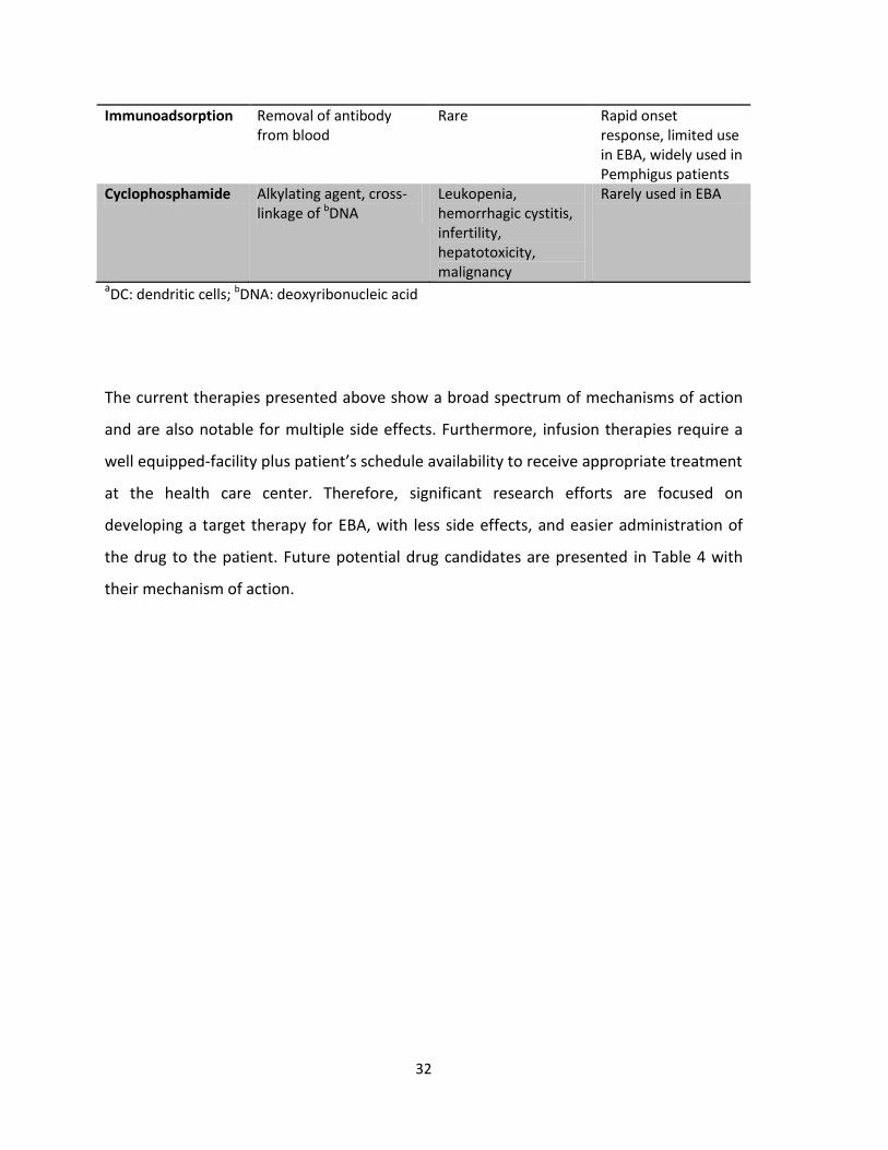
Table 3. Mechanism of action and adverse events of the current therapy used in EBA (11,
29-35).
Agent Mechanism of action Adverse events Comments
Glucocorticoids Immediate effects: inhibition of vasodilation. Late effects: Block the expression of pro-inflammatory genes and induce transcription of anti-inflammatory agents
Cushing’s syndrome diabetes mellitus, osteoporosis, avascular necrosis of the femoral head, cataracts, peptic ulcer, hypertension, acne
Main treatment for most of the EBA patients. Normally given initially to control severe disease.
Colchicine Inhibits neutrophils mobility and chemotaxis, interfering with the microtubules; decrease Ig secretion by plasma cells
Diarrhea, bone marrow suppression, myopathy, urticaria, toxic epidermal necrolysis
Given as monotherapy in mild cases of EBA, it is considered first line treatment
Dapsone Not well understood. It is thought to the blockade of reactive oxygen species synthesis
Hemolytic anemia, agranulocytosis, methemoglobinemia, peripheral
It has been reported in several case reports and occasionally as

31
neuropathy, headache, vertigo
monotherapy
Cyclosporine Inhibits T-cell activation through inhibiting transcription of IL-2
Nephrotoxicity, hypertension, diarrhea, hyperkaliemia, hypomagnasemia, hyperuricemia and hyperlipidemia, hypertrichosis
Case reports, however not usually indicated
Methotrexate Inhibits dihydrofolate reductase interfering with bDNA synthesis
Myelosupression, nausea, vomiting, myelosuppression, hepatotoxicity, pulmonary toxicity
Never used as monotherapy
Azathioprine Interferes with the synthesis of purines (bDNA synthesis)
Bone marrow suppression, macrocytosis, nausea, vomiting, hepatitis, pancreatitis
Never used as monotherapy.
Mycophenolate mofetil
Inhibits inosine monophosphate in the purine synthesis (bDNA synthesis)
Gastrointestinal upset, bone marrow suppression, infections, weakness, tiredness
Adult and pediatric case reports
High dose intravenous immunoglobulins
Unclear, increase clearance of antibodies, direct inhibitory effect on T- and B-cells functions
Headache, acute renal failure, aseptic meningitis, migraine, flu-like symptoms, hypotension, chest tightness
Used as second line therapy in refractory disease. Long-term use as monotherapy once response is achieved.
Plasmapheresis Removal of antibody from blood
Infections, hypotension, transfusion reactions, bleedings
Greater experience with pemphigus, limited experience in EBA
Rituximab Monoclonal antibody against CD20 (B-cell lymphocytes), avoiding development of plasma cells
Infusion reaction, hepatitis B reactivation, opportunistic infections, progressive multifocal leukoencephalopathy
Wide experience in pemphigus. Phase III clinical trial for pemphigus. EBA only used in a limited number of cases.
Extracorporeal photochemotherapy
Monocytes with maximal exposure to 8-methoxypsoralen develop into short-lived immature aDC with anti-inflammatory responses
Hypotension, syncope Case reports
32
Immunoadsorption Removal of antibody from blood
Rare Rapid onset response, limited use in EBA, widely used in Pemphigus patients
Cyclophosphamide Alkylating agent, cross-linkage of bDNA
Leukopenia, hemorrhagic cystitis, infertility, hepatotoxicity, malignancy
Rarely used in EBA
aDC: dendritic cells; bDNA: deoxyribonucleic acid
The current therapies presented above show a broad spectrum of mechanisms of action
and are also notable for multiple side effects. Furthermore, infusion therapies require a
well equipped-facility plus patient’s schedule availability to receive appropriate treatment
at the health care center. Therefore, significant research efforts are focused on
developing a target therapy for EBA, with less side effects, and easier administration of
the drug to the patient. Future potential drug candidates are presented in Table 4 with
their mechanism of action.

33
Table 4. Possible future therapies for EBA (11, 36-40).
Agents Mechanism of action Evidence Other conditions where it has been investigated
aHsp90 inhibitors
Inhibits antibody production by targeting autoreactive T-cells.
Prevented onset of antibody-transfer-induced EBA, and improved already established immunization-induced EBA
Phase I-II for multiple myeloma. Several adverse effects, including death-related treatment
SM101, soluble bFcgRIIB
Blockade bFcgR, which is a key feature for autoantibody blister formation
Potential candidate.
Phase II for primary immune thrombocytopenia, and systemic lupus erythematosus
Natalizumab Anti-α4 integrin, modulates leukocyte extravasation
Potential candidate. No literature evidence
Used in multiple sclerosis. Cases with progressive multifocal leukoencephalopathy
Efalizumab Anti-CD11a, inhibits leukocyte extravasation
Not successfully controlling skin inflammation
It has been withdrawn for psoriasis
Anti-cCCR4 (KW-0761)
Inhibits leukocyte recruitment into the skin
Non-tested in skin inflammatory conditions
Used for relapse adult T-cell leukemia lymphomas, cutaneous T-cell lymphoma
Modified heparins (dPS3)
Targets disruption of leukocyte extravasation
Has demonstrated anti-inflammatory and antimetastatic effect
It has less anticoagulant effect than heparin itself
Reparixin or eCXCR1/2 inhibitor
Inhibits neutrophil chemoatractant activity
Prevented onset of antibody-transfer-induced EBA, and improved already established immunization-induced EBA
Phase II for pancreatic islet transformation
Blockade of fGM-CSF
Inhibits colony stimulating factor of granulocytes, decrease antibody formation and neutrophil count
Beneficial in antibody transfer- and immunization –induced EBA.
Phase I-II for Rheumatoid arthritis
Anakinra Partial modulation of IL-6, which has anti-inflammatory effect
Potential candidate. Never used in EBA.
Used effectively in Schnitzler’s syndrome
aHsp: heat-shock protein; bFcgRIIB: receptor of the constant fragment of antibody; cCCR4: C-C motif chemokine receptor 4; dPS3: semisynthetic glucan sulfate; eCXCR1/2: C-C motif chemokine receptor 1/2. fGM-CSF: granulocyte macrophage colony-stimulating factor.

34
With all the treatments mentioned above, it seems that EBA patients have numerous
treatment options, however none of these new molecules can fully or partially
substituted for old treatments. Obviously, the efficacy of new agents is difficult to be
tested and compared with other available treatments due to low prevalence/incidence of
the disease. Thus, collaboration of multiple centers to increase study patient number is
necessary in order to gather enough and significant data regarding efficacy as well as the
drugs’ adverse events. Table 5 shows the well-accepted current treatment regimens for
EBA patients.
Table 5. Treatment suggested by EBA severity, defined as follows (41):
Mild EBA (<5% of body surface area
without mucous membrane involvement)
Moderate EBA (5 – 15% of body surface area and or two or more mucous
membranes involvement, not including eyes)
Severe EBA (Greater than 5% of body
surface area or greater than two mucous membranes
involved or ocular involvement )
Oral steroids (0.5 – 1 mg/kg/d) +
Colchicine 0.5 – 1 mg/d (mild) or 1-3 mg/d (moderate) ±
Dapsone 1 – 2 mg/kg/d
Oral steroids (0.5 – 1 mg/kg/d) +
Colchicine 1-3 mg/d (moderate)
± Dapsone 1 – 2 mg/kg/d
± IV steroids pulse
± Mycophenolate mofetil 1 – 2
g/d ±
Plasmapheresis ±
IVIG 1 – 2 g/kg over 3 – 5 every 4 weeks
± Rituximab 375 mg/m2 /wk for
4 wk

35
Although EBA is a rare condition, our extensive knowledge of its pathophysiology and
immunology are intriguing and offer the unique opportunity to investigate methods to
improve diagnosis and treatment options with the objective to improve patient’s quality
of life.
2.d. Experimental models of EBA
A summary of experimental models of EBA is presented in Table 6.
2.d.i. Ex vivo
There are two ex vivo experimental models described in the literature: the cryosection
model and the regenerated/organ cultured human skin.
The former has been comprehensively characterized compared with the latter. The
cryosection model technique takes human skin (normally neonatal foreskin) and exposes
it to patient’s serum, which is further incubated with leukocytes from healthy donors.
The same process is used for the regenerated/organ cultured human skin technique,
although the substrate to expose patient’s antibodies is cultured skin. Both techniques
directly demonstrate the pathogenicity of anti-CVII IgG in EBA patients, yet the organ
culture model has not further been used (42).
2.d.ii. In vivo
Two types of animal models have been described, the so-called passive and active
models. Passive-transfer experimental models are those where the disease is induced by
the injection of antibodies against the target protein, whereas in active models, animals
develop the disease by immunization with the target antigen, this is, by injection of the

36
target protein or peptide. Therefore, in the active model, animals do produce their own
pathogenic antibodies in response to the artificial immunization with an antigen.
Passive-transfer animal model of EBA. There are two types of passive transfer animal
models of EBA, in which the source of antibodies differs one from the other. One is the
rabbit IgG anti-CVII passive transfer animal model, in which antibodies come from
immunized rabbits with human or murine fragments of CVII. And the second is the human
IgG anti-CVII, in which antibodies come from EBA patients after plasma has been purified
and concentrated.
Passive transfer of rabbit IgG against CVII. Inbred mouse strains such as BALB/C, C57BL/6
and outbreed mice SKH-1 developed EBA phenotype after the subcutaneous injections of
the antibodies. Protocol consisted of the injection of antibodies every other day for a
total of 40 – 90 mg in six divided doses. The first lesions were seen 2 to 4 days after the
first dose of antibody, and full-blown disease, meaning the presence of erythema, blisters
and crust associated with alopecia, was seen 5 to 6 days after the first injection.
Moreover, disease extension correlated with the total IgG dose. After 12 days, mice were
sacrificed. Skin biopsies showed histological findings typically described in human EBA,
with a mixed inflammatory infiltrate rich in neutrophils, along with dermal-epidermal
separation. DIF of mice skin biopsies showed a linear deposition of rabbit IgG at the BMZ,
as well as linear deposition of mouse C3 at the BMZ. ELISA of mice sera extracted each
day or every other day revealed increasing levels of IgG (42, 43).
Passive transfer of patients’ IgG against CVII. The development of blisters by the passive
transfer of autoantibodies from EBA subjects to SHK-1 mice clearly demonstrates the
pathogenicity of the autoantibodies. However, the high amounts of human antibodies
that are needed to induce the disease phenotype make this type of animal model
impractical. This is due to two facts: first, EBA patients are exceedingly rare, and second,
it seems that human IgG antibodies from EBA patients show low affinity for mice Fc
gamma receptors. In short, these are the reasons why most authors use rabbit antibodies

37
after immunizing them with murine collagen VII peptides, instead of EBA patients’ sera
(43-45).
Active disease model of EBA. In these models, disease phenotype is achieved by
immunizing the animals by means of the injection of certain fragments of recombinant
murine CVII associated with an adjuvant (usually a non-ionic block copolymer, or other
adjuvant such as CpG, pertussis or cholera toxins). It requires repeated injections of the
antigen (at least two to three times) at 3-week intervals for a total of 12 weeks, to
achieve full-blown disease. Inbred mice, including BALB/C, and outbreed such as SJL-1, AJ
are able to develop an immune response after injection of the murine CVII. Interestingly,
the genetic background is strongly associated with the susceptibility for disease
development. For instance, SJL-1 mice have an incidence of 80 to 100%, whereas only
50% of the BALB/C mice develop the disease phenotype. Thus, outbreed SJL-1 is the
preferred strain for this type of animal model. This active model is useful to dissect the
cellular and molecular aspects of pathogenic autoantibody production, and to design
immunomodulatory treatments for EBA (42, 46).
Table 6. Summary of experimental models of EBA (42).
Type of experimental model
Source Phenotype induced by
Cryosection model (47)
Human neonatal foreskin
anti-CVII IgG from EBA patients, incubated with human leukocytes from healthy donors
Skin organ culture (48)
Regenerated human skin
anti-CVII IgG from EBA patients, incubated with human leukocytes from healthy donors
Passive transfer (45, 47)
BALB/C, C57BL/6, SHK-1 mice
Rabbit or murine anti-CVII IgG or human IgG from EBA patients
Active model (46) Preferably SJL-1 mice Murine CVII associated with antigenic adjuvant

38

39
3. BACKGROUND

40

41
3. BACKGROUND
The pathogenesis of EBA is still unclear even though the role of the autoantibodies seems
to be crucial in the development of the disease. EBA is genetically associated with the
MHC class II haplotype, in particular HLA-DR2, HLA-DRB1*15 in blacks and HLA-DRB1*13
in Koreans (9, 13-15, 49, 50). Overall, EBA pathogenesis can be summarized in three
consecutive steps; 1) loss of tolerance against CVII; 2) maintenance of antibody
production once loss of tolerance to anti-CVII has been established; and 3) the tissue
injury triggered by the deposition of autoantibodies at the BMZ (11, 51, 52).
In order to cover the pathogenesis of EBA, this section will discuss the structure and
function of CVII as a protein and target antigen, the loss of tolerance to CVII by
autoreactive T-cells, and the phenomena involved in the development of tissue injury.
The latter corresponds to the focus of this doctoral thesis.
3.a. Type VII collagen
The COL7A1 is the gene encoding the CVII protein, which contains two main domains:
collagenous and non-collagenous. The collagenous domain is formed by a triple helical
structure containing repeating glycine-proline-hydroxy proline or hydroxyl lysine (Gly-X-Y)
sequences. The non-collagenous (NC) domain can be divided as follows; 1) a 39-amino
acid domain forming the hinge region located in the center of the collagenous domain; 2)
an N-terminal domain of 145 kDa, also called NC1; and 3) a C-terminal domain of 34 kDa,
also known as NC2. NC1 domain has subdomains that are structurally similar to other
proteins with adhesion function such as cartilage matrix protein, nine fibronectin III-like
domains and von Willebrand factor A-like domain followed by a cystein and proline-rich
domain (11, 53, 54). NC2 domain is structurally similar to protease inhibitor molecules.
The CVII protein is organized as follows: headed by an N-terminal 145 kDa NC domain,
followed by a triple-helical of collagenous domain disrupted by a NC hinge region in the

42
center, and terminated with a C-terminal 34 kDa domain. When CVII is released by the
fibroblast to the extracellular matrix, NC2 domain is cleaved, and then two identical CVII
proteins align tail-to-tail to form a dimer linked by disulfide bonds. This structure
corresponds to the anchoring fibrils that are attached to different proteins of the lamina
densa of the BMZ. Specifically, fibronectin III-like domains bind to laminin 332, von
Willebrand factor A-like domains bind to collagen I; however, the specific NC1 subdomain
where collagen IV binds remains unknown (Figure 2) (55-57).
In the setting of an autoimmune condition, it is of interest to search for the specific areas
of the protein that induce more antigenic reaction than other regions. A second interest is
to determine the capability of those antibodies to induce disease. These have been well
characterized in EBA. First, the antigenicity of CVII in the setting of EBA patients has been
studied by epitope-distributed mapping. The major number of epitopes is located in the
NC1 domain, though antibodies against the collagen domain and NC2 have also been
described with less frequency (58-64). Second, the pathogenic relevance of the antibodies
against CVII has been demonstrated not only ex vivo (by cryosections), but also in vivo by
inducing EBA in mice with the passive transfer of rabbits’ and patients’ autoantibodies
(mentioned in Experimental animal model section). A relationship between the different
epitopes recognized and the different phenotypes of the disease (e.g. mechanobullous,
inflammatory) could not been demonstrated (43, 45, 65). Currently, and as discussed
above, the disease severity has been shown to correlate with the level of circulating
antibodies (19).
Upon characterization of the most antigenic areas of CVII and demonstration of the
pathogenicity of antibodies, a comparison of disease severity between the different
epitope-specific antibodies was performed in a passive transfer animal model (66). By a
functional epitope mapping with antibodies against different fragments of NC1 and NC2,
it was shown that autoantibodies against NC2 domain do not induce blistering skin
disease, which is in contrast to the antibodies against. The authors suggested a low
specificity of the IgG against the NC2. Therefore, a higher amount of IgG was required to

43
induce disease, which probably caused at the same time, a saturation of the neonatal Fc
receptor (which participates in hemostasis of immunoglobulins) and favored their
catabolism. Thus, pathogenic relevance of NC2 still needs to be elucidated. On the other
hand, mice injected with specific antibodies against epitopes located on the amino acids
(aa) 281 to 1125 of the NC1 domain showed more severity of the disease compared with
those mice that received antibodies against epitopes located on the aa 26 – 300 or 1108 –
1323 of NC1 (66). For this reason, fragments of human or murine NC1 domain localized
on the aa 281 to 1125 are used in the experimental animal models to induce either
disease in active animal model, or antibody formation for passive transfer animal models.
In this research project, we specifically used a reviewed fragment located in aa 759-980,
named as fragment C of the murine CVII (mCVIICr).

44
Figure 2. Type VII collagen. Headed by NC1 domain (N-terminal), followed by a triple-helical of collagenous domain disrupted by a NC hinge region in the center, and terminated by NC2 (C-terminal). NC2 domain is cleaved at the extracellular matrix, and then two identical CVII proteins align tail-to-tail to form a dimer linked by disulfide bonds. This structure corresponds to the anchoring fibrils that are attached to different proteins of the lamina densa of the BMZ. Specifically, fibronectin (Fn) III-like domains bind to laminin 332, and von Willebrand factor (vWF) A-like domains bind to collagen I. CMP: cartilage matrix protein (9).

45
3.b. Loss of immunological tolerance
The mechanism in which T or B-cells react against self-antigens is known as loss of
tolerance. This implies two steps throughout disease course, first its development and
second its maintenance. The latter favors the chronic nature of the disease.
Autoantibodies are the B-cell product generated from this mechanism and they can be
developed directly from autoreactive B-cells or indirectly from autoreactive T-cells
interacting with B-cells. It has been shown, that T-cell-deficient mice cannot develop EBA
phenotype by active immunization (induction of autoimmune response in experimental
animal model). Yet, EBA is clinically induced when the T-cell population is restored in
these mice (11, 67). In addition, autoreactive T-cells against the same epitopes of CVII
targeted by IgG autoantibodies have been demonstrated (51, 68). Further studies have
characterized those T-cells as CD4+ T-cells. However, the type of antigen presenting cell
that favors the clonal selection and expansion of the CD4+ T-cell remains unknown (11,
51, 67).
Neonatal Fc (fragment crystallizable, constant fragment of the antibody) receptor is a
subtype of Fc receptor that functions to transport mother’s IgG through the placenta, but
also monitors IgG homeostasis. Once loss of tolerance has been developed, neonatal Fc
receptor plays a major role in controlling serum levels of IgG against CVII, thus
maintaining the autoimmune response, and so, the maintenance of disease. Indeed, mice
deficient in neonatal Fc receptor were protected from EBA induction (52).
Heat-shock proteins (Hsp) are a family of proteins that are produced by cells in stressful
conditions, initially related to heat shock, and latter detected in other stressful conditions
such as wound healing. An inhibitor of Hsp subtype 90 (Hsp90) has been shown to induce
death of malignant plasma cells (for instance, in multiple myeloma). Kasperkiewicz et al.
raised the question whether if this inhibitor could also target non-transformed plasma
cells. Surprisingly, in EBA, Hsp90 inhibitor was found to target T-cell proliferation. By the
administration of Hsp90 inhibitors in passive transfer animal model or active animal
model, mice did not developed disease, or presented with milder severity of EBA.

46
Moreover, in the active animal model, a decrease of B-cell proliferation was detected, yet
no impact was seen in autoreactive plasma cells (69, 70).
3.c. Tissue injury
Different clinical phenotypes of EBA may result from different mechanisms of tissue
injury. As discussed above, in broad terms there are two types of clinical presentation of
EBA, inflammatory and mechano-bullous (non-inflammatory). In the mechano-bullous
EBA subtype, a reduction in the number of anchoring fibrils is seen in either lesional and
perilesional skin has been described, along with a scattered inflammatory infiltrate. It is
suspected that the direct effect of the antibody disrupts the joining of anchoring fibrils to
the lamina densa and the formation of the antiparallel dimer (71). As a matter of fact, the
pathogenesis of mechanobullous EBA is very difficult to investigate as all the
experimental animal models mimic the inflammatory form of EBA.
On the other hand, the innate immune system seems to play a major role in the
inflammatory form of EBA (the most frequent presentation in humans), triggered by the
autoantibodies bound to the dermal-epidermal BMZ. Overall, it consists on a complex
pathway with activation of multiple elements that subsequently induces tissue injury. A
disruption of this complex pathway seems to be the main target for the development of
new and more specific therapies. Among others, possible therapeutic targets include the
Fc (constant fragment of autoantibodies) or its receptors, alternative complement
pathway, neutrophils and the generation of reactive oxygen species (ROS). As mentioned
above, each distinct types of experimental EBA animal models represent the
inflammatory clinical phenotype, which results in reliable reproduction of the
inflammatory environment occurring in EBA (9).

47
3.c.i. Role of the complement system
The alternative complement pathway is the part of the innate immune system that
participates in opsonizing and killing pathogens through the membrane attack complex.
Specifically, complement 5a (C5a), a component of the alternative pathway, is a potent
leukocyte chemoattractant. Passive transfer of antibody against CVII in C5-deficient mice
failed to induce experimental EBA (9, 43, 51). Further studies with mice that received
passive transfer of IgY (chicken immunoglobulin -by itself incapable to activate murine
complement-) against CVII did not developed experimental EBA (72). In contrast, mice
deficient in classical pathway (C1q) or mannose binding lectin-complement components
showed the same severity in EBA phenotype compared with wild type mice with the
passive transfer of antibodies against CVII (73).
The synthesis of this information reveals, it is agreed that the alternative complement
pathway is an important key factor in the EBA pathogenesis. However, its role is more
important as an intermediate factor by recruiting neutrophils than a direct toxic effect on
the skin. The latter is shown by the fact that ex vivo dermal-epidermal separation is
induced by autoantibodies when incubated with granulocytes on cryosections of normal
skin, where complement is absent.(51).
3.c.ii. Neutrophils
With the attachment of antibodies and activation of complement, neutrophils are
recruited to the skin, which are considered as the most significant cells in tissue injury.
There are still some controversies regarding this statement. First, inhibition of colony-
stimulating factor (CSF), specifically granulocyte-macrophage(GM)-CSF (Csf2) that is the
most potent with regards to the recruitment of neutrophils and macrophages to sites of
inflammation, showed reduced skin blistering in a rabbit IgG anti-CVII passive transfer
animal model. Moreover analysis of the immunization-induced epidermolysis bullosa
acquisita animal model on GM-CSF KO mice, lower serum autoantibody titers suggesting,

48
that GM-CSF also modulates antibody production (74). This phenomenon has already
been shown in other conditions, where antibody generation is modulated by B cell helper
neutrophils. Another example is the contribution of GM-CSF to the formation of anti-
influenza IgG (40, 74). Anti-Gr1 is a monoclonal antibody that mainly depletes neutrophils
and monocytes. The injection of this monoclonal antibody showed protection in the
experimental antibody induced-EBA. However, the fact that anti-Gr1 can also deplete
monocytes questions the theory that tissue injury is limited to neutrophils (51, 75, 76).
Another finding suggesting the neutrophil involvement in tissue injury was the
involvement of IL-8 (which corresponds to CXCL-1 and CXCL-2 in mice). IL-8 is known to
induce neutrophil chemotaxis and phagocytosis. It was shown that they are (CXCl-2>CXCL-
1) are highly expressed in the mice’s skin when EBA is induced. Moreover, skin blistering
is not observed by blocking their receptors. More evidence of the neutrophil involvement
is the release of ROS and proteolytic enzymes (such as metalloproteases and elastases),
which finally induce blister formation and will be further examined in section 3.c.iv.
3.c.iii. Fc receptors
Fc receptors (FcR) have recently been an important target in the field of EBA research.
These receptors are localized on the surface of cells (e.g. neutrophils, macrophages,
natural killer cells) and have different mechanisms of action including facilitating antigen
presentation, inducing phagocytosis or antigen-dependent cellular cytotoxicity, and
releasing toxic oxygen metabolites and inflammatory mediators.
There are different subtypes of FcR that correspond to each subclass of the
immunoglobulins. For instance FcγR binds to IgG, FcεR to IgE, and FcαR to IgA. Focusing
on IgG, there are also different subtypes of FcγR with the aim of modulating the immune
response, meaning that there is no exclusive activation or inhibition of the immune
response, yet there is balance between both immune responses (77). In normal
conditions, this protects against an exaggerated immune response as inhibitory function
49
would balance activator response. This activator or inhibitor function is defined by two
types of receptor-associated signal-transducing molecules, such as immunoreceptor
tyrosine-based activation (ITAM) and inhibition motif (ITIM). ITAM is attached to
activating receptors such FcγRIA, FcγRIIA and FcγRIIIA, and ITIM is bind to the inhibitory
receptor FcγRIIB.
The role of FcγR in EBA tissue injury was demonstrated as follows. By removing the Fc
portion of IgG with pepsin, the injection of the isolated variable portion of the antibody
F(ab)2 with affinity to CVII showed no induction of EBA lesions in mice. And again, the
injection of IgY that lacks affinity for murine Fc receptors did not show induction of
blisters in mice (43, 78).
Recently, Kasperkiewicz et al. showed the exclusive importance of murine FcγRIV (which
corresponds to the orthologue of human FcγRIIIA) for the tissue injury in EBA, among all
others subtypes of FcγR. By gene expression profiling and weighted gene co-expression
network analysis, they assessed the expression of FcγRIV using quantitative reverse
transcription and polymerase chain reaction in the skin of wild type mice after treatment
with pathogenic IgG. They found 1) an increased expression of FcγRIV and to a lesser
extent expression of all the other FcγRs in murine skin biopsies, 2) a strong expression of
FcγRIIIA in two out of three of human skin biopsies of EBA patients, and 3) weak
expression of FcγRIIIA in control specimens or in EBA samples with scattered infiltrate
(79). Thus, FcγRIV is considered as the important activator factor in the immune response
(basically neutrophil activation) to develop tissue injury on EBA patients.
3.c.iv. Reactive-oxygen species
Nicotinamide adenine dinucleotide phosphate (NADPH) oxidase is an enzyme complex
located at the cell surface of neutrophils or in the membrane of the phagosome. It is
composed of 6 subunits: a Rho guanosine triphosphatase (GTPase) usually Rac2, and five
phagocytic oxidases (phox); gp91phox, p22phox, p40 phox, p47 phox (also called neutrophil

50
cytosolic factor 1), and p67 phox (80). NADPH oxidase generates superoxide anion by
transferring electrons from NADPH to the interior of the cell and coupling them to an
oxygen molecule. Superoxide anion is a free radical that can spontaneously form
hydrogen peroxide, which finally generates ROS. Deficiencies of NADPH in humans have
been described to be due to a mutation in either gp91 phox (X-linked) or p47 phox
(autosomal recessive), inducing chronic granulomatous disease (CGD) or Bridges-Good
syndrome. Diagnosis is performed by the nitroblue-tetrazolium (NBT) test, where the lack
of blue staining of neutrophils confirms an impairment of NADPH oxidase.
Figura 3. NADPH oxidase complex. a) Disassembled complex is present in the cytoplasm. b) Assembly of the complex and translocation to cytoplasmic membrane for the release of ROS (81).

51
The involvement of NADPH oxidase in EBA has been demonstrated by both ex vivo and in
vivo experiments. The incubation of autoantibodies of EBA patients in cryosections of
normal skin with neutrophils from patients with CGD showed no blue staining with NBT,
confirming the absence of NAPH oxidase (76). Mice deficient in neutrophil factor 1
(p47phox) were also protected from developing EBA lesions with the injection of IgG
against CVII (76). So, NADPH demonstrates an important role for tissue damaging in EBA
patients, which makes this molecule of greatest interest as a target for therapies.
A summary of EBA pathogenesis is illustrated in figure 4.

52
Figure 4. Pathogenesis of EBA. Tissue injury in EBA is developed as follows: 1) autoantibody binds to CVII at the dermal-epidermal BMZ through F(ab) fragments, 2) there is a complement activation that leads to recruitment of effector cells, including neutrophils, 3) Fc portion of the autoantibody binds to FcγRIV on immune cells causing their activation, and resulting with the release of ROS (by NADPH oxidase), serine and metalloproteases that mediate extracellular proteolysis (Courtesy of Dr. Sitaru, adapted image).

53
3.d. Rac2 protein
As explained above, Rac2 protein is one of the subunits that form the complex of NADPH
oxidase, although this protein has other functions of interest that might be important in
EBA.
Rac2 (ras-related C3 botulinum toxin substrate 2) protein belongs to Rho GTPases, which
are divided into families of proteins based on sequence homology, protein domains, and
function. These include Rac, RhoA, Cdc42, TC10 and TCL, Rnd, the Rho BTB subset and the
Miro subfamily. It is worth to mention, that Rho GTPases are subject to posttranslational
modifications at the C-terminal sequence. The C-terminal sequence contains the CaaL
sequence (C as cysteine, a as aliphatic aminoacid, and L as a Leucine), followed by a
‘polybasic domain’, in the majority of the protein of the Rho family. Posttranslational
modifications consist in lipid modification and ‘aaL’ removal by proteolysis, which are
critical in protein localization in the cell and its function (82).
The main functions of these proteins include cytoskeleton rearrangement, cell cycle
progression and cell survival, cell adhesion, cell motility, cytokinesis and membrane
trafficking, and regulation of gene transcription (82, 83). Focusing in the subfamily of Rac
proteins, there are three types of Rac proteins encoded by different genes. Rac 1 is the
most comprehensively studied and is ubiquitously expressed, while Rac2 is restricted to
hematopoietic cells. These two isoforms share 92% identity and are highly homologous
between murine and human species. Rac3 has been recently described and is less well
characterized. It is also expressed in a variety of tissues and shares 72% and 83% identity
with Rac1 and Rac2, respectively. It is important to remark that Rac2 protein differs from
Rac1 in the polybasic domain at the C-terminal sequence. Rac2 protein contains only 3
basic residues compared to the polybasic residues present in Rac1. This non-polybasic
domain gives unique functions to Rac2 protein (82, 84).
Rac proteins act as molecular switches that cycle between inactive (when associated to
guanosine diphosphate (GDP)) and active (when associated to guanosine triphosphate
(GTP)) states. Regulation of the active and inactive forms is mediated by: 1) Dbl-family

54
guanine nucleotide exchange factors (GEFs) that are activated by receptor-dependent
kinases; 2) GTPases activating proteins (GAPs) that remove the phosphate and return the
active GTPase to a GDP-bound (inactive) form of the protein; and 3) GDP dissociation
inhibitors (GDIs) by sequestering or stabilizing the inactive form of the protein (82) (Figure
5).
Figure 5. Rac protein regulation. Rac proteins cycle between inactive when associated to
GDP-bound and active when associated to GTP-bound states. Regulation of the active and
inactive forms is mediated by: 1) GEFs that are activated by receptor-dependent kinases;
2) GAPs remove the phosphate and return the active GTPase to a GDP (inactive) form of
the protein; and 3) GDIs by sequestering or stabilizing the inactive form of the protein.
The black dot shows the consequence of missense mutation found in human Rac2
deficiency disorder. GAPs: GTPase activating proteins. GEFs: guanine nucleotide exchange
factor. GDIs: guanosine diphosphate dissociation inhibitors.

55
While Rac1/Rac2 proteins seem to play an important role in regulating the inflammatory
response, Rac3 protein has been demonstrated to be more relevant in myelopoiesis.
Further understanding of the Rac proteins function has been obtained after
characterizing phenotypes of deficient mice for each isoform of Rac protein,
differentiating their unique, as well as overlapping functions. For instance, it has been
demonstrated that both Rac1 and Rac2 are required for functions related to the
cytoskeleton (migration), and Rac2 was absolutely required for the activation of the
NADPH oxidase, at least in mice (84, 85). Further studies on human neutrophils confirmed
these results and specifically showed that Rac1 initiates migration towards the
chemoattractant gradient, while Rac2 continues the migration and activation of NADPH
oxidase on the target tissue. Another group studied the specificity regarding NADPH
activation between Rac1 and Rac2 protein. The replacement of the Rac1 C-terminal
polybasic domain with the C-terminal domain of Rac2 showed a reconstitution of ROS
formation. Instead, the replacement of the Rac2 C-terminal domain with the Rac1 C-
terminal polybasic domain did not (86). Thus, Rac2 is the key to the mediation of oxidase
activation in neutrophils and downstream of chemoattractant (84).
In addition to neutrophils, Rac2 protein has been shown to be involved in T-cell
development, a function shared with Rac1. Mice lacking Rac1 and Rac2 were detected to
have a decreased number of mature CD4+ T-cells, compared to mice lacking Rac1 only, or
Rac2 only, which did not show to perturb T-cell development (82). Moreover, in Rac
deficient mice, abnormalities in B-cell development have been also observed (82).
Human disease caused by dysfunction of the Rac2 protein have been described, showing
combined clinical features of leukocytes adhesion deficiency (LAD) and CGD. The two
patients reported presented defects in neutrophil adhesion and migration, along with
neutrophilia and leukocytosis plus dramatic absence of pus in the infected areas. First,
suspicion for LAD was ruled out when expression of CD11b/CD18 (commonly affected in
LAD) was demonstrated. Further characterization of the neutrophils in these patients
showed deficiency in activation of NADPH oxidase, which mirrors CGD. Missense

56
mutation was found on 169bp G>A (D57N), which results in a change of amino acid
sequence (aspartate for asparagine). This mutation corresponds to the GTP binding
pocket, that results in inactivation of not only Rac2 protein, but also Rac1 protein as well
as other Rho GTPases (Figure 5) (82). Rac2 KO mice also show clinical features of LAD and
CGD, however some overlapping functions are compensated by the expression of Rac1, as
Rac2 KO mice preserve the GTP-binding pocket required for Rac1 to be active, compared
to the human mutation. Thus, compared with the human scenario, Rac2 KO mice show
normal T-cell development and lack of anemia.
The existence of Rac2 KO mice allowed for the in vivo study of the role of Rac2 in tissue
inflammation. Rac2 has been comprehensively studied in acute lung injury (ALI), a
common clinical disorder involving respiratory failure caused by injury to alveolar,
epithelial and endothelial barriers in the lung. It can cause an acute respiratory distress
syndrome that is associated with high morbidity and mortality, and the current available
therapies fail to improve outcomes. Neutrophil recruitment and activation are primarily
involved in ALI pathogenesis, leading to tissue-damage by degranulation and ROS release.
Arthus reaction (hypersensitivity type III) was induced by intratracheal injection of rabbit
anti-ovoalbumin IgG (which induces the immune complex response). Rac2 KO mice
showed less cellular inflammation regarding neutrophil count in response to increasing
doses of antibodies to ovoalbumin. The levels of cytokine release between wild-type mice
and Rac2 KO mice were also examined. Cytokines’ profiles did not show any significant
difference between Rac2 KO and wild-type, however levels of myeloperoxidasa (MPO;
granule-derived mediator) which were significantly diminished in the Rac2 KO mice (87).
Another group also investigated the role of Rac proteins in ALI, but instead of using Rac2
KO mice, they pharmacologically inhibited Rac proteins with the molecule NSC23766 and
compared its efficacy with dexamethasone (glucocorticoid). NSC23766 is a Rac-specific
small-molecule inhibitor that inhibits Rac binding and activation through Rac-specific GEF,
thus inhibiting both Rac1 and Rac2. Yao et al. studied the ALI induced by
lipopolysaccharide (LPS, a component of gram-negative bacteria cell wall), which not only
57
triggers neutrophil infiltration but also induces neutrophil release of pro-inflammatory
mediators, such as tumor necrosis factor-α, IL-1β. NSC23766 was injected to wild-type
mice using two different doses, one group at 1 mg/kg, and another group at 3 mg/kg.
Both groups showed a reduced number of total inflammatory cells and neutrophils as
compared with non-treated mice (positive control group). However, the reduced number
of inflammatory cells observed in Dexamethasone group was only similar to the group
receiving NSC23766 at 3 mg/kg, showing a dose-dependent efficacy of NSC23766. MPO
activity was also compared, achieving similar results at higher doses of NSC23766 to
those treated with dexamethasone at 1 mg/kg (88). Therefore, NSC23766 shows an anti-
inflammatory effect that is similar to those exerted by dexamethasone at high doses.
Even though ALI and EBA have different pathogenesis, neutrophils and NADPH oxidase
seems to participate similarly in tissue injury development. Following the hypothesis
studied in ALI, we decided to investigate the role of the Rac2 protein in tissue injury in the
passive transfer EBA animal model.

58

59
4. OBJECTIVES

60

61
4. OBJECTIVES
The primary objective of this project was to investigate the in vivo the role of the innate
immune system, specifically the role of neutrophils, in experimental EBA. As stated
above, the presence of neutrophils may induce ROS formation leading to BMZ disruption
and subsequent blister formation. We hypothesize that the absence of Rac2 protein
(which mainly participates in neutrophil recruitment and ROS release by these
leukocytes) may protect mice from disease development induced by the passive transfer
of pathogenic anti-CVII antibodies.
The secondary objective of this study was to assess the efficacy of the pharmacological
inhibition of Rac2 protein (NSC23766) in passively induced EBA in wild-type mice.
Avoiding disease development by using an inhibitor would potentially establish a new
therapeutic weapon. At the same time and assuming benefit of Rac2 protein inhibition,
we aim to compare it with the effect of current therapies known to benefit EBA patients,
such as dexamethasone and dapsone.

62

63
5 . MATERIALS AND METHODS

64

65
5. MATERIALS AND METHODS
5.a Mice
Six to 8-week-old C57BL6/J female mice with a bodyweight of ~17 g were obtained from
Charles River Laboratories (Wilmington, MA, USA). Breeding pairs of Rac2 KO mice with
C57BL6/J background were obtained from Boston Children’s Cancer Center and Blood
Disorders Center. KO mice were also mixed with WT mice. Inbred mice were genotyped.
Only those homozygous for either Rac2 KO or WT were used in each experiment. All
injections and blood draws were performed on mice anesthetized by inhalation of
isoflurane. The experiments were approved by local authorities of the Animal Care and
Use Committee (JHG-10-1250P2) and performed by certified personnel. Wild-type mouse
strains were reported to be susceptible for the induction of skin blistering by the passive
transfer of CVII-antibodies (43).
5.b. Mice genotyping
Genotyping of the mice was performed as follows. Samples of tails were taken from all
mice and DNA extracted for genotyping by PCR. All the primers for PCR genotyping were
used at 0.5 mM final concentration. With 8 µl of APEX Taq Red Master Mix 2.0X primers
used were: 1 µl for exon 1: CAC ACA CTT GAT GGC CTG CAT, 1 µl for control: GAC GCA
TGC TCC ACC CCC T; and 1 µl pGKneo plasmid: TGC CAA GTT CTA ATT CCA TCA GAA GC.
PCR buffer (10x): 750 mM Tris-HCl (pH 9.0), 500 mM KCl, 200 mM (NH4) 2SO4 and 2 mM
dNTPs. An aliquot of 0.6 U Biotools DNA polymerase (Biotools B&M Labs, SA, Madrid,
Spain) was used per sample. Thermocycling: initial step was set at 94οC for 5 min,
followed by 35 cycles of 30 seconds at 94 οC, 1 min at 62 οC, and 1 min 72 οC, followed by
7 min at 72 οC. The DNA bands were separated in a 1% agarose gel. Size identifying WT
mice was 245bp and for KO mice was 170 bp.

66
5.c. Recombinant protein CVII
The recombinant form of murine CVII corresponding to fragment Cr (referred below as
mCVIICr) (aa 759-980) was expressed as glutathione-S-transferase (GST) fusion protein in
Escherichia coli as previously described (89). The full-length form of murine collagen VII
was obtained following published protocols (47, 90). Briefly, the cDNA sequence coding
specifically for the fragment of the NC1 was cloned into pcDNA5/FRT vector and
expressed in Flp-In HEK 293T human embryonic kidney cells (Flp-InTM- 293; Invitrogen,
Carlsbad, CA, USA). The construction of the entire cDNA sequence utilized the
overlapping internal restriction sites including NheI, Asp, AgeI, AvrII and AarI from each
cDNA fragment. DNA sequence data for murine collagen VII was retrieved from GenBank
using the accession number NM_007738 http://www.ncbi.nlm.nih.gov/gene/?term=
NM_007738. Seventy per cent confluent Flp-In HEK 293T human embryonic kidney cells
were transfected with 3 µg of recombinant vector. When they reached 90% confluence,
they were grown in serum-free DMEM medium, without phenol red (Gibco, Darmstadt,
Germany) supplemented with L-glutamine, penicillin, streptomycin, hygromycin (all from
Biochrome, Berlin, Germany) and 100 µg/ml vitamin C. Two days after medium change
and vitamin C addition, the medium was collected, centrifuged, PMSF and EDTA were
added to a final concentration of 0.1 and 0.5 M respectively, and stored at 20°C. The
recombinant murine CVII fragment was concentrated from the harvested culture media
by 30% ammonium sulfate precipitation.
5.d. Rabbit immunization
Four New Zealand White rabbits (Eurogentec, Seraing, Belgium) were immunized
subcutaneously with 200 mg of purified, recombinant protein in Freund’s complete
adjuvant. The animals received two boosting injections with the same protein amount
suspended in incomplete Freund’s adjuvant at 2 week-intervals. Immune sera were
obtained at regular intervals and tested by immunofluorescence microscopy on

67
cryosections of murine skin. Unspecific rabbit IgG was obtained from C.C.pro (GmbH,
Neustadt, Deutschland).
5.e. Affinity purification of IgG
Total IgG from immune and normal rabbit serum was isolated using Protein G Sepharose
4 Fast Flow affinity column chromatography (GE Healthcare, Freiburg, Germany).
Antibodies were eluted with 0.1 M glycine buffer (pH 2.5), neutralized with 1.5 M Tris-HCl
(pH 10), and concentrated under extensive washing with PBS (pH 7.2) using Amicon Ultra-
15 filters (Millipore, Darmstadt, Germany). The concentration of the purified IgG was
measured spectrophotometrically at 280 nm (Nanodrop 1000 Spectophotometer, Thermo
Fisher Scientific, Waltham, Massachusetts).
5.f. Induction of experimental EBA
Experimental EBA was induced by passive transfer of rabbit anti-mCVIICr IgG in WT and
Rac2 KO mice, as described with minor modifications (43). A total of 60 mg of purified
rabbit anti-mCVIICr IgG was injected to each mouse, equivalent to the purified unspecific
rabbit IgG dose injected to negative controls. Mice were bled before the first
immunization and every other day thereafter for a period of 12 days. Mice were weighed
and evaluated every second day for clinical signs of EBA, such as erythema, blisters,
scarring, erosions, crusting and alopecia. The extent of the disease activity was
determined by quantifying the affected skin surface area (see scoring system in Table 7).
Because blood was drawn from tail before each antibody injection, tail was not
considered in the clinical assessments. Biopsies of lesional and perilesional skin were
collected after the euthanasia of mice, and subsequently prepared for
immunofluorescence microscopy and histopathology. Intact blisters, erosions crusts and
alopecia were quantified and the extent of skin disease was scored as follows: 0, no
lesions; 1, less than 1% of the skin surface; 2, 1–5% of the skin surface; 3, 5–10% of the

68
skin surface; 4, 10–20% of the skin surface; 5, greater than 20% of skin surface affected.
In our project, in order to avoid misdiagnosis between trauma induced and disease
development, tail was not included in the assessment as this was the location of the
blood draw.
Table 7. Clinical score depending on the body surface area (BSA) involved.
5.g. Drugs administration
NSC23766, (N6-[2-[(4-(Diethylamino)-1-methylbutyl]amino]-6-methyl- 4-pyrimidinyl]-2
methyl-4,6-quinolinediamine trihydrochloride), and dexamethasone (DEX) were
purchased from Tocris Bioscience (Bristol,UK). Dapsone (4,4′-Diaminodiphenyl sulfone)
was purchased on Sigma-Aldrich (St Louis, MO, USA). NSC23766 was administered
intraperitoneally at 7 mg/kg, Dexamethasone was administered subcutaneously at 1
mg/kg (88, 91, 92), and Dapsone was administered orally with a feeding needle (18-20
gauge) at 2 mg/kg (93). Mice were pre-treated with daily doses of each drug during one
week prior and afterwards to the passive transfer of rabbit anti-mCVIICr IgG and
unspecific rabbit IgG.
5.h. Histology
Biopsies of lesional skin were fixed in 3.7% buffered formalin. Sections from paraffin-
embedded tissues were stained by haematoxylin and eosin.
Clinical score BSA involved
0 0%, no lesions detected 1 <1% 2 1 – 5% 3 5 – 10% 4 10 – 20% 5 >20

69
5.i. Immunofluorescence microscopy
Serum and tissue bound rabbit or mouse antibodies were detected following published
protocols with minor modifications (43). Briefly, after incubating with serial dilutions of
rabbit immune sera or affinity-purified total rabbit IgG, the frozen sections of normal and/
or salt-split murine skin were treated with 100-fold diluted, FITC- conjugated Abs to rabbit
IgG (Sigma-Aldrich; St Louis, MO, USA). Direct IF microscopy was performed on frozen
sections, obtained from murine tissue biopsies, using 50-fold diluted FITC-conjugated Abs
to rabbit IgG (Sigma-Aldrich; St Louis, MO, USA), and murine C3 (MP Biomedicals Cappel,
Solon, OH, USA), respectively. Slides were observed with the fluorescence microscope
(Olympus BX51, Olympus, San Diego, CA).
5.j. ELISA
To measure the serum levels of rabbit antibodies against CVII, ELISA was performed as
described, with minor modifications (52). Briefly, each well of the polystyrene plates
(Maxisorb Immunoplate, Nunc, Germany) was coated overnight with 200 ng of
recombinant protein mCVIICr in 0.1 M bicarbonate buffer (pH 9.6). After blocking, wells
were incubated with a 100- fold dilution of mouse sera for 60 min. Bound Abs were
detected using a 10,000-fold horseradish peroxidase (HRP)-conjugated anti-rabbit IgG
(Sigma-Aldrich; St Louis, MO, USA), and secondary antibody union was detected by
chemiluminescence, by adding 3,3’,5,5’-tetramethylbenzidine (DAB) (Sigma-Aldrich, St
Louis, MO, United States). The reaction was stopped by adding 1M H2SO4 (50 μl/well),
afterwards optical densities (OD) were read at 450 nm using a microplate reader (Tecan
Infinite® 200; Tecan Group Ltd, Maennedorf, Switzerland).

70
5.h. Statistical analysis
Descriptive statistics including median (with range) and counts (with percentages) were
determined and compared using U Mann Whitney (two groups) or Kruskal-Wallis (for
multiple groups) and Fisher’s Exact tests, respectively. All tests were performed using
SPSS statistics software (Chicago, IL, USA) with a type I error rate of 5%. P-values less than
0.05 were considered indicative of a trend.

71
6. RESULTS

72

73
6. RESULTS
6.a. Rabbits immunized with mCVIICr produced high titers of IgG antibodies that
recognize mouse BMZ.
IIF was performed with rabbit sera against murine skin to assess the presence of
antibodies with affinity to the BMZ. Median titer of post-immunized rabbit sera was
1:20,000, with the highest titer detected at 1:40,000. IIF on salt-split murine skin showed
deposition on the dermal side of the blister (Figure 6).
Figure 6. Sera from immunized rabbit with mCVIICr showed IgG deposition at the murine BMZ by IIF. a) IIF (x10) at 1:5000 dilutions of rabbit sera with anti-CVII IgG against murine skin. Linear deposition of rabbit IgG against mCVIICr is seen along the BMZ of epidermis and follicles. b) IIF (x10) at dilution of 1:100 of rabbit sera with anti-CVIICr IgG on salt-split of murine skin. Linear deposition of rabbit IgG is seen on the dermal side of the epidermis.

74
6.b. Passive transfer of rabbit anti-mCVIICr IgG in wild-type mice induced EBA
phenotype.
Rabbit anti-mCVIICr IgG was tested in a pilot study. Group 1 (G1) including three WT
C57BL6/J mice were injected with 45 mg of rabbit anti-mCVIICr IgG (7.5 mg per day), and
group 2 (G2) including three WT C57BL6/J mice were injected with 90 mg (15 mg per day)
of the same preparation. A third group was injected with unspecific rabbit IgG as a
negative control group.
6.b.i. EBA phenotype
Lesions started to appear at the 3rd dose in both groups. Alopecia associated with
erythema was the most common type of lesion observed. Crusted patches and
occasionally serohemorrhagic crusts were developed afterwards. These were mainly
distributed on ears, eyes, upper and lower extremities, snout, neck and trunk. Disease
severity was different between both groups due to the amount of antibody received. No
skin lesions were observed on the negative control group.
6.b.ii. Clinical differences between G1 and G2 based on BSA involved (Figure 7 – 9)
At 3rd dose the median percentage of BSA was 1.16% (ranging from 0.49 to 2.47) for G1
and 3.02% (ranging from 1.1 to 5.05) for G2. Differences were not statistically significant
(p=.27) at this point. Instead, at the end of the experiment, median percentage of BSA
affected for G1 was 11.69% (raging from 8.4 to 12.07) and the median percentage of BSA
involved for G2 was 18.03% (ranging from 14.53 to 23.12). Median percentage of BSA of
G1 was significantly lower than G2 at 6th dose and at the end of experiment (both with a
p=.05).

75
Figure 7. Percentage of BSA involved in the three groups throughout disease development. The increase of the median percentage of body surface area for each group of mice is represented. Y-axis is % of body surface area, x-axis represents de number of consecutive doses until the end of experiment. BSA: body surface area in percentage.
Dose number until death
%BSA
Time-point dosage
0
2
4
6
8
10
12
14
16
18
20
1st 2nd 3rd 4th 5th 6th Dead
G1 (45 mg of anti-mCVIICr R-IgG)
G2 (90 mg of anti-mCVIICr R-IgG)
G3 (unspecific R-IgG)

76
Figure 8. Comparison of BSA involvement between groups showed a significant disease severity in G2 than in G1. Median of BSA at dose 6th was significantly higher in G2 (18.03%; ranging from 14.53 to 23.12) compared with G1 (11.69%; raging from 8.4 to 12.07) (p=.05). Wild-type mice receiving unspecific R-IgG did not developed any type of lesion.

77
Figure 9. Mice injected with IgG against mCVIICr showed crusted plaques, alopecia and erosions. a) Mouse injected with 45 mg of anti-mCVIICr R-IgG, showing alopecia and erosions around the right eye and the right front leg. b) Mouse injected with 45 mg of anti-mCVIICr R-IgG, showing left ear with scaly erythema. c and d) mice injected with 90 mg of anti-mCVIICr R-IgG, showing extensive alopecia, erosion and right ear thickening. As observed in the pictures greater disease severity is seen in the second group. Clinical pictures shown correspond to 6th dose.

78
6.b.iii. Clinical differences between G1 and G2 based on clinical score
Comparison of clinical score between both groups resulted in a significantly higher score
in G2 by the 6th dose (p=.03) (Table 8).
Tabe 8. Clinical score for each mouse throughout the experiment.
1st
Dose
2nd
Dose
3rd
Dose
4th
Dose
5th
Dose
6th
Dose
Dead
G1(NN) 0 0 1 1 2 3 4
G1 (1T) 0 0 1 2 2 2 3
G1 (2T) 0 0 1 2 2 3 4
G2 (NN) 0 0 1 3 4 4 5
G2 (1T) 0 0 1 2 3 4 4
G2 (2T) 0 0 1 2 2 4 4
G3 (NN) 0 0 0 0 0 0 0
G3 (1T) 0 0 0 0 0 0 0
6.b.iv. Passive transfer of rabbit anti-mCVIICr IgG did not induced significant changes in
weight between both groups receiving different amount of antibodies.
Weight loss was higher in G1 compared with G2, with no statistical significance (p=.09)
(Table 9).
Table 9. Changes in weight for each mouse.
Weight change throughout the
experiment (gr)
G1 (NN) -2.2
G1 (1T) -1
G1 (2T) -0.4
G2 (NN) 1
G2 (1T) 0
G2 (2T) 1
G3 (NN) 2
G3 (1T) 2

79
6.b.v. Injection of antibodies against mCVIICr induces neutrophil-rich interstitial infiltrate
and dermal-epidermal separation in skin biopsies of wild-type mice.
Skin biopsies of lesional skin (involved ears) from G1 and G2 showed moderate to severe
granulocyte infiltrate, ranging from moderate to severe, distributed from BMZ to deep
dermis, (Figure 10 a – b). Dermal-epidermal separation was a common finding sometimes
full-filled with neutrophils and forming intraepidermal neutrophil-abscesses (Figure 10 c –
d). Serohemorrhagic crusts were seen in severe cases (Figure 10e). No cartilage
involvement was seen in any of the specimens. Skin biopsies of G3 showed no
neutrophilic infiltrate or dermal-epidermal separation.

80
Figure 10. Skin biopsies of mice receiving IgG against mCVIICr showed neutrophil-rich infiltrate and dermal epidermal separation. G1 (a,c), G2 (b,d), G3 (f). a) Moderate granulocyte infiltrate at the dermis extending almost to cartilage, HE x10. b) Severe dermal infiltrate with thickening of the dermis, HE x20. c) Dermal epidermal separation, with neutrophils filling the bulla HE x10. d) Neutrophil abscess on the epidermis associated with a thick crust at the stratum corneum HE x40. e) Dermal-epidermal separation with serohemorrhagic crust formation HE x40). f) Normal skin biopsy of the mice ear, without dermal infiltrate or dermal-epidermal separation, and normal ear thickening.

81
6.b.vii. Rabbit anti-mCVIICr IgG and murine C3 were detected at the BMZ on the
perilesional skin of wild type mice with skin lesions.
Linear deposition of rabbit IgG was seen along the BMZ of the epidermis and hair follicles
in all skin biopsies of both groups G1 and G2 (Figure 11 a-b). Deposition of murine C3 was
detected on BMZ and also around hair follicles of both groups receiving rabbit anti-
mCVIICr IgG (Figure 11 c-d). G3 receiving unspecific rabbit IgG did not show any
deposition at the BMZ.
Figure 11. DIF of perilesional skin of mice receiving rabbit anti-mCVIICr IgG showed both, deposition of rabbit IgG and murine C3 at the BMZ of the epidermis and hair follicles. a) Linear deposition of IgG at the BMZ of hair follicles, DIF IgG x20. b) Linear deposition of IgG at the BMZ, DIF IgG x40. c) Linear deposition of C3 at the BMZ of the epidermis and hair follicles, DIF C3 x20. d) Linear deposition of C3 at the BMZ, DIF C3 x40.

82
6.b.viii. Rabbit antibodies against mCVIICr are detected in blood of wild-type mice with
EBA phenotype
Mice’ sera from all three groups were analyzed by ELISA for total circulating levels of IgG
against mCVIICr from before IgG was injected through days receiving 4th and 6th doses.
Increased levels of IgG were seen in both groups throughout all the experiment receiving
rabbit anti-mCVIICr IgG. No statistical differences were detected between both groups
(p=.27), G1 and G2 (Figure 12).
Figure 12. Total serum levels of rabbit anti-mCVIICr IgG for each group throughout the experiment. Sera from WT mouse injected with rabbit anti-mCVIICr IgG showed increased levels of circulating antibodies.
Based on these results, further sets of experiments were performed at a total dose of 60
mg (10 mg/day) of IgG (rabbit anti-mCVIICr and unspecific rabbit IgG).
UI/ml
Time-point dosage
0
1
2
3
4
5
6
7
8
9
Before IgG injection
4th 6th
G1 (NN)
G1 (1T)
G1 (2T)
G2 (NN)
G2 (1T)
G2 (2T)

83
6.c. Rac2 KO mice injected with rabbit anti-mCVIICr IgG did not developed blisters,
erythema, or serohemorrhagic crusts suggestive of EBA phenotype.
Two sets of Rac2 KO mice (first group) n=5; second group n=5) were injected with rabbit
anti-mCVIICr IgG. Both sets were injected with 60 mg (10 mg per dose) and 50 mg (10 mg
per dose) of rabbit anti-mCVIICr IgG, respectively. Two sets of 4 Rac2 KO mice were each
injected with unspecific rabbit IgG as negative controls, in addition to three WT mice that
were used as positive controls.
6.c.i. EBA phenotype
None of the Rac2 KO mice receiving rabbit anti-mCVIICr IgG showed blister, erosions or
alopecia formation after 5 or 6 doses of antibodies (Figure 13). Positive control group
showed erythematous alopecic patches on ears, around eyes, snout, upper and lower
extremities starting on the 3rd dose of rabbit anti-mCVIICr IgG. Median percentage of BSA
at this point was 1.79 (ranging from 0.2 – 2.42) (clinical score=2). Median percentage of
BSA at the end of the experiment was 12.35% (9.09 – 14.46) (clinical score=4) (Table 9).
Table 9. Clinical score of each mice of the positive control
Mice 1st Dose 2nd Dose 3rd Dose 4th Dose 5th Dose Dead
Positive Control 1
0 0 1 2 2 4
Positive Control 2
0 0 2 2 3 4
Positive Control 3
0 0 2 2 3 3

84
Figure 13. Rac2 KO (a) mouse and WT mouse (b), both injected with rabbit anti-mCVIICr IgG. a) Rac2 KO mouse showing completely healthy skin, with no blisters, erosions and crusted plaques at 6th dose. b) WT mice showing full-blown EBA phenotype manifested on the neck and ear.

85
6.c.ii. Rac2 KO mice injected with rabbit anti-mCVIICr IgG did not show significant weight
loss compared with positive control group.
No significant weight loss was detected in any of the Rac2 KO mice injected with rabbit
anti-mCVIICr IgG when compared with positive controls (p=.13) (Table 10).
Table 10. Weight changes for each mouse of all groups.
Groups Mouse Weight change throughout experiment (gr)
1st group of Rac2 KO + anti-mCVIICr R-IgG (n=5)
1 0 2 0 3 0 4 -1 5 -1
2nd group of Rac2 KO + anti-mCVIICr R-IgG (n=5)
6 2 7 0 8 0.5 9 1
10 2.1
1st group of Rac2 KO + unspecific R-IgG (n=4)
1 0 2 1 3 1 4 -1
2nd group of Rac2 KO + unspecific R-IgG (n=4)
5 2.3 6 -4.4 7 0.6 8 0.3
Positive control group (wild type mice) (n=3)
1 0.4 2 -3.9 3 -3.3

86
6.c.iii. Skin biopsies of Rac2 KO mice injected with rabbit anti-CVIICr IgG did not show
neither neutrophil-rich infiltrate nor dermal-epidermal separation.
Skin biopsies (involved ears and trunk) of both groups of Rac2 KO mice receiving rabbit
anti-mCVIICr IgG showed neither cellular infiltrate nor dermal-epidermal separation. Skin
biopsies of mice receiving unspecific rabbit IgG did not show any infiltrate either. Skin
biopsies of lesional skin of the positive control group showed the typical neutrophilic
infiltrate along the dermis, and dermal-epidermal separation, similar to what was
observed in the previous experiment (Figure 14).
Figure 14. Skin biopsies of Rac2 KO mice injected with rabbit anti-mCVIICr IgG showed neither infiltrate nor dermal-epidermal separation. Murine skin biopsies of the ears (a,b,c) and trunk (d), without abnormalities, (a) HE x10, b) HE x20, c) HE x2, d) HE x40).

87
6.c.iv. Skin biopsies of Rac2 KO mice injected with rabbit anti-mCVIICr IgG demonstrate
deposition of rabbit IgG and murine C3 at the BMZ by DIF.
Murine skin biopsies from both groups of Rac2 KO mice injected with rabbit anti-mCVIICr
IgG showed linear deposition of rabbit IgG and murine C3 at the BMZ of the epidermis
and hair follicle, as it was seen in the positive control group. Rac2 KO mice injected with
unspecific rabbit IgG did not show any deposition at the BMZ (Figure 15).
Figure 15. Skin biopsies of Rac2 KO mice receiving rabbit anti-mCVIICr IgG showed linear deposition of rabbit IgG and murine C3 by DIF. Rabbit IgG (d,f) and mouse C3 (e) on skin biopsies from Rac2 KO mice. d) Linear deposition of IgG at the BMZ of epidermis and adnexa is seen (x20). e) Deposition of murine C3 is seen at the BMZ of the epidermis and adnexa. Image (f) shows a skin biopsy from a Rac2 KO mouse injected with normal rabbit IgG (negative control).
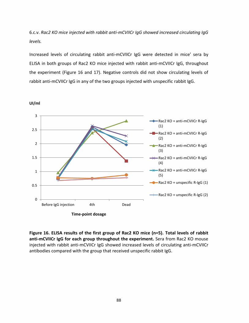
88
6.c.v. Rac2 KO mice injected with rabbit anti-mCVIICr IgG showed increased circulating IgG
levels.
Increased levels of circulating rabbit anti-mCVIICr IgG were detected in mice’ sera by
ELISA in both groups of Rac2 KO mice injected with rabbit anti-mCVIICr IgG, throughout
the experiment (Figure 16 and 17). Negative controls did not show circulating levels of
rabbit anti-mCVIICr IgG in any of the two groups injected with unspecific rabbit IgG.
Figure 16. ELISA results of the first group of Rac2 KO mice (n=5). Total levels of rabbit anti-mCVIICr IgG for each group throughout the experiment. Sera from Rac2 KO mouse injected with rabbit anti-mCVIICr IgG showed increased levels of circulating anti-mCVIICr antibodies compared with the group that received unspecific rabbit IgG.
0
0.5
1
1.5
2
2.5
3
Before IgG injection 4th Dead
Rac2 KO + anti-mCVIICr R-IgG (1)
Rac2 KO + anti-mCVIICr R-IgG (2)
Rac2 KO + anti-mCVIICr R-IgG (3)
Rac2 KO + anti-mCVIICr R-IgG (4)
Rac2 KO + anti-mCVIICr R-IgG (5)
Rac2 KO + unspecific R-IgG (1)
Rac2 KO + unspecific R-IgG (2)
UI/ml
Time-point dosage

89
Figure 17. ELISA results of the second group of Rac2 KO mice (n=5). Total levels of rabbit anti-mCVIICr IgG for each group throughout experiment. Sera from Rac2 KO mouse injected with rabbit anti-mCVIICr IgG showed increased levels of circulating anti-mCVIICr antibodies compared with the group that received unspecific rabbit IgG.
A summary of the all the results obtained for the experiments performed in Rac2 KO mice
are shown in table 11.
0
0.5
1
1.5
2
2.5
3
Before IgG injection 4th Dead
Rac2 KO + anti-mCVIICr R-IgG (6)
Rac2 KO + anti-mCVIICr R-IgG (7)
Rac2 KO + anti-mCVIICr R-IgG (8)
Rac2 KO + anti-mCVIICr R-IgG (9)
Rac2 KO + anti-mCVIICr R-IgG (10)
Rac2 KO + unspecific R-IgG (6)
Rac2 KO + unspecific R-IgG (7)
UI/ml
Time-point dosage

90
Table 11. Summary of clinical, histological, DIF and ELISA results obtained for both groups of Rac2 KO mice compared with wild type group.
Groups (number of mice per group)
Clinical score at death Median, range
Weight changes
Median, range DIF R-IgG
DIF murine C3
ELISA anti-mCVIICr R-IgG
Rac2 KO 1st group + anti-mCVIICr R-IgG (n=5)
0 0 (-1 - 0) Linear at BMZ Linear at
BMZ +
Rac2 KO 2nd group + anti-mCVIICr R-IgG (n=5)
0 1 (0.5 – 2.1) Linear at BMZ Linear at
BMZ +
Rac2 KO 1st group + unspecific IgG (n=4)
0 1 (0 - 2.1) Negative Negative -
Rac2 KO 2nd group + unspecific IgG (n=4)
0 0.6 (-4.4 - 2.3) Negative Negative -
Positive control group (wild-type mice) (n=3)
4 (3 – 4) -3.3 (-3.9-0.4) Linear at BMZ Linear at
BMZ +

91
6.d. Wild type mice pre-treated with NSC23766 developed blisters, erosions and alopecic
patches after the injection of rabbit anti-mCVIICr IgG.
Three groups of wild type mice were pre-treated with NCS23766 (n=3), dexamethasone
(n=3) and dapsone (n=5) before passive transfer of 10 mg of rabbit anti-mCVIICr IgG.
Positive control group (n=3) corresponded to wild-type mice receiving rabbit anti-mCVIICr
IgG without pre-treatment and the negative control group (n=2) corresponded to wild-
type mice receiving unspecific rabbit IgG.
6.d.i. EBA phenotype
Alopecia and erythematous patches, as well as, scaly and crusted patches were seen in
each group receiving rabbit anti-mCVIICr IgG starting at 3rd dose, yet being more evident
at 4th dose for the mice belonging to the pre-treated groups (Figure 18 – 19). At 6th dose
all presented the same clinical score.
6.d.ii. Disease severity was not significantly decreased in any of the pre-treated group
when compared with the positive control group.
BSA involvement at 6th dose was 5.64% (ranging from 5.06 – 11.31) (clinical score=3) in
the group receiving dexamethasone, 6.42% (ranging from 3.79 – 9.58) (clinical score=3) in
the group receiving dapsone, and 8.19% (ranging from 7.90 – 13.70) (clinical score=3) in
the group receiving NCS23766, and 9.88% (clinical score=3) in non-pre-treated mice.
Comparison of the median BSA involved performed at 6th dose did not show any
statistical significance in pre-treated groups when compared with the positive control
(non-pretreated group) (p=.27, p=.18 and p=.83, respectively). Even though, initially there
was a trend of milder disease in the pre-treated groups (meaning that disease appeared
to start later in the pre-treated group than in non-pre-treated group), at dose 6th
significant differences in BSA involvement between groups could not be demonstrated
(p=.08) (Figure 19 and 20).

92
Figure 18. WT mice pre-treated and positive control group (non-pretreated mice) are shown. a) Mouse pre-treated with
dexamethasone showing ear thicknening and erythema. b) Pre-treated mouse with dapsone showing ear thicknening and mild
scaling. c) Pre-treated mouse with NSC23766 showing ear thicknening and mild alopecia on the right face. d) Non-pretreated mice
showing extensive alopecia on the posterior trunk.

93
Figure 19. Comparison of body surface area involved throughout disease development for each group. The median of percentage of body surface area involved throughout disease showed a milder severity among pre-treated mice with dexamethasone (DEXA), dapsone (DAP) or the Rac2 inhibitor NSC23766, compared with the positive control group. Y-axis is the % of body surface area, x-axis is the doses that each mouse received until the end of the experiment. DEX: dexamethasone; PosC: positive control; NegC: negative control
% BSA
Time-point dosage
0
2
4
6
8
10
12
14
16
18
20
1st 2nd 3rd 4th 5th 6th Death
DEX
Dapsone
NSC23766
PosC
NegC

94
Figure 20. Disease severity was not statistically significant among different groups at dose 6th. Box plot showing the median and ranges of body surface area involved by disease for each group. The overlapping ranges show no statistical significant differences among all four groups.

95
6.d.iii. Weight loss was not significantly observed in the positive control group when
compared with the pre-treated groups.
No significant weight loss was observed in positive control group when compared with
each pre-treated groups; dexamethasone group (p=.18), dapsone group (p=.45), or
NSC23766 group (p=.27).
Table 12. Weight changes for each mouse of the pre-treated, positive control and
negative control groups.
Groups Mice Weight changes (gr)
Dexamethasone 1 -1.2 2 -1.4 3 -1.4
Dapsone 1 1.3 2 -0.3 3 -1.7 4 -2.6 5 -2.6
NSC23766 1 0.6 2 -0.1 3 0.2
Positive control 1 -1.2 2 0.3 3 -1.3
Negative control 1 -0.9 2 -0.9

96
6.d.iv. Murine skin biopsies from the different pre-treated groups showed neutrophil-rich
infiltrate and dermal-epidermal separation as was observed in the positive control group.
Skin biopsies of pre-treated mice showed dense infiltrates predominantly of neutrophils
in the dermis, and dermal-epidermal separation. Serohemorrhagic crust formation was
also observed in all different groups. Neutrophil-rich infiltrates were demonstrated in the
group pre-treated with NSC23766, as well as in group pre-treated with dapsone (Figure
21). The same histological features were seen in the positive control group.

97
Figure 21. Murine skin biopsies from each group of pre-treated mice, H&E. a) Dermal-epidermal separation with moderate dermal
infiltrate in a mouse pre-treated with dexamethasone (x20). b) Subepidermal bulla formation and dermal inflammatory infiltrate in a
mouse pre-treated with dapsone. c) Dermal-epidermal separation with moderate dermal infiltrate in a mouse pre-treated with
NSC23766. d) Infiltrate in a pre-treated mouse with NSC23766 showing the presence of neutrophils.

98

99
7. DISCUSSION

100

101
7. DISCUSSION
The passive transfer of 45 or 90 mg of rabbit anti-mCVIICr IgG was capable of inducing the
EBA phenotype in WT mice. Disease severity was demonstrated to be IgG dose-
dependent, when evaluated by either both, BSA involvement or clinical score. Skin
biopsies of lesional skin demonstrated the dermal granulocytic infiltrate associated with
dermal-epidermal separation. Deposition of rabbit IgG and murine C3 was demonstrated
in perilesional skin showing the activation of murine immune response to induce tissue
damage. Clinical scores of our mice were similar to the ones obtained from the group of
Sitaru et al. (43). However, weight loss and levels of specific anti-mCVIICr antibodies were
not statistically different when comparing both groups receiving different amount of
antibodies. For the purpose of our project, and considering that both groups showed
disease development, a 10 mg/day of rabbit anti-mCVIICr IgG was considered enough to
carry out further assays.
Based on the results obtained on the two sets of experiments performed in Rac2 KO mice,
the deficiency of Rac2 protein has been shown to protect against EBA development after
the passive transfer of rabbit anti-mCVIICr IgG. Skin biopsies showed scattered infiltrate
with no dermal-epidermal separation. With the deposition of rabbit IgG and mouse C3 at
the BMZ (demonstrated both by DIF), a first step activation of murine immune response
was demonstrated. Moreover the increased levels of circulating rabbit anti-mCVIICr IgG
throughout the observation time (by ELISA) showed the persistent impregnation of the
mice with rabbit IgG. However, skin blistering, erosions, alopecia, serohemorrhagic crusts
or weight loss did not develop in Rac2 KO mice. Surprisingly, studies of ALI induced in
Rac2 KO mice showed the presence of neutrophils in the interstitium of the lung, yet not
within alveolar spaces, which were mainly observed in sick WT mice (87). Compared to
our results migration of neutrophils in the target tissue were not observed. The degree of
inflammation in ALI studies were further assessed by: 1) the levels of chemokines and
cytokines related with neutrophils recruitment, which were not significantly decreased

102
between WT and Rac2 KO mice; and 2) the levels of MPO were significantly decreased in
Rac2 KO mice, which is what damage the lung tissue along with the secreted
metalloproteinases. In our assays no experiments assessing chemokines and cytokines
involved in neutrophil recruitment were performed, however no neutrophilic infiltrate
was observed.
In contrast, the set of experiments performed with Rac2 inhibitor (NSC23766) did not
affect or prevent the development of EBA after antibody exposure. Skin biopsies showed
abundant neutrophilic infiltrate and dermal-epidermal separation, as was seen in the
positive control group (non-pre-treated mice). However, there were many issues
occurred while performing this set of experiment that will be further explained below.
Yao et al. used the NSC23766 to prevent ALI induced on WT mice (88). Their results
showed significant decrease in inflammatory cells infiltrate, such as neutrophils among
others, as well as decrease in MPO activity (p=.01 and p=.05, respectively). Greater
inhibition of the inflammatory infiltrate was observed at higher doses (3mg/kg) of
NSC23766 with similar results obtained in Dexamethasone group at 1 mg/Kg. Yet, marked
decrease of neutrophils in the infiltrate was also observed at a lower dose (1 mg/kg) of
NSC23766 (88). Regarding to our results, none of the pre-treated mice with NSC23766
had a significantly milder disease compared with the positive control group, and the skin
biopsy showed moderate neutrophilic infiltrate similar to the positive control group.
Understanding the main pathways where Rac2 protein is involved, helps to elucidate and
demonstrates the important role of neutrophils in EBA tissue injury, as well as the non-
pathological effect of the isolated presence of antibodies and/or complement at the
dermal-epidermal BMZ. Actually, recent research in EBA has been focused on neutrophils
blockade, and its effect on chemotaxis and cellular function. This was also noted when
reviewing each of the potential agents that are currently investigated for providing a
benefit to EBA patients (mentioned in Table 4) (11).

103
As explained in the Background section, upon antibody-complement complex formation,
neutrophils become crucial in tissue damage. This can be summarized in three steps: first,
their recruitment to the skin; second, granulocyte activation; and third ROS formation
and release. Some examples of molecules acting in the first step are GM-CSF as well as
CXCL1/2 (orthologue of IL-8 in human). It has already been demonstrated that blockade
of GM-CSF or CXCL1/2 or other molecules that induce the depletion of neutrophils such
as neutrophil-depleting anti-Gr-1 antibody protects against disease development (74, 76,
94). The second step is started by the binding of the antibody-complement complex to
the FcγRIV (orthologue of FcγRIIIA in human) of neutrophils, which induces activation of
the immune response, and thus activation of neutrophils. Kasperkiewicz et al. could
demonstrate that among all subtypes of FcγR, FcγRIV is exclusively required for EBA tissue
injury, when compared with the other activating FcyR (79). Third, the ROS formation is
induced by neutrophils by the limiting enzyme of NADPH oxidase. The best example for
its requirement was the use of neutrophils from patients with CGD, as they lack NADPH
oxidase. The in vitro incubation of those defective neutrophils with human skin and IgG
against CVII failed to induce dermal-epidermal separation (76). Rac2 GTPase is involved in
neutrophil recruitment and ROS release; therefore, by blocking Rac2 GTPase, previous
step 1 and 3 are blocked as well. It is important to mention that EBA research experts are
aware that there are still other non-well recognized cells intervening in tissue injury,
however current assays show that the complete blockade of neutrophils are enough to
avoid disease development, as our results confirmed (95).
Additionally, a current well-accepted EBA treatment is colchicine plus dapsone as first line
therapy for mild and moderate disease (30, 96). Since colchicine inhibits the migration of
neutrophils (by interfering with microtubules), and dapsone supposedly inhibits ROS
formation (which mechanism is not yet clear), their combination might exert a similar
effect as the one obtained from Rac2 deficiency or its blockade. This suggests the
importance of this protein as a therapy target.

104
As shown herein, NSC23766 blocks Rac2 protein. However, this effect is not specific for
Rac2, as it also inhibits Rac1, which is ubiquitously expressed and not limited to immune
cells. Indeed, the main limitation of this study falls into the pharmacological inhibition of
Rac2 protein, which could not demonstrate efficacy in neither avoiding nor inducing a
milder EBA phenotype in mice. Several issues emerged while performing the set of
experiments, including the administered drug dosage (a drug that has been used for many
different conditions, including inflammation and cancer with distinct origins and
behavior) and regimen of administration (ranging from single dosage for inflammatory
conditions, to continuous administration with a pump (subcutaneous) for
myeloproliferative disorders) (88, 91, 92). Furthermore, we suspect that NSC23766 has a
narrow therapeutic window, as in our current study, intraperitoneal doses higher of 10
mg/d showed the sudden death in mice (this effect was not mentioned in the reviews
published), and 7 mg/day could not show efficacy (87, 91, 92). Considering the natural
course of EBA in this study’s experimental animal model, it is possible that a continuous
administration of NSC23766 (i.e. by subcutaneous implantation of a pump on mouse’s
back) may have produced improved outcomes. However, considering a drug with such a
narrow therapeutic window along with the requirement of a pump in order to reach
constant blood levels does not make this agent attractive for further investigation. While
an increased interest in NSC23766 has been extremely related in its capability to inhibit
Rac1 protein, no specific agents solely blocking Rac2 are currently available.
In regards to pre-treated mice with dapsone and dexamethasone, it is suspected that a
longer pre-treatment period was likely required to prevent the development of EBA or to
induce a milder EBA phenotype. Dose of dapsone used in our study was obtained from a
study published regarding an experimental animal model of dermatitis herpetiformis.
Two groups of mice were sensitized with gluten and treated afterwards. The first group
was treated with gluten-free diet and dapsone, and the second group with gluten-free
diet only. Resolution of blisters formation was observed after 1 week in the first group
and after 2 – 3 weeks in the second group (93). It is difficult then to compare our results

105
with this study since our mice were initially healthy and no combined therapy was
performed. Finally, because the inhibitor of the target protein was not successful; this
part of the experiment was withdrawn after all the issues surfaced. Moreover, the benefit
of dexamethasone and dapsone in humans has already been demonstrated in the
literature.
From this point, further research in the development of more specific molecules that can
effectively block Rac2 protein is required. For instance, 1) by developing new molecules
with a higher affinity to GEFs that activate specifically Rac2, or 2) targeting the binding
point between GTP and Rac2 (as occurs in the human Rac2 mutation, causing Rac2
deficiency), or 3) by directly competing with Rac2 protein when assembling to NADPH
oxidase. The latter could be centered on the C-terminal sequence of Rac2 that differs
from Rac1, and which has been exclusively related to the NADPH activation as
demonstrated by the group of Yamauchi et al (84). Finding a specific blocker of this
domain would increase specificity in inhibiting the Rac2 protein.
Additional research on Rac2 protein involvement in EBA pathogenesis would be by
inducing the active animal model of EBA in Rac2 KO mice (by injecting mCVIICR protein
instead of the specific IgG), since Rac2 protein is involved in T-cell development, and a
CD4+ T-cell clone has been shown to participate in EBA development. It was mentioned
already that exclusive Rac2 KO mice does not produce T-cell development abnormalities,
however this has been studied in mice inducing immune response by bacteria products
(such as fMLP a chemotactic peptide of neutrophils released by bacteria) instead of
inducing immune response with self-proteins (loss of tolerance). A group studying the
genetic diversity of RAC2 gene has demonstrated susceptibility to certain autoimmune
disease such as Multiple sclerosis (p=.022) and Chron’s Disease (p=048), yet, the authors
affirmed that these results required of further validation (97). Other observations from
this experimental active animal model could also answer whether Rac2 protein has an

106
additional inhibitory effect in neutrophils similar to GM-CSF, which has shown to affect
antibody production by blocking the B-cell helper neutrophils (74).
The main limitation of this research project is whether these data obtained from mice can
be extrapolated to human EBA. A starting point to answer this question could be
performing in vitro experiments by cryosection models, incubating patient’s anti-CVII IgG
with normal skin and further adding neutrophils lacking Rac2 protein. This experiment
would focus only on the ROS formation induced by NADPH oxidase (since migration of
neutrophils would be the step already passed over). Due to the deficiency of Rac2
protein, ROS would not be released; and dermal-epidermal separation would be
prevented.
To finalize, the pathophysiology of EBA remains only partially understood and requires
further studies to clarify ambiguities. Research is centered to investigate target therapies
that involve different areas of EBA pathogenesis, going from the initial loss of immune
tolerance to the last effector phase of tissue damaging. With the current animal models
available, chances to research new target molecules are high. Moreover, due to the rarity
of the disease in the population, we foresee difficulties in testing new developed
molecules in the human context.

107
8. CONLUSIONS

108

109
8. CONCLUSIONS
EBA is a rare and refractory autoimmune skin disease that requires attention in
specialized health centers in order to achieve a correct and fast diagnosis and a proper
treatment. The chronicity of this disease, as well as its refractoriness to most of the
today’s available treatments, makes EBA patients’ management extremely challenging. All
current therapies are not specific and associated with multiple short and long-term side
effects.
Innate immunity is strongly involved in EBA, with the activation of neutrophils and ROS
formation being essential events in the development of clinical manifestations of EBA. In
the present study we demonstrate that the Rac2 protein, which is involved in both
neutrophil chemotaxis and activation (including ROS synthesis by activation of NADPH
oxidase and release of proteolytic enzymes), is crucial in the pathogenesis of
experimental EBA in animal models. This protein represents a new therapeutic target
(Rac2) for developing new systems to treat patients with EBA and other IgG-mediated
organ-specific autoimmune diseases, where neutrophils play a central role.
We have been unable to demonstrate the efficacy of the pharmacological inhibition of
Rac2 protein (NSC23766) in passively induced EBA in wild-type mice. Possible underlying
mechanisms that may explain this lack of efficacy are discussed. Further efforts are
required to develop effective, more selective and non-toxic drugs for these patients.

110

111
9. REFERENCES

112

113
9. REFERENCES
1. Hashmi S, Marinkovich MP. Molecular organization of the basement membrane
zone. Clin Dermatol. 2011;29(4):398-411.
2. Turcan I, Jonkman MF. Blistering disease: insight from the hemidesmosome and
other components of the dermal-epidermal junction. Cell Tissue Res.
2015;360(3):545-69.
3. Baum S, Sakka N, Artsi O, Trau H, Barzilai A. Diagnosis and classification of
autoimmune blistering diseases. Autoimmun Rev. 2014;13(4-5):482-9.
4. Dainichi T, Koga H, Tsuji T, Ishii N, Ohyama B, Ueda A, et al. From anti-p200
pemphigoid to anti-laminin gamma1 pemphigoid. J Dermatol. 2010;37(3):231-8.
5. Yayli S, Pelivani N, Beltraminelli H, Wirthmuller U, Beleznay Z, Horn M, et al.
Detection of linear IgE deposits in bullous pemphigoid and mucous membrane
pemphigoid: a useful clue for diagnosis. Br J Dermatol. 2011;165(5):1133-7.
6. Hashimoto T, Ishii N, Ohata C, Furumura M. Pathogenesis of epidermolysis bullosa
acquisita, an autoimmune subepidermal bullous disease. J Pathol. 2012;228(1):1-
7.
7. Elliot G. Two cases of epidermolysis bullosa. Journal of Cutaneous Genitourinary
Disease. 1895;13:10.
8. R. K. Ein Beitrog zur Frage der Epidermolysis Bullosa (Hereditary et Aquisita).
These Inaug Rostock Diss. 1904.
9. Kim JH, Kim SC. Epidermolysis bullosa acquisita. J Eur Acad Dermatol Venereol.
2013;27(10):1204-13.
10. Zhu XJ, Niimi Y, Bystryn JC. Epidermolysis bullosa acquisita. Incidence in patients
with basement membrane zone antibodies. Arch Dermatol. 1990;126(2):171-4.
11. Ludwig RJ. Clinical presentation, pathogenesis, diagnosis, and treatment of
epidermolysis bullosa acquisita. ISRN Dermatol. 2013;2013:812029.
12. Gupta R, Woodley DT, Chen M. Epidermolysis bullosa acquisita. Clin Dermatol.
2012;30(1):60-9.

114
13. Lee CW. Prevalences of subacute cutaneous lupus erythematosus and
Epidermolysis bullosa acquisita among Korean/Oriental populations. Dermatology.
1998;197(2):187.
14. Zumelzu C, Le Roux-Villet C, Loiseau P, Busson M, Heller M, Aucouturier F, et al.
Black patients of African descent and HLA-DRB1*15:03 frequency overrepresented
in epidermolysis bullosa acquisita. J Invest Dermatol. 2011;131(12):2386-93.
15. Lee CW, Kim SC, Han H. Distribution of HLA class II alleles in Korean patients with
epidermolysis bullosa acquisita. Dermatology. 1996;193(4):328-9.
16. Callot-Mellot C, Bodemer C, Caux F, Bourgault-Villada I, Fraitag S, Goudie G, et al.
Epidermolysis bullosa acquisita in childhood. Arch Dermatol. 1997;133(9):1122-6.
17. Mayuzumi M, Akiyama M, Nishie W, Ukae S, Abe M, Sawamura D, et al. Childhood
epidermolysis bullosa acquisita with autoantibodies against the noncollagenous 1
and 2 domains of type VII collagen: case report and review of the literature. Br J
Dermatol. 2006;155(5):1048-52.
18. Abrams ML, Smidt A, Benjamin L, Chen M, Woodley D, Mancini AJ. Congenital
epidermolysis bullosa acquisita: vertical transfer of maternal autoantibody from
mother to infant. Arch Dermatol. 2011;147(3):337-41.
19. Kim JH, Kim YH, Kim S, Noh EB, Kim SE, Vorobyev A, et al. Serum levels of anti-type
VII collagen antibodies detected by enzyme-linked immunosorbent assay in
patients with epidermolysis bullosa acquisita are correlated with the severity of
skin lesions. J Eur Acad Dermatol Venereol. 2013;27(2):e224-30.
20. Marzano AV, Cozzani E, Fanoni D, De Pita O, Vassallo C, Berti E, et al. Diagnosis and
disease severity assessment of epidermolysis bullosa acquisita by ELISA for anti-
type VII collagen autoantibodies: an Italian multicentre study. Br J Dermatol.
2013;168(1):80-4.
21. Ishii N, Recke A, Mihai S, Hirose M, Hashimoto T, Zillikens D, et al. Autoantibody-
induced intestinal inflammation and weight loss in experimental epidermolysis
bullosa acquisita. J Pathol. 2011;224(2):234-44.

115
22. Terra JB, Meijer JM, Jonkman MF, Diercks GF. The n- vs. u-serration is a learnable
criterion to differentiate pemphigoid from epidermolysis bullosa acquisita in direct
immunofluorescence serration pattern analysis. Br J Dermatol. 2013;169(1):100-5.
23. Vodegel RM, Jonkman MF, Pas HH, de Jong MC. U-serrated immunodeposition
pattern differentiates type VII collagen targeting bullous diseases from other
subepidermal bullous autoimmune diseases. Br J Dermatol. 2004;151(1):112-8.
24. Komorowski L, Muller R, Vorobyev A, Probst C, Recke A, Jonkman MF, et al.
Sensitive and specific assays for routine serological diagnosis of epidermolysis
bullosa acquisita. J Am Acad Dermatol. 2013;68(3):e89-95.
25. Saleh MA, Ishii K, Kim YJ, Murakami A, Ishii N, Hashimoto T, et al. Development of
NC1 and NC2 domains of type VII collagen ELISA for the diagnosis and analysis of
the time course of epidermolysis bullosa acquisita patients. J Dermatol Sci.
2011;62(3):169-75.
26. De Jong MC, Bruins S, Heeres K, Jonkman MF, Nieboer C, Boorsma DM, et al.
Bullous pemphigoid and epidermolysis bullosa acquisita. Differentiation by
fluorescence overlay antigen mapping. Arch Dermatol. 1996;132(2):151-7.
27. Kazama T, Yamamoto Y, Hashimoto T, Komai A, Ito M. Application of confocal
laser scanning microscopy to differential diagnosis of bullous pemphigoid and
epidermolysis bullosa acquisita. Br J Dermatol. 1998;138(4):593-601.
28. Yaoita H, Briggaman RA, Lawley TJ, Provost TT, Katz SI. Epidermolysis bullosa
acquisita: ultrastructural and immunological studies. J Invest Dermatol.
1981;76(4):288-92.
29. Bozeman PM, Learn DB, Thomas EL. Inhibition of the human leukocyte enzymes
myeloperoxidase and eosinophil peroxidase by dapsone. Biochem Pharmacol.
1992;44(3):553-63.
30. Intong LR, Murrell DF. Management of epidermolysis bullosa acquisita. Dermatol
Clin. 2011;29(4):643-7.
31. Gurcan HM, Ahmed AR. Current concepts in the treatment of epidermolysis
bullosa acquisita. Expert Opin Pharmacother. 2011;12(8):1259-68.
116
32. Arora KP, Sachdeva B, Singh N, Bhattacharya SN. Remission of recalcitrant
epidermolysis bullosa acquisita (EBA) with colchicine monotherapy. J Dermatol.
2005;32(2):114-9.
33. Maize JC, Jr., Cohen JB. Cyclosporine controls epidermolysis bullosa acquisita co-
occurring with acquired factor VIII deficiency. Int J Dermatol. 2005;44(8):692-4.
34. Kowalzick L, Suckow S, Ziegler H, Waldmann T, Ponnighaus JM, Glaser V.
Mycophenolate mofetil in epidermolysis bullosa acquisita. Dermatology.
2003;207(3):332-4.
35. Tran MM, Anhalt GJ, Barrett T, Cohen BA. Childhood IgA-mediated epidermolysis
bullosa acquisita responding to mycophenolate mofetil as a corticosteroid-sparing
agent. J Am Acad Dermatol. 2006;54(4):734-6.
36. Kasperkiewicz M, Muller R, Manz R, Magens M, Hammers CM, Somlai C, et al.
Heat-shock protein 90 inhibition in autoimmunity to type VII collagen: evidence
that nonmalignant plasma cells are not therapeutic targets. Blood.
2011;117(23):6135-42.
37. Alban S, Ludwig RJ, Bendas G, Schon MP, Oostingh GJ, Radeke HH, et al. PS3, a
semisynthetic beta-1,3-glucan sulfate, diminishes contact hypersensitivity
responses through inhibition of L- and P-selectin functions. J Invest Dermatol.
2009;129(5):1192-202.
38. Citro A, Cantarelli E, Maffi P, Nano R, Melzi R, Mercalli A, et al. CXCR1/2 inhibition
enhances pancreatic islet survival after transplantation. J Clin Invest.
2012;122(10):3647-51.
39. Magnusson SE, Andren M, Nilsson KE, Sondermann P, Jacob U, Kleinau S.
Amelioration of collagen-induced arthritis by human recombinant soluble
FcgammaRIIb. Clin Immunol. 2008;127(2):225-33.
40. Chen Q, He F, Kwang J, Chan JK, Chen J. GM-CSF and IL-4 stimulate antibody
responses in humanized mice by promoting T, B, and dendritic cell maturation. J
Immunol. 2012;189(11):5223-9.

117
41. Ishii N, Hamada T, Dainichi T, Karashima T, Nakama T, Yasumoto S, et al.
Epidermolysis bullosa acquisita: what's new? J Dermatol. 2010;37(3):220-30.
42. Sitaru C. Experimental models of epidermolysis bullosa acquisita. Exp Dermatol.
2007;16(6):520-31.
43. Sitaru C, Mihai S, Otto C, Chiriac MT, Hausser I, Dotterweich B, et al. Induction of
dermal-epidermal separation in mice by passive transfer of antibodies specific to
type VII collagen. J Clin Invest. 2005;115(4):870-8.
44. Woodley DT, Chang C, Saadat P, Ram R, Liu Z, Chen M. Evidence that anti-type VII
collagen antibodies are pathogenic and responsible for the clinical, histological,
and immunological features of epidermolysis bullosa acquisita. J Invest Dermatol.
2005;124(5):958-64.
45. Woodley DT, Ram R, Doostan A, Bandyopadhyay P, Huang Y, Remington J, et al.
Induction of epidermolysis bullosa acquisita in mice by passive transfer of
autoantibodies from patients. J Invest Dermatol. 2006;126(6):1323-30.
46. Sitaru C, Chiriac MT, Mihai S, Buning J, Gebert A, Ishiko A, et al. Induction of
complement-fixing autoantibodies against type VII collagen results in
subepidermal blistering in mice. J Immunol. 2006;177(5):3461-8.
47. Sitaru C, Kromminga A, Hashimoto T, Brocker EB, Zillikens D. Autoantibodies to
type VII collagen mediate Fcgamma-dependent neutrophil activation and induce
dermal-epidermal separation in cryosections of human skin. Am J Pathol.
2002;161(1):301-11.
48. Gammon WR, Inman AO, 3rd, Wheeler CE, Jr. Differences in complement-
dependent chemotactic activity generated by bullous pemphigoid and
epidermolysis bullosa acquisita immune complexes: demonstration by leukocytic
attachment and organ culture methods. J Invest Dermatol. 1984;83(1):57-61.
49. Gammon WR, Heise ER, Burke WA, Fine JD, Woodley DT, Briggaman RA. Increased
frequency of HLA-DR2 in patients with autoantibodies to epidermolysis bullosa
acquisita antigen: evidence that the expression of autoimmunity to type VII
collagen is HLA class II allele associated. J Invest Dermatol. 1988;91(3):228-32.

118
50. Woodley DT, Briggaman RA, O'Keefe EJ, Inman AO, Queen LL, Gammon WR.
Identification of the skin basement-membrane autoantigen in epidermolysis
bullosa acquisita. N Engl J Med. 1984;310(16):1007-13.
51. Ludwig RJ, Zillikens D. Pathogenesis of epidermolysis bullosa acquisita. Dermatol
Clin. 2011;29(3):493-501, xi.
52. Sesarman A, Sitaru AG, Olaru F, Zillikens D, Sitaru C. Neonatal Fc receptor
deficiency protects from tissue injury in experimental epidermolysis bullosa
acquisita. J Mol Med (Berl). 2008;86(8):951-9.
53. Christiano AM, Rosenbaum LM, Chung-Honet LC, Parente MG, Woodley DT, Pan
TC, et al. The large non-collagenous domain (NC-1) of type VII collagen is amino-
terminal and chimeric. Homology to cartilage matrix protein, the type III domains
of fibronectin and the A domains of von Willebrand factor. Hum Mol Genet.
1992;1(7):475-81.
54. Gammon WR, Abernethy ML, Padilla KM, Prisayanh PS, Cook ME, Wright J, et al.
Noncollagenous (NC1) domain of collagen VII resembles multidomain adhesion
proteins involved in tissue-specific organization of extracellular matrix. J Invest
Dermatol. 1992;99(6):691-6.
55. Villone D, Fritsch A, Koch M, Bruckner-Tuderman L, Hansen U, Bruckner P.
Supramolecular interactions in the dermo-epidermal junction zone: anchoring
fibril-collagen VII tightly binds to banded collagen fibrils. J Biol Chem.
2008;283(36):24506-13.
56. Chen M, Marinkovich MP, Veis A, Cai X, Rao CN, O'Toole EA, et al. Interactions of
the amino-terminal noncollagenous (NC1) domain of type VII collagen with
extracellular matrix components. A potential role in epidermal-dermal adherence
in human skin. J Biol Chem. 1997;272(23):14516-22.
57. Chen M, Marinkovich MP, Jones JC, O'Toole EA, Li YY, Woodley DT. NC1 domain of
type VII collagen binds to the beta3 chain of laminin 5 via a unique subdomain
within the fibronectin-like repeats. J Invest Dermatol. 1999;112(2):177-83.

119
58. Woodley DT, Burgeson RE, Lunstrum G, Bruckner-Tuderman L, Reese MJ,
Briggaman RA. Epidermolysis bullosa acquisita antigen is the globular carboxyl
terminus of type VII procollagen. J Clin Invest. 1988;81(3):683-7.
59. Lapiere JC, Woodley DT, Parente MG, Iwasaki T, Wynn KC, Christiano AM, et al.
Epitope mapping of type VII collagen. Identification of discrete peptide sequences
recognized by sera from patients with acquired epidermolysis bullosa. J Clin
Invest. 1993;92(4):1831-9.
60. Gammon WR, Murrell DF, Jenison MW, Padilla KM, Prisayanh PS, Jones DA, et al.
Autoantibodies to type VII collagen recognize epitopes in a fibronectin-like region
of the noncollagenous (NC1) domain. J Invest Dermatol. 1993;100(5):618-22.
61. Chen M, Doostan A, Bandyopadhyay P, Remington J, Wang X, Hou Y, et al. The
cartilage matrix protein subdomain of type VII collagen is pathogenic for
epidermolysis bullosa acquisita. Am J Pathol. 2007;170(6):2009-18.
62. Ishii N, Yoshida M, Hisamatsu Y, Ishida-Yamamoto A, Nakane H, Iizuka H, et al.
Epidermolysis bullosa acquisita sera react with distinct epitopes on the NC1 and
NC2 domains of type VII collagen: study using immunoblotting of domain-specific
recombinant proteins and postembedding immunoelectron microscopy. Br J
Dermatol. 2004;150(5):843-51.
63. Ishii N, Yoshida M, Ishida-Yamamoto A, Fritsch A, Elfert S, Bruckner-Tuderman L, et
al. Some epidermolysis bullosa acquisita sera react with epitopes within the triple-
helical collagenous domain as indicated by immunoelectron microscopy. Br J
Dermatol. 2009;160(5):1090-3.
64. Chen M, Keene DR, Costa FK, Tahk SH, Woodley DT. The carboxyl terminus of type
VII collagen mediates antiparallel dimer formation and constitutes a new antigenic
epitope for epidermolysis Bullosa acquisita autoantibodies. J Biol Chem.
2001;276(24):21649-55.
65. Vorobyev A, Ujiie H, Recke A, Buijsrogge JJ, Jonkman MF, Pas HH, et al.
Autoantibodies to Multiple Epitopes on the Non-Collagenous-1 Domain of Type VII
Collagen Induce Blisters. J Invest Dermatol. 2015;135(6):1565-73.

120
66. Csorba K, Chiriac MT, Florea F, Ghinia MG, Licarete E, Rados A, et al. Blister-
inducing antibodies target multiple epitopes on collagen VII in mice. J Cell Mol
Med. 2014;18(9):1727-39.
67. Sitaru AG, Sesarman A, Mihai S, Chiriac MT, Zillikens D, Hultman P, et al. T cells are
required for the production of blister-inducing autoantibodies in experimental
epidermolysis bullosa acquisita. J Immunol. 2010;184(3):1596-603.
68. Muller R, Dahler C, Mobs C, Wenzel E, Eming R, Messer G, et al. T and B cells
target identical regions of the non-collagenous domain 1 of type VII collagen in
epidermolysis bullosa acquisita. Clin Immunol. 2010;135(1):99-107.
69. Tukaj S, Hellberg L, Ueck C, Hansel M, Samavedam U, Zillikens D, et al. Heat shock
protein 90 is required for ex vivo neutrophil-driven autoantibody-induced tissue
damage in experimental epidermolysis bullosa acquisita. Exp Dermatol.
2015;24(6):471-3.
70. Tukaj S, Zillikens D, Kasperkiewicz M. Heat shock protein 90: a pathophysiological
factor and novel treatment target in autoimmune bullous skin diseases. Exp
Dermatol. 2015;24(8):567-71.
71. Woodley DT, Remington J, Chen M. Autoimmunity to type VII collagen:
epidermolysis bullosa acquisita. Clin Rev Allergy Immunol. 2007;33(1-2):78-84.
72. Sesarman A, Mihai S, Chiriac MT, Olaru F, Sitaru AG, Thurman JM, et al. Binding of
avian IgY to type VII collagen does not activate complement and leucocytes and
fails to induce subepidermal blistering in mice. Br J Dermatol. 2008;158(3):463-71.
73. Mihai S, Chiriac MT, Takahashi K, Thurman JM, Holers VM, Zillikens D, et al. The
alternative pathway of complement activation is critical for blister induction in
experimental epidermolysis bullosa acquisita. J Immunol. 2007;178(10):6514-21.
74. Samavedam UK, Iwata H, Muller S, Schulze FS, Recke A, Schmidt E, et al. GM-CSF
modulates autoantibody production and skin blistering in experimental
epidermolysis bullosa acquisita. J Immunol. 2014;192(2):559-71.
75. Simon D, Borradori L, Simon HU. Glucocorticoids in autoimmune bullous diseases:
are neutrophils the key cellular target? J Invest Dermatol. 2013;133(10):2314-5.

121
76. Chiriac MT, Roesler J, Sindrilaru A, Scharffetter-Kochanek K, Zillikens D, Sitaru C.
NADPH oxidase is required for neutrophil-dependent autoantibody-induced tissue
damage. J Pathol. 2007;212(1):56-65.
77. Nimmerjahn F, Ravetch JV. Antibody-mediated modulation of immune responses.
Immunol Rev. 2010;236:265-75.
78. Ambrosius H, Hadge D. Chicken immunoglobulins. Vet Immunol Immunopathol.
1987;17(1-4):57-67.
79. Kasperkiewicz M, Nimmerjahn F, Wende S, Hirose M, Iwata H, Jonkman MF, et al.
Genetic identification and functional validation of FcgammaRIV as key molecule in
autoantibody-induced tissue injury. J Pathol. 2012;228(1):8-19.
80. Filip-Ciubotaru F, Manciuc C, Stoleriu G, Foia L. Nadph Oxidase: Structure and
Activation Mechanisms (Review). Note I. Rev Med Chir Soc Med Nat Iasi.
2016;120(1):29-33.
81. Bedard K, Krause KH. The NOX family of ROS-generating NADPH oxidases:
physiology and pathophysiology. Physiol Rev. 2007;87(1):245-313.
82. Pai SY, Kim C, Williams DA. Rac GTPases in human diseases. Dis Markers.
2010;29(3-4):177-87.
83. Gu Y, Williams DA. RAC2 GTPase deficiency and myeloid cell dysfunction in human
and mouse. J Pediatr Hematol Oncol. 2002;24(9):791-4.
84. Zhang H, Sun C, Glogauer M, Bokoch GM. Human neutrophils coordinate
chemotaxis by differential activation of Rac1 and Rac2. J Immunol.
2009;183(4):2718-28.
85. Pick E. Role of the Rho GTPase Rac in the activation of the phagocyte NADPH
oxidase: outsourcing a key task. Small GTPases. 2014;5:e27952.
86. Yamauchi A, Marchal CC, Molitoris J, Pech N, Knaus U, Towe J, et al. Rac GTPase
isoform-specific regulation of NADPH oxidase and chemotaxis in murine
neutrophils in vivo. Role of the C-terminal polybasic domain. J Biol Chem.
2005;280(2):953-64.

122
87. Dooley JL, Abdel-Latif D, St Laurent CD, Puttagunta L, Befus D, Lacy P. Regulation
of inflammation by Rac2 in immune complex-mediated acute lung injury. Am J
Physiol Lung Cell Mol Physiol. 2009;297(6):L1091-102.
88. Yao HY, Chen L, Xu C, Wang J, Chen J, Xie QM, et al. Inhibition of Rac activity
alleviates lipopolysaccharide-induced acute pulmonary injury in mice. Biochim
Biophys Acta. 2011;1810(7):666-74.
89. Csorba K, Sesarman A, Oswald E, Feldrihan V, Fritsch A, Hashimoto T, et al. Cross-
reactivity of autoantibodies from patients with epidermolysis bullosa acquisita
with murine collagen VII. Cell Mol Life Sci. 2010;67(8):1343-51.
90. Olaru F, Mihai S, Petrescu I, Zillikens D, Sitaru C. Generation and characterization
of monoclonal antibodies against the intracellular domain of hemidesmosomal
type XVII collagen. Hybridoma (Larchmt). 2006;25(3):158-62.
91. Binker MG, Binker-Cosen AA, Gaisano HY, Cosen-Binker LI. Inhibition of Rac1
decreases the severity of pancreatitis and pancreatitis-associated lung injury in
mice. Exp Physiol. 2008;93(10):1091-103.
92. Thomas EK, Cancelas JA, Chae HD, Cox AD, Keller PJ, Perrotti D, et al. Rac
guanosine triphosphatases represent integrating molecular therapeutic targets for
BCR-ABL-induced myeloproliferative disease. Cancer Cell. 2007;12(5):467-78.
93. Marietta E, Black K, Camilleri M, Krause P, Rogers RS, 3rd, David C, et al. A new
model for dermatitis herpetiformis that uses HLA-DQ8 transgenic NOD mice. J Clin
Invest. 2004;114(8):1090-7.
94. Sadeghi H, Lockmann A, Hund AC, Samavedam UK, Pipi E, Vafia K, et al. Caspase-1-
independent IL-1 release mediates blister formation in autoantibody-induced
tissue injury through modulation of endothelial adhesion molecules. J Immunol.
2015;194(8):3656-63.
95. Kasperkiewicz M, Sadik CD, Bieber K, Ibrahim SM, Manz RA, Schmidt E, et al.
Epidermolysis Bullosa Acquisita: From Pathophysiology to Novel Therapeutic
Options. J Invest Dermatol. 2016;136(1):24-33.

123
96. Hughes AP, Callen JP. Epidermolysis bullosa acquisita responsive to dapsone
therapy. J Cutan Med Surg. 2001;5(5):397-9.
97. Sironi M, Guerini FR, Agliardi C, Biasin M, Cagliani R, Fumagalli M, et al. An
evolutionary analysis of RAC2 identifies haplotypes associated with human
autoimmune diseases. Mol Biol Evol. 2011;28(12):3319-29.