affinity-based, biophysical methods to detect and analyze ligand
TRANSCRIPT

Journal of Structural Biology 172 (2010) 142–157
Contents lists available at ScienceDirect
Journal of Structural Biology
journal homepage: www.elsevier .com/ locate/y jsbi
Affinity-based, biophysical methods to detect and analyze ligand binding torecombinant proteins: Matching high information content with high throughput
Geoff A. Holdgate a, Malcolm Anderson a, Fredrik Edfeldt b, Stefan Geschwindner b,*
a Lead Generation Sciences, AstraZeneca R&D Alderley Park, 50F49 Mereside, Alderley Park, United Kingdomb Lead Generation Sciences, AstraZeneca R&D Mölndal, S-43183 Mölndal, Sweden
a r t i c l e i n f o
Article history:Available online 4 July 2010
Keywords:AffinityThermodynamicInteractionFragmentLigandScreening
1047-8477/$ - see front matter � 2010 Elsevier Inc. Adoi:10.1016/j.jsb.2010.06.024
Abbreviations: BACE, b-secretase; DBA, direct binscanning calorimetry; ED, equilibrium dialysis; EDCnopropyl)carbodiimide; ESI, electrospray ionization;tography; HTS, high-throughput screening; IMS, ioninhibition in solution assay; ITC, isothermal titrachromatography; MS, mass spectrometry; MW, mhydroxysuccinimide; NMR, nuclear magnetic resonOWG, optical waveguide grating; SAR, structure activplasmon resonance; TDC, target definition compound
* Corresponding author. Address: AstraZeneca R&Mölndal, Sweden. Fax: +46 31 7763792.
E-mail address: stefan.geschwindner@astrazeneca
a b s t r a c t
Affinity-based technologies have become impactful tools to detect, monitor and characterize molecularinteractions using recombinant target proteins. This can aid the understanding of biological functionby revealing mechanistic details, and even more importantly, enables the identification of new improvedligands that can modulate the biological activity of those targets in a desired fashion. The selection of theappropriate technology is a key step in that process, as each one of the currently available technologiesoffers a characteristic type of biophysical information about the ligand-binding event. Alongside theindisputable advantages of each of those technologies they naturally display diverse restrictions thatare quite frequently related to the target system to be studied but also to the affinity, solubility andmolecular size of the ligands. This paper discusses some of the theoretical and experimental aspects ofthe most common affinity-based methods, what type of information can be gained from each one of thoseapproaches, and what requirements as well as limitations are expected from working with recombinantproteins on those platforms and how those can be optimally addressed.
� 2010 Elsevier Inc. All rights reserved.
1. Introduction
Biophysical methods can have an impact in several valuableareas in early drug discovery. The available technologies haveevolved in recent years such that the reliability, throughput,high-quality and orthogonality of approaches now compromise atool-box of methods essential to modern drug discovery programs.Biophysics can allow rapid and reliable quality control checks onrecombinant target proteins, and the assays in which they areused. This forms an important first step in establishing a suite ofapproaches focused on finding hits and leads.
While high-throughput screening of corporate compound col-lections has been the main approach used within the pharmaceu-tical industry to identify hits and leads, these methods have had
ll rights reserved.
ding assay; DSC, differential, N-ethyl-N0-(3-dimethylami-FAC, frontal affinity chroma-mobility spectrometry; ISA,
tion calorimetry; LC, liquidolecular weight; NHS, N-
ance; RU, resonance units;ity relationship; SPR, surface; TS, thermal shift.D Mölndal, SC263, S-43183
.com (S. Geschwindner).
limited success in identifying novel drug candidates. This fact,along with an increase in the number of biophysical approachesthat can be applied, both to primary and secondary screening, aswell as in lead optimization, has led the pharmaceutical industryto invest heavily in biophysical screening approaches in recentyears. Some biophysical methods have the required throughputto compete directly with traditional biochemical screens such thatthey can be considered as truly primary hit finding assays. Yetmore methods have sufficient throughput to allow focused screen-ing for particular targets or for using selected compound libraries.
Although suitable for characterizing interactions of compoundscovering a wide range of molecular weights, biophysical technolo-gies are most often employed to focus on smaller libraries of low-molecular weight compounds. These so-called fragment-basedlead generation approaches are being used increasingly, alongsideor even instead of traditional high-throughput screening (see Al-bert et al. (2007) for a detailed review about the philosophy andstrategy for fragment-based lead generation within AstraZeneca).The reasons for this are twofold: high throughput biochemical as-says are already established as a route to screen larger compounds,but the probability of finding compounds showing optimal interac-tions is low; and perhaps more importantly, highly sensitive, high-quality biophysical assays are essential in order to detect theinteractions of smaller compounds, due to their often weaker affin-ities. Given that the mean molecular weight of a drug molecule isaround 335 Da, (median around 320 Da) and the mean molecular

G.A. Holdgate et al. / Journal of Structural Biology 172 (2010) 142–157 143
weight of a bioactive compound is 455 Da (median around 450 Da,see Tyrchan et al., 2009), biophysical methods capable of utilizingfragment start points with molecular weights in the range 150–250 Da may be an extremely useful primary screening approach.Identifying lower molecular weight start points may support sev-eral rounds of medicinal chemistry design-make-test cycles, dur-ing which the tendency is usually to increase molecular weight(Smith, 2009). This contrasts with using HTS approaches, whichmay successfully identify larger bioactive compounds, but thenecessity will be to optimize compound properties with little orno change in molecular weight, in order to fit the profile for mar-keted drugs. Of course, this oversimplifies the situation somewhat,as there are many other considerations of compound propertiesthat are important in drug discovery, although it does provide aconvenient backdrop for the increased application of biophysicalmethods coupled to fragment screening.
Thus, biophysical methods are becoming increasingly estab-lished as complementary approaches to traditional hit findingroutes, and are being actively exploited across the industry. Thehope is that these biophysical methods will add an extra dimen-sion to drug discovery by providing an opportunity to create hitsand leads, rather than just finding them from within the corporatecompound collection.
Thus, coupled to the incorporation of these new screeningmethods have been efforts to improve compound libraries for usewith these technologies. These improvements include buildingfragment libraries which can be used as chemical start points, ex-tended fragment libraries exploiting protein–ligand recognitionprinciples, and target-specific focused libraries.
The result is that there are now a large number of method-li-brary combinations which can be employed to monitor ligandbinding in drug discovery.
In order to exploit these developments in technology and li-brary design most effectively, it is necessary to consider the systemand the information required before choosing which approach touse. Important considerations are the availability of the proteinand well-characterized reference compounds, including the ame-nable concentration range, the functionality and the stability. Theavailability of tool compounds should also be investigated, as evenif these are not essential, they may provide routes to additionalvaluable experiments for screening or evaluation. Different bio-physical methods also offer a range of information content, so itis important to determine what information is critical to the stageof the project, and employ the most suitable method to extract thatinformation from the collected data.
It may be necessary or desirable to combine approaches in orderto identify and characterize compounds, to access the informationrequired, in the most resource and time efficient manner. Consid-eration should be given to the most appropriate combinations ofmethods with the appropriate read-outs and level of confidencein order to achieve the desired goals. By combining techniques inthis way, it should be feasible to provide medicinal chemists withdata on the kinetics and thermodynamics of an interaction, whichcan then be interpreted alongside available structural information.This, almost full characterization of a binding event (lacking maystill be the mean structures and in most cases the dynamic ensem-ble populations of one or both of the free interacting partners),should be invaluable in assigning some rules for guiding optimiza-tion of the appropriate parameters to meet the required candidatedrug target profile.
So, the pharmaceutical industry is realizing that front-loadingbiophysical screening, or using it in conjunction with establishedHTS methods can be advantageous, as these methods can provideimportant information early in the drug discovery process aboutthe required routes for lead generation for particular targets, andthe potential success rates of HTS. This knowledge can be useful
in helping to reduce the rate of attrition for valuable targets. Itcan also be useful in providing a more thorough description of pro-tein–ligand interactions allowing attempts to optimize compoundstowards profiles that appear to be favored in marketed drugs, forexample larger negative enthalpies (Freire, 2008) and slower off-rates (Swinney, 2009).
This review highlights some of the available biophysical ap-proaches that can be used to identify hits, provide data and infor-mation on the fundamental properties of the target protein–ligandinteraction, and to give insights into how the thermodynamic andkinetic properties of that interaction may be modified in order toimprove potency during the medicinal chemistry phase of aproject.
2. Thermodynamic methods – ITC
Over the past decade ITC has been established as the gold stan-dard method for directly measuring ligand binding affinity andthermodynamics (for a review see Freyer and Lewis, 2008). Thetechnique often allows the affinity, enthalpy and stoichiometry ofa binding interaction to be measured in a single experiment usu-ally taking under one hour. Recent advances in sensitivity, reduc-tion in cell volume, and automation have allowed the approachto evolve from a technique predominantly used for bespoke com-pound thermodynamic characterization, to one which can now be-gin to be applied in compound screening. The combination ofthermodynamic and structural data has always been powerful inhelping to guide molecular design, but the opportunity to charac-terize increased compound numbers relatively quickly, will seethe use of ITC extended in medicinal chemistry design-make-testcycles.
The ITC experiment involves the monitoring of the heat pro-duced (for an exothermic binding event) or absorbed (for an endo-thermic binding event) during the binding reaction (for acomprehensive protocol see Holdgate, 2010). As the name sug-gests, the experiment occurs at (almost) constant temperaturewith the ligand solution usually titrated from the injection syringeinto the protein solution contained within the calorimeter cell.Modern calorimeters operate via power compensation, wherebythe difference in the variable power, proportional to the bindingheat, applied to the sample cell and the constant power appliedto the thermal reference cell (in order to maintain a zero temper-ature difference between the two cells) is monitored by theinstrument.
During the titration, in which small, typically 2–5 lL aliquots ofthe ligand solution are added, the first injections generate the larg-est heat change as the largest number of moles of protein–ligandcomplex are generated. As the titration progresses through subse-quent injections, the protein becomes increasingly saturated withligand, and the amount of newly generated complex falls (althoughthe total amount of complex increases), resulting in a lower mea-surement of instrumental power. Finally, once all of the proteinbinding sites are occupied by ligand at the end of the titration,no further incremental complexation occurs and no further heatchange is detected. Sometimes significant, non-zero heats follow-ing saturation are observed. These are often attributable to the heatassociated with dilution of the ligand, as this is often larger thanthat associated with protein dilution (see Fig. 1).
Depending upon the binding affinity and the amounts of avail-able reagents, it is often possible to arrange the experimental con-ditions so that a single experiment can provide precise estimates ofthe affinity (Kd), the enthalpy (DH) and the stoichiometry (n) of thebinding interaction. This also allows calculation of the entropy (DS)from the Gibbs–Helmholtz equation:
DG ¼ DH � TDS ¼ RT ln Kd:

Fig. 1. Typical ITC data. Shown is a thermogram for a test compound binding to a24 kDa fragment of the DNA gyrase B subunit. The top panel presents the raw data,whereas the bottom panel presents the integrated data with the binding isotherm.
144 G.A. Holdgate et al. / Journal of Structural Biology 172 (2010) 142–157
Useful concentrations of protein are typically estimated by rear-ranging the relationship 5 6 c 6 100, where the value of c is givenby: c = n � [protein]/Kd.
However, experiments with c values outside of this range canstill yield valuable information. Low c values are often encounteredfor fragments, since they bind only weakly to the target protein.Under usual titration conditions this low c value would yield a flat,featureless binding isotherm, from which it would be difficult toextrapolate values for the binding parameters. However, affinityvalues can often be accessed for these low c value situations as longas the stoichiometry of the interaction is known and the final li-gand concentration ensures almost full saturation of the protein(Turnbull and Daranas, 2003).
High c values, typical of tight-binding compounds, lead to ther-mograms taking the shape of step functions where information onthe DH is not only amenable, but very precise, but little informa-tion about affinity can be determined. In this case, it may be possi-ble to lower the protein concentration, within the confines of themagnitude of DH, in order to bring the c value back on scale. Alter-natively, displacement experiments with lower affinity competingcompounds may allow the affinity of the tight-binder to bemeasured.
ITC experiments within the range of c values above typically re-quire protein concentrations of around 5–10 lM, which can trans-late into 0.1 mg of protein per titration with the newer low volumeinstruments, or up to 1 mg of protein per titration in the olderinstruments with large cell volumes, for a protein Mr of 50,000.These relatively high protein requirements have tended to limitthe application of ITC to a few high interest compounds during a
drug discovery project. However the prevailing difficulty in pre-dicting ligand binding thermodynamics, along with an improvingability to generate large amounts of purified protein and recent ad-vances in instrumentation provide the motivation to study largerlibraries of related compounds, in order to build up more completeunderstanding of the structure–activity relationships. This willfacilitate exploitation of thermodynamic signatures during leadgeneration and optimization.
ITC has a wide application in drug discovery focusing on pro-tein–ligand interactions (Ward and Holdgate, 2001; Weber andSalemme, 2003), ranging from protein quality control applications(where it can allow an evaluation of protein functionality) throughits use in crystallization protocols (allowing demonstration of com-plex formation with test compounds prior to set-up of crystalliza-tion trials), and of course including the use of the thermodynamicdata gathered on compounds or series of interest to inform molec-ular design during lead optimization. ITC also can be applied to di-verse applications including enzyme kinetics, examination ofprotonation effects, nucleic acid recognition and even in certaincircumstances in the determination of association and dissociationrate constants for reversible binding interactions (Bjelic and Jelesa-rov, 2007).
We have viewed ITC for a number of years as part of a tool-boxof techniques which can be employed to evaluate a range of prop-erties associated with assessing the quality of protein preparations.We believe investigating the identity, purity, concentration, func-tionality and stability of proteins is an essential first step in anybiophysical study of protein–ligand interactions. We first usedITC in this way to assess the quality and reproducibility of recom-binant protein preparations for work carried out on the enoyl (acylcarrier protein) reductase (Ward et al., 1999). The ability to assessthe functionality of the protein by observing substrate or tool com-pound binding with the expected affinity, as well as determiningthe functional protein concentration from the stoichiometry valueallows batch to batch variation in different protein batches, ob-tained from different purification protocols, or produced at differ-ent times during the life-span of the project to be assessed. ITC canalso be used to assess the suitability of different storage conditions,by monitoring changes in these parameter values. The identifica-tion and removal of non-functional protein, which may be presentin some purified protein preparations, allows the use of fully func-tional protein which reduces the risk of detecting compounds withartefactual activity.
The high precision of ITC data means that it can be used to as-sess the validity of other assays, which is useful for drug discoveryas the requirement for high throughput and low reagent consump-tion dictates that other, perhaps less rigorous, assay methods haveto be used. This is of particular value in situations where modelsubstrates are employed in the assay, either for convenience orfor availability or cost purposes. For example, peptides are oftenused as alternatives to full length proteins because they are easierto produce and purify. ITC can test the reliability of peptides asmodels of the full length protein, by comparison of the bindingparameters of the full length protein with that of the modelsubstrate.
In a similar manner, recombinant DNA technology is often usedto produce protein samples for structural studies and assays duringdrug discovery. Sometimes these purified protein constructs maylack the full-length wild-type functionality. ITC can be used to ver-ify the validity of using these recombinant proteins, by evaluatingthe SAR of ligand binding. If the same SAR for the authentic and re-combinant protein is observed, then the model protein can be usedwithin the drug discovery project with increased confidence. Wehave used ITC in this way during several drug discovery projects,for example by investigating the binding thermodynamics of ATPand the antibiotic novobiocin to 24 and 43 kDa fragments of the
G.A. Holdgate et al. / Journal of Structural Biology 172 (2010) 142–157 145
antibacterial target DNA gyrase. These fragments of the B subunitof DNA gyrase lack the topoisomerase-linked ATPase activity ofthe intact A2B2 heterotetramer, and so traditional biochemical as-says following catalytic activity were impossible. The similar bind-ing affinities observed for these and other ligands binding to thesetwo recombinant proteins, compared to the wild-type protein al-lowed us to substitute for the full length gyrase tetramer duringthe structural studies (Holdgate et al., 1997).
Similarly, ITC has been used to characterize the binding of li-gands to non-activated kinases. Non-activated kinases typicallyshow little catalytic activity. Again, following test compound bind-ing by steady state enzyme assays would result in assays havinglow signal:noise. Thus biophysical methods, such as ITC, allow li-gand binding to be characterized without having to set-up complexenzyme assays, in order to search for and characterize compoundspreventing kinase activation. It is possible to configure preventionof activation assays, which can identify binders to the non-acti-vated kinase present in the assay. However, because the concentra-tion response relationship for binding to the non-activating kinaseis the same as that for test compound binding to the upstream acti-vating kinase, ITC offers a simpler assay format for measuring affin-ities. Examples from our work include MEK protein kinase andp38a MAP kinase (VanScyoc et al., 2008; Sullivan et al., 2005).
Perhaps the most useful application of ITC is in the characteriza-tion of compounds produced by medicinal chemists during thelead optimization phase of drug discovery. Understanding the ther-modynamics of a molecular interaction is key in drug discovery, asit allows modifications to be made to test compounds in moremeaningful way. Thermodynamic measurements are fundamentalin trying to understand molecular interaction, and in applying thatlearning in the pursuit of compounds, not only with higher affinity,but with the appropriate thermodynamic and kinetic profiles fortheir biological function (Holdgate, 2007). The binding affinity ofa test compound is related to the free energy of the interaction,which is dependent upon the enthalpic and entropic components.The situation is complicated further by other factors such as influ-ence of the solvent water on the binding thermodynamics and thechange in dynamics and conformation of the ligand and the proteinbetween the free and bound states, making these parametersincredibly difficult to predict. ITC allows the quantification of theenthalpic and entropic contributions to ligand binding. BecauseITC is effectively a dual probe technique, allowing measurementof affinity and enthalpy in a single experiment, it can be useful inhighlighting discontinuities in SAR which affinity only techniquesmay miss. It has been suggested that the change in enthalpy partic-ularly can provide a valuable additional tool for the selection ofcompounds in lead identification and for helping to guide leadoptimization. Examples of enthalpic optimization, where the en-thalpy of interaction is increased from early drugs to later com-pounds with advantages in the clinic have been presented for thestatins and the HIV protease inhibitors (Freire, 2008; Ladburyet al., 2010). The approach is theoretically simple: rather thanestablishing SAR on the binding affinity alone to improve the affin-ity of test compounds, more efficient optimization can be achievedif the contributions of enthalpy and entropy are considered andimproved simultaneously (Freire, 2009). The thermodynamics ofthe AstraZeneca statin, Crestor, were evaluated in the context ofboth the structure and kinetics of the complex with the target pro-tein, HMG-CoA Reductase (Holdgate et al., 2003). However, itshould be remembered that although improving enthalpy is a use-ful strategy for medicinal chemists, sometimes interpretingchanges, even relatively small changes in structure can be difficult.In a recent study of pyrazole and azaindoles binding to p38 MAPkinase, using van’t Hoff analysis no discernible relationship be-tween compound IC50 and enthalpy or entropy could be estab-lished (Papalia et al., 2008).
Only by the increased use of methods able to access thermody-namic parameters, such as ITC, and by exploring the relationshipbetween thermodynamics and structure, will we really begin tobe able to increase and exploit our knowledge of molecular inter-action. In order to do this we will need to make use of databasesproviding structural and thermodynamic data. A recently de-scribed database, PDBcal (Li et al., 2008), has been created to pro-vide a single source of structural and calorimetrically derivedthermodynamic data. This database may be useful in developingour understanding of the relationship between structure and ther-modynamics and may provide some impetus for the developmentof improved models to predict binding affinity from computationalapproaches.
3. Thermodynamic methods – thermal shift
The ligand-induced thermodynamic stabilization of proteins isthe biophysical basis of the technique commonly known as ther-mofluor, introduced by 3DP, now part of Johnson & Johnson (Pan-toliano et al., 2001; Cummings et al., 2006). The enhanced stabilityof a protein can be monitored in a variety of ways, utilizing eitherphysical (temperature, pressure) or chemical denaturants (guani-dine hydrochloride or urea). In thermofluor, the stabilization ismanifest as an increase in the thermal stability conferred to theprotein following the ligand-binding event. The assay has a homo-geneous format, without the need for labeling protein or com-pound and can be applied in both 96- and 384-well plateformats. The approach has been demonstrated for a wide rangeof protein classes and has been applicable to both allosteric and ac-tive site binding compounds.
Further benefits of the method are its rapidity, as well as thelack of any requirement for custom assay development or the needfor expensive instrumentation. This allows broad applicability bothin the sense of amenable target proteins, but also in terms ofnon-biophysical laboratory settings (for a general introductionsee Pantoliano et al., 2001). The protein thermal unfolding in thethermofluor approach is monitored indirectly utilizing extrinsicfluorescence, whereas other thermal unfolding approaches makeuse of direct read-outs, such as the change in heat capacity(DSC), optical rotation (CD) or the degree of light-scattering (Star-gazer-384 from Harbinger Biotech).
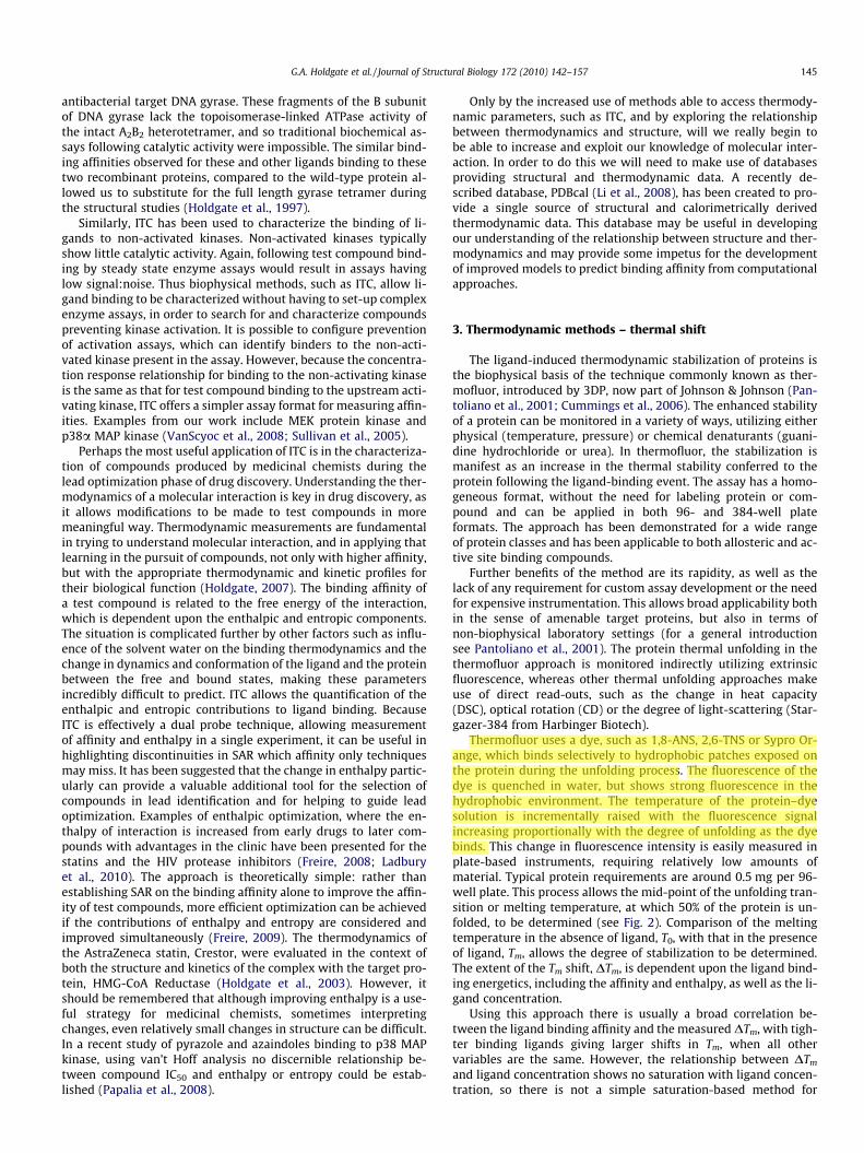
Thermofluor uses a dye, such as 1,8-ANS, 2,6-TNS or Sypro Or-ange, which binds selectively to hydrophobic patches exposed onthe protein during the unfolding process. The fluorescence of thedye is quenched in water, but shows strong fluorescence in thehydrophobic environment. The temperature of the protein–dyesolution is incrementally raised with the fluorescence signalincreasing proportionally with the degree of unfolding as the dyebinds. This change in fluorescence intensity is easily measured inplate-based instruments, requiring relatively low amounts ofmaterial. Typical protein requirements are around 0.5 mg per 96-well plate. This process allows the mid-point of the unfolding tran-sition or melting temperature, at which 50% of the protein is un-folded, to be determined (see Fig. 2). Comparison of the meltingtemperature in the absence of ligand, T0, with that in the presenceof ligand, Tm, allows the degree of stabilization to be determined.The extent of the Tm shift, DTm, is dependent upon the ligand bind-ing energetics, including the affinity and enthalpy, as well as the li-gand concentration.
Using this approach there is usually a broad correlation be-tween the ligand binding affinity and the measured DTm, with tigh-ter binding ligands giving larger shifts in Tm, when all othervariables are the same. However, the relationship between DTm
and ligand concentration shows no saturation with ligand concen-tration, so there is not a simple saturation-based method for
Fig. 2. Typical TS data. Shown is the thermal unfolding transition for BACE-1. Rawdata is displayed by a solid line, with the fitted curve in red.
146 G.A. Holdgate et al. / Journal of Structural Biology 172 (2010) 142–157
determining ligand binding affinities in the thermofluor approach(Matulis et al., 2005). Converting changes in Tm to a binding affinityat the relevant temperature requires knowledge of the ligand bind-ing enthalpy as well as the protein unfolding enthalpy. The exactrelationship between the ligand binding and the Tm shift is givenby:
KL;NðTrelÞ ¼ KL;NðTmÞ exp½ð�DHLðTrelÞ=RÞð1=Trel � 1=TmÞ þ ðDCPL=RÞ� ðlnðTrel=TmÞ þ 1� ðTrel=TmÞÞ�
where KL,N denotes the equilibrium association constant for ligandbinding to the native protein at a relevant temperature (Trel) or atthe Tm; DHL is the enthalpy of ligand binding; R is the gas constant;DCPL is the change in heat capacity for ligand binding.
In principle the thermofluor approach appears to be a simpleand generic approach, but there are instances when the methodis less useful, because of issues such as protein precipitation, or re-duced unfolding of the protein. Both of these situations may lead toa lack of dye binding and hence a reduced thermofluor read-out.There may also be difficulty in interpreting small shifts in Tm.Our experience with thermofluor shows that the standard devia-tion for control assays of target proteins used in drug discoveryprojects can often be around 0.4 �C, and that even in situationswhere the standard deviation is lower (in 96 controls for the kinaseprotein p38, the standard deviation was 0.12 �C, data not shown),the range of measured Tm values can still be reasonably large. Inthe p38 example the measured control Tm values covered a rangediffering by 0.7 �C. This would suggest that confidence in measur-ing a real difference in Tm may only be achieved if the DTm is largerthan around 1–1.5 �C, especially in cases where there may be rela-tively few control wells. This potentially limits the use of the ap-proach for fragments, where the affinity may be low, relative tothe solubility limit of the compound, and hence the expected Tm
shift may not be in this range.This problem may be compounded by the fact that at high li-
gand concentrations, non-specific effects may also occur moreprevalently as well as increasing risk of possible artifacts to anyauto-fluorescence of the compounds at these elevated concentra-tions. Also the method will tend to give positive shifts in Tm, notjust for reversible, noncovalent binders, but also for covalent orirreversible compounds. Thus, by using the technique cautiously,as guide rather than an as an absolute determination of binding
for Tm shifts which are below 1.5 �C, confidence in the interpreta-tion of the data and classification of the compounds may be en-hanced. As with other methods, orthogonal approaches to verifyand characterize binding are recommended.
As well as identifying compounds that stabilize the protein, it isalso feasible to identify compounds which destabilize proteinsthrough non-specific aggregation or other types of promiscuous ef-fects. It is possible for the protein in the presence of these com-pounds to exhibit a negative DTm.
The ability of thermofluor and other thermal unfolding basedmethods to identify compounds binding to target proteins, as wellas their ability to help to identify optimal buffer and additive con-ditions conferring stability for assay and storage has led to wide-spread use in primary (especially fragment) compound libraryscreening, buffer screening for assays and optimization of condi-tions for crystallization and NMR studies.
An obvious use of the thermal unfolding approach is in thescreening of compound libraries for binders and potential inhibi-tors of target proteins. Lo et al. (2004) report the use of the iCyclerinstrument (BioRad Laboratories) for thermal unfolding measure-ments for hit identification versus the pharmaceutical targetBACE-1. Thirteen compounds from four chemical structural classeswere evaluated in thermal unfolding and ITC assays. The resultsillustrate that true hits can be identified based on thermal shiftsunder appropriate conditions. The determined binding affinitieswere shown to be similar to those measured by ITC.
An extension to this screening approach for proteins of knownfunction is to use thermal shift assays in order to search for ligandsthat bind proteins of unknown function. Binding information ob-tained in the thermofluor approach can then be combined withbiochemical, sequence and structural evidence in order to assignputative functions to those orphan proteins. An example of thisapplication of thermofluor has been published for an essential genefrom Streptococcus pneumoniae (Carver et al., 2005). Here, the ther-mofluor method was used to screen 3000 compounds specificallyselected to provide information about speculative biologicalfunctions.
The production and storage of large amounts of suitable qualityprotein is a challenge that must be overcome in order to allow thebiophysical and biochemical studies that make up a drug discoverycampaign to be completed. An approach that has been taken to dothis is to engineer multiple constructs of the target protein in orderto assess which may lead to optimal expression, purification andstability. Testing the stability in parallel of multiple constructscan easily be achieved using thermal unfolding. A recent review(Bommarius et al., 2006) highlights some examples where proteinshave been engineered for increased stability.
A related approach is then to evaluate the most favorable con-ditions in which to produce and store the wild-type, or a chosenprotein construct, which shows behavior indistinguishable interms of ligand-binding characteristics from the wild-type. Thethermofluor approach is well suited to scan buffer conditions, suchas concentration and identity of buffer salts and pH for proteinstorage and assay, since it allows a variety of buffer conditions tobe evaluated in a single run. The approach can also be used to mon-itor the effects of other additives and excipients such as detergents,cofactors, metal ions or in the case or crystallization trials, precip-itants that may be included in the buffer solution. This applicationhas been demonstrated for several proteins of interest to the phar-maceutical industry, and the systematic approach is illustrated ni-cely for Akt-3 and cFMS (Mezzasalma et al., 2007).
Thermofluor has also been used in identification and prioritiza-tion of ligands for cocrystallization trials. The premise is that thosecompounds showing large stability shifts are likely to be tighterbinders and may be better start points for cocrystallization trialsthan those compounds displaying small or no thermal shift. Of

G.A. Holdgate et al. / Journal of Structural Biology 172 (2010) 142–157 147
course, there will be systems where both strong and weak binderswill tend to yield crystals, so again it is suggested that the approachshould be used as a guide rather than as an absolute surrogate forsuccess. The method has been applied to 25 Escherichia coli pro-teins, where the thermofluor results were used to identify stabiliz-ing and destabilizing additives. The results showed a twofoldimprovement in the number of crystallization leads identifiedwhen the stabilizing additives were included (Ericsson et al., 2006).
In a recent extension to the thermofluor method the combina-tion of thermal unfolding with the different fluorescent propertiesof flavin-containing proteins in the folded and unfolded state hasbeen used to explore stability and crystallizability (Forneris et al.,2009). This method exploits the properties of the flavin prostheticgroup present in 2–3% of proteins, and potentially highlights theapplicability of other fluorescent cofactors and prosthetic groupsfor use in this way. In this technique, the flavin cofactor is usedas an intrinsic probe to monitor protein folding and stability, tak-ing advantage of the different fluorescent properties of flavin-con-taining proteins between the folded and denatured state.
Thermofluor can also be used to probe mechanism of action, bymeasuring multiple independent binding events. For example,monitoring the change in Tm occurring under a matrix of substrateand inhibitor concentrations may allow classification of the mech-anism of inhibition. Uncompetitive inhibitors would be expected toshow larger Tm shifts in the presence of substrate (as the effectwould be dependent upon the additivity of the individual bindingfree energies, whereas competitive compounds would tend to-wards the showing the Tm shift of the ligand giving the greatest de-gree of saturation.
We have used a range of fluorescent plate readers with heatingfunctions to access many of the applications described above.Many of the target proteins we have worked with show unfoldingtransitions in the range 40–50 �C. Obviously, for proteins having Tm
values 10–15� lower than this, the thermal unfolding approachprovides relatively little useful information without the compara-tive data on different conditions or different constructs, since pro-teins with melting temperatures only slightly above roomtemperature would be inherently thermodynamically unstable.Thus, we have tended to use the technique in a comparative man-ner mostly to probe pH and additive effects.
4. Optical biosensors – SPR
Optical biosensors typically generate a measureable change insome characteristic property of light that is coupled to the sensorsurface by making use of the evanescent-wave phenomenon. SPRis by far the best-known optical biosensor that makes use of thatphenomenon to enable the real-time measurement of protein–li-gand interactions. During the last two decades SPR biosensor tech-nology has seen a rapid evolution starting with the launch of thefirst commercial Biacore instrument by Pharmacia Biosensor in1990 (Jonsson et al., 1991). This evolution relates not only to theemerging and newly established types of alternative instrumentplatforms (see Rich and Myszka, 2007 for available biosensor plat-forms) but even more to the expanding range of applications: tomention but two, the advances in working with membrane pro-teins (Navratilova et al., 2006; Karlsson and Lofas, 2002) andsmall-molecule work including fragment screening (Danielson,2009).
SPR is a phenomenon that occurs when plane-polarized lighthits a metal film under conditions of total internal reflection. TheSPR angle, which is the angle of the incident light that results inthe lowest intensity of reflected light at a constant wavelength, de-pends mainly on the properties of the metal film and the refractiveindex of the medium that is close to that film. By monitoring alter-
ations in the refractive index, SPR is able to measure changes in themass of dissolved material in the aqueous layer (biosensor surface)close to the metal film, which allows the interaction of proteinswith other molecules or ligands to be monitored in real-time (fora detailed review of the basics of SPR see (Huber and Mueller,2006). This enables the kinetic parameters and equilibrium con-stants for a given system, i.e. on-rate, off-rate and apparent disso-ciation constants, to be determined. If these systems can be studiedwithin a large temperature interval it allows even for the determi-nation of thermodynamic binding parameters in conjunction witha van’t Hoff analysis and can provide a sound alternative to ITCmethods (Papalia et al., 2008).
A critical success factor in conducting SPR experiments is thecareful design and execution of the assay. Key to all SPR assaysemploying a DBA format is the successful tethering of the recombi-nant target protein to the biosensor surface by using different cou-pling strategies without compromising the activity/functionality ofthe protein or the access to the ligand binding pocket(s). The mostcommon approach is random-orientation immobilization via acces-sible primary amines on the protein surface by activating the carbo-xymethylated dextran-matrix with a mixture of EDC and NHS inorder to create NHS-esters that can react with amino-containingmolecules. Besides other available chemistries (OShannessy et al.,1992) this approach works quite well for the majority of soluble re-combinant target proteins and only a small fraction require a differ-ent coupling chemistry or in some cases even more sophisticatedstrategies. These strategies involve quite often the generation ofsuitable protein constructs that permit a more directed and orienta-tion-controlled immobilization process via engineered tags (e.g.Streptavidin affinity tags for the use with Streptavidin-coated Bio-sensors (Li et al., 2006) or fusion proteins (e.g. hAGT fusion proteinsHuber et al., 2004). For work with membrane proteins, and in partic-ular G-protein coupled receptors, a labor-intensive strategy involv-ing an antibody-capturing step with detergent-solubilized receptorcan be required (Navratilova et al., 2006). To make use of a keyadvantage of SPR technology, namely the ability to utilize the samesensor surface during multiple cycles thus consuming very smallamounts of recombinant protein, it is also essential to establish suit-able conditions that allow for the reproducible regeneration of thesensor surface without losing binding activity and functionality ofthe immobilized protein after multiple ligand binding experiments.This becomes even more important in working with ligands or com-pounds that show an unspecific binding component (promiscuousor aggregation-based inhibitors) either to the sensor surface or theprotein, which makes an accurate analysis of subsequent ligandbinding experiments unfeasible.
As most of these central factors can be addressed in one way orthe other by applying different strategies or approaches, there isone remaining but significant aspect that is related to the attain-able sensitivity and thus the dynamic range of the measurementsand is partly out of the control of the scientist as it is dictated bythe system to be studied. The SPR signal intensity is dependenton several factors, namely the MW, the immobilized amount andremaining binding capacity (i.e. functionality) of the protein aswell as the MW, the total concentration [LT] and dissociation bind-ing constant KD of the ligand. Simulations displaying the minimumrequired ratio [LT]/KD for a reliable detection of the binding as afunction of the MW of the ligand and the protein show that the dy-namic range gets smaller with increasing MW of the protein anddecreasing MW of the ligand (Dalvit, 2009). This obvious sensitiv-ity limit presents a real challenge for working with fragments orfragment-like ligands but also with ligands that display low affin-ities combined with low solubility and will thus exclude this typeof studies for larger protein systems.
The flexibility in the SPR assay format allows for the design of atailored assay format and enables some of the key aspects

148 G.A. Holdgate et al. / Journal of Structural Biology 172 (2010) 142–157
described earlier to be tackled. The DBA, where the protein isimmobilized on the sensor surface and compounds are passed overthe surface, is commonly used and has been published for a largenumber of different systems. Alternative assay formats such asthe surface competition assay (SCA) or the ISA are viable alterna-tives to a DBA (Karlsson et al., 2000). In particular the ISA formathelps to deal appropriately with the challenges for setting up andconducting a DBA as well as the limitations in the affinity rangeand analyte masses and thus makes systems amenable for small-molecule work that are usually deemed unfeasible for a DBA. Wehave thus employed a general strategy to assess ligand bindingby making wide-ranging use of the ISA format.
The fundamental difference between the ISA and the DBA for-mat is the tethering of a target definition compound (TDC) to thebiosensor surface which serves as a probe for the binding site, in-stead of immobilizing the recombinant protein and monitoringthe binding directly (see Fig. 3). The interaction with the TDCand the ligand to be investigated occurs simultaneously and dueto this competition it is possible to derive the dissociation bindingconstant KD for this particular ligand (Karlsson, 1994). If the anal-ysis is performed on a biosensor surface with very high bindingcapacity and thus density of the TDC (typically 200–400 pg/mm2)in conjunction with very low flow rates (<20 ll/min) it is feasibleto generate conditions of mass transport limitations. Somethingthat one usually would try to avoid in a DBA as it will obscurethe kinetic analysis helps actually to simplify the analysis of theISA data as the observed binding is solely determined by the masstransfer of the protein to the surface and not anymore by the inter-action kinetics, and as a result the signal becomes concentration-dependent. This situation is typically characterized by a constantinitial binding rate (RU/s) over a certain period of time which al-lows for the precise determination of the free protein concentra-tion as the initial binding rate (RU/s) is directly proportional tothat. Usually, the inhibition studies are carried out by pre-incuba-tion of the recombinant protein with the ligands and subsequentinjection over the TDC-modified sensor surface. The initial bindingrate is used to determine the percentage of free protein in solutionwhich will change by varying the concentration of the competingligand. By plotting the free protein concentration against the loga-rithm of the ligand concentration one can apply sigmoidal dose–re-sponse curve-fit models available in standard software packages todetermine the KD-value. The assay can even be used for a morequalitative affinity ranking of different ligands without the needto determine their affinity, if experiments are conducted at similarligand concentrations.
Fig. 3. Assay flexibility in SPR. Shown is the general principle of a direct bindingassay (A) versus an inhibition in solution assay (B).
An important step in the development of an ISA is the identifi-cation of a suitable ligand to be used as a TDC. This ligand shouldideally cover the entire binding site, should display a rapid associ-ation phase to enable binding studies under conditions of masstransport limitations and ideally a slow dissociation phase so thatthis becomes negligible during the initial association. A high affin-ity (KD < 1 lM) of the TDC is desirable as the affinity dictates theconcentration of the protein to be used in the assay (protein con-centrations are typically in the range of 20–200 nM) in order toachieve an good binding signal (ideally RU/s > 1) and thus deter-mines the overall protein consumption. A good choice of a ligandto be used as a TDC could be a substrate-analog or a commerciallyavailable compound. An absolute prerequisite is the possibility toimmobilize the ligand preferably via primary amines without com-promising the binding to the target protein, which presents a chal-lenge if those functionalities are involved in direct contacts in thebinding pocket. A successful strategy in our hands has been thechemical modification of appropriate ligands by attaching a carbo-hydrate- or ethylenglycol-linker that contain a free primary amineat their end. Great care has to be taken in choosing the correctlength of the linker as it needs to sufficiently protrude out fromthe protein surface to enable efficient binding without facing sterichindrance, but should not be too long to avoid reduction of the freebinding energy owing to the entropic penalty that arises uponbinding of a molecule with increased flexibility and mobility.
There are obvious benefits of the ISA format compared to themore traditional DBA. First of all and most importantly one gainsfull control of the dynamic range of the assay via variation of theprotein concentration as there are no MW-limitations for eitherthe recombinant protein or the ligands to be studied. The interac-tion of the ligand and the protein occurs truly in solution and theassay set-up enables read-out of the free protein concentrationwithout disturbing the equilibrium, thus enabling the determina-tion of exact KD-values. The procedures to establish protocols forthe immobilization as well as the regeneration are much more pre-dictable and usually straightforward, as quite harsh and powerfulconditions for the immobilization of such small organic ligandsas well as the regeneration of the modified sensor surface can beapplied, which are usually not compatible with a DBA using immo-bilized protein. Clear shortcomings are of course that the describedapproach will not allow for the determination of the kinetic param-eters of the protein–ligand interaction and that the consumption ofrecombinant protein will be significantly larger.
We have effectively applied the concept of the ISA as a follow-upfor an NMR-based screening of a fragment library against BACE-1(Geschwindner et al., 2007) but have recently also used a similar ap-proach for direct screening of larger fragment libraries, which eveninvolved the interrogation of fragment pools to increase thethroughput. For the characterization and analysis of fragment bind-ing to BACE, we designed an ISA using an P1 (S)-statine substitutedsubstrate analog (sequence: KTEEISEVN(Sta)DAEF, where Sta is thetransition state mimetic), which was reported to be a nanomolarinhibitor of BACE activity and binds specifically to the active site,as TDC. In order to show that the fragments that have been identi-fied by the NMR-based screening might also act as broad asparticprotease warheads we also configured an ISA using the renin-inhib-itor H-142 (sequence: PHPFHLRVIHK, where R depicts the reducedisostere of the scissile peptide bond between residue Leu10 andVal11 in human angiotensinogen) as TDC and probed the fragmentbinding to the aspartic protease endothiapepsin in a similar fashion.Those substrate analogs have been particularly useful as TDCs, asthey display very high affinities with their respective bindingpartners and could be readily immobilized using amine-couplingwithout any modifications but retained binding activity. For theligand-binding studies we made use of BACE protein that has beenproduced as a C-terminal fusion protein with the Fc part of human

G.A. Holdgate et al. / Journal of Structural Biology 172 (2010) 142–157 149
IgG1, which increases the MW of BACE to >100 kDa and conse-quently would not have been considered as a suitable protein con-struct for a DBA. The NMR screen helped to identify a low-molecularweight isocytosine fragment hit (MW = 153.19 Da) that displayedvery weak affinity (KD = 4.5 mM) but could be easily detected andvalidated in the ISA. By using analogs of the original hits at a singleconcentration an emerging SAR could be determined that correlatedreally well with the NMR results and quickly helped to identify mol-ecules with higher affinity that could subsequently developed intoinhibitors with nanomolar potency and cellular activity (Edwardset al., 2007).
The ISA has been a very efficient tool, not only for the rapidaffinity ranking which guided the selection of suitable fragmenthits, but most importantly for the determination of quantitativeSAR of those very weak inhibitors during the ‘analoging phase’ ofthe fragment screening campaign. As such the ISA helped to effec-tively bridge the existing affinity gap until those fragment-derivedinhibitors became potent enough to be picked up in a conventionalenzymatic assay. By looking again at simulations displaying theminimum required ratio [LT]/KD for a reliable detection of fragmentbinding (Dalvit, 2009) and using both the MW of the untaggedBACE construct (approx. 45 kDa) and the MW of the originalfragment hit we would have needed a concentration of around3–4 mM to reliably detect this fragment in a DBA, a concentrationthat would never be considered for screening purposes. The in-creased dynamic range of the ISA tolerates performing those exper-iments at a much lower ligand concentration as it obviously allowsfor the detection of much weaker interactions compared to a DBA.Consequently the ISA can also be used as an attractive alternativefor the reliable detection of weak fragment binding during a pri-mary fragment screen.
5. Optical biosensors – OWG
As an emerging complement to SPR-technology (and probablyalso owing to the big impact of SPR in opening up the entire fieldof label-free analysis of protein–ligand interactions), alternativebiosensor platforms have been developed that exploit related phys-ical phenomena but offer different throughput and approaches to
Fig. 4. The principles of OWG to measure ligand-binding events as exemplified for the BINan increase in the reflected wavelength both upon immobilization of a protein (2) and t
study those interactions. Evanescent field sensing provided by opti-cal waveguides has recently found a wider acceptance within theaffinity-based community as two technology providers, Corningand SRU Biosystems, have recently developed and successfullylaunched related systems (EPIC and BIND system, respectively) thatallow for increased sample throughput by using a plate-based tech-nology platform.
In optics, electromagnetic evanescent waves are formed whenlight waves that are traveling through a boundary between twomedia of different refractive indices undergo total internal reflec-tion because they strike it at a critical angle of incidence. Evanes-cent wave coupling is usually accomplished by placing two ormore electromagnetic elements such as optical waveguides closetogether, thus enabling propagation of a wave from one elementto the next. In order to perform as a biosensor, some portion ofthose elements needs to be in contact with the liquid test sample.The change in the refractive index at the interface between thesensor surface and the liquid which occurs as a consequence of aligand-binding event will modify the wave coupling and triggersa change in the reflected or transmitted output. This permits accu-rate determination of the alterations in mass at the sensor surface,thus allowing highly sensitive measurements of changes in bindingor adherence in the proximity of the sensor surface (see Fig. 4 andfor a review of waveguide-based biosensors and their principlesplease see Mukundan et al., 2009). Most of the related technologiesoperate with planar optical waveguides, with the EPIC systemmaking use of resonant waveguide grating (Fang et al., 2006) whilethe BIND system applies nanostructured optical grating alsoknown as ‘photonic crystals’ (Cunningham et al., 2004) whichcan be used to conduct either biochemical or even cell-based appli-cations in various standard assay formats such as 96-, 384- and1536-well microtitre plates.
The latter leads to some very interesting and novel approachesin working with GPCRs. In brief, ligand binding to GPCRs leadssubsequently to trafficking of molecules and protein complexes,receptor internalization and translocation. This movement ofproteins within a cell has been termed dynamic mass redistribu-tion and can be followed by applying OWG, thus permitting studiesin cell signaling as well as the screening of compounds against
D system. The increase in mass within the proximity of the biosensor surface causeshe specific binding of a ligand after protein immobilization (3).

150 G.A. Holdgate et al. / Journal of Structural Biology 172 (2010) 142–157
endogenous receptors (Fang et al., 2007). Studies have shown thatthis can be successfully used both for primary screening as well asan orthogonal approach to enable rapid follow-up from other hitfinding sources (Dodgson et al., 2009). This furnishes the interest-ing opportunity to find novel ligands that would not have beenidentified using a classical screening approach, thus enhancingsuccess rates.
For biochemical assays, i.e. assays that use biosensor-coated re-combinant proteins to study protein–ligand interactions, one natu-rally tends to compare OWG with SPR but one needs to be awarethat there are fundamental differences from existing SPR-technol-ogy that can present particular opportunities as well as unique chal-lenges for the application of OWG platforms in those studies. Wehave used both platforms in multiple projects and this puts us in aposition to form an initial judgment about the possibilities as wellas limitations of this approach. The most striking difference, whichis due to the plate-based nature of the device, is the absence of anymicrofluidics and liquid flow. The consequent inability to measureaccurate kinetics (while being able to determine accurate KD-values)in a biochemical assay is somewhat compensated by the possibilityto add several binding components (cofactors, substrates or sub-strate-analogs, competitive and noncompetitive inhibitors, etc.)either simultaneously or sequentially into the same well of the plateand study their interdependency. This possibility to sequentially addor take away different components is a unique advantage that can beoptimally utilized by adopting a similar assay strategy as describedfor SPR, i.e. using an ISA format instead of a DBA format.
But why should one consider using an ISA format on OWG plat-forms? First of all, most of the restrictions described earlier for SPRare also applicable for OWG platforms, and in particular the MW-limitations for either the protein or the ligands to be studied pres-ent a severe sensitivity issue. Secondly, all the sensors should ide-ally be resistant to non-specific binding of the sample, as this willeither mask the specific binding signal or even lead to wrong inter-pretations of ligand binding. All the in-house data that we havecollected so far point towards a much larger degree of unspecificligand binding in OWG platforms as compared to others which isreflected in a significantly increased hit rate in primary screeningdue to a larger fraction of false positive hits. In our opinion, thisis for the most part related to the difference in the protein densityon the sensor surface as compared to SPR. As opposed to Biacoretechnology, which uses a 3-dimensional hydrogel with a heightof approximately 100 nm (Karlsson, 1994) for the covalent attach-ment of proteins, the sensor surfaces of the currently availableOWG biosensors are rather 2-dimensional in nature and thus re-quire a significantly higher immobilization density in order toachieve an attainable ligand binding signal. This high local proteinconcentration can be in the range of up to 100 mg/ml or more andcan lead to non-productive binding of ligands to aggregated orpoorly folded proteins. Using immobilized compounds insteadand making use of the ISA format will enable to appropriately ad-dress those key challenges and preliminary in-house results inapplying that concept in fragment screening are very encouraging.Another important aspect to consider is of course the increasedconsumption of recombinant proteins as compared to SPR, whichcan be several orders of magnitude higher for a DBA using OWGplatforms. In addition there is currently only a very narrow rangeof immobilization surface chemistries available, which needs tobe addressed as it limits the application of this technology for bio-chemical assays.
6. Spectroscopic methods – NMR
Since the introduction of the SAR by NMR method by Fesik andco-workers in 1996 (Shuker et al., 1996), NMR has evolved into an
important tool for ligand screening of expressed proteins. In thepharmaceutical industry setting ligand screening is now a moreimportant application of biomolecular NMR than protein–ligandcomplex structure determination, which is almost exclusively doneby X-ray crystallography. NMR has been and remains a reliablework horse among affinity-based techniques in detecting, monitor-ing and characterizing molecular interactions using recombinanttarget proteins.
NMR binding studies can be conducted over a wide range ofaffinity regimes. However, the major strength of NMR is that bind-ing of ligands with weak affinity, with KD values in the lM to mMrange, can be reliably detected. This defines one of the main nichesfor NMR, namely in fragment screening for which affinities indeedtend to be weak due to the low complexity of the compounds. Mosttarget proteins can be subjected to ligand-binding studies by NMRprovided that sufficient quantities of expressed protein can be ob-tained. NMR assays tend to be simple and robust, and generallygenerate few false negatives or false positives. NMR ligand-bindingstudies have two main applications: in fragment screening and inmode of action studies of compounds coming from, for instance,HTS or medicinal chemistry programs. Both protein-observe 2DNMR and ligand-observe 1D NMR techniques are routinely used.We typically deploy three NMR approaches to detect ligand bind-ing: 2D NMR chemical shift mapping, 1D NMR direct binding and1D NMR reference displacement (see Fig. 5).
Protein-observe 2D NMR chemical shift mapping experimentscan be conducted either using 15N–1H or 13C–1H correlations inHSQC experiments (Shuker et al., 1996; Hajduk et al., 2000). Thisgives a fingerprint of the amide or methyl groups in the protein,respectively. Residue specific changes in the protein are then mon-itored upon addition of ligands. This means that specific bindingsite information can be observed directly in the protein. Residuepeak assignments can be obtained through a series of additional3D NMR experiments, although this is not a strict requirement.With such assignments more advanced analyses of binding modecan be performed. Even in the absence of such assignments resi-dues in the binding site can generally be found, although notexplicitly identified, as was done in the case of prostaglandin Dsynthase (PGDS) described below. With 2D NMR methods screen-ing can therefore be performed without prior knowledge aboutbinding sites.
In addition to being highly information-rich, 2D NMR bindingassays tend to be very robust with low false positive rates. Non-specific binding is generally not an issue since such binders areeither not detected at all or give rise to general non-specific linebroadening. While high affinity (nM) binding is readily detected,quantitative KD measurements are most reliably determined inthe low affinity (lM to mM) and fast ligand exchange regime. 2DNMR offers some technical challenges on the expressed protein,since it requires uniform or amino acid specific 15N or 13C isotopelabeling, or both if residue assignments are to be obtained (for acomprehensive review see Wagner, 2009). In practice this typicallymeans that 2D NMR methods are limited to proteins that can beexpressed in E. coli, although for instance cell-free expression sys-tems have also been used successfully (Kigawa et al., 1995). This isindeed the main blocker to the deployment of 2D NMR for a giventarget. Another disadvantage of 2D NMR is the high demand for ex-pressed protein, typically on the order of 0.5 mg per sample for a30 kDa protein. The size of the protein is also limiting. For routineapplications the size limit is on the order of 30–40 kDa. Uniformdeuteration of the target protein, achieved by expressing the pro-tein in D2O instead of H2O, reduces dipolar relaxation effects andenhance spectral quality. This can roughly double the routineapplicable size range to 60–80 kDa. A rather unique feature of 2DNMR is that a single domain that has been labeled can be studiedas part of a multi-domain complex.

Fig. 5. NMR binding assays. (A) 2D NMR chemical shift mapping experiment showing spectrum of ligand bound form in red on top of spectrum of apo protein in black, withresidue assignments shown in small rectangle. (B) 1D NMR direct binding experiment. Ligand signals are observed directly (top blue spectrum) with reduction of intensityupon binding to protein (middle red spectrum), followed by regained intensity upon displacement by high affinity ligand (bottom green spectrum). (C) 1D NMR referencedisplacement experiment. Different levels of displacement of signal from reference ligand with twelve different ligand additions. The blank (DMSO) addition, far left, and highaffinity ligand addition, second from left, define minimum and maximum displacement, respectively.
G.A. Holdgate et al. / Journal of Structural Biology 172 (2010) 142–157 151
The most common ligand-observe 1D NMR techniques arewaterLOGSY, saturation transfer difference (STD), and T1q and T2filtered experiments (Dalvit et al., 2000; Mayer and Meyer, 1999;Hajduk et al., 1997). Generally 1H detection is used for 1D NMRbut other nuclei such as 19F may also be used (Dalvit et al.,2003). 1D NMR techniques are unique in that the ligand signalsare observed directly, which results in a built-in quality controland solubility measure for the ligands under investigation. Anothermajor advantage is that a robust ligand binding assay can be set upfor most protein targets given that sufficient quantities have beenexpressed. Protein consumption varies with set-up but is on the or-der of 10–50 lg per sample for a typical 30 kDa protein. The pro-tein material can be derived from bacteria, as well as morecomplex sources such as insect and mammalian cells. There is nolimit to protein size. In fact, it is actually advantageous to use lar-ger proteins due to the increased relaxation effects, which in turnresult in magnified ligand binding effects. 1D NMR techniquesare generally not adversely affected by the various tags and fusionproteins used to increase protein expression and solubilization,although non-specific ligand binding interactions may then be-come more pronounced. In the case of BACE described in theSPR-section, expression with the C-terminal fusion with the Fc partof human IgG1, actually meant that screening could be performedwith very low protein concentrations (3 lM).
1D NMR screening can be run either in direct binding modewhere putative binding of each individual ligand is observed di-rectly, or by monitoring the modulation in binding of an establishedreference ligand, sometimes referred to as spy molecule or reporter,upon the addition of ligands of interest. Both approaches rely oncompetition with a ligand that is known to bind to a site of interest,in order to establish that the binding interaction is indeed specificand to a distinct site (Dalvit et al., 2002a,b; Jahnke et al., 2002; Sir-iwardena et al., 2002). The direct binding approach is quite uniqueamong affinity-based methods in that significant binding effectsare observed even at ligand concentrations much below the KD. Thisin turn means the ligand solubility becomes less of an issue, whichcan be of major importance when analyzing the output from, for in-stance, an HTS. In direct binding experiments, the magnitude ofbinding effects is non-linear, which makes it difficult to extract KD
values from such experiments. Simply observing binding effects ina 1D NMR experiments without any sort of competition is not rec-
ommended as binding effects are highly dependent on the natureof the ligand, particularly in terms of lipophilicity. Such an assayset-up would run the risk of simply measuring non-specific interac-tions. Certain expressed proteins are particularly sticky and prone tosuch effects. In addition, the magnitude of the binding effects de-crease when the binding approaches the high affinity (nM) and slowligand exchange regime. This is a major limitation of the 1D NMR di-rect binding approach, and could potentially lead to false negatives.In contrast, reference displacement experiments are not subject tosuch affinity limitations and the accompanying potential for falsenegatives. KD values for competing ligands can be determined read-ily, given that the KD of the reference is known (Dalvit et al., 2002a;Wang, 1995). A drawback of both competition approaches is ofcourse that some prior knowledge of ligands is required. However,even in the absence of known small molecule ligands these experi-ments can be informative. Competing ligands could comprise of, forinstance, interacting protein domains. Cross-wise competition ofnovel identified ligands can also be used. First the binding of one li-gand is observed, a second binding ligand is then added to see ifbinding of the first ligand is perturbed. The reversed experiment isthen performed and if any competition between the two ligands isobserved then the binding is most likely to a distinct site.
Advances such as cryoprobe technology and advanced roboticssystems (Folmer and Fetzer, 2004) have increased sensitivity andthroughput of NMR over the past decade. Thousands of compoundsmay be screened against a given target protein in a relatively shorttime period. Yet even with efficient robotics that prepare samplesfresh and make measurements in a fully automated fashion thethroughput for NMR screening is modest compared to HTS. Screen-ing of mixtures is routinely done and offers way to increasethroughput significantly. In 1D NMR direct binding experimentsmixtures can be screened without subsequent deconvolution, pro-vided that reference spectra for the ligands in the mixture havebeen recorded. Both 2D NMR screening and 1D NMR reference dis-placement screening requires deconvolution to identify putativehits in a mixture. This can be done in several ways, for instanceby the subsequent addition of each component of the mixture untilthe hit has been identified. In general terms when using mixtures,hundreds of compounds can be screened per day, which means thata full NMR screen of 3000–4000 compounds generally takes any-where from a few days to several weeks depending on screening

152 G.A. Holdgate et al. / Journal of Structural Biology 172 (2010) 142–157
mode. Data analysis of either 1D or 2 NMR screening data is straightforward and can be done with standard NMR software. However,semi-automated solutions may be beneficial when dealing withlarge quantities of data. Screening libraries are generally dividedinto general sets and target specific sets based on prior knowledgeabout the protein structure and/or known ligands. A typical NMRscreen will encompass 500–4000 fragments. In order to perform a1D NMR screen typically at least 10 mg of expressed protein is re-quired. Typical conditions are 1–10 lM protein and 30–1000 lM li-gand depending on which experimental method is used, but ligandconcentration may be even higher with the reference displacementmethod. A 2D NMR screen typically requires at least 50 mg of iso-tope labeled protein, sometimes significantly more. Typical condi-tions are 50 lM protein and 400–1000 lM ligand. In some casesit may be possible to reduce protein consumption by recyclingthe protein, provided that the protein is sufficiently stable overtime. NMR screening can be conducted over wide range of bufferconditions. Although many buffer components, as well as DMSO,give rise to NMR signals, these are either not observed in theNMR spectrum, as is the case with 2D NMR, or can be suppressedduring 1D NMR experiments. Many buffer components are alsoavailable in deuterated form, making them NMR-silent.
A rather unique niche for NMR screening is the ability to iden-tify binders to a second adjacent site once binders to a primary sitehave been identified. A major challenge to such experiments is thatthe affinity for a second site tends to be very weak. Furthermore, itmay be difficult to distinguish second site binding from binding tothe primary site. There are NMR methods that cope with both ofthese challenges. Inter-ligand NOE measurements can be used toscreen for binders adjacent to an identified binder (Li et al.,1999). Another approach is to chemically link a spin-label to anidentified binder. Only ligands binding simultaneously to thespin-labeled compound are identified in regular 1D NMR screeningexperiments (Jahnke et al., 2001).
The recent example of prostaglandin D synthase (PGDS) illus-trates how NMR screening can be used to drive a fragment-basedlead generation campaign (Hohwy et al., 2008). The modest sizeof the protein (23 kDa) and the fact that it could be produced fromE. coli routinely yielding up to 75 mg/l, made this target readilyamenable to 2D NMR. This approach was also particularly suitabledue to the lack of high affinity reference compounds at start of thecampaign, and allowed for the exploration of alternative bindingsites to the substrate binding pocket. Chemical sift assignmentswere not obtained but HSQC peaks corresponding to residues inor near the active site could readily be identified when referencecompounds were added. Initially a set of 2500 fragments, 450 froma targeted set and 2000 general fragments, was screened. Com-pounds were screened in mixtures of 12 at 400 lM with 100 lMof 15 N-labeled PGDS, giving a KD cut-off of roughly 500 lM. Theprimary screen was effectively completed in 9 days. This resultedto the identification of 24 primary hits (a 1% hit rate), which weresubjected to KD determination in the NMR assay, with affinities inthe range of KD 50–500 lM. X-ray structure determination of pro-tein–ligand complexes with the hits enabled a hit optimizationprocess whereby increasingly more potent inhibitors from ourcompound collection were identified. Two iterative cycles of ana-log screening were carried out, comprising NMR screening, molec-ular modeling, X-ray crystallography, and in vitro biochemicaltesting for PGDS glutathione-S-transferase activity. Nine high-res-olution PGDS complex structures were determined and 300 hitanalogs were tested in total, resulting in a detailed pharmacophoremodel. This rational drug design procedure culminated in the dis-covery of 24 compounds with an IC50 below 1 lM in the in vitrobiochemical assay. The best inhibitor (IC50 = 21 nM) is one of themost potent inhibitors of PGDS described to date with an excellentligand efficiency of 0.65 kcal mol�1 per heavy atom.
The example of BACE-1 discussed earlier (Geschwindner et al.,2007; Edwards et al., 2007) illustrates another effective applicationof NMR in fragment screening. A set of 2000 general fragmentswere screened using 1D NMR waterLOGSY competition experi-ments. Compounds were screened in mixtures of six at 300 lMeach with 3–5 lM BACE, resulting in a hit rate of 0.5%. One ofthe primary hits, an isocytosine, that gave a KD of 4.5 mM in theSPR assay was investigated further. Initial fragment SAR could begenerated using related analogs available from our compound col-lection. The magnitude of the displacement effects observed in thewaterLOGSY experiments correlated very well with the single-point SPR measurements. This example illustrates nicely how thetwo orthogonal affinity screening methods complemented eachother and increased confidence in the results. Combining multipleaffinity-based methods is indeed a common way to guide the evo-lution and design of fragments, until affinity has improved suffi-ciently such that activity can be measured in conventionalbiochemical and cell-based assays.
7. Spectrometric methods – MS
The application of mass spectrometry to ligand screening hasdeveloped in a number of directions over the past 20 years. Ad-vances in both instrumentation and techniques have broadenedthe scope of MS such that protein–ligand binding affinities, bindingsite information, and even induced conformational shifts can bestudied. The diversity of MS methods encompasses many widelyvarying approaches that cannot be satisfactorily addressed withinthe scope of this review. Our intention is to briefly outline the prin-ciples of MS techniques, focusing attention on the relative meritsand disadvantages in the context of ligand screening and analysis,rather than MS instrumentation or experimental detail. We havemade an attempt to bias the article towards what we subjectivelyperceive to be the more relevant MS strategies offering potentialfor screening, while acknowledging that this categorization is in it-self an area open for discussion and debate.
MS methods for ligand study may be loosely grouped into fourbasic approaches: (1) direct detection/identification of the ligand,(2) detection of the protein:ligand complex, (3) analysis of the li-gand binding site and in the case of an enzymatic assay, ligandsmay be identified indirectly by monitoring substrate or reactionproduct concentration (4).
The first of these four categories itself encompasses a number ofdiscrete techniques that share a common principle, whereas thelatter three approaches are more easily defined.
7.1. Direct detection of ligand
The first and most highly populated group of techniques is char-acterized by MS analysis of the ligand itself. MS analysis of small or-ganic molecules is well-established, routine technology. One clearstrength of MS data in ligand detection is that hits can be identifiedby mass (or by mass fragmentation signature with MS/MS) from amixture of candidate compounds. The novel part of these tech-niques is derived from the initial separation of ligands from non-binding compounds prior to the MS analysis step, and has been tack-led in different ways via size exclusion chromatography using eithercentrifugation or liquid chromatography, ultrafiltration, equilib-rium dialysis, frontal affinity chromatography and ligand quantita-tion. The first four of these approaches are better suited to LC–ESI-MS analysis since LC or ionization parameters can be adjusted topromote protein denaturation thereby releasing bound ligand priorto MS detection. In the case of the latter two techniques, the analysisrelies only on detection of free ligand in solution and is amenable toa wider range of ionization mechanisms.
G.A. Holdgate et al. / Journal of Structural Biology 172 (2010) 142–157 153
For size exclusion based separations, target protein is incubatedwith one or a mixture of potential ligands before chromatographyfacilitated by either centrifugation or an LC pump (Annis et al.,2007; Siegel, 2007). In both cases the chromatographic step rapidlyseparates unbound small molecules from ligands bound to the tar-get protein. The latter are analyzed and identified by LC–ESI-MS.Naturally these two subtly different approaches share a numberof pre-requisites, advantages and drawbacks. A relatively puresource of target protein must be available, since the presence ofsignificant quantities of contaminating proteins will lead to anunnecessarily high level of false positives. The size exclusion ap-proach is amenable to higher throughput screening by virtue ofits ability to deal with incubations with multiple compounds,and this is a parameter which heavily influences the consumptionof target protein per compound tested. The centrifugation-basedseparation can be performed in a 96-column well plate format,the consumable hardware for which is commercially available(e.g. Pierce Zeba 96-well Spin Desalting Plates, Thermo Fisher Sci-entific, Rockford IL USA).
Owing to the simplicity of the read-out, assay development isminimal when compared to more conventional screening methods.Size exclusion chromatography has excellent compatibility withincubation buffer components. Ligand binding affinity estimationscan be made via saturation titration experiments, though in prac-tical terms this becomes difficult when the binding affinity be-comes weaker (Kd P 20 lM; Siegel, 2007). This binding affinityrestriction probably also represents the practical limit for liganddetection, making size exclusion approaches unsuitable for frag-ment screening. However, it has been amply demonstrated thatthese size exclusion/MS approaches can be effective and are estab-lished methods for drug discovery screening (Siegel et al., 1998;Muckenschnabel et al., 2004; Blom et al., 1999; Annis et al., 2004).
Using ultrafiltration the target protein is incubated with poten-tial ligands, typically subsets from combinatorial libraries. Postincubation the sample is concentrated in a centrifugal device fittedwith a semi-permeable membrane. The sample is treated to twosuccessive rounds of dilution and concentration, such that un-bound compound able to pass through the membrane will becomediluted to an insignificant concentration. Bound ligands are re-tained in the protein-containing sample, and are identified byLC–ESI-MS. The implementation of this screening technique hasbeen demonstrated as part of a drug discovery process (Comesset al., 2006; Wendt et al., 2007), and shares similarities with sizeexclusion chromatography in its applicability and capabilities.Again, throughput and protein consumption depend largely onthe number of compounds used per incubation. Ultrafiltration isalso compatible with a wide range of incubation buffer compo-nents, and assay development is straightforward. The limit of li-gand binding affinity detectable by this method is considered tobe �10 lM, and Kd estimations are possible by analyzing filtratesamples in addition to the protein-containing sample (Cloutierand Comess, 2007).
Equilibrium dialysis (ED) entails an alternative use of the semi-permeable membrane, in which compound mixtures are allowedto fully equilibrate between two chambers separated by a mem-brane, one chamber of which contains target protein. The contentsof each pair of chambers are analyzed quantitatively by LC–ESI-MS; bound ligands should be identifiable by a bias in concentrationtowards the protein-containing chamber (Wan and Rehngren,2006). ED has the advantages that it has good compatibility witha wide range of buffer components and requires no modificationsto the protein, simplifying assay development. However, proteinconsumption will be high and throughput relatively low unlesslarge combinatorial compound libraries are used.
Frontal affinity chromatography is a fundamentally differentapproach in which the target protein is immobilized onto a liquid
chromatography column bed. Compounds are injected and de-tected post column by ESI-MS; modulation in retention volumeindicating a degree of binding to the column enables ligands tobe identified (Chan et al., 2003). The introduction of known ligandsinto the system allows competition experiments for the estimationof binding affinity of a second ligand. This technique has beenshown suitable to produce more refined, lower throughput datasuch as ligand ranking (Chan et al., 2007). FAC has been imple-mented as a form of high-throughput screening with the use ofcombinatorial compound libraries (Ng et al., 2005), but is notwidely regarded as a universally applicable screening techniquedue to the unpredictable step of protein immobilization. This stepcan be time-consuming and lead to a relatively high requirementfor target protein, and depending on the form of implementation,the chromatography step is not necessarily compatible with thefull range of buffer components.
The final category of direct ligand quantitation covers a widespectrum of potential assay formats in which the utilization ofMS for the final quantitation seems relatively incidental. It is abroad topic that has been fully discussed in another publication(Höfner et al., 2007), from which this article cites a number of ref-erences and is recommended as a further source of informationregarding all uses of MS in ligand study or screening. A centraltheme among the possible assay formats is to incubate compoundswith target protein, separate the protein (e.g. by precipitation), andmonitor the modulation in free (non-bound) compound concentra-tion by quantitative MS. Alternatively, the separated protein frac-tion may be treated to release bound ligand, and this may bedetected quantitatively by MS. Variations in experimental designinclude competition experiments using known ligands, which pro-vides binding affinity data. The technique does not lend itself to theuse of combinatorial compound libraries which has a negative ef-fect on throughput, and a greater cost in the amount of target pro-tein consumed.
7.2. Detection of protein–ligand complex
A relatively recent enhancement of electrospray ionization (andnanoelectrospray ionization) has been the development of robustsystems that allow folded proteins (or multi-subunit assemblies)to be ionized without disrupting tertiary or quaternary structure(Ashcroft, 2005; Tito et al., 2000). There is evidence to suggest thatalterations to protein structure due to desolvation under vacuumcan be sufficiently subtle that the use of mass spectrometry tostudy intra- and intermolecular interactions is valid (Patrikssonand Marklund, 2007). This technique (sometimes referred to as ‘na-tive mass spectrometry’ (van den Heuvel and Heck, 2004) wasfound capable of identifying and studying noncovalent protein li-gand complexes under near-physiological conditions (Danielet al., 2002; Heck, 2008). The concept of using native MS as meansof screening for ligands is well documented (Hofstadler and San-nes-Lowery, 2007; Vivat Hannah et al., 2010) and has been sug-gested as a viable approach for the more challenging task ofscreening fragment compounds (Erlanson et al., 2004).
Typically, the protein concentration required for native MStends to be in the region of �10 lM. In addition, the practical limitin employing combinatorial libraries has been found to be limited,particularly if weaker ligands are to be identified. When individualincubation volumes are taken into account, all these factors meanthat native MS is not easily employed in high-throughput screen-ing due to the amount of highly purified target protein required(author’s own findings, unpublished data). However, native MS’sability to identify relatively weak interactions (Swayze et al.,2002) together with the relatively small size of fragment com-pound libraries mean that native MS has potential for fragmentcompound screening.

154 G.A. Holdgate et al. / Journal of Structural Biology 172 (2010) 142–157
A future direction for small molecule ligand study by native MSis in the use of ion mobility spectrometry coupled to mass spec-trometry (IMS-MS). This configuration of instrument enables anion’s average cross-sectional area to be estimated along with itsmass. This allows discrete conformations of a given protein to bedifferentiated (Smith et al., 2007), and where a ligand induces aspecific conformational shift, it may be possible to characterizethe ligand binding mode as a result of the conformational shiftidentified.
The tethering concept requires the target protein to have anaccessible cysteine residue on its surface, and ideally, close to aknown site of potential ligand binding (either a naturally occurringcysteine or an artificially introduced mutation). The technique uti-lizes compound libraries in which members all contain a thiolgroup (typically, relatively low-molecular weight compounds oftenreferred to as fragments) and a low concentration of a reducingagent, such that binding of fragments via a disulfide bond is revers-ible. However, if the fragment has some affinity for binding to theprotein in addition to the disulfide bond, this will stabilize the pro-tein-fragment conjugate to allow detection and identification byMS. Fragments can subsequently be elaborated or combined withone another to yield higher affinity ligands (Erlanson et al.,2004). As a consequence of the specific modifications to the targetprotein that may be necessary, and also of the specialized com-pound library required, developing this form of assay requires sig-nificant effort. While not being amenable to high-throughputscreening, tethering is ideally suited to fragment screening (Cancil-la et al., 2008).
7.3. Analysis of the ligand binding site
When a protein is exposed to an environment of D2O solvent,the backbone amide hydrogen atoms tend to exchange to deute-rium at a rate that depends on their individual environments.The presence of a ligand bound to the protein can offer a level ofprotection from the solvent and therefore slow the rate of H/D ex-change for the backbone amides at its binding site. This differencein H/D exchange (between ligand-bound and ligand-free samplesof protein) is detectable by mass spectrometry, and the concepthas been used to design different variations of experiment. Oneof the more prominent is the use of enzymatic digestion post deu-terium exchange to fragment the target protein, and to identify thepeptide(s) whose deuterium exchange mass shift is affected bypre-incubation with ligand (prior to H/D exchange and digestion).This latter technique allows the site of ligand binding to be mappedwith considerable precision (Chalmers et al., 2006). Protein con-sumption is high and experiments tend to be designed with singleligands, making this technique much more suited to ligand charac-
Table 1Summary of important parameters to be considered for ligand screening and analysis for
Technique Throughput Consumption oftarget protein
Covalent modificatags immobilizati
SEC/centrifugation Excellent Intermediate NoneSEC/LC driven Good Low NoneUltrafiltration Good Low NoneEquilibrium dialysis Poor Intermediate to high NoneFrontal affinity
chromatographyPoor Intermediate to high Immobilization
Ligand quantitation Good High NoneNative MS Intermediate Low NoneTethering Intermediate Intermediate Introduction of
surface cysteinesDeuterium exchange/MS Very poor High NoneEnzymatic assay/MS
readoutExcellent Target and screening
strategy dependentVariable
terization than screening. The sizeable topic of H/D exchange isdiscussed much more fully elsewhere (Zhu et al., 2007; Hamuroet al., 2007).
7.4. Enzymatic assay/MS
The principle behind this approach can be readily appreciated.Briefly, an enzymatic process in which a substrate is converted intoa product can be monitored by measuring the concentrations ofsubstrate, product, or both. When screening compounds in anenzymatic assay, these may modulate substrate/product concen-trations and thereby identify potential inhibitory (or effector) li-gands. In this approach the use of MS is merely the means ofquantitatively monitoring those concentrations. It does, however,have the advantage that the use of fluorescent labels, radioactiveisotopes, or other labeling systems are not required for assaydevelopment. This screening technology is established and com-mercially available (Jonas et al., 2009). The potential for high-throughput screening with this technology is clearly good,although the rate of consumption of target protein will dependon whether the screen incubates with individual compounds orcombinatorial libraries. However, both approaches will use cata-lytic amounts of enzyme, which may be advantageous comparedto the other biophysical methods exemplified in this review. Com-patibility with buffer components may not be as flexible as othertechniques, but ligand activity measurements can clearly be madeusing this approach.
Table 1 provides a superficial summary of each the MS ap-proaches in the context of ligand screening/analysis, but is neces-sarily subjective and open to debate. In particular, combinatorialcompound libraries that are commonly applied to these techniquesgreatly affect both the throughput potential and the consumptionof protein. The column designating potential for KD estimationcould also be a contentious subject; some techniques require muchmore effort or consume much greater quantities of protein thanothers to achieve a similar quality of affinity estimation. However,in compiling the table, an attempt was made to reflect the mannerin which the techniques have been implemented in publishedliterature.
8. Summary and future perspectives
The biophysical methods and approaches described here havebecome a widely accepted and well-implemented tool-box that al-low for a detailed interrogation of protein–ligand interaction. If se-lected carefully they can provide a set of highly complementarybut unique data that enlights the protein–ligand interaction frommultiple viewpoints. The value of applying orthogonal methods
the different MS approaches.
tionson
Compatibilitywith cofactors,salts, metals etc.
Ligandidentification
KD estimation Fragment screeningcompatibility
Excellent Yes Intermediate PoorExcellent Yes Intermediate PoorExcellent Yes Intermediate PoorExcellent Yes Good PoorIntermediate Yes Good Poor
Good Yes Good PoorVery poor Partial Good Intermediate/goodGood Yes No Excellent
Good No Poor PoorPoor No Good Poor

G.A. Holdgate et al. / Journal of Structural Biology 172 (2010) 142–157 155
can been easily envisaged via the exclusion of model system inher-ent artifacts, as some of those technologies can provide a moreintegrative response including multiple effects like, e.g. ITC. Theversatility of the available technology platforms is making a criticalcontribution to the full mechanistic understanding of ligand bind-ing by making use of the unique strengths that each of those bio-physical methods provide and will in some cases even serve asan enabling approach in the screening for novel ligands, e.g. infragment-based lead generation.
Furthermore one can not underestimate the high value of someof the described biophysical methods for the quality control of re-combinant proteins that are subsequently used for biophysicalscreening and/or in structural studies. A thorough investigationusing preferably a combination of different biophysical methodswill provide confidence in the reagents and will ensure that spe-cific and relevant results are obtained. Very often the productionof recombinant protein is primarily focusing on increasing theamount of soluble target protein and sometimes lacks the proofof functionality early in that optimization process. As an example,biophysical tools like ITC or Biacore ISA can be used to assess thefunctionality by simultaneously determining the ligand bindingcapability and capacity. A comparison of the stoichiometry ob-tained by ITC with the known number of binding sites allows esti-mation of the proportion of functional protein. If the proteinconcentration that is used in the Biacore ISA is higher than theKD value of the test ligand (meaning it is above the tight bindinglimit), an estimate of functional protein concentration can be ob-tained as well. Additionally, some of these methods can be veryeffectively applied during the optimization of recombinant proteinproduction and/or protein storage conditions. As an example, mea-suring the thermodynamic stability using thermal shift assays isvery suitable for quickly optimizing reagent buffer conditions forlong-term protein storage or for identifying an appropriate proteinconstruct to be moved forward into large-scale production.
The recent technology development as well as method expan-sion, the significant reduction of reagent consumption as well asthe availability of high throughput biophysical platforms havepositioned them very well in the drug discovery process, wherethose biophysical parameters can and will be used to optimallydrive the rational design of novel, improved lead compounds. Animportant aspect in that process is the understanding of the bio-logical relevance of those parameters and how those link to im-proved efficacy and we still have some way to go in order tounleash the full potential of all the data for drug discovery. Thereis the associated risk that the identified binders will not displaythe desired activity, in particular if not great care has been takenin producing a biologically relevant protein construct or if the pro-tein structure is compromised in any way during the binding pro-cess. The use of OWG in cell-applications can serve as a tool thathelps bridging that gap between in vitro and in vivo, as many ofthe systems that are studied by affinity-based methods are inten-tionally artificial and simplistic in nature due to the applicabilityof many orthogonal methods and the clear linkage between actionsand responses. Also other affinity-based technologies are havingtheir play that have not been covered in this review, but it isimperative to make a careful technology choice based on the typeof information that is required as well as the limitations that areset by the system to be studied. The use of some of those technol-ogies can be severely constrained to a small number of high-valuemeasurements so increasing the throughput and minimizing theprotein consumption have become a continuous trend within theindustry. For example recent efforts in thermodynamic approachesusing enthalpy arrays are holding some promise to apply the con-cept of thermodynamic profiling onto a wider range of biomolecu-lar interactions and systems (Recht et al., 2008). Also some of thekey limitations of optical methods that relate to the requirements
for immobilization as well as the sensitivity limit can be hopefullyaddressed in the near future by techniques that allow for the exactmeasurement of refractive index changes in free-solution irrespec-tive of the size of the protein–ligand complex. Back-scatteringinterferometry can display such an opportunity as it is capable toprovide a label-free, homogenous and mass-independent detectionin free-solution with much lower requirements on sample quantityand purity (Bornhop et al., 2007). This could open up for biophys-ical studies on systems that are currently not amenable for smallmolecule ligand-binding studies using currently implementedaffinity-based technologies. So the future looks bright for the appli-cation of affinity-based technologies in drug discovery as well asrecombinant protein quality control and it is expected that the im-pact and scope of those methodologies will significantly grow overthe next couple of years.
Acknowledgments
The authors did not receive any financial contributions or writ-ing assistance for the production of this manuscript and declare nocompeting interests in the writing of this review. We would like tothank Tomas Lundqvist and Alex Breeze for critical feedback on thecontent and form of this manuscript and all the researchers whosework has been well documented and published so we could get in-spired to conduct our studies as well as writing this manuscript.We would further like to thank SRU Biosystems for the provisionof Fig. 4.
References
Albert, J.S., Blomberg, N., Breeze, A.L., Brown, A.J., Burrows, J.N., Edwards, P.D., et al.,2007. An integrated approach to fragment-based lead generation: philosophy,strategy and case studies from AstraZeneca’s drug discovery programmes. Curr.Top. Med. Chem. 7, 1600–1629.
Annis, D.A., Athanasopoulos, J., Curran, P.J., Felsch, J.S., Kalghatgi, K., Lee, W.H., et al.,2004. An affinity selection–mass spectrometry method for the identification ofsmall molecule ligands from self-encoded combinatorial libraries: discovery ofa novel antagonist of E. coli dihydrofolate reductase. Int. J. Mass Spectrom. 238,77–83.
Annis, A., Chuang, C., Nazef, N., 2007. ALIS: an affinity selection-mass spectrometrysystem for the discovery and characterization of protein–ligand interactions. In:Wanner, K.T., Höfner, G. (Eds.), Mass Spectrometry in Medicinal Chemistry, firsted. Wiley-VCH Verlag GmbH & Co KGaA, pp. 121–156.
Ashcroft, A.E., 2005. Recent developments in electrospray ionisation massspectrometry: noncovalently bound protein complexes. Nat. Prod. Rep. 22,452–464.
Bjelic, S., Jelesarov, I., 2007. A survey of the year 2007 literature on applications ofisothermal titration calorimetry [Review]. J. Mol. Recognit. 21, 289–311.
Blom, K.F., Larsen, B.S., McEwen, C.N., 1999. Determining affinity-selected ligandsand estimating binding affinities by online size exclusion chromatography/liquid chromatography–mass spectrometry. J. Comb. Chem. 1, 82–90.
Bommarius, A.S., Broering, J.M., Chaparro-Riggers, J.F., Polizzi, K.M., 2006. High-throughput screening for enhanced protein stability. Curr. Opin. Biotechnol. 17,606–610.
Bornhop, D.J., Latham, J.C., Kussrow, A., Markov, D.A., Jones, R.D., Sorensen, H.S.,2007. Free-solution, label-free molecular interactions studied by back-scattering interferometry. Science 317, 1732–1736.
Cancilla, M.T., He, M.M., Viswanathan, N., Simmons, R.L., Taylor, M., Fung, A.D., et al.,2008. Discovery of an Aurora kinase inhibitor through site-specific dynamiccombinatorial chemistry. Bioorg. Med. Chem. Lett. 18, 3978–3981.
Carver, T.E., Bordeau, B., Cummings, M.D., Petrella, E.C., Pucci, M.J., Zawadzke, L.E.,et al., 2005. Decrypting the biochemical function of an essential gene fromStreptococcus pneumoniae using ThermoFluor (R) technology. J. Biol. Chem. 280,11704–11712.
Chalmers, M.J., Busby, S.A., Pascal, B.D., He, Y., Hendrickson, C.L., Marshall, A.G.,et al., 2006. Probing protein ligand interactions by automated hydrogen/deuterium exchange mass spectrometry. Anal. Chem. 78, 1005–1014.
Chan, N.W., Lewis, D.F., Rosner, P.J., Kelly, M.A., Schriemer, D.C., 2003. Frontalaffinity chromatography–mass spectrometry assay technology for multiplestages of drug discovery: applications of a chromatographic biosensor. Anal.Biochem. 319, 1–12.
Chan, N., Lewis, D., Kelly, M., Ng, E.S.M., Schriemer, D.C., 2007. Frontal affinitychromatography–mass spectrometry for ligand discovery and characterization.In: Wanner, K.T., Höfner, G. (Eds.), Mass Spectrometry in Medicinal Chemistry,first ed. Wiley-VCH Verlag GmbH & Co KGaA, pp. 217–246.
Cloutier, T.E., Comess, K.M., 2007. Library screening using ultrafiltration and massspectrometry. In: Wanner, K.T., Höfner, G. (Eds.), Mass Spectrometry in

156 G.A. Holdgate et al. / Journal of Structural Biology 172 (2010) 142–157
Medicinal Chemistry, first ed. Wiley-VCH Verlag GmbH & Co KGaA, pp. 157–183.
Comess, K.M., Schurdak, M.E., Voorbach, M.J., Coen, M., Trumbull, J.D., Yang, H.,et al., 2006. An ultraefficient affinity-based high-throughout screening process:application to bacterial cell wall biosynthesis enzyme MurF. J. Biomol. Screen.11, 743–754.
Cummings, M.D., Farnum, M.A., Nelen, M.I., 2006. Universal screening methods andapplications of ThermoFluor (R). J. Biomol. Screen. 11, 854–863.
Cunningham, B.T., Li, P., Schulz, S., Lin, B., Baird, C., Gerstenmaier, J., et al., 2004.Label-free assays on the BIND system. J. Biomol. Screen. 9, 481–490.
Dalvit, C., 2009. NMR methods in fragment screening: theory and a comparisonwith other biophysical techniques. Drug Discov. Today 14, 1051–1057.
Dalvit, C., Pevarello, P., Tato, M., Veronesi, M., Vulpetti, A., Sundstrom, M., 2000.Identification of compounds with binding affinity to proteins via magnetizationtransfer from bulk water. J. Biomol. NMR 18, 65–68.
Dalvit, C., Flocco, M., Knapp, S., Mostardini, M., Perego, R., Stockman, B.J., et al.,2002a. High-throughput NMR-based screening with competition bindingexperiments. J. Am. Chem. Soc. 124, 7702–7709.
Dalvit, C., Fasolini, M., Flocco, M., Knapp, S., Pevarello, P., Veronesi, M., 2002b. NMR-Based screening with competition water-ligand observed via gradientspectroscopy experiments: detection of high-affinity ligands. J. Med. Chem.45, 2610–2614.
Dalvit, C., Fagerness, P.E., Hadden, D.T., Sarver, R.W., Stockman, B.J., 2003. Fluorine-NMR experiments for high-throughput screening: theoretical aspects, practicalconsiderations, and range of applicability. J. Am. Chem. Soc. 125, 7696–7703.
Daniel, J.M., Friess, S.D., Rajagopalan, S., Wendt, S., Zenobi, R., 2002. Quantitativedetermination of noncovalent binding interactions using soft ionization massspectrometry. Int. J. Mass Spectrom. 216, 1–27.
Danielson, U.H., 2009. Fragment library screening and lead characterization usingSPR biosensors. Curr. Top. Med. Chem. 9, 1725–1735.
Dodgson, K., Gedge, L., Murray, D.C., Coldwell, M., 2009. A 100 K well screen for amuscarinic receptor using the Epic label-free system–a reflection on thebenefits of the label-free approach to screening seven-transmembranereceptors. J. Recept. Signal Transduct. Res. 29, 163–172.
Edwards, P.D., Albert, J.S., Sylvester, M., Aharony, D., Andisik, D., Callaghan, O., et al.,2007. Application of fragment-based lead generation to the discovery of novel,cyclic amidine beta-secretase inhibitors with nanomolar potency, cellularactivity, and high ligand efficiency. J. Med. Chem. 50, 5912–5925.
Ericsson, U.B., Hallberg, B.M., Detitta, G.T., Dekker, N., Nordlund, P., 2006.Thermofluor-based high-throughput stability optimization of proteins forstructural studies. Anal. Biochem. 357, 289–298.
Erlanson, D.A., McDowell, R.S., O’Brien, T., 2004. Fragment-based drug discovery. J.Med. Chem. 47, 3463–3482.
Erlanson, D.A., Wells, J.A., Braisted, A.C., 2004. Tethering: fragment-based drugdiscovery. Annu. Rev. Biophys. Biomol. Struct. 33, 199–223.
Fang, Y., Ferrie, A.M., Fontaine, N.H., Mauro, J., Balakrishnan, J., 2006. Resonantwaveguide grating biosensor for living cell sensing. Biophys. J. 91, 1925–1940.
Fang, Y., Li, G., Ferrie, A.M., 2007. Non-invasive optical biosensor for assayingendogenous G protein-coupled receptors in adherent cells. J. Pharmacol.Toxicol. Methods 55, 314–322.
Folmer, R.H., Fetzer, J., 2004. Automated NMR screening in drug development.BIOspektrum 10, 687–688.
Forneris, F., Orru, R., Bonivento, D., Chiarelli, L.R., Mattevi, A., 2009. ThermoFAD, aThermofluor-adapted flavin ad hoc detection system for protein folding andligand binding. FEBS J. 276, 2833–2840.
Freire, E., 2008. Do enthalpy and entropy distinguish first in class from best in class?Drug Discov. Today 13, 869–874.
Freire, E., 2009. A thermodynamic approach to the affinity optimization of drugcandidates. Chem. Biol. Drug Des. 74, 468–472.
Freyer, M.W., Lewis, E.A., 2008. Isothermal titration calorimetry: experimentaldesign, data analysis, and probing macromolecule/ligand binding and kineticinteractions. Methods Cell Biol. 84, 79–113.
Geschwindner, S., Olsson, L.L., Albert, J.S., Deinum, J., Edwards, P.D., de Beer, T., et al.,2007. Discovery of a novel warhead against beta-secretase through fragment-based lead generation. J. Med. Chem. 50, 5903–5911.
Hajduk, P.J., Olejniczak, E.T., Fesik, S.W., 1997. One-dimensional relaxation- anddiffusion-edited NMR methods for screening compounds that bind tomacromolecules. J. Am. Chem. Soc. 119, 12257–12261.
Hajduk, P.J., Augeri, D.J., Mack, J., Mendoza, R., Yang, J., Betz, S.F., et al., 2000. NMR-based screening of proteins containing 13C-labeled methyl groups. J. Am. Chem.Soc. 122, 7898–7904.
Hamuro, Y., Coales, S.J., Woods Jr., V., 2007. Protein-targeting drug discovery guidedby hydrogen/deuterium exchange mass spectrometry (DXMS). In: Wanner, K.T.,Höfner, G. (Eds.), Mass Spectrometry in Medicinal Chemistry, first ed. Wiley-VCH Verlag GmbH & Co KGaA, pp. 377–398.
Heck, A.J., 2008. Native mass spectrometry: a bridge between interatomics andstructural biology. Nat. Methods 5, 927–933.
Höfner, G., Zepperitz, C., Wanner, K.T., 2007. MS binding assays – an alternative toradioligand binding. In: Wanner, K.T., Höfner, G. (Eds.), Mass Spectrometry inMedicinal Chemistry, first ed. Wiley-VCH Verlag GmbH & Co KGaA, pp. 247–283.
Hofstadler, S.A., Sannes-Lowery, K.A., 2007. Interrogation of noncovalent complexesby ESI-MS: a powerful platform for high throughput drug discovery. In:Wanner, K.T., Höfner, G. (Eds.), Mass Spectrometry in Medicinal Chemistry, firsted. Wiley-VCH Verlag GmbH & Co KGaA, pp. 321–338.
Hohwy, M., Spadola, L., Lundquist, B., Hawtin, P., Dahmen, J., Groth-Clausen, I., et al.,2008. Novel prostaglandin D synthase inhibitors generated by fragment-baseddrug design. J. Med. Chem. 51, 2178–2186.
Holdgate, G.A., 2007. Thermodynamics of binding interactions in the rational drugdesign process. Exp. Opin. Drug Discov. 2, 1103–1114.
Holdgate, G.A., 2010. Isothermal titration calorimetry and differential scanningcalorimetry. In: Roque, A.C.A. (Ed.), Ligand–macromolecular Interactions inDrug Discovery. Human Press Inc., Totowa, NJ.
Holdgate, G.A., Tunnicliffe, A., Ward, W.H., Weston, S.A., Rosenbrock, G., Barth, P.T.,et al., 1997. The entropic penalty of ordered water accounts for weaker bindingof the antibiotic novobiocin to a resistant mutant of DNA gyrase: athermodynamic and crystallographic study. Biochemistry 36, 9663–9673.
Holdgate, G.A., Ward, W.H., McTaggart, F., 2003. Molecular mechanism forinhibition of 3-hydroxy-3-methylglutaryl CoA (HMG-CoA) reductase byrosuvastatin. Biochem. Soc. Trans. 31, 528–531.
Huber, W., Mueller, F., 2006. Biomolecular interaction analysis in drug discoveryusing surface plasmon resonance technology. Curr. Pharm. Des. 12, 3999–4021.
Huber, W., Perspicace, S., Kohler, J., Muller, F., Schlatter, D., 2004. SPR-basedinteraction studies with small molecular weight ligands using hAGT fusionproteins. Anal. Biochem. 333, 280–288.
Jahnke, W., Rudisser, S., Zurini, M., 2001. Spin label enhanced NMR screening. J. Am.Chem. Soc. 123, 3149–3150.
Jahnke, W., Floersheim, P., Ostermeier, C., Zhang, X., Hemmig, R., Hurth, K., et al.,2002. NMR reporter screening for the detection of high-affinity ligands. Angew.Chem. Int. Ed. Engl. 41, 3420–3423.
Jonas, M., LaMarr, W.A., Ozbal, C., 2009. Mass spectrometry in high-throughputscreening: a case study on acetyl-coenzyme a carboxylase using RapidFire–mass spectrometry (RF–MS). Comb. Chem. High Throughput Screen. 12, 752–759.
Jonsson, U., Fagerstam, L., Ivarsson, B., Johnsson, B., Karlsson, R., Lundh, K., et al.,1991. Real-time biospecific interaction analysis using surface plasmonresonance and a sensor chip technology. BioTechniques 11, 620–627.
Karlsson, R., 1994. Real-time competitive kinetic analysis of interactions betweenlow-molecular-weight ligands in solution and surface-immobilized receptors.Anal. Biochem. 221, 142–151.
Karlsson, O.P., Lofas, S., 2002. Flow-mediated on-surface reconstitution of G-proteincoupled receptors for applications in surface plasmon resonance biosensors.Anal. Biochem. 300, 132–138.
Karlsson, R., Kullman-Magnusson, M., Hamalainen, M.D., Remaeus, A., Andersson,K., Borg, P., et al., 2000. Biosensor analysis of drug-target interactions: directand competitive binding assays for investigation of interactions betweenthrombin and thrombin inhibitors. Anal. Biochem. 278, 1–13.
Kigawa, T., Muto, Y., Yokoyama, S., 1995. Cell-free synthesis and amino acid-selective stable isotope labeling of proteins for NMR analysis. J. Biomol. NMR 6,129–134.
Ladbury, J.E., Klebe, G., Freire, E., 2010. Adding calorimetric data to decision makingin lead discovery: a hot tip. Nat. Rev. Drug Discov. 9, 23–27.
Li, D., DeRose, E.F., London, R.E., 1999. The inter-ligand Overhauser effect: apowerful new NMR approach for mapping structural relationships ofmacromolecular ligands. J. Biomol. NMR 15, 71–76.
Li, Y.J., Bi, L.J., Zhang, X.E., Zhou, Y.F., Zhang, J.B., Chen, Y.Y., et al., 2006. Reversibleimmobilization of proteins with streptavidin affinity tags on a surface plasmonresonance biosensor chip. Anal. Bioanal. Chem. 386, 1321–1326.
Li, L., Dantzer, J.J., Nowacki, J., O’Callaghan, B.J., Meroueh, S.O., 2008. PDBcal: acomprehensive dataset for receptor-ligand interactions with three-dimensionalstructures and binding thermodynamics from isothermal titration calorimetry.Chem. Biol. Drug Des. 71, 529–532.
Lo, M.C., Aulabaugh, A., Jin, G., Cowling, R., Bard, J., Malamas, M., et al., 2004.Evaluation of fluorescence-based thermal shift assays for hit identification indrug discovery. Anal. Biochem. 332, 153–159.
Matulis, D., Kranz, J.K., Salemme, F.R., Todd, M.J., 2005. Thermodynamic stability ofcarbonic anhydrase: measurements of binding affinity and stoichiometry usingThermoFluor. Biochemistry 44, 5258–5266.
Mayer, M., Meyer, B., 1999. Characterization of ligand binding by saturation transferdifference NMR spectroscopy. Angew. Chem. Int. Ed. 38, 1784–1788.
Mezzasalma, T.M., Kranz, J.K., Chan, W., Struble, G.T., Schalk-Hihi, C., Deckman, I.C.,et al., 2007. Enhancing recombinant protein quality and yield by proteinstability profiling. J. Biomol. Screen. 12, 418–428.
Muckenschnabel, I., Falchetto, R., Mayr, L.M., Filipuzzi, I., 2004. SpeedScreen: label-free liquid chromatography–mass spectrometry-based high-throughputscreening for the discovery of orphan protein ligands. Anal. Biochem. 324,241–249.
Mukundan, H., Anderson, A.S., Grace, W.K., Grace, K.M., Hartman, N., Martinez, J.S.,et al., 2009. Waveguide-based biosensors for pathogen detection. Sensors 9,5783–5809.
Navratilova, I., Dioszegi, M., Myszka, D.G., 2006. Analyzing ligand and smallmolecule binding activity of solubilized GPCRs using biosensor technology.Anal. Biochem. 355, 132–139.
Ng, E.S., Yang, F., Kameyama, A., Palcic, M.M., Hindsgaul, O., Schriemer, D.C., 2005.High-throughput screening for enzyme inhibitors using frontal affinitychromatography with liquid chromatography and mass spectrometry. Anal.Chem. 77, 6125–6133.
OShannessy, D.J., Brigham-Burke, M., Peck, K., 1992. Immobilization chemistriessuitable for use in the BIAcore surface plasmon resonance detector. Anal.Biochem. 205, 132–136.
G.A. Holdgate et al. / Journal of Structural Biology 172 (2010) 142–157 157
Pantoliano, M.W., Petrella, E.C., Kwasnoski, J.D., Lobanov, V.S., Myslik, J., Graf, E.,et al., 2001. High-density miniaturized thermal shift assays as a general strategyfor drug discovery. J. Biomol. Screen. 6, 429–440.
Papalia, G.A., Giannetti, A.M., Arora, N., Myszka, D.G., 2008. Thermodynamiccharacterization of pyrazole and azaindole derivatives binding to p38mitogen-activated protein kinase using Biacore T100 technology and van’tHoff analysis. Anal. Biochem. 383, 255–264.
Patriksson, A., Marklund, E., van der Spoel, D., 2007. Protein structures underelectrospray conditions. Biochemistry 46, 933–945.
Recht, M.I., De Bruyker, D., Bell, A.G., Wolkin, M.V., Peeters, E., Anderson, G.B., et al.,2008. Enthalpy array analysis of enzymatic and binding reactions. Anal.Biochem. 377, 33–39.
Rich, R.L., Myszka, D.G., 2007. Higher-throughput, label-free, real-time molecularinteraction analysis. Anal. Biochem. 361, 1–6.
Shuker, S.B., Hajduk, P.J., Meadows, R.P., Fesik, S.W., 1996. Discovering high-affinityligands for proteins: SAR by NMR. Science 274, 1531–1534.
Siegel, M.M., 2007. Drug screening using gel permeation chromatography spincolumns coupled with ESI-MS. In: Wanner, K.T., Höfner, G. (Eds.), MassSpectrometry in Medicinal Chemistry, first ed. Wiley-VCH Verlag GmbH & CoKGaA, pp. 63–120.
Siegel, M.M., Tabei, K., Bebernitz, G.A., Baum, E.Z., 1998. Rapid methods forscreening low molecular mass compounds non-covalently bound to proteinsusing size exclusion and mass spectrometry applied to inhibitors of humancytomegalovirus protease. J. Mass Spectrom. 33, 264–273.
Siriwardena, A.H., Tian, F., Noble, S., Prestegard, J.H., 2002. A straightforward NMR-spectroscopy-based method for rapid library screening. Angew. Chem. Int. Ed.Engl. 41, 3454–3457.
Smith, G.F., 2009. 1 medicinal chemistry by the numbers: The physicochemistry,thermodynamics and kinetics of modern drug design. In: Lawton, G., Witty, D.R.(Eds.), Progress in Medicinal Chemistry. Elsevier, pp. 1–29.
Smith, D.P., Giles, K., Bateman, R.H., Radford, S.E., Ashcroft, A.E., 2007. Monitoringcopopulated conformational states during protein folding events usingelectrospray ionization-ion mobility spectrometry–mass spectrometry. J. Am.Soc. Mass Spectrom. 18, 2180–2190.
Sullivan, J.E., Holdgate, G.A., Campbell, D., Timms, D., Gerhardt, S., Breed, J., et al.,2005. Prevention of MKK6-dependent activation by binding to p38alpha MAPkinase. Biochemistry 44, 16475–16490.
Swayze, E.E., Jefferson, E.A., Sannes-Lowery, K.A., Blyn, L.B., Risen, L.M., Arakawa, S.,et al., 2002. SAR by MS: a ligand based technique for drug lead discovery againststructured RNA targets. J. Med. Chem. 45, 3816–3819.
Swinney, D.C., 2009. The role of binding kinetics in therapeutically useful drugaction. Curr. Opin. Drug Discov. Devel. 12, 31–39.
Tito, M.A., Tars, K., Valegard, K., Hajdu, J., Robinson, C.V., 2000. Electrospray time-of-flight mass spectrometry of the intact MS2 virus capsid. J. Am. Chem. Soc. 122,3550–3551.
Turnbull, W.B., Daranas, A.H., 2003. On the value of c: can low affinity systems bestudied by isothermal titration calorimetry? J. Am. Chem. Soc. 125, 14859–14866.
Tyrchan, C., Blomberg, N., Engkvist, O., Kogej, T., Muresan, S., 2009. Physicochemicalproperty profiles of marketed drugs, clinical candidates and bioactivecompounds. Bioorg. Med. Chem. Lett. 19, 6943–6947.
van den Heuvel, R.H., Heck, A.J., 2004. Native protein mass spectrometry: fromintact oligomers to functional machineries. Curr. Opin. Chem. Biol. 8, 519–526.
VanScyoc, W.S., Holdgate, G.A., Sullivan, J.E., Ward, W.H., 2008. Enzyme kinetics andbinding studies on inhibitors of MEK protein kinase. Biochemistry 47, 5017–5027.
Vivat Hannah, V., Atmanene, C., Zeyer, D., Van Dorsselaer, A., Sanglier-Cianférani, S.,2010. Native MS: an ‘ESI’ way to support structure- and fragment-based drugdiscovery. Future Med. Chem. 2, 35–50.
Wagner, G., 2009. A topical issue: production and labeling of biologicalmacromolecules for NMR investigations. J. Biomol. NMR. 46, 1–2.
Wan, H., Rehngren, M., 2006. High-throughput screening of protein binding byequilibrium dialysis combined with liquid chromatography and massspectrometry. J. Chromatogr. A 1102, 125–134.
Wang, Z.X., 1995. An exact mathematical expression for describing competitivebinding of two different ligands to a protein molecule. FEBS Lett. 360, 111–114.
Ward, W.H., Holdgate, G.A., 2001. Isothermal titration calorimetry in drugdiscovery. Prog. Med. Chem. 38, 309–376.
Ward, W.H., Holdgate, G.A., Rowsell, S., McLean, E.G., Pauptit, R.A., Clayton, E., et al.,1999. Kinetic and structural characteristics of the inhibition of enoyl (acylcarrier protein) reductase by triclosan. Biochemistry 38, 12514–12525.
Weber, P.C., Salemme, F.R., 2003. Applications of calorimetric methods to drugdiscovery and the study of protein interactions. Curr. Opin. Struct. Biol. 13, 115–121.
Wendt, M.D., Sun, C., Kunzer, A., Sauer, D., Sarris, K., Hoff, E., et al., 2007. Discoveryof a novel small molecule binding site of human survivin. Bioorg. Med. Chem.Lett. 17, 3122–3129.
Zhu, M.M., Hambly, D., Gross, M.L., 2007. Quantification of protein–ligandinteractions in solution by hydrogen/deuterium exchange (PLIMSTEX). In:Wanner, K.T., Höfner, G. (Eds.), Mass Spectrometry in Medicinal Chemistry, firsted. Wiley-VCH Verlag GmbH & Co KGaA, pp. 339–375.