afpp – palm pest mediterranean conference nice … · afpp – palm pest mediterranean conference...
TRANSCRIPT

AFPP – PALM PEST MEDITERRANEAN CONFERENCE NICE – 16, 17 AND 18 JANUARY 2013
INITIATION,CHARACTERIZATION AND KARYOTYPING OF A NEW CELL LINE FROM
RED PALM WEEVIL RHYNCHOPHORUS FERRUGINEUS ADAPTED AT 27°C
O. KHAMISS AND A. ABDEL BADEEA Animal Biotech. Dept., Genetic Engineering and Biotechnology Institute-Minufiya University;
ABSTRACT Since many species of Coleoptera are serious agricultural and palm pests specially the Red Palm Weevil (Rhynchophorus ferrugineus) regarding the difficulties and obscurities in field study of these pests, cell culture initiation and rearing systems were established, effects have been made to utilize some of pathogens as bio pesticides. Cell cultures are important in this endeavour since viruses require a living cell. Primary cultures were initiated from pupae ovaries and another from Embryos of R. ferrugineus which were reared in the laboratory in GEBRI for several generations as describe by Steve et al (2002) with some modifications. The medium employed in this study to initiate primary cultures was modified Grace’s medium supplemented with 10% inactivated foetal bovine serum. The cell cultures were observed daily using a phase contrast inverted microscopy at 400x magnification and were morphologically characterized as spherical at the beginning then most of them were turned to be spindle shaped. Profiles of Rhynchophorus ferrugineus in vivo and in vitro were characterized by using the RAPD PCR, SDS-PAGE, Isozymes and karyotyping, in addition to testing different baculovirus. Key words: RPW cell culture, RAPD PCR, SDS-PAGE, Isozymes and karyotyping. RESUME Attendu que de nombreuses espèces de coléoptères sont de graves ravageurs des cultures, et en particulier le charançon rouge des palmiers (Rhynchophorus ferrugineus) et au vu des difficultés des étude sur le terrain de ces ravageurs, des cultures de cellules et des systèmes d'élevage ont été établis pour utiliser certains agents pathogènes comme pesticides biologiques. Les cultures cellulaires sont importantes puisque les virus exigent une cellule vivante. Des cultures primaires ont été initiées à partir d'ovaires de pupes et également à partir d'embryons de R. ferrugineus, élevés en laboratoire GEBRI depuis plusieurs générations comme décrit par Steve et al. (2002) avec quelques modifications. Le milieu utilisé dans cette étude pour initier des cultures primaires a été le milieu modifié de Grace supplémenté avec 10% de sérum de veau fœtal inactivé. Les cultures cellulaires ont été observées quotidiennement en utilisant un microscope à contraste de phase inversée à un grossissement de 400x et ont été caractérisés morphologiquement comme sphériques au début, puis la plupart d'entre elles se sont été transformées en forme de fuseau. Des profils de Rhynchophorus ferrugineus in vivo et in vitro ont été caractérisées par l'utilisation de la PCR RAPD, SDS-PAGE, isozymes et caryotypage pour tester différents baculovirus. Mots-clés : RPW culture cellulaire,RAPD PCR, SDS-PAGE, Isozymes et caryotype.
Introduction The red palm weevil (RPW), Rhynchophorus ferrugineus (Oliver) is a beetle belonging to the order Coleoptera Family Curculionidae. The origin of the red palm weevil is south and Southeast Asia. It has been named Asiatic palm weevil, coconut weevil, red palm weevil, red stripe weevil, hidden enemy and also called AIDS palm because of the damage and the slow death of the palm tree. The red palm weevil (RPW) is an economically important, tissue-boring pest of date palm world wide (Kaakeh et al. 2001a,b , Kaakeh 2005 and Monzer and Abdel-Rahman 2003). It is a major pest of date palm in some of the Arabian Gulf States including Saudi Arabia, United Arab Emirates, Sultanate of Oman, and Egypt (Cox, 1993; Abraham et al; 1998).The pest is widely distributed, occurring in Oceania, Asia, Africa, Europe (Spain) and the Middle East, Australia (isolated record in Queensland), In Pakistan. RPW is also found in other countries like Bahrain, Bangladesh, India, Indonesia, Iran, Iraq, Kuwait, Malaysia, Oman, Palestine, Philippines, Saudi Arabia, Sri Lanka, Syria, Thailand, U.A.E., and Vietnam Taiwan (CABI/EPPO 2003). The first successfully reared red palm weevil on sugarcane stem was by (Rahalkar et al; 1972), RPW was also reared on sugarcane in field conditions which showed significant results (Salama and Abdel-Razek 2002 Shahina et al; 2009). The first continuous insect cell lines established from the emperor gum moth ovary, Antheraea eucalypti (Since Grace, 1962). Insect cells have been successfully cultured in vitro as continuous cell lines for over 35 years. Over 500 insect cell lines reported so far are derived from Lepidopteran and Dipteran insects. In contrast, only 22 cell lines have been established from eight Scarabedidae, Chrysomelidae, Curculionidae, and Cerambycidae. Taking into consideration the higher degree of species divergence within the order Coleoptera, more coleopteran cell lines should be established to enable study of insects in this order (Hoshino et al; 2009). The media, culture methodology and conditions have been well resolved such that, for many insects, new cells lines can be routinely developed. One of the major rationales for developing insect cell lines was for the study of insect viruses and other (natural or chemical) agent's impacts or mechanism. This was particularly true for species of Lepidoptera from which over 900 viruses have been reported (Lynn 1999). The in vitro insect tissues cultures offer considerable potential for studying various aspects of the pathogenic cycles of protozoan, bacteria, and viral agents having insect vectors or reservoir (Petcharawan et al; 2005). Established procedures that can be used for developing new cell lines exist, these usually require some fine-tuning for various tissue source. RAPD-PCR Random amplified polymorphic DNA profile analysis technique has been widely used to elucidate the geographical origin of gene flow among insect populations (Vandewoestijne and Baguette, 2002; Ayres et al., 2003) and consider as one of the important parameters in the characterization. Similarly Isozyme analyses were described for characterization and discrimination in addition Isozymes are quicker, less labour intensive than traditional methods and more reliable since the expressions of isozyme loci are co-dominant and not altered by environmental factors (Arus 1983).Also, Cytogenetic techniques have been utilized to distinguish among homologous chromosomes and to investigate the longitudinal differentiation of chromosomes of invertebrates. (McNally et al; 2000) Materials and Methods Rearing system: This study was carried out in the Animal Biotechnology Department (the insectariums Rearing Room and laboratories) at the Genetic Engineering and Biotechnology Research Institute (GEBRI), Minufiya University, Sadat City, Egypt. R. ferrugineus were originally obtained from infested palm trees in Sharkyia Governorate and Sadat City, in Egypt. Rearing of different stages was carried out in a strictly controlled rearing room at 25 ± 2ºC and 60-70% RH and photoperiod approximately 12:12 L:D. Primary cell culture Embryos: Primary cultures were initiated from 2-3 day old eggs of R. ferrugineus which had been reared in the laboratory for several generations. Embryonated eggs were collected using the procedure described by Mitsuhashi (2001) with some modification created on GEBRI-Animal Biotechnology Dept. cell culture lab. All procedures of preparation were
performed under sterile conditions in a laminar flow hood (EdgeGARD Hood, U.S.). RPW Eggs surfaces were sterilized by submersion in 70% ethanol for 5 min, then eggs were rinsed with sterile double distilled water Sterilized and rinsed eggs were transferred into Divalent ion-free phosphate buffered removed using needles, embryos collected, and washed with fresh physiological saline. Eggs were desiccated by cutting the chorion, and squeeze the content of the eggs out under a dissecting microscope, the yolk surrounding the embryos was transferred into culture medium placed in 1ml Eppendorf tube to be crushed by the use of "mortar and pestle". The suspension of homogenized embryos was placed in a 25ml tissue culture T-flask containing growth medium. Ovaries: Cells were originated from pupae obtained from the healthy insect rearing unit in GEBRI. The method used for establishing primary cell cultures was derived from that of Volk et al. (1996) and Khamiss (2005).Three female pupae were surface sterilized by immersed successively in 70% ethyl alcohol for 2 min, 5.25% sodium hypochlorite (Clorox R) for 5 min, and 70% ethyl alcohol for 2 min and finally rinsed in sterile distilled water for 4 min. Pupae were placed in a sterile Petri dish and allowed to air dry in a laminar flow hood each pupa was then dissected in a small amount of Divalent ion-free phosphate buffered saline and dissected. The ovaries tissue from each pupae were removed into culture medium and placed in Eppendorf tube and homogenized with "mortar and pestle" Then suspension seeded into 25cm² flasks containing 3 ml of growth medium. Cells were counted within 15 days post seeding, cell viability was detected by Trypan blue staining, observed and photographed under phase contrast inverted microscope at 450x magnification. Maintenance and subcultures: Cells were maintained and incubated at 28°C, culture medium was added (or replaced) with fresh medium on each culture at 5 day interval until cells were (106
cell/ T-flask of 25 ml) available for subculture. Several media was screened to optimize the growth (Grace's Insect medium (Applichem) TNM-FH Insect medium (modified Grace's Insect medium) Applichem )-TC 100 Insect medium (safc biosciences) -HyClone SFX Insect medium (Thermo Scientific). Media were supplemented with 1.5 ml ampicillin (10 mg/ml), 1.5 ml of gentamycin (50 mg/ml) and 1 ml of penicillin streptomycin solution (5,000 units penicillin/5,000m g streptomycin, Sigma, St. Louis, MO) per 100 ml of medium. The medium was sterilized by passage through a 1μm, 0.45-μm, 0.22-μm millipore filter. The ampicillin and gentamycin were gradually reduced over several weeks following initiation of the primary culture and finally omitted from the growth medium. The percentages of FBS (inactivated fetal bovine serum at 56°C for 30 min) were used in this study 30 % 20% -15 % -10% -5%). Cells were dislodged from the flask surfaces by repeatedly pipetting until a uniform cell suspension was obtained. For subculture, 2.5 ml of suspended cells were transferred to a new 25 ml flask containing 2.5 ml of fresh medium and incubated at 28 °C. Karyotyping: The chromosomes were prepared in vivo from male and female gonads according to the method described by Rozek (1994) with minor modification Sónia Proença et al. (2004), and in vitro from cell cultured Petcharawan et al. (2005). Cells in the logarithmic growth phase were incubated for 3 hrs in medium containing 20 μg/ml of Colcemid at 28°C. Cells were detached and centrifuged at 3,500 rpm for 10 min. The pellets were suspended and submerged in 0.56% KCl hypotonic solution for 30 min. After centrifugation, cells were fixed with methanol and glacial acetic acid (3:1). The fixed cells were dropped vertically onto glass slides. After air drying, cells were stained 24hrs later in two individually staining 7% Giemsa phosphate buffer (pH 6.8) for 10 min or 1% Aceto-orcein for 15 min the chromosome numbers were counted and photographed under a light microscope. PAGE Protein and Isozyme profile: The protein were extracted from RPW adult, RPW Larva, RPW Embryo, RPW Embryo cells, RPW Ovary cells, and from three cell lines as control for comparing the results as a references C636 cell line, Ld cell line, SF9 cell line. Extraction started by homogenized 100mg of samples in 300 ul of extraction buffer (62.5 mM Tris-HCL, pH 7.5, 5mM EDTA), centrifuged at 10.000g for 15 min at 4°C, (Biofuge primo R centrifuge, Kendro la products Heraeus) and the concentration of soluble proteins was

determined. SDS–Polyacrylamide gel electrophoresis 12% (SDS-PAGE) was performed for total proteins according to the method described in Laemmli (1970). Isozyme analysis was applied for identification of species and biotype, and population genetic studies. Estrases was identified by using method as describe by Sawaby et al. (2009) by grinding 0.5 g samples in one ml extraction buffer using a mortar and pestle. The extract was then centrifuged at 10,000 rpm for five minutes. Supernatants were used for α-and β – Esterases enzymes (α-and β–Est) separation patterns which was achieved by polyacrylamide gel electrophoresis (PAGE) technique. Gel was soaked in 0.5M borate buffer (pH: 4.1) for 90 min at 4 °C, then was rinsed twice in double distilled water and was stained for esterolytic activity by incubation at 25°C in a solution of 100mg α-or β – naphthyl acetate (as a substrate) and 100 mg fast blue salt BN (as Diazo coupler) in 200 ml of 0.1 M phosphate buffer pH 6.5. After incubation, the gel was de-stained in 7% acetic acid. Gels were photographed, scanned and analysed using the Syngene gel documentation system. RAPD PCR : All the procedure of PCR and the separation of amplified products were carried out as described by Hallett et al. (2004) DNA was extracted from the five cell lines and purification was carried out using method as describe by Léry et al. (2003). The PCR cycling condition involved initial denaturation at 94°C for 4 min followed by 35 cycles of amplification under the parameters, template denaturation at 94°C for 1 min, primer annealing at 60°C for 50 sec, and primer extension at 72°C for 2 min., final extension at 72°C for 5 min was given, followed by storage at 4°C. Amplified products were analyzed by electrophoresis in 1.5 % agarose gel and analyzed by gel documentation system. The random primers (Bio NEER) sequences which were used in this study are demonstrated in table (1).
Table (1): Primers which were used in this study.
Results & Discussion In the present study Red Palm Weevil was reared as previously mentioned in insectariums’ unit at the Animal Biotechnology Department in the Genetic Engineering and Biotechnology Research Institute (GEBRI), Menofiya University, Sadat City, Egypt, under strictly conditioned on sterilized sugarcane as reported by Salama and Ismail (2007) with some home modifications . The whole life cycle was completed on cut pieces of sterilized matured sugarcane stem. Data indicated that, adult females laid about 220-380 milky white, 2 mm long eggs in sugarcane pieces. The total larval period ranged from 32 to 75 days with an average of 55.69 days which agreed with the results of Prabhu and Patil (2009). Results revealed that totally there were seven to eleven larval instars which is in line with the results of Shahina et al. (2009) who recorded that development of seven larval instars on sugarcan stem. El-Sebay et al. (2003) recorded eleven larval instars which varied with the results of Prabhu and Patil (2009) who recorded nine and eight larval instars, respectively. This variation may be due to early instars are not accounted by earlier authors or changes in the development of larvae due to other factors via injury during transfer of larvae. Adult emergence was 85% to 90%. In general, both the sexes were dark reddish brown in color and are having black spots on the pronotum, which varied from 1 to 6 spots. Variations were found in adult correlatively with the presence or absence of spots. This variation may be
No Primer name 3'—Sequence (5 1 OPK-11 G T G C A A C G T G
2 OPK-8 G A A C A C T G G G
3 OPK-7 A G C G A G C A A G
4 OPK-6 C C C G T C A G C A
5 OPG-05 C T G A G A C G G A
6 OPC-07 G T C C C G A C G A
7 OPR-09 T G A G C A C G A G
8 UBC-300 G G C T A G G G C G
9 A01 C A G G C C C T T C
10 A04 A A T C G G G C T G
11 A07 G A A A C G G G T G
12 A18 A G G T G A C C T G
13 D19 C T G G G G A C T T

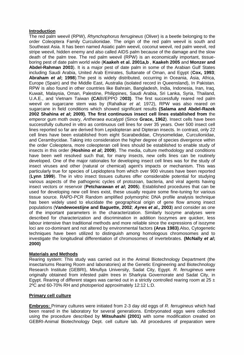
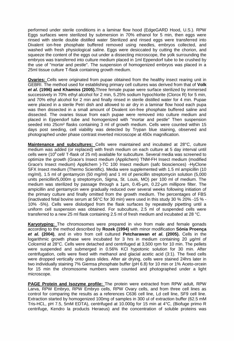
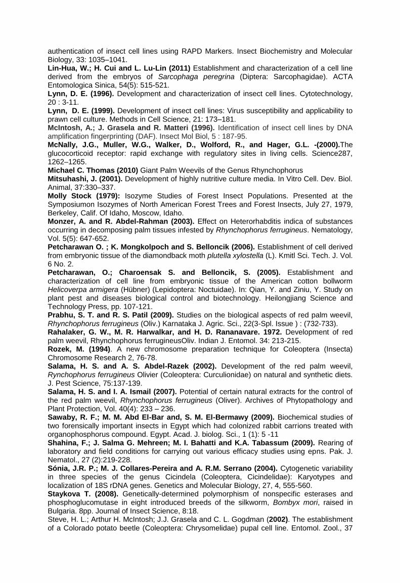
attributed to amount of feeding ,nutritional factors, Temperature and humidity must have played role in these variations. Establishment of cell line: Among the four tested media, Grace's insect medium (Applichem), TNM-FH insect medium (modified Grace's Insect medium) (Applichem), TC100 insect medium (safe biosciences) and HyClone SFX-Insect medium (Thermo Scientific). The Grace's insect medium and TNM-FH insect medium supported higher percentage attachment of cells leading to cell proliferation than TC100 and HyClone SFX-Insect that no results were obtained with them.The success of Primary Cell Culture establishment and Cell line development was not dependent on the species but on the tissues selected as reported by Goodman et al. (2001) and Lynn, (1999). The tissue types that were used in our study whole embryonic homogenates and pupae reproductive tissues (ovary), the embryonic tissues of RPW Rhynchophorus ferrugineus began to attach to the culture flask after incubating at 28°C for 24 h. Figure (2) showed RPW embryo cell culture post seeding and RPW ovary originated cell culture post seeding, at 24 h, 48h, 7 d, 30 d and 60 d post seeding and cell migration or cell division. many tissue fragments become attached to the scratches formed by the scalpel in the plastic. The explant shrank gradually and cell mass liberated from it. Then, more cells migrated from the explant and a number of cells were mitotic during subsequent days. At this point, cell size and morphology was varied. The early migrated cells were mostly spindle and spherical in shape with prominent networks attaching to the bottom of the culture flask, this results agreed with the results that obtained by Petcharawan et al. (2006) also with Lynn (1996). Cell migrated out of the tissue fragments also occurred with fibroblast-like and neuroblast-like cells within 30 day after explantation. By the end of 2 months the bottoms of the flasks were nearly covered with networks of the culture and the culture developed slowly until 5 months of explantation. After this interval the culture developed fast and the bottom of flasks were nearly covered with layer of cells, this results were agreed with the results that obtained by Guoxn et al; (1999) and agreed with the results that obtained by Lin-Hua et al. (2011) they established and characterized a cell line derived from the embryos of Sarcophaga peregrina (Diptera: Sarcophagidae) at 28°C. Cells currently grow with a five days doubling time at 27oC, which is consider slow comparing with diptran and lepidopteran cell lines and are sub-cultured by pibetting at one week intervals at a 1: 3 to 1: 5 split ratios. Cell morphology: The morphological and growth characters of this cell culture were consisting of six major morphologically different cell types (companion cell, neuroblast like cells, round cells, myoblast cells, spindle-shaped cell and epithelial-like cell); nevertheless, after the 3rd subculture, the cells in the monolayer were predominantly epitheloid and spindle-shaped cells (Fig.2). Characterization of RPW in vivo and in vitro : RAPD analysis: RAPD-PCR analysis was performed to detect the genetic similarities and dissimilarities at the DNA levels among the two different forms of R. ferrugineus (a-without spots b-with spots) and to identify a character profile of the local Egyptian RPW. The random primers which were previously mentioned in (table 1) were successfully amplify the weevil genome only five primers gave sufficient polymorphism among the different DNA samples OPK-11, OPK-8, OPK-7, OPC-07 and UBC-300 which gave reproducible results with the weevil genome. RAPD patterns demonstrated in Figs(1B) there were different bands in the profile of RPW without spots when tested with OPK-11 primer this bands of 1121 bp in molecular weight and 1036 bp, and with OPK-7 at the bands of 1279 bp and 788 bp., also demonstrated that there were different bands in the profile of RPW with spots when tested with UBC-300 at the level of 1161bp, with OPK-8 at the band of 533bp and with OPC-07 of 392bp. Results indicated different profile for the RPW without spots which differed in some bands The RAPD profile with spots was clearly showed homology with the majority of bands in the profile of RPW without spots which could be a finger print to characterize or to be a reference with this type of primers. This is not similarly when using another group of primers that demonstrated a clear homology of all profiles which will be so difficult to differentiate the
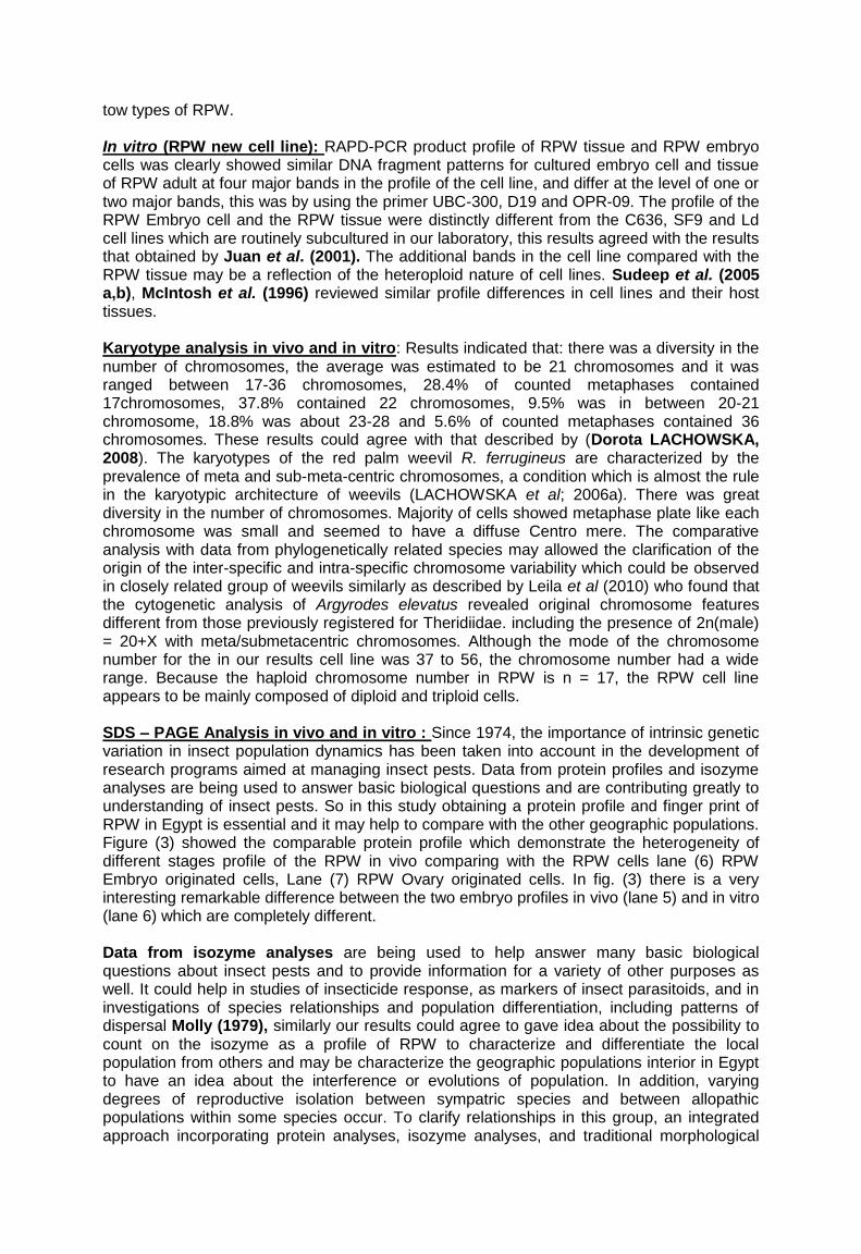
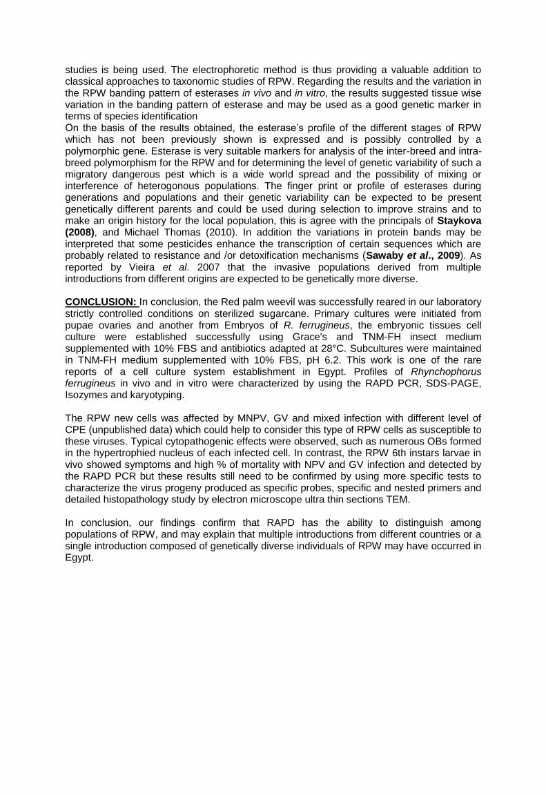
tow types of RPW. In vitro (RPW new cell line): RAPD-PCR product profile of RPW tissue and RPW embryo cells was clearly showed similar DNA fragment patterns for cultured embryo cell and tissue of RPW adult at four major bands in the profile of the cell line, and differ at the level of one or two major bands, this was by using the primer UBC-300, D19 and OPR-09. The profile of the RPW Embryo cell and the RPW tissue were distinctly different from the C636, SF9 and Ld cell lines which are routinely subcultured in our laboratory, this results agreed with the results that obtained by Juan et al. (2001). The additional bands in the cell line compared with the RPW tissue may be a reflection of the heteroploid nature of cell lines. Sudeep et al. (2005 a,b), McIntosh et al. (1996) reviewed similar profile differences in cell lines and their host tissues. Karyotype analysis in vivo and in vitro: Results indicated that: there was a diversity in the number of chromosomes, the average was estimated to be 21 chromosomes and it was ranged between 17-36 chromosomes, 28.4% of counted metaphases contained 17chromosomes, 37.8% contained 22 chromosomes, 9.5% was in between 20-21 chromosome, 18.8% was about 23-28 and 5.6% of counted metaphases contained 36 chromosomes. These results could agree with that described by (Dorota LACHOWSKA, 2008). The karyotypes of the red palm weevil R. ferrugineus are characterized by the prevalence of meta and sub-meta-centric chromosomes, a condition which is almost the rule in the karyotypic architecture of weevils (LACHOWSKA et al; 2006a). There was great diversity in the number of chromosomes. Majority of cells showed metaphase plate like each chromosome was small and seemed to have a diffuse Centro mere. The comparative analysis with data from phylogenetically related species may allowed the clarification of the origin of the inter-specific and intra-specific chromosome variability which could be observed in closely related group of weevils similarly as described by Leila et al (2010) who found that the cytogenetic analysis of Argyrodes elevatus revealed original chromosome features different from those previously registered for Theridiidae. including the presence of 2n(male) = 20+X with meta/submetacentric chromosomes. Although the mode of the chromosome number for the in our results cell line was 37 to 56, the chromosome number had a wide range. Because the haploid chromosome number in RPW is n = 17, the RPW cell line appears to be mainly composed of diploid and triploid cells. SDS – PAGE Analysis in vivo and in vitro : Since 1974, the importance of intrinsic genetic variation in insect population dynamics has been taken into account in the development of research programs aimed at managing insect pests. Data from protein profiles and isozyme analyses are being used to answer basic biological questions and are contributing greatly to understanding of insect pests. So in this study obtaining a protein profile and finger print of RPW in Egypt is essential and it may help to compare with the other geographic populations. Figure (3) showed the comparable protein profile which demonstrate the heterogeneity of different stages profile of the RPW in vivo comparing with the RPW cells lane (6) RPW Embryo originated cells, Lane (7) RPW Ovary originated cells. In fig. (3) there is a very interesting remarkable difference between the two embryo profiles in vivo (lane 5) and in vitro (lane 6) which are completely different. Data from isozyme analyses are being used to help answer many basic biological questions about insect pests and to provide information for a variety of other purposes as well. It could help in studies of insecticide response, as markers of insect parasitoids, and in investigations of species relationships and population differentiation, including patterns of dispersal Molly (1979), similarly our results could agree to gave idea about the possibility to count on the isozyme as a profile of RPW to characterize and differentiate the local population from others and may be characterize the geographic populations interior in Egypt to have an idea about the interference or evolutions of population. In addition, varying degrees of reproductive isolation between sympatric species and between allopathic populations within some species occur. To clarify relationships in this group, an integrated approach incorporating protein analyses, isozyme analyses, and traditional morphological
studies is being used. The electrophoretic method is thus providing a valuable addition to classical approaches to taxonomic studies of RPW. Regarding the results and the variation in the RPW banding pattern of esterases in vivo and in vitro, the results suggested tissue wise variation in the banding pattern of esterase and may be used as a good genetic marker in terms of species identification On the basis of the results obtained, the esterase’s profile of the different stages of RPW which has not been previously shown is expressed and is possibly controlled by a polymorphic gene. Esterase is very suitable markers for analysis of the inter-breed and intra-breed polymorphism for the RPW and for determining the level of genetic variability of such a migratory dangerous pest which is a wide world spread and the possibility of mixing or interference of heterogonous populations. The finger print or profile of esterases during generations and populations and their genetic variability can be expected to be present genetically different parents and could be used during selection to improve strains and to make an origin history for the local population, this is agree with the principals of Staykova (2008), and Michael Thomas (2010). In addition the variations in protein bands may be interpreted that some pesticides enhance the transcription of certain sequences which are probably related to resistance and /or detoxification mechanisms (Sawaby et al., 2009). As reported by Vieira et al. 2007 that the invasive populations derived from multiple introductions from different origins are expected to be genetically more diverse. CONCLUSION: In conclusion, the Red palm weevil was successfully reared in our laboratory strictly controlled conditions on sterilized sugarcane. Primary cultures were initiated from pupae ovaries and another from Embryos of R. ferrugineus, the embryonic tissues cell culture were established successfully using Grace's and TNM-FH insect medium supplemented with 10% FBS and antibiotics adapted at 28°C. Subcultures were maintained in TNM-FH medium supplemented with 10% FBS, pH 6.2. This work is one of the rare reports of a cell culture system establishment in Egypt. Profiles of Rhynchophorus ferrugineus in vivo and in vitro were characterized by using the RAPD PCR, SDS-PAGE, Isozymes and karyotyping. The RPW new cells was affected by MNPV, GV and mixed infection with different level of CPE (unpublished data) which could help to consider this type of RPW cells as susceptible to these viruses. Typical cytopathogenic effects were observed, such as numerous OBs formed in the hypertrophied nucleus of each infected cell. In contrast, the RPW 6th instars larvae in vivo showed symptoms and high % of mortality with NPV and GV infection and detected by the RAPD PCR but these results still need to be confirmed by using more specific tests to characterize the virus progeny produced as specific probes, specific and nested primers and detailed histopathology study by electron microscope ultra thin sections TEM. In conclusion, our findings confirm that RAPD has the ability to distinguish among populations of RPW, and may explain that multiple introductions from different countries or a single introduction composed of genetically diverse individuals of RPW may have occurred in Egypt.
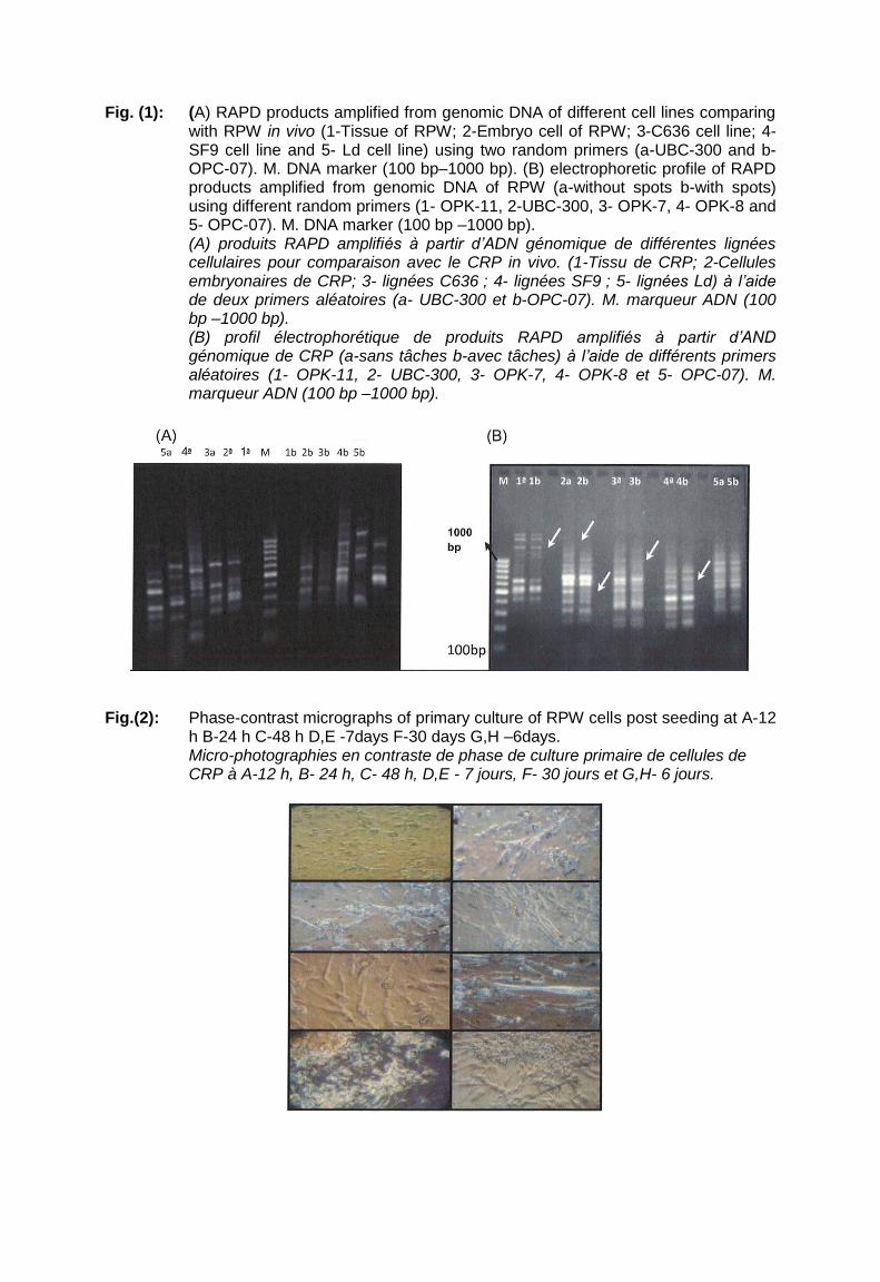
Fig. (1): (A) RAPD products amplified from genomic DNA of different cell lines comparing with RPW in vivo (1-Tissue of RPW; 2-Embryo cell of RPW; 3-C636 cell line; 4-SF9 cell line and 5- Ld cell line) using two random primers (a-UBC-300 and b- OPC-07). M. DNA marker (100 bp–1000 bp). (B) electrophoretic profile of RAPD products amplified from genomic DNA of RPW (a-without spots b-with spots) using different random primers (1- OPK-11, 2-UBC-300, 3- OPK-7, 4- OPK-8 and 5- OPC-07). M. DNA marker (100 bp –1000 bp). (A) produits RAPD amplifiés à partir d’ADN génomique de différentes lignées cellulaires pour comparaison avec le CRP in vivo. (1-Tissu de CRP; 2-Cellules embryonaires de CRP; 3- lignées C636 ; 4- lignées SF9 ; 5- lignées Ld) à l’aide de deux primers aléatoires (a- UBC-300 et b-OPC-07). M. marqueur ADN (100 bp –1000 bp). (B) profil électrophorétique de produits RAPD amplifiés à partir d’AND génomique de CRP (a-sans tâches b-avec tâches) à l’aide de différents primers aléatoires (1- OPK-11, 2- UBC-300, 3- OPK-7, 4- OPK-8 et 5- OPC-07). M. marqueur ADN (100 bp –1000 bp).
Fig.(2): Phase-contrast micrographs of primary culture of RPW cells post seeding at A-12
h B-24 h C-48 h D,E -7days F-30 days G,H –6days. Micro-photographies en contraste de phase de culture primaire de cellules de CRP à A-12 h, B- 24 h, C- 48 h, D,E - 7 jours, F- 30 jours et G,H- 6 jours.

Fig.(3): The SDS-Polyacrylamide gel (12.5%) of electrophoretic profile shows protein patterns of: Lanes : 1 (M) protein marker, 2 (Con) water as control, (3) RPW adult, (4) RPW Larva, (5) RPW Embryo, (6) RPW Embryo cell, (7) RPW Ovary cell, (8) C636 cell line, (9) Ld cell line and (10) SF9 cell line. Profil d’électrophorèse sur gel SDS-polyacrylamide (12.5%) montrant (A) un patron de protéines de : Bandes : 1 (M) marqueur, 2 (Con) témoin eau, (3) CRP adulte, (4) larve de CRP, (5) embryon de CRP, (6) cellule embryonnaire de CRP, (7) cellule ovarienne de CRP, (8) lignée C636, (9) lignées Ld et (10) lignées SF9.
Fig. (23): Polyacrylamide gel (7.5%) electrophoresis shows (A) Esterase banding pattern
using α-naphthyl and β-naphthyl (B) as a substrate. Lanes: 1- RPW adult, 2- RPW Larva, 3- RPW Embryo, 4- RPW Embryo cell, 5- RPW Ovary cell, 6- C636 cell line, 7 Ld cell line, and 8- SF9 cell line. Electrophorèse sur gel de polyacrylamide (7.5%) montrant (A) un patron de bandes d’estérases (B) sur substrat α-naphthyl et β-naphthyl. Bandes : 1- CRP adulte, 2- larve de CRP, 3- embryon de CRP, 4- cellule embryonnaire de CRP, 5- cellule ovarienne de CRP, 6- lignée C636, 7- lignées Ld et 8- lignées SF9.
Fig. 25. Karyotype for red palm weevil R. ferrugineus constructed from a male and showing Mitotic metaphase Caryotype du CRP, R. ferrugineus, en métaphase mitotique. construit à partir d’un adulte mâle.
Fig. 27. Karyotype for red palm weevil, R. ferrugineus, constructed from RPW cell and showed Mitotic metaphase. Caryotype du CRP, R. ferrugineus, en métaphase mitotique, construit à partir d’une cellule en culture.

REFERENCES:. Abraham, V.A.; Al Shuaibi, M.A.; Faleiro, J.R.; Abozuhairah, R.A. and Vidyasagar, P.S.P.V. (1998). An integrated management approach for red palm weevil, Rhynchophorus ferrugineus Oliv. a key pest of date palm in the Middle East. Agricultural Sciences, 3: 77–83. Arus P. Orton TJ (1983) Isozyme and linkage relationships of Isozyme loc' in Brassica oleracea. J Hered 74:405--412 Ayres, C.F.J.; M.A.V. Melo–Santos; A.M. Solé–Cava and A.F. Furtado (2003). genetic differentiation of Aedes aegypti (Diptera: Culicidae) the major dengue vector in Brazil. J. Med. Entomol., 40: 430–5. CABI/EPPO (2003). Rhynchophorus ferrugineus. Distribution maps of plant pests No.259 CAB International Wallingford (GB). Cox, M.L. (1993). Red palm weevil, Rhynchophorus ferrugineus, in Egypt. FAO Plant Prot. Bull, 41:30-31. Dorota, L.; M. Rozek and Milada Holecova (2007): New data on the cytology of parthenogenetic weevils (Coleoptera, Curculionidae. Genetica DOI 10.1007/s10709-007-9230-x. El-Sebay, Y.; M. K. Abd El-Lattef and T. M. Makhlouf (2003). Laboratory rearing of red palm weevil Rhynchophorus ferrugineus Oliver (Coleoptera: Curculionidae) on artificial diet. Egypt. J. Agric. Res., 81(2). Goodman, C.L.; G. N. EL Sayed; A. H. Mcintosh; J. J. Grasela and B. Stiles (2001). Establishment and Characterization of Insect Cell Lines from 10 Lepidopteran Species. In Vitro Cell. Dev. Biol.—Animal, 37:367–373. Grace, T.D.C. (1962). Establishment of four strains of cells from insect tissue grown in vitro. Nature 195: 788-789. Hallett R. H. ; B. J. Crespi and J. H. Borden (2004): Synonymy of Rhynchophorus ferrugineus (Olivier), 1790 and R.vulneratus (Panzer), 1798 (Coleoptera, Curculionidae, Rhynchophorinae) Journal of Natural History, , 38, 2863–2882. Hoshino, K.; M. Hirose and K. Iwabuchi (2009). A new insect cell line from the longicorn beetle Plagionotus christophi (Coleoptera: Cerambycidae). In Vitro Cell. Dev. Biol.-Animal, 45:19–22. Juan, J. G.; G. Li; P. Wang; J. Zhong and R. R. Granados (2001). Primary and continuous midgut cell cultures from pesudaletia unipuncta (Lepidoptera: Noctuidae). In Vitro Cell. Dev. Biol-Animal, 37:353-359. Kaakeh, W. (2005). Longevity, fecundity, and fertility of the red palm weevil, Rhynchophorus ferrugineus Olivier (Coleoptera: Curculionidae) on natural and artificial diets. Emir. J. Agric. Sci. 17 (1): 23-33. Kaakeh, W.; A. A. Khamis and M. Aboul-Anour (2001b). Life parameters of the red palm weevil, Rhynchophorus ferrugineus Oliv., on sugarcane and artificial diet. pp. 310-324. Proceedings of the Second International Conference on Kaakeh, W.; M. Aboul-Anour and A. A. Khamis (2001a). Mass rearing of the red palm weevil, Rhynchophorus ferrugineus Oliv., on sugarcane and artificial diets for laboratory studies, the Second International Conference on Date Palm (refereed), Al-Ain, UAE. Proceedings, pp. 344-357. Khamiss, O. (2005). Establishment and characterization of Spodoptera littoralis cell line (SL 96), adapted at 19 degrees C permissive for different baculoviruses. J Egypt Soc Parasitol, 35(3):751-60. LACHOWSKAD., HOLECOVÁM. and ROĪEKM. 2006a: Cytogenetic differences between Peritelus familiaris and Centricnemus leucogrammus (Coleoptera: Curculionidae: Entiminae: Perite-lini). Eur. J. Entomol.103: 687–690. Laemmli, U.K. (1970). Cleavage of structure proteins during assembly of head bacteriophage T4. Nature Vol., 227. Leila M. S.; M. C. Schneider; D. Araujo; A. D. Brescovit and D. M.Cella (2010). Chromosomes of Theridiidae spiders (Entelegynae): Interspecific karyotype diversity in Argyrodes and diploid number intraspecific variability in Nesticodes rufipes. Genetics and Molecular Biology, 33, 4, 663-668 Léry, X.; B. LaRue; J. Cossette and G. Charpentier (2003). Characterization and
authentication of insect cell lines using RAPD Markers. Insect Biochemistry and Molecular Biology, 33: 1035–1041. Lin-Hua, W.; H. Cui and L. Lu-Lin (2011) Establishment and characterization of a cell line derived from the embryos of Sarcophaga peregrina (Diptera: Sarcophagidae). ACTA Entomologica Sinica, 54(5): 515-521. Lynn, D. E. (1996). Development and characterization of insect cell lines. Cytotechnology, 20 : 3-11. Lynn, D. E. (1999). Development of insect cell lines: Virus susceptibility and applicability to prawn cell culture. Methods in Cell Science, 21: 173–181. McIntosh, A.; J. Grasela and R. Matteri (1996). Identification of insect cell lines by DNA amplification fingerprinting (DAF). Insect Mol Biol, 5 : 187-95. McNally, J.G., Muller, W.G., Walker, D., Wolford, R., and Hager, G.L. -(2000).The glucocorticoid receptor: rapid exchange with regulatory sites in living cells. Science287, 1262–1265. Michael C. Thomas (2010) Giant Palm Weevils of the Genus Rhynchophorus Mitsuhashi, J. (2001). Development of highly nutritive culture media. In Vitro Cell. Dev. Biol. Animal, 37:330–337. Molly Stock (1979): Isozyme Studies of Forest Insect Populations. Presented at the Symposiumon Isozymes of North American Forest Trees and Forest Insects, July 27, 1979, Berkeley, Calif. Of Idaho, Moscow, Idaho. Monzer, A. and R. Abdel-Rahman (2003). Effect on Heterorhabditis indica of substances occurring in decomposing palm tissues infested by Rhynchophorus ferrugineus. Nematology, Vol. 5(5): 647-652. Petcharawan O. ; K. Mongkolpoch and S. Belloncik (2006). Establishment of cell derived from embryonic tissue of the diamondback moth plutella xylostella (L). Kmitl Sci. Tech. J. Vol. 6 No. 2. Petcharawan, O.; Charoensak S. and Belloncik, S. (2005). Establishment and characterization of cell line from embryonic tissue of the American cotton bollworm Helicoverpa armigera (Hübner) (Lepidoptera: Noctuidae). In: Qian, Y. and Ziniu, Y. Study on plant pest and diseases biological control and biotechnology. Heilongjiang Science and Technology Press, pp. 107-121. Prabhu, S. T. and R. S. Patil (2009). Studies on the biological aspects of red palm weevil, Rhynchophorus ferrugineus (Oliv.) Karnataka J. Agric. Sci., 22(3-Spl. Issue ) : (732-733). Rahalaker, G. W., M. R. Harwalkar, and H. D. Rananavare. 1972. Development of red palm weevil, Rhynchophorus ferrugineusOliv. Indian J. Entomol. 34: 213-215. Rozek, M. (1994). A new chromosome preparation technique for Coleoptera (Insecta) Chromosome Research 2, 76-78. Salama, H. S. and A. S. Abdel-Razek (2002). Development of the red palm weevil, Rynchophorus ferrugineus Olivier (Coleoptera: Curculionidae) on natural and synthetic diets. J. Pest Science, 75:137-139. Salama, H. S. and I. A. Ismail (2007). Potential of certain natural extracts for the control of the red palm weevil, Rhynchophorus ferrugineus (Oliver). Archives of Phytopathology and Plant Protection, Vol. 40(4): 233 – 236. Sawaby, R. F.; M. M. Abd El-Bar and, S. M. El-Bermawy (2009). Biochemical studies of two forensically important insects in Egypt which had colonized rabbit carrions treated with organophosphorus compound. Egypt. Acad. J. biolog. Sci., 1 (1): 5 -11 Shahina, F.; J. Salma G. Mehreen; M. I. Bahatti and K.A. Tabassum (2009). Rearing of laboratory and field conditions for carrying out various efficacy studies using epns. Pak. J. Nematol., 27 (2):219-228. Sónia, J.R. P.; M. J. Collares-Pereira and A. R.M. Serrano (2004). Cytogenetic variability in three species of the genus Cicindela (Coleoptera, Cicindelidae): Karyotypes and localization of 18S rDNA genes. Genetics and Molecular Biology, 27, 4, 555-560. Staykova T. (2008). Genetically-determined polymorphism of nonspecific esterases and phosphoglucomutase in eight introduced breeds of the silkworm, Bombyx mori, raised in Bulgaria. 8pp. Journal of Insect Science, 8:18. Steve, H. L.; Arthur H. McIntosh; J.J. Grasela and C. L. Gogdman (2002). The establishment of a Colorado potato beetle (Coleoptera: Chrysomelidae) pupal cell line. Entomol. Zool., 37

(3): 447-450. Sudeep, A.B.; D.T. Mourya and A.C. Mishra (2005b). Insect cell culture in research: Indian scenario. Indian J Med Res, 121:pp 725-738. Sudeep, A.B.; R. Khushiramani; S.S. Athawale; A.C. Mishra and D.T. Mourya (2005a). Characterization of a newly established potato tuber moth (Phthorimaea operculella Zeller) cell line. Indian J Med Res 121, pp 159-163. Vandewoestijne, S. and M. Baguette, (2002). The genetic structure of endangered populations in the cranberry fritillary, Boloria aquilonaris (Lepidoptera, Nymphalidae): RAPDs vs allozymes. Heredity, 89: 439–45. Volk C.J.; C.B. Volk; and L. A. Kaplan (1997): Chemical composition of biodegradable dissolved organic matter in stream water Limnol. Oceunogr,42 (l) 39-44 . by the American Society of Limnology and Oceanography, Inc