agenesis of the corpus callosum: genetic, developmental ...than the corpus callosum, may provide a...
TRANSCRIPT
Agenesis of the Corpus Callosum:
Genetic, Developmental and Functional Aspects of Connectivity
Lynn K. Paul1,2, Warren S. Brown2, Ralph Adolphs1, J. Michael Tyszka1, Linda J. Richards3, Pratik Mukherjee4, and Elliott H. Sherr4.
1 California Institute of Technology
2 Travis Research Institute 3University of Queensland
and 4University of California, San Francisco
Address correspondence to: Lynn K Paul HSS 228-77 Caltech, Pasadena, CA 91125 Website: http://www.emotion.caltech.edu/AgCC And to: Elliott H. Sherr Department of Neurology, UCSF 350 Parnassus Ave, Suite 609 San Francisco, CA 94143-0137 Website: http://www.ucsf.edu/brain/callosum/callosum.htm
Agenesis of the Corpus Callosum Page 2 of 33
Agenesis of the corpus callosum (AgCC), a failure to develop the large bundle of fibers that connect the cerebral hemispheres, occurs in 1:4000 individuals. Genetics, animal models and detailed structural neuroimaging are now providing insights into the developmental and molecular bases of AgCC. Neuropsychological, EEG, and fMRI approaches are examining the resulting impairments in emotional and social functioning, and have begun to explore the functional neuroanatomy underlying impaired higher-order cognition. The study of AgCC could provide insight into the integrated cerebral functioning of healthy brains, and may offer a model for understanding certain psychiatric illnesses, such as schizophrenia and autism.
Introduction The brain’s complexity arises from its connectivity – this is highlighted by the disproportionate increase in white matter volume through primate evolution 1. The largest connective structure in the brain is the corpus callosum, consisting of over 190 million axons that transfer information between the two cerebral hemispheres (Figure 1) 2. The corpus callosum contains homotopic and heterotopic interhemispheric connections. Although there has been debate about whether the connections are primarily excitatory (integrating information across hemispheres) or inhibitory (allowing the hemispheres to inhibit each other to maximize independent function) 3 it appears to be primarily excitatory and this is the focus of most research on interhemispheric transfer (IHT).
Corpus callosum function in humans was first investigated in classic studies of “split brain” patients, whose callosum is severed surgically for the treatment of epilepsy (Box 1) 4, 5. However, there is another population that provides valuable insight about the functions of the corpus callosum and the role of altered connectivity in neurodevelopmental disorders: individuals with developmental absence (agenesis) of the corpus callosum (AgCC) (Box 2).
AgCC encompasses complete absence as well as hypogenesis (partial absence) of the corpus callosum (Box 3). This review covers a broad range of findings from research into AgCC, including animal models of callosal development, genetic and environmental contributions to AgCC, neuroimaging in acallosal humans, and neuropsychological outcomes in individuals with Primary AgCC. Therefore, the interdisciplinary nature of this review provides a framework for bridging once largely non-overlapping domains of neuroscience: genetics and neuropsychology. AgCC is a complex condition, which may result from disruption in any one of the multiple steps of callosal development, such as cellular proliferation and migration, axonal growth, or glial patterning at the midline. We review the molecular mechanisms underlying these processes. Later sections address behavioral and neuropsychological aspects of AgCC. We briefly examine research on IHT and alternative hypotheses regarding behavioral symptoms. Although the contribution of AgCC to our understanding of callosal function is complicated by concomitant anatomic changes (Box 2), we suggest that AgCC may be a powerful model for understanding cortico-cortical plasticity in other neurological and psychiatric populations. Development of the Corpus Callosum Corpus callosum formation involves multiple steps, including correct midline patterning, formation of telencephalic hemispheres, birth and specification of commissural neurons and axon guidance across the midline to their final target in the contralateral hemisphere. Much of
Agenesis of the Corpus Callosum Page 3 of 33
what we know about the stages of callosal development comes from animal models 6, 7. Several principal mechanisms have been hypothesized to regulate callosal formation. Guidance by pre-exisiting axon tracts The first axons to cross the midline arise from neurons in the cingulate cortex. In mice, these pioneer axons cross the rostral midline at embryonic day (E)15.5 8, providing a path for the fasciculation of later-arriving neocortical axons. In humans, pioneer axons express the guidance receptor Neuropilin -1 9, which may guide these axons themselves or the later-arriving callosal neurons from the neocortex. Cingulate cortex neurons also project axons into the rostrolateral cortex, perhaps to guide neocortical axons toward the midline initially. In more caudal regions of the corpus callosum, the hippocampal commissure, which in mice is formred a day earlier than the corpus callosum, may provide a growth substrate 10, 11. Midline glial structures Multiple glial structures including the glial wedge, midline zipper glia, and indusium griseum (Figure 3), are present at the developing midline and are likely required for corpus callosum formation 12-15. The glial wedge is a bilaterally symmetrical structure composed of radial glial cells that reside ventral to the corpus callosum at the corticoseptal boundary. It prevents callosal axons from entering the ventrally located septum, and once callosal axons have crossed the midline, repels the axons away from the midline into the contralateral hemisphere 13, 16. Guidance by the glial wedge occurs through both Slit-Robo and Wnt-Ryk signaling 13, 16-18. Midline zipper glia are found ventral to the developing corpus callosum at the septal midline. Their fusion at the midline has been hypothesized to be necessary for subsequent callosal axons to grow across the midline 19.
The indusium griseum glia (IGG), which are dorsal to the developing corpus callosum, also express Slit-2 and may help in guiding commissural axons toward their site of midline crossing 13. Recent work in conditional FGFR1/GFAP Cre knockout mice has shown the importance of these glia in corpus callosum formation 14. When FGFR1 is selectively eliminated from glia (and not neurons), the corpus callosum fails to form. Further analysis showed that FGFR1 is required for the migration of the IGG and selectively for the development of this midline glial structure 14. However, when FGFR1 is knocked out earlier in development, all midline glial structures at the corticoseptal boundary fail to develop, suggesting that FGFR1 plays a signaling role at multiple stages of callosal development 15. Similarly, Nfia and Nfib knockout mice 20, 21, whose IGG and glial wedge are absent or significantly reduced in size, do not form a corpus callosum. However, midline glia are not the only guidance mechanisms at the midline. FGFR1 heterozygote (KO/WT) 15 and GAP43 knockout mice 22 do form midline glial structures (and express Slit 2), but callosal axons fail to cross, suggesting that multiple mechanisms regulate callosal development.
The subcallosal sling lies dorsal to the glial wedge and ventral to the developing corpus callosum. When the sling is severed experimentally, the corpus callosum fails to form 19, suggesting a role for this structure in callosal guidance, although the first callosal axons cross prior to the formation of the subcallosal sling. In mice, the majority of cells that make up the sling prenatally are neurons 23, but in humans, whose subcallosal sling contains a large number of glia, the cellular origins of this structure are more complex 9 (Figure 3). Finally, additional neurons that have been identified within the corpus callosum 24 may play a role in axon guidance. Target recognition and selective pruning in the contralateral hemisphere
Agenesis of the Corpus Callosum Page 4 of 33
After crossing the midline, callosal axons grow into the contralateral hemisphere toward their designated target region, usually homotopic to their region of origin, and then innervate the appropriate cortical layer. Such processes probably involve both molecular recognition of the appropriate target region and activity-dependent mechanisms that regulate axon targeting to the correct layer and the subsequent refinement of the projection 25. In cats and ferrets, refinement of callosal visual projections occurs through the selective pruning of axons after eye opening 25-27. Correct pruning and stabilization at the border of areas 17 and 18 (but to a lesser extent in other areas) requires visual input 28. A similar refinement of developmentally exuberant projections occurs in the somatosensory cortex 25. It is not yet clear whether defects in axonal pruning may affect corpus callosum size and contribute to callosal hypoplasia (See Box 3) in humans. Animal Models of AgCC Animal models of AgCC provide a basis for identifying genes that may be involved in human AgCC. The inactivation of genes that cause AgCC in mice often also trigger neurological deficits in other large fiber-tracts such as the internal capsule, and consequently leads to death at birth in many cases. These phenotypes, mostly resulting from gene deletions, may be too severe to model human AgCC, and such gene deletions may also result in embryonic or perinatal death in humans. However, a number of mouse models exist in which AgCC is partially or fully penetrant, but the animals have normal lifespans. Strains such as 129 and BTBR have been used to map quantitative trait loci that affect corpus callosum size 29. Recent studies have shown that the gene DISC1 is homozygously inactivated in all 129 strains 30 and this genetic mutation may be causally linked to AgCC in these mice. Interestingly, this gene has also been implicated in schizophrenia, and may be an important mechanistic link between the two disorders 31.
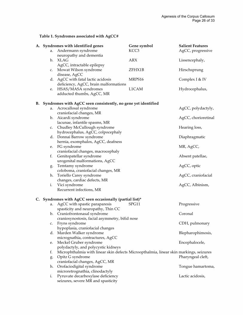
Finally, recent studies have used novel tools for labeling and isolating functional subsets of neurons to identify markers that are unique to callosal projection neurons 32. These studies have identified the gene Lmo4 as a candidate transcription factor for specifying callosal “identity” to projection neurons. This approach will potentially lead to a greater understanding of how neurons acquire their functional identities. Causes of AgCC in humans Genetic causes The genetics of AgCC in humans are quite variable and reflect the underlying complexity of callosal development. Current evidence suggests that a combination of genetic mechanisms, including single-gene Mendelian, single-gene sporadic mutations and complex genetics (which may have a mixture of inherited and sporadic mutations) may have a role in the aetiology of AgCC. Retrospective chart reviews and cross-sectional cohort studies report that 30-45% of individuals with AgCC have identifiable causes for AgCC. Approximately 10% have chromosomal anomalies and the remaining 20-35% have recognizable genetic syndromes (Table 1) 33. However, if we only consider individuals with complete AgCC, then the percentage of patients with identifiable syndromes drops to 10-15%, and thus 75% of patients with complete AgCC do not have an identified cause.
Agenesis of the Corpus Callosum Page 5 of 33
One example of AgCC associated with a Mendelian disorder is X-linked lissencephaly with AgCC and ambiguous genitalia (XLAG), which results from a mutation in the Aristaless related homeobox gene (ARX). The first description of this disorder included only male patients. However, females with mutations in ARX, who, because of X-inactivation, have clinical symptoms that range from normal to those with spasticity, mental retardation and seizures. MRI scans of these female patients are either normal or show Isolated AgCC with Probst bundles (Box 3) 34. Male ARX knockout mice also have AgCC and mimic many of the other clinical and anatomic findings in XLAG 35, including a significant reduction in cortical interneurons, which likely explains the severe and uncontrollable seizures in this condition 36.
Another syndrome caused by a single-gene mutation with considerable overlap between the human and animal phenotype is CRASH syndrome (corpus callosum agenesis, retardation, adducted thumbs, spastic paraplegia and hydrocephalus), which is accompanied by diminutive cortico-spinal tracts within the brainstem. CRASH is caused by mutations in the L1CAM gene that codes for a transmembrane cell adhesion protein broadly expressed in the central nervous system. Mice with L1CAM gene inactivation show complete or partial AgCC, hydrocephalus, small corticospinal tracts, reduced neuron numbers, and additional abnormalities in the elaboration of apical dendrites from cortical pyramidal neurons 37. Recent work suggests that inhibition of homophilic binding of the L1CAM protein can cause hydrocephalus, but that preventing corpus callosum formation also requires disruption of heterophilic interactions with other proteins including integrins 38. Gene dosage effects have also been observed in mouse knockout models for the genes DCC and GAP-43. Here, heterozygote mice show partial AgCC whereas homozygote knockout mice have complete AgCC with additional anomalies 22.
Andermann syndrome, an autosomal recessive condition prevalent in the Saguenay-Lac-St-Jean region of Quebec, presents with callosal hypoplasia or AgCC, cognitive impairment, episodes of psychosis and a progressive central and peripheral neuropathy. It is caused by mutation of the potassium-chloride cotransporter, KCC3 39. KCC3 knockout mice display neurodegeneration, and also have hearing loss and progressive neuropathy 40. However, in contrast to ARX and L1 mouse mutants, they have normally formed corpora callosa. Interestingly, some patients with KCC3 mutations also have a fully formed corpus callosum, and there is even phenotypic variability within families, suggesting that additional genetic influences are at play.
The variable effects of gene inactivation on callosal development in mice and humans is also evident in Meckel Gruber syndrome. In humans, mutation of Meckelin (the gene in MKS3) causes occipital encephaloceles, hepatic ductal cysts and polydactyly. In mice, mutation of the same gene causes AgCC, hydrocephalus and cysts within the kidney 41. Table 1 shows other disorders associated with callosal agenesis that show a clear recessive pattern of inheritance but for which the causative gene has not been identified, (Table 1).
In spite of the progress of research into single-gene Mendelian causes of AgCC, most individuals with AgCC do not have a clearly inherited cause or a recognized genetic syndrome, suggesting that AgCC can be caused by sporadic (de novo) genetic events. One salient example of this is Mowat-Wilson syndrome (MWS), which, in addition to AgCC, causes Hirschsprung disease, congenital heart disease, genitourinary anomalies, microcephaly, epilepsy and severe cognitive impairment 42. MWS is caused by heterozygous inactivating mutations in the gene ZFHX1B on chromosome 2q22, which codes for SIP1 (Smad interacting protein 1) 43. AgCC is not observed in all MWS cases, suggesting that haploinsufficiency or gene dosage of SIP1 interacts with other genetic polymorphisms to alter callosal development 42.
Aicardi syndrome is another disorder likely caused by sporadic mutations, in this case on the X chromosome. Only observed in females and XXY males with Klinefelter syndrome, this
Agenesis of the Corpus Callosum Page 6 of 33
disorder consists of AgCC, infantile spasms and chorioretinal lacunae. Patients with Aicardi syndrome show a constellation of additional cerebral and ophthalmologic abnormalities, so it is likely that the mutation participates widely and early in prosencephalic development. By inference, it is likely that other cases of AgCC are caused by haploinsufficiency at other genetic loci. This is exemplified by many reports of patients with AgCC who have sporadic chromosomal mutations, with particular loci identified repeatedly 44. Recent data using microarray-based comparative genomic hybridization demonstrate that patients with AgCC have chromosomal deletions or duplications that are smaller than those that can be detected using conventional cytogenetics 45. Indeed, in collaboration with the California Birth Defects Monitoring Program, we have demonstrated that the risk of having a child with AgCC is nearly three-fold higher for mothers aged 40 and above, consistent with causal sporadic chromosomal changes (E. Sherr, unpublished observations).
As noted for the single-gene disorders discussed above, not every patient displays AgCC, suggesting that many cases of AgCC are caused by polygenic and other complex interactions. Moreover, the abundance of case reports of AgCC associated with specific diseases (partially listed in Table I) probably also reflects complex underlying mechanisms. This is exemplified by a recent report examining the MRI findings of individuals with Sotos syndrome, which is caused by haploinsufficiency of the NSD1 gene. In this study, only one patient had complete AgCC and the rest (35/36) had either diffuse hypoplasia or thinning of the posterior body of the corpus callosum 46. These findings suggest that some genes that are often referred to as modifier genes only partially contribute to callosal formation. Common disorders that affect behavior and for which the influence of many modifier genes is the likely mode of inheritance include autism and schizophrenia 47, 48. Moreover, as there are many reports of AgCC or abnormally formed corpora callosa in patients with autism and schizophrenia 49, 50, it is possible that the modifier genes that affect callosal development overlap significantly with those that cause these neuropsychiatric disorders. Environmental factors Finally, it is important to note that environmental factors may contribute to AgCC as well. While much less is known about these than the genetic factors we have reviewed above, one clear example of environmental influences on callosal development is provided by fetal alcohol syndrome (FAS). Both clinical and experimental evidence indicates that alcohol exposure in utero decreases gliogenesis 51 and glial-neuronal interactions 52, processes that are critical to corpus callosum development 53. Additionally, a growing body of literature suggests that ethanol disrupts the transcription and biochemical function of L1CAM 54-56, implicating an interplay of environment and genetics in AgCC. The incidence of AgCC in FAS is approximately 6.8% 57, with an even higher incidence of corpus callosum malformations short of complete AgCC. In many FAS cases, the corpus callosum is hypoplastic; this may result not only from disruption of early events in callosal formation, but also from later dysregulation of axonal pruning. Such mechanisms may also cause callosal hypoplasia in other disorders such as schizophrenia and autism 25, 31. Other environmental factors may also influence post-natal and prenatal callosal development, including musical training 58-60, hypothyroidism 61, 62, and enrichment or deprivation of experience 63. Behavioral Impairment in AgCC Consistent with the broad range of genetic factors involved in AgCC, the cognitive, behavioral and neurological consequences of AgCC are wide-ranging. One approach to defining clinical subsets of the AgCC patient population is to categorize individuals according to specific
Agenesis of the Corpus Callosum Page 7 of 33
neuroanatomical findings, and subsequently relate these to the behavioral symptoms within these groups. For example, a number of studies have suggested that the presence of polymicrogyria, pachygyria (abnormally broad gyri), heterotopia, detected using MRI, correlates with moderate to severe developmental delay 64, 65. However, creating accurate anatomical groupings is very difficult given the diversity of symptoms in patients with a particular anatomical abnormality.
The comparison between complete and partial AgCC has revealed conflicting data, with multiple studies showing no difference in behavioral and medical outcomes between the two conditions, whereas one earlier study reported a worse outcome for individuals with complete AgCC 66. One hospital-based study reported that just under a third of AgCC patients were developmentally “normal” or only mildly delayed 65. A longitudinal study of 17 children prenatally diagnosed with isolated AgCC, showed that nearly all patients had at least mild behavioral problems 67. This suggests that even isolated AgCC when not ascertained clinically, still causes behavioural and cognitive impairment. Parents often report that when their child was diagnosed with AgCC, they were told that the outcome was unclear, ranging from severely delayed to “perfectly normal”. However, as more individuals with Primary AgCC are identified and assessed with sensitive standardized neuropsychological measures, a pattern of deficits in higher-order cognition and social skills has become apparent even in the so-called “normal” individuals with AgCC. Connectivity deficits Historically, most research in subjects with AgCC focused on the consequences of callosal absence, with the expectation that AgCC patients would exhibit a “disconnection syndrome” similar to that seen in commissurotomy patients4. The classic disconnection syndrome (Box 1) involves complete lack of IHT and interhemispheric integration of sensory and motor information presented independently to each of the hemispheres 4, with surprisingly subtle behavioral consequences in everyday life.
Studies using tachistoscopic presentation of visual stimuli and studies of evoked potentials provide the most compelling information about functional connections and IHT in AgCC at various stages of sensory processing. Figure 4 illustrates the visual cortical disconnection effects in AgCC, as well as the limits of these disconnection symptoms. First, as illustrated by visual evoked potentials, there is a complete lack of IHT at the level of early visual processing in AgCC 68. The hemispheric disconnection of the primary visual system in AgCC patients results in a unique pattern of deficits in laboratory tasks that involve comparisons across the two visual fields: intact comparisons of simple stimuli and impaired comparisons of complex stimuli. First, despite the lack of transfer of early visual information, individuals with AgCC exhibit a normal ability to make comparisons of simple and easily encoded stimuli, implying intact interhemispheric transfer of simple or familiar information. For example, they exhibit an intact interhemispheric Stroop interference effect 69 and the typical bilateral field advantage for comparison of familiar and easily encoded visual information across hemifields 68. These findings confirm that information can be transferred between the hemispheres in AgCC. One theory to explain the preserved capacity for IHT of simple stimuli in AgCC patients is that simple information can be transferred via other connecting pathways, such as the anterior commissure. Structural and functional exploration of these alternate pathways for IHT is a critical frontier in AgCC research.
In contrast, the capacity for IHT may be limited by task complexity. For example, the performance of patients with AgCC when comparing tachistoscopically presented visual information is significantly less accurate for more visually complex, less familiar, and less easily
Agenesis of the Corpus Callosum Page 8 of 33
verbalized stimuli 68 (Figure 4). Similar limitations in IHT in AgCC patients were evident on other tasks that required transfer or integration of the products of more complex cognitive operations, required more rapid processing and relied on less prior experience 68, 70-72. Taken together, these studies indicate that there is greater reliance on the corpus callosum for rapid and effective interhemispheric interactions as task requirements increase (e.g., when stimuli become more complex or response criteria become more constrained).
The question remains, however, about what causes the behavioral disturbances evident in Primary AgCC. Studies using dichotic listening 73, 74, PET 75, and fMRI 76 have revealed that language lateralization is normal in Primary AgCC patients and is in some cases even exaggerated. Although there is no published evidence for normal or abnormal localization of other higher cognitive functions in this population, we may hypothesize that if localized functioning in the cortex in Primary AgCC patients is normal, a lack of information transfer between localized processing regions in opposite hemispheres could contribute to behavioral difficulties. This leaves callosal disconnection as a viable explanation for the behavioural disturbances in Primary AgCC patients. Neuropsychological impairment The major anatomic feature of Primary AgCC is the absence of the corpus callosum, and it is presumed to be the cause of the cognitive and behavioral changes in Primary AgCC patients. However, colpocephaly (Box 3) and Probst bundles are common in people with Primary AgCC (and never without AgCC), and together with other more subtle anatomical changes, likely also affect behavior. Functional and anatomic imaging approaches, coupled with incisive neuropsychological assessments, may in the future be able to map the neural processes and neuropathology associated with AgCC onto the diverse behavioral anomalies. For now, we begin by describing the symptom profile. Primary AgCC has surprisingly limited impact on general cognitive ability. Although the full-scale IQ may be lower than expected based on family history, scores frequently remain within the average range 77. Interestingly, in an unexpectedly large number of persons with Primary AgCC (as many as 60%), performance IQ and verbal IQ are significantly different 77, 78. However, there is no consistency with respect to which of the two is more affected. Impairments in abstract reasoning 79, 80, problem solving 81-83, generalization (the ability to extrapolate from one case to others) 84, and category fluency (the ability to list multiple items that belong to a semantic category, e.g., names of animals)80 have all been consistently observed in patients with Primary AgCC. Data from large sample sizes suggest that problem solving abilities become more impaired as task complexity increases (Brown and Paul, unpublished observations). While neuropsychological research into domains such as memory, attention, and spatial skills is under way in large samples of patients with Primary AgCC, currently published results in these domains are limited to a few case-studies that do not yet provide consistent findings. The most comprehensively examined higher cognitive domain in AgCC patients is language. Overall, individuals with Primary AgCC have intact general naming (e.g. naming objects from line drawings 85, 86), receptive language (e.g. comprehension of sentences 78, 86), and lexical reading skills 87. However, impairments have been reported in comprehension of syntax and linguistic pragmatics 88, 89, and in phonological processing and rhyming 86, 88-90. With respect to linguistic pragmatics, persons with Primary AgCC are impaired in comprehension of idioms, proverbs, vocal prosody 91, 92, and narrative humor 93. Within humor, they exhibit difficulty in over-riding literal interpretation bias and are poor at using context to infer meaning 92-94. Primary AgCC patients also show marked difficulties with expressive language, for
Agenesis of the Corpus Callosum Page 9 of 33
example in the verbal expression of emotional experience, which is consistent with a diagnosis of alexithymia 95. In a study of a large sample of AgCC patients with adequate expressive language skills, parents consistently described “meaningless” or “out-of-place” conversation as particularly common 96. Interestingly, recent studies of language support the dynamic dual pathway model according to which syntax and semantics are lateralized to the left hemisphere and prosody to the right hemisphere 97-102. In this model, the corpus callosum is the main path for coordination of this lateralized information, particularly for coordinating syntactic and prosodic information 97-99, the very areas of linguistic weakness evident in AgCC.
Parents of individuals with Primary AgCC consistently describe impaired social skills and poor personal insight as the features that interfere most prominently with the daily lives of their children 96, 103-105. Specific traits include emotional immaturity, lack of introspection, impaired social competence, general deficits in social judgment and planning, and poor communication of emotions (e.g. prefer much younger friends, marked difficulty generating and sustaining conversation, take all conversation literally, do not take perspective of others, inability to effectively plan and execute daily activities such as homework, showering, or paying bills 96, 105). Consequently, Primary AgCC patients often have impoverished and superficial relationships, suffer social isolation, and have interpersonal conflict both at home and at work due to misinterpretation of social cues.
Responses of adults with Primary AgCC on self-report measures also suggest diminished self-awareness 104. The patients’ self-reports are often in direct conflict with observations from friends and family. One potential factor contributing to poor self-awareness may be a more general impairment in comprehension and description of social situations. For instance, when asked to generate stories about social pictures, adults with Primary AgCC provided stories lacking in logic, narrative content, and social understanding 106. It appeared that they had difficulty recognizing the implications of pictures depicting social scenes, imagining a sequence of events, and organizing relevant ideas in order to present an appropriate narrative. Similarly, when presented with highly provocative social pictures (e.g., photos of mutilations), adults with AgCC tended to under-estimate the emotional valence and intensity of the pictures, particularly for negatively valenced pictures 107. Taken together, the neuropsychological findings in Primary AgCC highlight a pattern of deficits in problem solving and in social pragmatics of language and communication. AgCC and neuropsychiatric disorders The deficits in social communication and social interaction in Primary AgCC patients overlap with the diagnostic criteria for autism (DSM-IV)103. Furthermore, people with Primary AgCC may display a variety of other social, attentional, and behavioral symptoms that may resemble those of certain psychiatric disorders. Psychiatric diagnoses are based on clusters of behaviors, which are very complex and likely involve multiple neural mechanisms 108. Examination of symptom overlap between psychiatric disorders and AgCC may help isolate the symptoms that are directly caused by a dysfunction in cortico-cortical connectivity. There are also structural similarities between AgCC and some psychiatric disorders. Indeed, structural correlates of abnormal brain connectivity are evident in essentially every psychiatric disorder that has been examined. For instance, several studies have found altered morphology of the corpus callosum in schizophrenia patients, including changes in size and shape, as well as microstructural changes in callosal regions that are reflected in diffusion MRI (dMRI) 31. There are also a number of reports of complete AgCC in schizophrenia patients 50, 80, 109, underscoring a direct connection between AgCC and schizophrenia and countering claims that the smaller anatomic changes in the corpora callosa in schizophrenic patients are not causally related to the
Agenesis of the Corpus Callosum Page 10 of 33
condition. Corpus callosum size is also decreased in some cases of autism, especially its anterior sectors 110 111. Moreover, in one study, 8.5% of AgCC individuals had a diagnosis of autism, compared to only 1% of their siblings112. Microstructural changes in the corpus callosum have also been found in patients with Tourette syndrome 113 and attention deficit hyperactivity disorder 114, 115. One recent study provides evidence linking genetic changes in KCC3 (the gene mutated in Andermann syndromed this is defined in the genetics section) with bipolar disorder 116, even though these patients did not have evident changes in callosal anatomy. As more causes of AgCC are identified, we anticipate further genetic correlations between AgCC and neuropsychiatric disorders will be found. The functional consequences of structural changes in brain connectivity, which may be revealed by dMRI analysis117 including diffusion tensor imaging (DTI), contribute to cognitive impairment. Functional connectivity studies show that the strength of the correlations between brain activation in different regions and anatomical abnormalities is strikingly task-dependent. For example, children with attention deficit hyperactivity disorder show a disturbed transcallosally mediated motor inhibition 118. Functional connectivity studies in AgCC patients may reveal the means by which these highly atypical brains attain such apparently “typical” interhemispheric interaction. In turn, understanding the functional limits of such connectivity may contribute to knowledge about psychopathological conditions with apparent corpus callosum involvement. AgCC, like many psychiatric disorders, but unlike callosotomy, results from abnormal development of connectivity. It may therefore be able to shed light on the behavioral and cognitive consequences of abnormal connectivity during development in general, as well as on potential compensation due to early intervention—a topic that is now receiving much interest, especially in studies of autism 119. Of course, most people with autism, schizophrenia or ADHD do not typically have gross absence of the corpus callosum. Nonetheless, insofar as AgCC models one specific component (namely, altered connectivity) of what is likely to contribute to the cognitive symptoms of these psychiatric diseases, it may allow us to isolate a subset of symptoms that arise primarily from altered connectivity. As disorders as complex as autism are not likely to have a single correct explanation 108, finding clear genetic and neuroanatomical models that can dissect particular aspects of such disorders may be invaluable. Taken together, AgCC might be a powerful model for studying behavioural and cognitive aspects of a number of psychiatric disorders. Integrating Findings Across Disciplines We have emphasized the genetic and developmental nature of AgCC, and described its cognitive neuropsychology. How can data from these different domains best be synthesized? In linking genes and development to behavior and cognition, one approach is to postulate multiple intermediate traits or “endophenotypes”, a compelling concept that has been developed to dissect the causes of complex psychiatric disorders 120. One type of endophenotype category is the anatomy. We propose that in AgCC, the principal anatomic endophenotype is the absence of the corpus callosum. Regardless of the diverse genetic and developmental factors that result in AgCC, callosal absence in itself may directly lead to the behavioral and cognitive symptoms we have described in this review. However, in AgCC additional neuroanatomical factors such as Probst Bundles, colpocephaly, abnormal ipsilateral connections and abnormal cortical folding may well contribute separately to the clinical outcome of AgCC patients, functioning as independent endophenotypes within the AgCC clinical complex, as well as contributing to other disorders like schizophrenia and autism.
Agenesis of the Corpus Callosum Page 11 of 33
Thus, the endophenotype concept as applied to AgCC proposes that the abnormal neuroanatomy is generated by genetic and environmental factors operating on development, and that the neuroanatomy in turn generates the behavioral phenotype seen in AgCC. Such a picture offers intriguing possibilities for drawing parallels with psychiatric illness. Are there sets of genetic mutations or environmental factors that might contribute both to AgCC and to psychiatric illnesses such as schizophrenia and autism? Are there sets of cognitive and behavioral impairments that are common both AgCC and those psychiatric illnesses? Commonalities at either the genetic/environmental, anatomic or the behavioral level would provide preliminary support for hypotheses that callosal and other cortico-cortical white matter tract impairments may be central to these disorders. We thus suggest that geneticists, anatomists, cognitive neuroscientists, and psychiatrists need to collaborate closely in order to take full advantage of the insights that AgCC can offer to understanding psychiatric illness. Conclusions and Future Directions Research on AgCC holds great promise for multiple scientific disciplines. In the field of genetics, much needs to be learned about the mode of inheritance. Most current data point to sporadic and polygenic inheritance. Identification of additional fully penetrant genetic causes will provide important insight into callosal development and function. As these genetic causes are illuminated, understanding the range of behavioral phenotypes that correlate with the genetics may be particularly useful for informing family planning decisions, for understanding related psychiatric conditions and for developing early intervention strategies for children whose developmental trajectories may be more accurately predicted.
The biological basis of AgCC is complex; this is reflected by the large number of human congenital syndromes associated with AgCC. It is perhaps one of the most complicated neurological birth defects simply because so many developmental processes are involved in the final readout of a fully formed corpus callosum. It is this observation that makes AgCC a plausible model for many other neurological and psychiatric illnesses with neurodevelopmental components. Callosal development can be affected by defects in cellular proliferation and migration, axon growth and guidance, glial development and patterning at the midline. Understanding the basis of the many disorders associated with AgCC, such as schizophrenia and autism, requires not only the identification of genes that regulate each of these processes but also a deep understanding of the function of each of the genes and how they work together in separate and overlapping molecular pathways to produce a corpus callosum.
AgCC is also particularly interesting to those studying network plasticity and compensation, as it does not result in the classic disconnection syndrome seen following surgical disconnection in adulthood. Careful integration of imaging and electrophysiology methods may provide important information about the intra and inter-hemispheric connections in AgCC, similar to current work with split-brain patients 5. In turn, AgCC provides a powerful test-bed for the integration of methods such as dMRI, fMRI, and EEG to examine effective connectivity: given the demonstrable gross absence of specific structural connectivity, how does this translate to the functional deficits? Generating a functional map of AgCC brains will inform critical questions about cortical and subcortical re-organization: where are particular functional regions located (e.g., specific visual areas, areas involved in language)? To what extent do their locations differ from those in healthy brains? Are there some functional regions whose anatomical location remains relatively invariant, whereas others can shift location more
Agenesis of the Corpus Callosum Page 12 of 33
variably? Such questions have been much investigated in studies of plasticity in animal brains; next to nothing is known about this in the human brain.
Of course, the people most invested in AgCC research are the individuals and families who deal with this condition. Neuropsychological and behavioral characterization of AgCC may help clarify distinctions between it and various behavioral diagnoses (for example autism, Tourette syndrome, and ADHD). Methods from cognitive neuroscience will be the most fruitful route to understand the mechanisms underlying the cognitive and psychosocial characteristics that are common in Primary AgCC. In turn utilizing this information, clinicians can develop more nuanced interventions for key deficits in AgCC such as social skills, problem solving, and planning, with the goal of enhancing the everyday lives of these individuals.
Agenesis of the Corpus Callosum Page 13 of 33
References 1. Schoenemann, P. T., Sheehan, M. J. & Glotzer, L. D. Prefrontal white matter volume is
disproportionately larger in humans than in other primates. Nature Neuroscience 8, 242-252 (2005).
2. Tomasch, J. Size, distribution, and number of fibres in the human corpus callosum. The Anatomical Record 119, 119-135 (1954).
3. Bloom, J. S. The role of the corpus callosum in interhemispheric transfer of information: excitation or inhibition? Neuropsychology Review 15, 59-71 (2005).
4. Sperry, R. W. in The Neurosciences (eds. Schmitt, F. O. & Worden, F. G.) 5-19 (MIT Press, Cambridge, MA, 1974).
5. Gazzaniga, M. S. Forty-five years of spilt-brain research and still going strong. Nature Reviews Neuroscience 6, 653-659 (2005).
6. Lindwall, C., T, F. & Richards, L. Commissure formation in the mammalian forebrain. Curr Opin Neurobiol (2007 (in press)).
7. Richards, L. J., Plachez, C. & Ren, T. Mechanisms regulating the development of the corpus callosum and its agenesis in mouse and human. Clin Genet 66, 276-289 (2004).
8. Rash, B. G. & Richards, L. J. A role for cingulate pioneering axons in the development of the corpus callosum. J Comp Neurol 434, 147-157 (2001).
9. Ren, T. et al. Imaging, anatomical, and molecular analysis of callosal formation in the developing human fetal brain. Anat Rec A Discov Mol Cell Evol Biol 288, 191-204 (2006).
10. Livy, D. J. & Wahlsten, D. Retarded formation of the hippocampal commissure in embryos from mouse strains lacking a corpus callosum. Hippocampus 7, 2-14 (1997).
11. Ozaki, H. S. & Wahlsten, D. Prenatal formation of the normal mouse corpus callosum: a quantitative study with carbocyanine dyes. J Comp Neurol 323, 81-90 (1992).
12. Shu, T., Puche, A. C. & Richards, L. J. Development of midline glial populations at the corticoseptal boundary. J Neurobiol 57, 81-94 (2003).
13. Shu, T. & Richards, L. J. Cortical axon guidance by the glial wedge during the development of the corpus callosum. J Neurosci 21, 2749-2758 (2001).
14. Smith, K. et al. Midline radial glia translocation and corpus callosum formation require FGF signaling. Nature Neuroscience 9, 787-797 (2006).
15. Tole, S., Gutin, G., Bhatnagar, L., Remedios, R. & Hebert, J. M. Development of midline cell types and commissural axon tracts requires Fgfr1 in the cerebrum. Dev Biol 289, 141-151 (2006).
16. Shu, T., Sundaresan, V., McCarthy, M. M. & Richards, L. J. Slit2 guides both precrossing and postcrossing callosal axons at the midline in vivo. J Neurosci 23, 8176-8184 (2003).
17. Andrews, W. et al. Robo1 regulates the development of major axon tracts and interneuron migration in the forebrain. Development 133, 2243-2252 (2006).
18. Keeble, T. R. & Cooper, H. M. Ryk: A novel Wnt receptor regulating axon pathfinding. Int J Biochem Cell Biol 38, 2011-2017 (2006).
19. Silver, J. & Ogawa, M. Y. Postnatally induced formation of the corpus callosum in acallosal mice on glia-coated cellulose bridges. Science 220, 1067-9 (1983).
Agenesis of the Corpus Callosum Page 14 of 33
20. Shu, T., Butz, K. G., Plachez, C., Gronostajski, R. M. & Richards, L. J. Abnormal development of forebrain midline glia and commissural projections in Nfia knock-out mice. J Neurosci 23, 203-212 (2003).
21. Steele-Perkins, G. et al. The transcription factor gene Nfib is essential for both lung maturation and brain development. Mol Cell Biol 25, 685-698 (2005).
22. Shen, Y., Mani, S., Donovan, S. L., Schwob, J. E. & Meiri, K. F. Growth-associated protein-43 is required for commissural axon guidance in the developing vertebrate nervous system. J Neurosci 22, 239-47 (2002).
23. Shu, T., Li, Y., Keller, A. & Richards, L. J. The glial sling is a migratory population of developing neurons. Development 130, 2929-2937 (2003).
24. Riederer, B. M., Berbel, P. & Innocenti, G. M. Neurons in the corpus callosum of the cat during postnatal development. Eur J Neurosci 19, 2039-46 (2004).
25. Innocenti, G. M. & Price, D. J. Exuberance in the development of cortical networks. Nat Rev Neurosci 6, 955-965 (2005).
26. Elberger, A. J. Distribution of transitory corpus callosum axons projecting to developing cat visual cortex revealed by DII. J Comp Neurol 333 (1993).
27. Innocenti, G. M., Manger, P. R., Masiello, I., Colin, I. & Tettoni, L. Architecture and callosal connections of visual areas 17, 18, 19 and 21 in the ferret (Mustela putorius). Cereb Cortex 12, 411-422 (2002).
28. Olavarria, J. F. & Van Sluyters, R. C. Overall pattern of callosal connections in visual cortex of normal and enucleated cats. J Comp Neurol 363, 161-176 (1995).
29. Kusek, G. K., Wahlsten, D., Herron, B. J., Bolivar, V. J. & Flaherty, L. Localization of two new X-linked quantitative trait loci controlling corpus callosum size in the mouse. Genes Brain Behav (in press (doi 10.1111/j.1601-183X.2006.00264.x)).
30. Clapcote, S. & Roder, J. C. Deletion polymorphism of Disc 1 is common to all 129 mouse substrains: Implications for gene-targeting studies of brain function. Genetics 173, 2407-2410 (2006).
31. Innocenti, G. M. Schizophrenia, neurodevelopment and corpus callosum. Molecular Psychiatry 8, 261-274 (2003).
32. Arlotta, P. et al. Neuronal subtype-specific genes that control corticospinal motor neuron development in vivo. Neuron 45, 207-221 (2005).
33. Bedeschi, M. F. et al. Agenesis of the corpus callosum: clinical and genetic study in 63 young patients. Pediatr Neurol 34, 186-93 (2006).
34. Bonneau, D. et al. X-linked lissencephaly with absent corpus callosum and ambiguous genitalia (XLAG): clinical, magnetic resonance imaging, and neuropathological findings. Ann Neurol 51, 340-349 (2002).
35. Sherr, E. H. The ARX story (epilepsy, mental retardation, autism, and cerebral malformations): one gene leads to many phenotypes. Curr Opin Pediatr 15, 567-571 (2003).
36. Kitamura, K. et al. Mutation of ARX causes abnormal development of forebrain and testes in mice and X-linked lissencephaly with abnormal genitalia in humans. Nat Genet 32, 359-369 (2002).
37. Demyanenko, G. P., Tsai, A. Y. & Maness, P. F. Abnormalities in neuronal process extension, hippocampal development, and the ventricular system of L1 knockout mice. J Neurosci 19, 4907-4920 (1999).
Agenesis of the Corpus Callosum Page 15 of 33
38. Itoh, K. et al. Brain development in mice lacking L1-L1 homophilic adhesion. J Cell Biol 165, 145-154 (2004).
39. Dupre, N. et al. Hereditary motor and sensory neuropathy with agenesis of the corpus callosum. Ann Neurol 54, 9-18 (2003).
40. Boettger, T., Rust, M. B. & Maier, H. Loss of K-Cl co-transporter KCC3 causes deafness, neurodegeneration and reduced seizure threshold. EMBO Journal 22, 5422-5434 (2003).
41. Smith, K. M. et al. Midline radial glia translocation and corpus callosum formation require FGF signaling. Nat Neurosci 9, 787-797 (2006).
42. Mowat, D. R., Wilson, M. J. & Goossens, M. Mowat-Wilson syndrome. J Med Genet 40, 305-310 (2003).
43. Zweier, C. et al. Clinical and mutational spectrum of Mowat-Wilson syndrome. Eur J Med Genet 48, 97-111 (2005).
44. Dobyns, W. B. Absence makes the search grow longer. American Journal of Human Genetics 58, 7-16 (1996).
45. Sherr, E. H. et al. Genomic microarray analysis identifies candidate loci in patients with corpus callosum anomalies. Neurology 65, 1496-1498 (2005).
46. Schaefer, G. B., Bodensteiner, J. B., Buehler, B. A., Lin, A. & Cole, T. R. The neuroimaging findings in Sotos syndrome. Am J Med Genet 68, 462-465 (1997).
47. Hu-Lince, D., Craig, D. W., Huentelman, M. J. & Stephan, D. A. The Autism Genome Project: goals and strategies. Am J Pharmacogenomics 5, 233-246 (2005).
48. Karayiorgou, M. & Gogos, J. A. Schizophrenia genetics: uncovering positional candidate genes. Eur J Hum Genet 14, 512-519 (2006).
49. Baron-Cohen, S. The cognitive neuroscience of autism. J Neurol Neurosurg Psychiatry 75, 945-948 (2004).
50. Chinnasamy, D., Rudd, R. & Velakoulis, D. A case of schizophrenia with complete agenesis of the corpus callosum. Australas Psychiatry 14, 327-330 (2006).
51. Rubert, G. Ethanol exposure during embryogenesis decreases the radial glial progenitor pool and affects the generation of neurons and astrocytes. J Neurosci Res 84, 483 (2006).
52. Evrard, S. G. Altered neuron-glia interactions in a low, chronic prenatal ethanol exposure. Brain Res Dev Brain Res 147, 119-133 (2003).
53. Guerri, C., Pascual, M. & Renau-PIqueras, J. Glia and fetal alcohol syndrome. Neurotoxicology 22, 593-599 (2001).
54. Greenberg, D. A. Linking acquired neurodevelopmental disorders to defects in cell adhesion. Proceedings of the National Academy of Sciences of the United States of America 100, 8043 (2003).
55. Minana, R. Alcohol exposure alters the expression pattern of neural cell adhesion molecules during brain development. J Neurochem 75, 954 (2000).
56. Wilkemeyer, M. F. Differential effects of ethanol antagonism and neuroprotection in peptide fragment NAPVSIPQ prevention of ethanol-induced developmental toxicity. Proceedings of the National Academy of Sciences of the United States of America 100, 8543 (2003).
57. Roebuck, T. M., Mattson, S. N. & Riley, E. P. A review of the neuroanatomical findings in children with fetal alcohol syndrome or prenatal exposure to alcohol. Alcohol Clin Exp Res 22, 339-344 (1998).
Agenesis of the Corpus Callosum Page 16 of 33
58. Münte, T. F., Altenmüller, E. & Jäncke, L. The musician's brain as a model of neuroplasticity. Nat Rev Neurosci 3, 473-478 (2002).
59. Satoh, M., Furukawa, K., Takeda, K. & Kuzuhara, S. Left hemianomia of musical symbols caused by callosal infarction. J Neurol Neurosurg Psychiatry 77, 705-706 (2006).
60. Schlaug, G., Jancke, L., Huang, Y., Staiger, J. & Steinmetz, H. Increased corpus callosum size in musicians. Neuropsychologia 33, 1047-1055 (1995).
61. Alvarez-Dolado, M. et al. Regulation of the L1 cell adhesion molecule by thyroid hormone in the developing brain. Mol Cell Neurosci 16, 499-514 (2000).
62. Berbel, P., Guadano-Ferraz, A., Angulo, A. & Ramon Cerezo, J. Role of thyroid hormones in the maturation of interhemispheric connections in rats. Behav Brain Res 64, 9-14 (1994).
63. Innocenti, G. M., Frost, D. O. & Illes, J. Maturation of visual callosal connections in visually deprived kittens: a challenging critical period. J Neurosci 5, 255-67. (1985).
64. d'Ercole, C. et al. Prenatal diagnosis of fetal corpus callosum agenesis by ultrasonography and magnetic resonance imaging. Prenat Diagn 18, 247-253 (1998).
65. Shevell, M. I. Clinical and diagnostic profile of agenesis of the corpus callosum. J Child Neurol 17, 896-900 (2002).
66. Goodyear, P. W., Bannister, C. M., Russell, S. & Rimmer, S. Outcome in prenatally diagnosed fetal agenesis of the corpus callosum. Fetal Diagn Ther 16, 139-145 (2001).
67. Moutard, M. L. et al. Agenesis of corpus callosum: prenatal diagnosis and prognosis. Childs Nerv Syst 19, 471-476 (2003).
68. Brown, W. S., Jeeves, M. A., Dietrich, R. & Burnison, D. S. Bilateral field advantage and evoked potential interhemispheric transmission in commissurotomy and callosal agenesis. Neuropsychologia 37, 1165-1180 (1999).
69. Brown, W. S., Thrasher, E. D. & Paul, L. K. Interhemispheric Stroop effects in partial and complete agenesis of the corpus callosum. J Int Neuropsychol Soc 7, 302-311. (2001).
70. Jeeves, M. A. in Structure and function of the cerebral commissures (eds. Russell, I., Hof, M. v. & Berlucchi, G.) 449 - 474 (Macmillian Co., New York, 1979).
71. Meerwaldt, J. D. Disturbances of spatial perception in a patient with agenesis of the corpus callosum. Neuropsychologia 21, 161-165 (1983).
72. Sauerwein, H. C. & Lassonde, M. Intra- and inter-hemispheric processing of visual information in callosal agenesis. Neuropsychologia 21, 167-171 (1983).
73. Lassonde, M. & Bryden, M. P. Dichotic listening, callosal agenesis and cerebral laterality. Brain and Language 39, 475-481 (1990).
74. Lassonde, M., Bryden, M. P. & Demers, P. The corpus callosum and cerebral speech lateralization. Brain and Language 38, 195-206 (1990).
75. Kessler, J., Huber, M., Pawlik, G., Heiss, W. D. & Markowitsch, H. J. Complex sensory cross integration deficits in a case of corpus callosum agenesis with bilateral language representation: Positron-Emission-Tomography and neuropsychological findings. International Journal of Neuroscience 58, 275-282 (1991).
76. McAndrews, M. B. Language representation and sensory-motor mapping absent a corpus callosum. NeuroImage 13, S816 (2001).
77. Chiarello, C. A house divided? Cognitive functioning with callosal agenesis. Brain and Language 11, 128-158 (1980).
Agenesis of the Corpus Callosum Page 17 of 33
78. Sauerwein, H. C., Nolin, P. & Lassonde, M. in Callosal agenesis: A natural split brain? (eds. Lassonde, M. & Jeeves, M. A.) 221-233 (Plenum, New York, 1994).
79. Brown, L. N. & Sainsbury, R. S. Hemispheric equivalence and age-related differences in judgments of simultaneity to somatosensory stimuli. J Clin Exp Neuropsychol 22, 587-598 (2000).
80. David, A. S., Wacharasindhu, A. & Lishman, W. A. Severe psychiatric disturbance and abnormalities of the corpus callosum: Review and case series. Journal of Neurology, Neurosurgery, and Psychiatry 56, 85-93 (1993).
81. Aalto, S. et al. Neuroanatomical substrata of amusement and sadness: a PET activation study using film stimuli. NeuroReport 13, 67-73 (2002).
82. Fischer, M., Ryan, S. B. & Dobyns, W. B. Mechanisms of interhemispheric transfer and patterns of cognitive function in acallosal patients of normal intelligence. Arch Neurol 49, 271-277 (1992).
83. Imamura, T., Yamadori, A., Shiga, Y., Sahara, M. & Abiko, H. Is disturbed transfer of learning in callosal agenesis due to a disconnection syndrome? Behavioural Neurology 7, 43-48 (1994).
84. Solursh, L. P., Margulies, A. I., Ashem, B. & Stasiak, E. A. The relationships of agenesis of the corpus callosum to perception and learning. J Nerv Ment Dis 141, 180-189 (1965).
85. Liederman, J., Merola, J. & Martinez, S. Interhemispheric collaboration in response to simultaneous bilateral input. Neuropsychologia 23, 673-683 (1985).
86. Temple, C. M., Jeeves, M. A. & Vilarroya, O. Ten pen men: Rhyming skills in two children with callosal agenesis. Brain and Language 37, 548-564 (1989).
87. Temple, C. M., Jeeves, M. A. & Vilarroya, O. Reading in callosal agenesis. Brain and Language 39, 235-253 (1990).
88. Banich, M. T. & Brown, W. S. A life-span perspective on interaction between the cerebral hemispheres. Dev Neuropsychol 18, 1-10 (2000).
89. Sanders, R. J. Sentence comprehension following agenesis of the corpus callosum. Brain and Language 37, 59-72 (1989).
90. Temple, C. M. & Ilsley, J. Phonemic discrimination in callosal agenesis. Cortex 29, 341-348 (1993).
91. Brown, W. S., Symington, M., Van Lancker-Sidtis, D., Dietrich, R. & Paul, L. K. Paralinguistic processing in children with callosal agenesis: emergence of neurolinguistic deficits. Brain and Language 93, 135-139 (2005).
92. Paul, L. K., Van Lancker-Sidtis, D., Schieffer, B., Dietrich, R. & Brown, W. S. Communicative deficits in agenesis of the corpus callosum: nonliteral language and affective prosody. Brain Lang 85, 313-324 (2003).
93. Brown, W. S., Paul, L. K., Symington, M. & Dietrich, R. Comprehension of humor in primary agenesis of the corpus callosum. Neuropsychologia 43, 906-916 (2005).
94. Huber-Okrainec, J., Blaser, S. E. & Dennis, M. Idiom comprehension deficits in relation to corpus callosum agenesis and hypoplasia in children with spina bifida meningomylocele. Brain and Language 93, 349-368 (2005).
95. Buchanan, D. C., Waterhouse, G. J. & West Jr., S. C. A proposed neurophysiological basis of alexithymia. Psychotherapy and Psychosomatics 34, 248-255 (1980).
96. O'Brien, G. in Callosal agenesis: A natural split brain? (eds. Lassonde, M. & Jeeves, M. A.) 235-246 (Plenum, New York, 1994).
Agenesis of the Corpus Callosum Page 18 of 33
97. Eckstein, K. & Friederici, A. D. It's early: Event-related potential evidence for initial interaction of syntax and prosody in speech comprehension. J Cogn Neurosci 18, 1696-1711 (2006).
98. Eckstein, K. & Friederici, A. D. Late interaction of syntactic and prosodic processes in sentence comprehension as revealed by ERPs. Cognitive Brain Research 25, 130-143 (2005).
99. Friederici, A. D. & Alter, K. Lateralization of auditory language functions: A dynamic dual pathway model. Brain and Language 89, 267-276 (2004).
100. Borod, J. C., Bloom, R. L., Brickman, A. M., Nakhutina, L. & Curko, E. A. Emotional processing deficits in individuals with unilateral brain damage. Applied Neuropsychology 9, 23-36 (2002).
101. Josse, G. & Tzourio-Mazoyer, N. Hemispheric specialization for language. Brain Res Reviews 44, 1-12 (2004).
102. Tabibnia, G. & Zaidel, E. Alexithymia, interhemispheric transfer, and right hemispheric specialization: a critical review. Psychotherapy and Psychosomatics 74, 81-92 (2005).
103. Badaruddin, D. H. et al. Social and Behavioral Problems of Children with Agenesis of the Corpus Callosum. Child Psychiatry and Human Development (under review).
104. Brown, W. S. & Paul, L. K. Cognitive and psychosocial deficits in agenesis of the corpus callosum with normal intelligence. Cognitive neuropsychiatry 5, 135-157 (2000).
105. Stickles, J. L., Schilmoeller, G. L. & Schilmoeller, K. J. A 23-year review of communication development in an individual with agenesis of the corpus callosum. International Journal of Disability, Development and Education 49, 367-383 (2002).
106. Paul, L. K., Schieffer, B. & Brown, W. S. Social processing deficits in agenesis of the corpus callosum: Narratives from the Thematic Apperception Test. Archives of Clinical Neuropsychology 19, 215-225 (2004).
107. Paul, L. K. et al. Emotional Arousal in Agenesis of the Corpus Callosum. International Journal of Psychophysiology 61, 47-56 (2006).
108. Happe, F. Time to give up on a single explanation for autism. Nature Neuroscience 9, 1218-1220 (2006).
109. Motomura, N. H. Monozygotic twin cases of the agenesis of the corpus callosum with schizophrenic disorder. Psychiatry and clinical neurosciences 56, 199 (2002).
110. Hardan, A. Y., Minshew, N. J. & Keshavan, M. S. Corpus callosum size in autism. Neurology 55, 1033-1036 (2000).
111. Vidal, C. N. et al. Mapping Corpus Callosum Deficits in Autism: An Index of Aberrant Cortical Connectivity. Biological Psychiatry 60, 218-225 (2006).
112. Doherty, D., Tu, S., Schilmoeller, K. & Schilmoeller, G. Health-related issues in individuals with agenesis of the corpus callosum. Child Care Health Dev 32, 333-342 (2006).
113. Plessen, K. J. et al. Reduced white matter connectivity in the corpus callosum of children with Tourette syndrome. J Child Psychol Psychiatry 47, 1013-1122 (2006).
114. Seidman, L. J., Valera, E. M. & Makris, N. Structural brain imaging of attention-deficit/hyperactivity disorder. Biological Psychiatry 57, 1263-1272 (2005).
115. Roessner, V., Banaschewski, T., Uebel, H., Becker, A. & Rothenberger, A. Neuronal network models of ADHD - lateralization with respect to interhemispheric connectivity reconsidered. Eur Child Adolesc Psychiatry 13 Suppl 1, I71-I79 (2004).
Agenesis of the Corpus Callosum Page 19 of 33
116. Meyer, J. Rare variants of the gene encoding the potassium chloride co-transporter 3 are associated with bipolar disorder. International Journal of Psychopharmacology 8, 495-504 (2005).
117. Le Bihan, D. Looking into the functional architecture of the brain with diffusion MRI. Nature Reviews Neuroscience 4, 469-480 (2003).
118. Buchmann, J. et al. Modulation of transcallosally mediated motor inhibition in children with attention deficits hyperactivity disorder (ADHD) by medication with methylphenidate (MPH). Neuroscience Letters 405, 14-18 (2006).
119. Barbaresi, W. J. Autism - A review of the state of the science for pediatric primary health care clinicians. Archives of Pediatrics & Adolescent Medicine 160, 1167-1175 (2006).
120. Gottesman, I. I. & Gould, T. D. The endophenotype concept in psychiatry: Etymology and strategic intentions. American Journal of Psychiatry 160, 636-645 (2003).
121. Bogen, J. E. in Clinical Neuropsychology (eds. Heilman, K. M. & Valenstein, E.) 308-359 (Oxford Univ. Press, New York, 1979).
122. Bogen, J. E. in Handbook of Clinical Neurology (ed. Frederiks, J. A. M.) 99-106 (Elsevier, Amsterdam, 1985).
123. Zaidel, D. & Sperry, R. W. Some long-term motor effects of cerebral commissurotomy in man. Neuropsychologia 15, 193-204 (1977).
124. Zaidel, D. & Sperry, R. W. Memory impairment after commissurotomy in man. Brain 97, 263-272 (1974).
125. Hoppe, K. D. & Bogen, J. E. Alexithymia in twelve commissurotomized patients. Psychotherapy and Psychosomatics 28, 148-155 (1977).
126. Jeeves, M. A., Silver, P. H. & Milne, A. B. Role of the corpus callosum in the development of a bimanual skills. Developmental Neuropsychology 4, 305-323 (1988).
127. Lassonde, M., Sauerwein, H., Chicoine, A. J. & Geoffroy, G. Absence of disconnexion syndrome in callosal agenesis and early callosotomy: brain reorganization or lack of structural specificity during ontogeny? Neuropsychologia 29, 481-495 (1991).
128. Njiokiktjien, C. in Pediatric behavioural neurology: The child's corpus callosum (eds. Ramaekers, G. & Njiokiktjien, C.) 235 - 250 (Suyi Publicaties, Amsterdam, 1991).
129. Guillem, P., Fabre, B., Cans, C., Robert-Gnansia, E. & Jouk, P. S. Trends in elective terminations of pregnancy between 1989 and 2000 in a French county (the Isere). Prenat Diagn 23, 877-83 (2003).
130. Wang, L. W., Huang, C. C. & Yeh, T. F. Major brain lesions detected on sonographic screening of apparently normal term neonates. Neuroradiology 46, 368-373 (2004).
131. Bodensteiner, J., Schaefer, G. B., Breeding, L. & Cowan, L. Hypoplasia of the corpus callosum: a study of 445 consecutive MRI scans. J Child Neurol 9, 47-49 (1994).
132. Jeret, J. S., Serur, D., Wisniewski, K. & Fisch, C. Frequency of agenesis of the corpus callosum in the developmentally disabled population as determined by computerized tomography. Pediatr Neurosci 12, 101-103 (1985).
133. Conover, P. & Roessmann, U. Malformational complex in an infant with intrauterine influenza viral-infection. Archives of Pathology & Laboratory Medicine 114, 535-538 (1990).
134. Mehta, N. & Hartnoll, G. Congenital CMV with callosal lipoma and agenesis. Pediatr Neurol 24, 222-224 (2001).
Agenesis of the Corpus Callosum Page 20 of 33
135. Wang, X., Jakobs, C. & Bawle, E. V. D-2-Hydroxyglutaric aciduria with absence of corpus callosum and neonatal intracranial haemorrhage. J Inherit Metab Dis 26, 92-4 (2003).
136. Hetts, S. W., Sherr, E. H., Gobuty, S., Chao, S. & Barkovich, A. J. Anomalies of the corpus callosum: MR analysis of the phenotypic spectrum. American Journal of Roentgenology 187 (2006).
137. Rauch, R. A. & Jinkins, J. R. in Callosal Agenesis: A Natural Split Brain? (eds. Lassonde, M. & Jeeves, M. A.) 83-95 (Plenum Press, New York, 1994).
138. Baker, L. L. & Barkovich, A. J. The large temporal horn: MR analysis in developmental brain anomalies versus hydrocephalus. AJNR Am J Neuroradiol 13, 115-22 (1992).
139. Mori, K. Giant interhemispheric cysts associated with agenesis of the corpus callosum. J Neurosurg 76, 224-230. (1992).
140. Tovar-Moll, F. et al. Neuroplasticity in Human Callosal Dysgenesis: A diffusion tensor imaging study. Cereb Cortex April (2006).
141. Parrish, M. L., Roessmann, U. & Levinsohn, M. W. Agenesis of the corpus callosum: a study of the frequency of associated malformations. Ann Neurol 6, 349-354. (1979).
142. Fazeli, A. et al. Phenotype of mice lacking functional Deleted in colorectal cancer (Dcc) gene. Nature 386, 796-804 (1997).
143. Richards, L. J. Surrounded by Slit--how forebrain commissural axons can be led astray. Neuron 33, 153-5 (2002).
144. Hofer, S. & Frahm, J. Topography of the human corpus callosum revisited - Comprehensive fiber tractography using diffusion tensor magnetic resonance imaging. Neuroimage 32, 989-994 (2006).
Agenesis of the Corpus Callosum Page 21 of 33
Box 1. AgCC and the classic “split-brain” Surgical commissurotomies (“split-brain”) are typically conducted in adulthood for the treatment of intractable epilepsy, while AgCC is a brain abnormality present at birth. In commissurotomy patients all cerebral commissures including the anterior commissure, are severed, whereas the anterior commissure is intact in almost all Primary AgCC patients. In callosotomy patients the anterior commissure is not surgically severed 5. Individuals with commissurotomy manifest a “disconnection syndrome” that includes (1) absence of callosal transfer of sensory information (e.g. Refs 121, 122), and (2) deficiency in bimanually coordinated motor activity (e.g. Ref 123). The pre-surgical existence of a seizure disorder complicates interpretation of higher cognitive functions in split-brain cases. However, Roger Sperry comments that “speech, verbal intelligence, calculation, motor coordination, verbal reasoning and recall, personality and temperament are all preserved to a surprising degree in the absence of hemispheric interconnection” 4. Nevertheless, deficits have been noted in cognitive processing time, arithmetic, abstract reasoning 4, and short-term memory 124. Commissurotomized patients also may exhibit alexithymia 125. Overall, AgCC patients are have better, though limited, interhemispheric integration than commissurotomy patients on many forms of visual and tactile information (e.g., 68, 126). The relative importance of age at onset of AgCC versus commissurotomy for interhemispheric transfer (IHT) is illustrated by the finding that patients with early callosotomy and children with AgCC show little evidence of a disconnection syndrome on IHT tests with simple tactile information, while adolescent and adult callosotomy patients show marked transfer deficits 127. This suggests that neural plasticity in children may allow for reinforcement of alternative neural pathways and that presence of the anterior commissure alone may not be sufficient to explain residual IHT in AgCC. The extent to which this compensatory plasticity involves unique recruitment of anterior commissure fibers remains unclear. Despite the difference in functional interhemispheric connectivity, commissurotomy and AgCC both result in impairments of reasoning in complex novel situations128 Social situations require extremely rapid processing of very complex information that is typically handled within lateralized regions (i.e. lexical and affective processes) and therefore may be particularly sensitive to corpus callosum abnormality.
Agenesis of the Corpus Callosum Page 22 of 33
Box 2. Prevalence and Features of AgCC
Agenesis of the corpus callosum (AgCC) encompasses a broad range of diagnoses. A synthesis of recent neonatal and prenatal imaging studies suggested that AgCC occurs in at least 1:4,000 live births129, 130, and other imaging studies 131, 132 demonstrated that 3-5% of individuals assessed for neurodevelopmental disorders have AgCC. Complete and partial AgCC likely result from disruption of the early stages callosal development that could result from genetic, infectious, vascular, or toxic causes 65, 133-135. Further heterogeneity in AgCC can arise from concomitant abnormalities in the anterior commissure. A recent study reported that the anterior commissure was small or absent in 60%, yet enlarged in 10% of AgCC cases 136. The latter may provide insight into brain plasticity, as it has been hypothesized that interhemispheric connections in AgCC could be re-routed through the anterior commissure 68, 137. This idea is indirectly supported by better clinical outcomes in individuals with a normal or large anterior commissure (E. Sherr, unpublished observations).
The contribution of AgCC to our understanding of callosal function is complicated by concomitant anatomic changes including colpocephaly and Probst bundles. It is possible that cognitive and behavioral differences between AgCC and split-brain patients arise from these other anatomic differences. Colpocephaly refers to the dilatation of the posterior aspect of the lateral ventricles, frequently including the temporal horns. This does not represent hydrocephalus 138 but may represent reduction of ipsilateral cortical association tracts 139. Probst bundles are the misrouted callosal axons that run parallel to the interhemispheric fissure and can also be observed in cases of partial AgCC. Apparently within the Probst bundles, a structure called the sigmoid bundle has been recently identified in several cases of partial AgCC (Figure 2, 140). This long, heterotopic commissural tract appears to connect the left frontal lobe with the right occipitoparietal cortex.
Other brain malformations can also be associated with AgCC 136. One AgCC autopsy study documented a lack of pyramidal tract decussation, suggesting a more global disorder of midline crossing 141. This pattern is also observed in many animal models of callosal agenesis 7,
142, 143. All concomitant anatomical abnormalities, including changes in commissural fibers outside the corpus callosum, may be relevant to clinical outcome.
Agenesis of the Corpus Callosum Page 23 of 33
Box 3. Key Diagnostic Definitions in AgCC Complete AgCC A congenital condition characterized by total absence of the corpus callosum. Hypogenesis of the Corpus Callosum (Also known as Partial AgCC) A congenital condition characterized by partial absence of the corpus callosum. The absence must be evident from birth and not be representation of a degenerative condition. Hypoplasia of the Corpus Callosum Condition in which the corpus callosum is fully formed, but is thinner than expected for age and sex of the individual. Isolated AgCC Neuroanatomic description which includes complete absence of the corpus callosum, without other confounding brain abnormalities such as polymicrogyria, heterotopia or schizencephaly. Individuals with isolated AgCC frequently have colpocephaly and Probst Bundles. Primary AgCC Primary AgCC refers to a symptom profile which includes isolated AgCC and generally intact intellectual functioning as indicated by full-scale IQ >/= 80. Anterior commissure Small band of approximately 50,000 axons that connect the cerebral hemispheres. The anterior commissure connects the temporal lobes and is located at the base of the fornix. Probst Bundles Misrouted callosal axons that run parallel to the interhemispheric fissure and can also be observed in cases of partial AgCC. Colpocephaly Dilatation of the posterior aspect of the lateral ventricles, frequently including the temporal horns. This does not represent hydrocephalus but may represent reduction of ipsilateral cortical association tracts. Sigmoid Bundle A long heterotopic commissural tract found in some cases of partial AgCC. It appears to connect the left frontal lobe with the right occipitoparietal cortex.
Agenesis of the Corpus Callosum Page 24 of 33
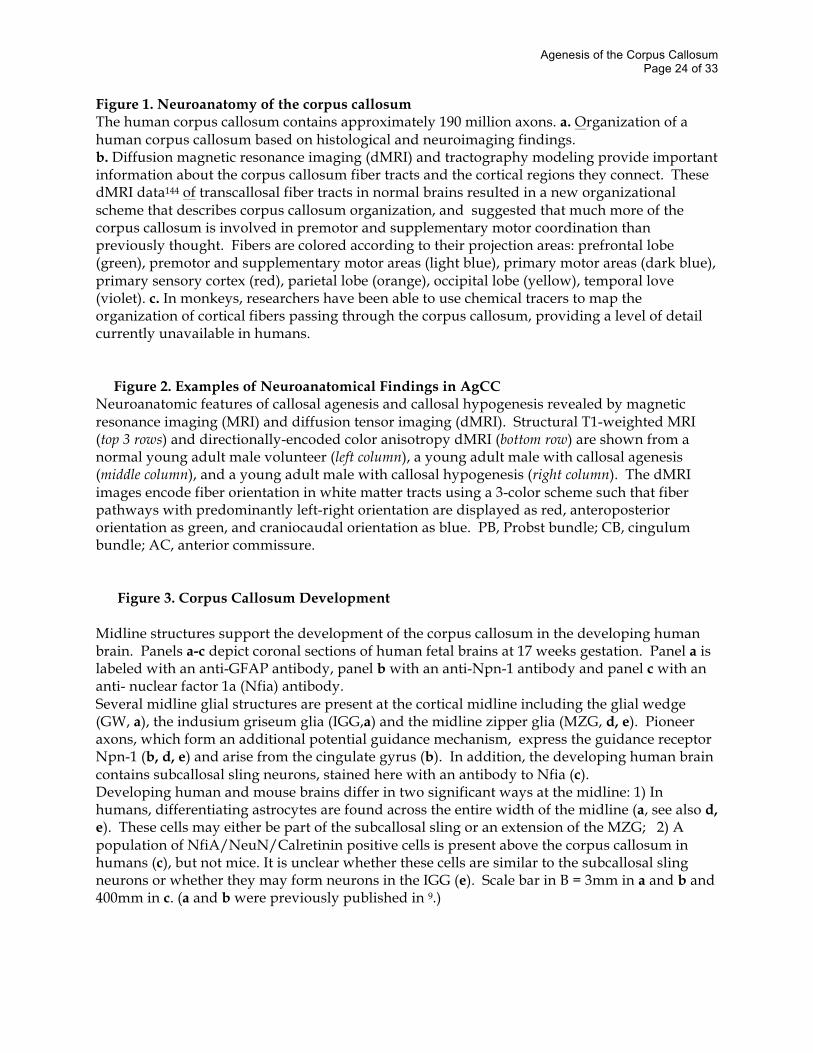
Figure 1. Neuroanatomy of the corpus callosum The human corpus callosum contains approximately 190 million axons. a. Organization of a human corpus callosum based on histological and neuroimaging findings. b. Diffusion magnetic resonance imaging (dMRI) and tractography modeling provide important information about the corpus callosum fiber tracts and the cortical regions they connect. These dMRI data144 of transcallosal fiber tracts in normal brains resulted in a new organizational scheme that describes corpus callosum organization, and suggested that much more of the corpus callosum is involved in premotor and supplementary motor coordination than previously thought. Fibers are colored according to their projection areas: prefrontal lobe (green), premotor and supplementary motor areas (light blue), primary motor areas (dark blue), primary sensory cortex (red), parietal lobe (orange), occipital lobe (yellow), temporal love (violet). c. In monkeys, researchers have been able to use chemical tracers to map the organization of cortical fibers passing through the corpus callosum, providing a level of detail currently unavailable in humans.
Figure 2. Examples of Neuroanatomical Findings in AgCC
Neuroanatomic features of callosal agenesis and callosal hypogenesis revealed by magnetic resonance imaging (MRI) and diffusion tensor imaging (dMRI). Structural T1-weighted MRI (top 3 rows) and directionally-encoded color anisotropy dMRI (bottom row) are shown from a normal young adult male volunteer (left column), a young adult male with callosal agenesis (middle column), and a young adult male with callosal hypogenesis (right column). The dMRI images encode fiber orientation in white matter tracts using a 3-color scheme such that fiber pathways with predominantly left-right orientation are displayed as red, anteroposterior orientation as green, and craniocaudal orientation as blue. PB, Probst bundle; CB, cingulum bundle; AC, anterior commissure.
Figure 3. Corpus Callosum Development
Midline structures support the development of the corpus callosum in the developing human brain. Panels a-c depict coronal sections of human fetal brains at 17 weeks gestation. Panel a is labeled with an anti-GFAP antibody, panel b with an anti-Npn-1 antibody and panel c with an anti- nuclear factor 1a (Nfia) antibody. Several midline glial structures are present at the cortical midline including the glial wedge (GW, a), the indusium griseum glia (IGG,a) and the midline zipper glia (MZG, d, e). Pioneer axons, which form an additional potential guidance mechanism, express the guidance receptor Npn-1 (b, d, e) and arise from the cingulate gyrus (b). In addition, the developing human brain contains subcallosal sling neurons, stained here with an antibody to Nfia (c). Developing human and mouse brains differ in two significant ways at the midline: 1) In humans, differentiating astrocytes are found across the entire width of the midline (a, see also d, e). These cells may either be part of the subcallosal sling or an extension of the MZG; 2) A population of NfiA/NeuN/Calretinin positive cells is present above the corpus callosum in humans (c), but not mice. It is unclear whether these cells are similar to the subcallosal sling neurons or whether they may form neurons in the IGG (e). Scale bar in B = 3mm in a and b and 400mm in c. (a and b were previously published in 9.)
Agenesis of the Corpus Callosum Page 25 of 33
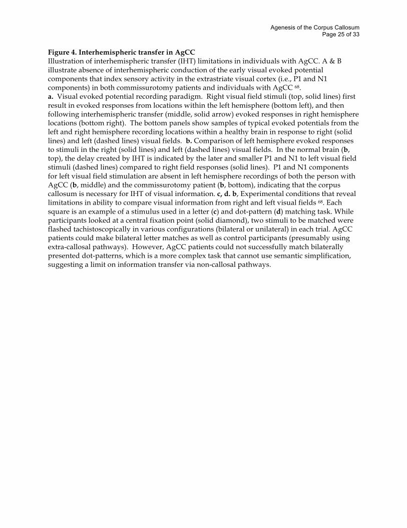
Figure 4. Interhemispheric transfer in AgCC Illustration of interhemispheric transfer (IHT) limitations in individuals with AgCC. A & B illustrate absence of interhemispheric conduction of the early visual evoked potential components that index sensory activity in the extrastriate visual cortex (i.e., P1 and N1 components) in both commissurotomy patients and individuals with AgCC 68. a. Visual evoked potential recording paradigm. Right visual field stimuli (top, solid lines) first result in evoked responses from locations within the left hemisphere (bottom left), and then following interhemispheric transfer (middle, solid arrow) evoked responses in right hemisphere locations (bottom right). The bottom panels show samples of typical evoked potentials from the left and right hemisphere recording locations within a healthy brain in response to right (solid lines) and left (dashed lines) visual fields. b. Comparison of left hemisphere evoked responses to stimuli in the right (solid lines) and left (dashed lines) visual fields. In the normal brain (b, top), the delay created by IHT is indicated by the later and smaller P1 and N1 to left visual field stimuli (dashed lines) compared to right field responses (solid lines). P1 and N1 components for left visual field stimulation are absent in left hemisphere recordings of both the person with AgCC (b, middle) and the commissurotomy patient (b, bottom), indicating that the corpus callosum is necessary for IHT of visual information. c, d. b, Experimental conditions that reveal limitations in ability to compare visual information from right and left visual fields 68. Each square is an example of a stimulus used in a letter (c) and dot-pattern (d) matching task. While participants looked at a central fixation point (solid diamond), two stimuli to be matched were flashed tachistoscopically in various configurations (bilateral or unilateral) in each trial. AgCC patients could make bilateral letter matches as well as control participants (presumably using extra-callosal pathways). However, AgCC patients could not successfully match bilaterally presented dot-patterns, which is a more complex task that cannot use semantic simplification, suggesting a limit on information transfer via non-callosal pathways.
Agenesis of the Corpus Callosum Page 26 of 33
Table 1. Syndromes associated with AgCC#
A. Syndromes with identified genes Gene symbol Salient Features Refs
a. Andermann syndrome KCC3 AgCC, progressive neuropathy and dementia
b. XLAG ARX Lissencephaly, AgCC, intractable epilepsy
c. Mowat Wilson syndrome ZFHX1B Hirschsprung disease, AgCC
d. AgCC with fatal lactic acidosis MRPS16 Complex I & IV deficiency, AgCC, brain malformations
e. HSAS/MASA syndromes L1CAM Hydrocephalus, adducted thumbs, AgCC, MR
B. Syndromes with AgCC seen consistently, no gene yet identified
a. Acrocallosal syndrome AgCC, polydactyly, craniofacial changes, MR
b. Aicardi syndrome AgCC, chorioretinal lacunae, infantile spasms, MR
c. Chudley McCullough syndrome Hearing loss, hydrocephalus, AgCC, colpocephaly
d. Donnai Barrow syndrome Diaphragmatic hernia, exomphalos, AgCC, deafness
e. FG syndrome MR, AgCC, craniofacial changes, macrocephaly
f. Genitopatellar syndrome Absent patellae, urogenital malformations, AgCC
g. Temtamy syndrome AgCC, optic coloboma, craniofacial changes, MR
h. Toriello Carey syndrome AgCC, craniofacial changes, cardiac defects, MR
i. Vici syndrome AgCC, Albinism, Recurrent infections, MR
C. Syndromes with AgCC seen occasionally (partial list)*
a. AgCC with spastic paraparesis SPG11 Progressive spasticity and neuropathy, Thin CC
b. Craniofrontonasal syndrome Coronal craniosynostosis, facial asymmetry, bifid nose
c. Fryns syndrome CDH, pulmonary hypoplasia, craniofacial changes
d. Marden Walker syndrome Blepharophimosis, micrognathia, contractures, AgCC
e. Meckel Gruber syndrome Encephalocele, polydactyly, and polycystic kidneys
f. Microphthalmia with linear skin defects Microopthalmia, linear skin markings, seizures g. Opitz G syndrome Pharyngeal cleft,
craniofacial changes, AgCC, MR h. Orofaciodigital syndrome Tongue hamartoma,
microretrognathia, clinodactyly i. Pyruvate decarboxylase deficiency Lactic acidosis,
seizures, severe MR and spasticity
Agenesis of the Corpus Callosum Page 27 of 33
j. Rubinstein-Taybi syndrome Broad thumbs and great toes, MR, microcephaly
k. Septo-optic dysplasia (DeMorsier syndrome) Hypoplasia of septum pellucidum and optic chiasm
l. Sotos syndrome Physical overgrowth, MR, craniofacial changes
m. Warburg Micro syndrome Microcephaly, microopthalmia, microgenitalia, MR
n. Wolf-Hirschhorn syndrome Microcephaly, seizures, cardiac defects, 4p-
#reliable incidence data is unavailable for these very rare syndromes *Many of these may also consistently have a thin or dysplastic corpus callosum, such as Sotos syndrome or AgCC with spastic paraparesis (SPG11). The overlap between AgCC and these conditions is still under investigation. Other gene symbols are omitted from section C.
Agenesis of the Corpus Callosum Page 28 of 33
Glossary hypoplasia See Box 3 interhemispheric transfer (IHT) Transmission of information between the cerebral hemispheres, typically assessed with laterally presented stimuli (see Box 1 and Figure 4). pioneer axons These are axons that innervate targets early in development and form a substrate for the guidance of later developing axons. retrospective chart reviews A retrospective analysis of medical records of a group of individuals with a particular condition or disease, typically used to study rare diseases for which prospective identification and follow up are difficult. Mendelian disorder A disorder controlled by a single genetic locus which is inherited according to Mendel’s Laws. X-inactivation Early embryonic inactivation of the genes in each cell on one of a female’s X chromosomes (may also occur in males with Klinefelter syndrome who have more than one X-chromosome). X-inactivation results in dosages of the proteins from genes on the X-chromosomes being equivalent to those in typical males (who only have one X chromosome to begin with). isolated AgCC Neuroanatomic description which includes complete absence of the corpus callosum, without other confounding brain abnormalities such as polymicrogyria, heterotopia or schizencephaly. Individuals with isolated AgCC frequently have colpocephaly and Probst Bundles. MKS3 encephaloceles A neural tube defect (NTD) that results in a sac-like protrusion of brain tissue and overlying meninges. These NTD’s are frequently associated with other brain or craniofacial malformations and clinically with broad-ranging neurologic problems. polydactyly The anatomic variant of having more than the normal number of digits on the hands or feet. This is observed in approximately 1:500 births and is usually inherited as an autosomal dominant trait with variable penetrance. Hirschsprung disease A developmental disorder of the enteric nervous system resulting in absence of the neuronal ganglion cells in the distal colon, resulting in a functional obstruction of the colon. Can present
Agenesis of the Corpus Callosum Page 29 of 33
with a dramatically distended colon (megacolon) or with bowel perforation. It is a cardinal feature of Mowat-Wilson syndrome. haploinsufficiency A clinically evident symptom arising when one of the two copies of a gene is mutated, leaving a single functional copy and a presumed reduction in the level of the encoded protein. Klinefelter syndrome A genetic syndrome defined as a 47, XXY karyotype in a phenotypic male. Patients frequently have small testes, minimal sperm production, breast enlargement in puberty and psychosocial problems. chorioretinal lacunae Punched out lesions in the pigmented layer of the retina that cluster around the optic disc that are pathognomonic for Aicardi syndrome. microarray-based comparative genomic hybridization (CGH) A method that compares the quantity of DNA across the whole genome between two individuals. Two DNA samples are labeled red and green respectively and both hybridized to a slide that has arrayed many thousands of spots containing DNA from unique places in the genome. The color ratio at each spot determines the relative amount of DNA present between the two samples. Sotos syndrome Also known as cerebral gigantism, is a genetic disorder that results in early physical overgrowth and cognitive impairment. Most cases are caused by haploinsufficiency of the gene NSD-1, which is a coregulator for steroid receptors. heterotopia In general, this terms refers to the displacement of neuronal cell bodies into the white matter. commissurotomy Surgical procedure that involves severing the corpus callosum as well as the anterior commissures (may also include severing of posterior and hippocampal commissures). This is the original “split-brain” procedure as reported by Roger Sperry 4. tachistoscopic Presentation of visual stimuli more rapidly than the eyes can move. Tachistoscopic presentation thus results in a visual stimulus being perceived in only one hemisphere and representation of the image in the opposite hemisphere will require interhemispheric transfer of information. Stroop interference effect A reaction time measure when identifying one feature of a stimulus, while inhibiting a dominant tendency to identify it according to an interfering feature (for example the normally increased reaction time when naming the ink color of the word Red printed in green ink). bilateral field advantage
Agenesis of the Corpus Callosum Page 30 of 33
The normal decrease in reaction time when comparing two stimuli presented in opposite visual hemifields, compared to presentation of both within one hemifield. The reason for this advantage is dual processing, i.e. each hemisphere only has to process on stimulus. Without efficient IHT, there cannot be such an advantage. anterior commissure Small band of approximately 50,000 axons that connect the cerebral hemispheres. The anterior commissure connects the temporal lobes and is located at the base of the fornix. dichotic listening An research method testing language lateralization by simultaneously presenting different auditory input to each ear. The degree to which subjects preferentially recall information from one ear or the other is an indication of which hemisphere is dominant in language processing. syntax Grammatical arrangement of words and phrases in a sentence, which impacts relationships of meaning. For example changing the placement of a word or phrase can change the meaning. linguistic pragmatics The processes that allows one to go beyond literal meaning of language and actually interpret the speaker’s intended meaning. This may involve utilizing second-order meanings, body language, vocal inflection, context, and other factors. alexithymia Impairment in expression of one’s feelings and mood-states. A dominant hypothesis is that alexithymia arises from compromised connection between the language processing in the left hemisphere and affect processing in the right. valence A continuous scale from pleasant to aversive. dMRI: diffusion magnetic resonance imaging This broad term covers both diffusion weighted MRI data acquisition and image analysis of this data 117, including diffusion tensor imaging (DTI:TERM). The MRI signal is weighted by the amount of water diffusion within tissues. The weighting can vary with direction allowing diffusion anisotropy arising from microscopic restrictions in biological tissues to be observed. Andermann syndrome This is defined in the genetics section DTI: diffusion tensor imaging Anisotropic diffusion within tissues is modeled as a second-rank tensor which can be calculated from diffusion-weighted MRI acquired in six or more non-collinear directions. The tensor at each point in the image can be visualized as an oriented and scaled ellipsoid. More simply, quantities such as the mean diffusivity and fractional anisotropy can be calculated from the tensor and visualized as conventional images. The tensor contains information about likely axonal fiber direction and can be used to create virtual fiber tracts through the DTI, reflecting structural connectivity in white matter.
Agenesis of the Corpus Callosum Page 31 of 33
callosotomy Surgical procedure that involves severing only the corpus callosum, either in part or in its entirety, leaving other commissures intact. This has also been described by some as a “split-brain” procedure 5. Endophenotypes Hereditary characteristics that are associated with a condition, but are not a direct symptom of that condition
Agenesis of the Corpus Callosum Page 32 of 33
Author Biographies
Lynn K. Paul – Dr. Paul began research on agenesis of the corpus callosum with Dr. Warren Brown in 1993, as part of her doctoral work in clinical psychology at Fuller Theological Seminary. Her research has focused on the cognitive and psychosocial characteristics in individuals with Primary AgCC. In 2004, she began the Corpus Callosum Research Program within Dr. Adolph’s laboratory and is now a Senior Research Fellow at Caltech. This program involves histological, neuroimaging, and social-cognition studies of AgCC. Dr. Paul was also the founding president of the National Organization for Disorders of the Corpus Callosum, a non-profit that gathers and disseminates information about these conditions (www.nodcc.org). Elliott H. Sherr - Dr. Sherr is an Assistant Professor in Neurology and Pediatrics at UCSF. He directs the Brain Development Research Program, studying the genetics and biology of AgCC and other developmental brain malformations. He obtained his undergraduate degree at Stanford University and his M.D. and Ph.D. at Columbia University. Ralph Adolphs - Dr. Adolphs received his undergraduate degree from Stanford University, and his graduate training in neurobiology from the California Institute of Technology. Since 1993 he has been investigating the neural basis of emotion and social behavior in humans. Much of this work studied neurological patients with focal brain lesions, research he conducted first as a postdoc and subsequently as faculty at the University of Iowa. Since 2004, Dr. Adolphs has been professor of psychology and neuroscience at the California Institute of Technology, while maintaining an adjunct appointment also at the University of Iowa. His long-term goals are to understand how emotion modulates perception, attention, memory, and social cognition. Dr. Adolphs is the recipient of awards from the Sloan Foundation, the Klingenstein Fund, the EJLB Foundation, and currently holds a 21st Century Science Award from the James S. McDonnell Foundation. Warren Brown - Warren S. Brown (Ph.D., University of Southern California) is currently Professor of Psychology at the Fuller Graduate School of Psychology, where he is Director of the Lee Travis Research Institute. Prior to Fuller, Brown was at the UCLA Department of Psychiatry, where he was involved in research on childhood schizophrenia, Alzheimer’s disease, and autism. He continues to be a member of the UCLA Brain Research Institute. His primary research interest is the role of the corpus callosum in human higher cognitive and social processes. For the last 15 years, Brown’s laboratory has been studying the cognitive and psychosocial consequences of agenesis of the corpus callosum in individuals who have intelligence within the normal range. Pratik Mukherjee - Dr. Mukherjee is a neuroradiologist and assistant professor of radiology and bioengineering at the University of California, San Francisco. His research over the past 8 years has focused on applications of diffusion tensor imaging (DTI) to the noninvasive microstructural analysis of biological tissues with special emphasis on the developing brain. He published some of the first DTI studies in the areas of stroke, hypertensive encephalopathy, and childhood brain development and is currently conducting prospective studies of DTI in
Agenesis of the Corpus Callosum Page 33 of 33
traumatic brain injury and callosal dysgenesis. Dr. Mukherjee continues to develop diffusion MRI methods that surpass DTI for non-invasive mapping of human white matter.
Linda Richards – Dr. Richards completed her PhD in 1994 at The Walter and Eliza Hall Institute, Melbourne, Australia in the laboratory of Prof. Perry Bartlett and postdoctoral training at The Salk Institute for Biological Studies, California, with Prof. Dennis O’Leary. She began her laboratory at The University of Maryland in 1997, where she remained until 2005 when she was appointed as an Associate Professor at The University of Queensland, Australia. Her laboratory investigates the development of the cerebral cortex and midline commissure formation in the brain. The focus is on the development and patterning of the cortical midline and aspects of glial and neuronal development that impact the formation of midline structures such as the corpus callosum.
J. Michael Tyszka – Dr. Tyszka is a graduate of Cambridge University where he developed a long-standing interest in magnetic resonance imaging under Laurie Hall. He began his collaboration with Scott Fraser's Biological Imaging Center at Caltech in the mid 1990s before joining the group full-time in 2001. He has published widely in the magnetic resonance imaging technical literature and his research interests range from engineering in MR microscopy, through MR diffusion histology, to the integration of structural and effective connectivity models of human brain function. He is currently Director of Magnetic Resonance Physics at the Caltech Brain Imaging Center in Pasadena, California.