agrobacterium-mediated genetic transformation of ... papers/jst vol... · the reporter system....
TRANSCRIPT

ISSN: 0128-7680Pertanika J. Sci. & Technol. 16 (2): 129 - 139 (2008) © Universiti Putra Malaysia Press
Agrobacterium-mediated Genetic Transformation of Phalaenopsisbellina Using GFP and GUS Reporter Genes
Mahmood Maziah* and Chew Yee ChernDepartment of Biochemistry, Faculty of Biotechnology and Biomolecular Sciences,
Universiti Putra Malaysia, 43400 UPM, Serdang, Selangor, Malaysia*E-mail: [email protected]
ABSTRACTGenetic transformation protocols of Phalaenopsis bellina protocorm-like bodies (PLBs)were established using gfp (green fluorescent protein) and gus (!-glucuronidase) genes asthe reporter system. Agrobacterium tumefaciens strain, LBA 4404 containing the binaryvector, pCAMBIA 1304, with the hptII gene as the selectable marker and gfp and gus-introngenes as the reporter genes. Horizontally dissected PLBs were immersed in A. tumefacienssuspensions with 200uM acetosyringone (AS) for 45 minutes. This was followed by co-cultivation until the growth of A. tumefaciens was observed surrounding the PLBs on theco-cultivation medium. GFP detection and GUS histochemical assay were carried out toinvestigate the transient expression of both GFP and GUS reporter genes. The selectionof proliferating PLBs was carried out on 4 mg/L hygromycin and 100 mg/L cefotaxime.GFP could be used as the reporter system as it is an effective, rapid and non-destructivesystem to monitor the transformed tissues.
Keywords: Agrobacterium tumefaciens, genetic transformation, gfp, gus, Phalaenopsis bellina
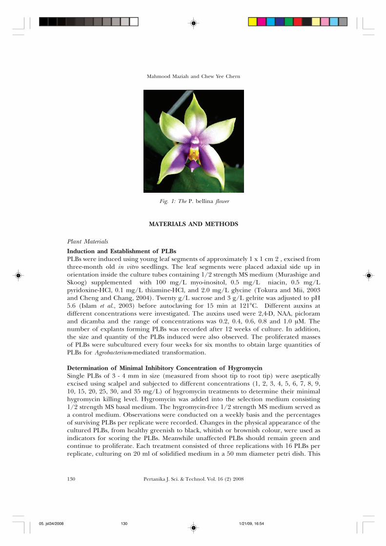
INTRODUCTIONPhalaenopsis bellina (Fig. 1) is a commercially important fragrant orchid species endemicto Borneo and Peninsular Malaysia. The attractive features of this orchid are that theyflower freely all year round and produce strong sweet-flora fragrance. Genetic improvementof P. bellina through sexual hybridization is, however, restricted by a long growth periodand limited genetic pool within the germplasm. Genetic engineering offers a promisingapproach in improving the orchid quality.
A protocol was developed to obtain transient expression of P. bellina via Agrobacteriumtumefaciens (Strain LBA 4404 harboring vector pCAMBIA 1304). Prior to the genetictransformation study, hygromycin sensitivity of the PLBs was investigated to determinethe minimal concentration required to sufficiently inhibit the growth of PLBs. Transientexpression of gfp gene in PLBs was observed under the florescence microscope after co-cultivation period. The effect of inoculation time on transient GFP expression was alsoevaluated in this study. The putative transformants displayed distinguishingly strongfluorescence when observed under a GFP stereomicroscope using GFP2 filter.
Received : 2 August 2007Accepted: 21 April 2008* Corresponding Author
05. jst34/2008 1/21/09, 16:54129
Mahmood Maziah and Chew Yee Chern
Pertanika J. Sci. & Technol. Vol. 16 (2) 2008130
MATERIALS AND METHODS
Plant MaterialsInduction and Establishment of PLBsPLBs were induced using young leaf segments of approximately 1 x 1 cm 2 , excised fromthree-month old in vitro seedlings. The leaf segments were placed adaxial side up inorientation inside the culture tubes containing 1/2 strength MS medium (Murashige andSkoog) supplemented with 100 mg/L myo-inositol, 0.5 mg/L niacin, 0.5 mg/Lpyridoxine-HCl, 0.1 mg/L thiamine-HCl, and 2.0 mg/L glycine (Tokura and Mii, 2003and Cheng and Chang, 2004). Twenty g/L sucrose and 3 g/L gelrite was adjusted to pH5.6 (Islam et al., 2003) before autoclaving for 15 min at 121°C. Different auxins atdifferent concentrations were investigated. The auxins used were 2,4-D, NAA, picloramand dicamba and the range of concentrations was 0.2, 0.4, 0.6, 0.8 and 1.0 µM. Thenumber of explants forming PLBs was recorded after 12 weeks of culture. In addition,the size and quantity of the PLBs induced were also observed. The proliferated massesof PLBs were subcultured every four weeks for six months to obtain large quantities ofPLBs for Agrobacterium-mediated transformation.
Determination of Minimal Inhibitory Concentration of HygromycinSingle PLBs of 3 - 4 mm in size (measured from shoot tip to root tip) were asepticallyexcised using scalpel and subjected to different concentrations (1, 2, 3, 4, 5, 6, 7, 8, 9,10, 15, 20, 25, 30, and 35 mg/L) of hygromycin treatments to determine their minimalhygromycin killing level. Hygromycin was added into the selection medium consisting1/2 strength MS basal medium. The hygromycin-free 1/2 strength MS medium served asa control medium. Observations were conducted on a weekly basis and the percentagesof surviving PLBs per replicate were recorded. Changes in the physical appearance of thecultured PLBs, from healthy greenish to black, whitish or brownish colour, were used asindicators for scoring the PLBs. Meanwhile unaffected PLBs should remain green andcontinue to proliferate. Each treatment consisted of three replications with 16 PLBs perreplicate, culturing on 20 ml of solidified medium in a 50 mm diameter petri dish. This
Fig. 1: The P. bellina flower
05. jst34/2008 1/21/09, 16:54130
131Pertanika J. Sci. & Technol. Vol. 16 (2) 2008
Agrobacterium-mediated Genetic Transformation of Phalaenopsis bellina
experiment was repeated three times. Eventually, the specific concentration that wassufficient to completely inhibit growth of PLBs or kill the PLBs would be determined andlater applied for screening of putative transformed PLBs in the genetic transformationstudy.
Preparation of A. tumefaciens Strain and PlasmidA. tumefaciens strain LBA 4404 (pCAMBIA 1304) was used for the transformation study.The T-DNA region of the binary vector pCAMBIA 1304 contains the selectable markerhptII gene, encoding hygromycin phosphotransferase, the reporter gfp, and intron-gusgenes. The !-glucuronidase gene was disrupted by an intron. This intron-gus reportergene expresses GUS activity in plant cells but not in the cell of A.tumefaciens. Expressionof gfp, gus and hptII genes are under the control of the cauliflower mosaic virus (CaMV)35S promoter.
The Effect of Inoculation TimeThe A. tumefaciens strain LBA 4404 was grown overnight at 28°C in liquid LB brothmedium containing 100 mg/L kanamycin. The following day, 500 ul of the bacterialsuspension was spread over the surface of LB agar solid medium, and incubated at 28°Cfor 2 days. A. tumefaciens cells were collected with a aseptic flame inoculums andsuspended in 30 ml LB liquid medium containing 100 mg/L kanamycin and 200 uM ASto and OD600 at 0.7 - 1.0 and agitated (100 rpm) in a shaker, at 25°C for 30 min beforeinoculating. PLBs of P. bellina were cut into pieces, 3-4 mm in diameter. The PLBs werethen immersed in the A. tumefaciens suspension for 15, 30, 45, 60, 75 and 90 minrespectively, with 20 PLBs per treatment. These PLBs were blotted dry on sterile filterpaper, and co-cultured on 1/2 MS medium containing 200 µM AS at 25°C in the darkfor 3 days until A. tumefaciens growth was observed. This experiment was repeated 3 times.
GFP MonitoringGFP-expressing cells were detected using a fluorescence microscope (Leica MZFL III)equipped with GFP2 filter (Excitation filter: 480/40 nm) to mask the red fluorescence ofchlorophyll, thereby permitting the visualization of the green fluorescence GFP-expressingcells. The PLBs observed with green fluorescent sections (using magnification 25X) wereconsidered GFP positive. An imaging system (Laica DC 200) was attached to thefluorescence microscope to capture the image in real time using the Leica DC Viewersoftware.
Selection of Putative TransformantsAfter 2-3 days of co-cultivation, the cultures were transferred to selective medium (1/2MS medium containing 100 mg/L cefotaxime, 4 mg/L hygromycin) and incubated at25°C under 16 photoperiod. Previous research indicated that 4 mg/L hygromycin wassufficient to inhibit the growth of non-transformed tissues of P. bellina. Tissues weresubcultured in a new selective medium every month. Cefotaxime were omitted after twomonths of selection. After three months of being cultured on the hygromycin-containingmedium, the surviving and growing cell clusters were picked out and cultured on 1/2 MSmedium supplemented with 4 mg/L hygromycin but without phytohormones for plantregeneration.
05. jst34/2008 1/21/09, 16:54131
Mahmood Maziah and Chew Yee Chern
Pertanika J. Sci. & Technol. Vol. 16 (2) 2008132
GUS Histochemical AssayGUS activity assays were performed on PLBs, hygromycin-resistent PLBs and then on leavesand roots of the regenerated transformants using the method of Jefferson et al., (1986).Tissues were immersed in X-gluc solution, (1 mM EDTA, 50 mM NaH2PO4 (pH 7.0), 10mM -mercaptoethanol and 0.1% Triton X-100) and incubated overnight at 37°C. Afterstaining, the materials were treated with 70% ethanol to remove chlorophyll beforeobservation. Transient GUS expression of PLBs was examined after 2 days of co-cultivation.
RESULTS AND DISCUSSION
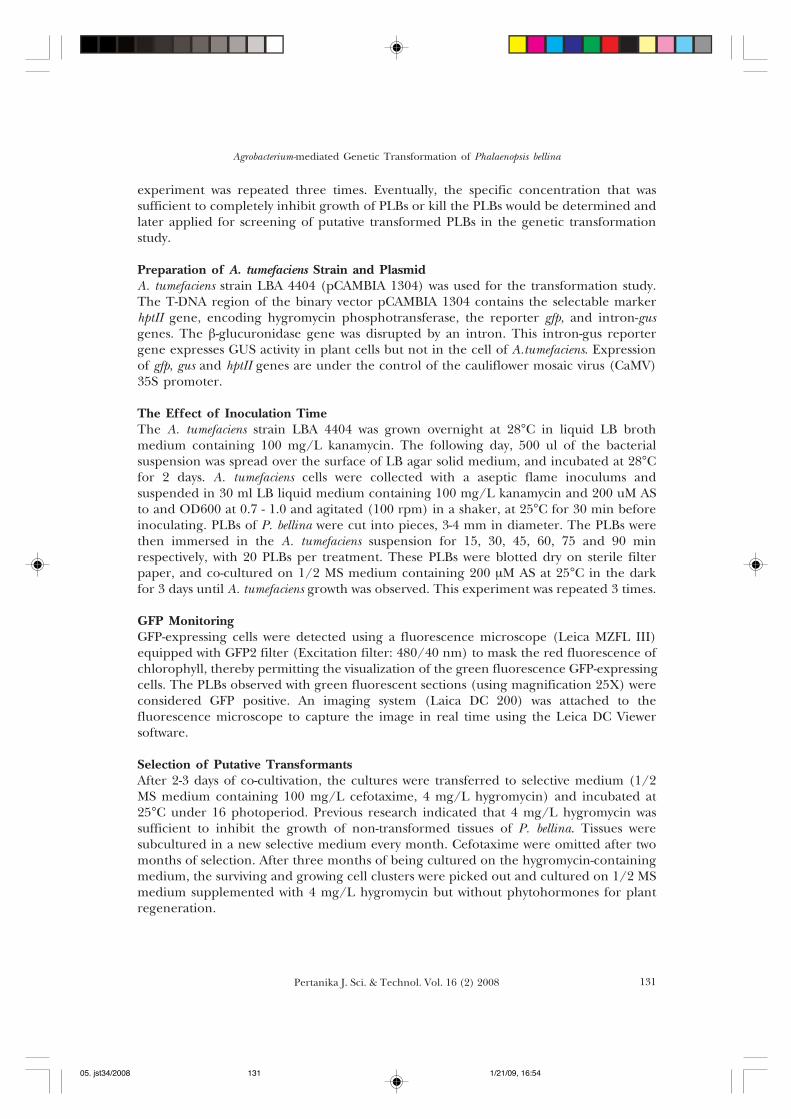
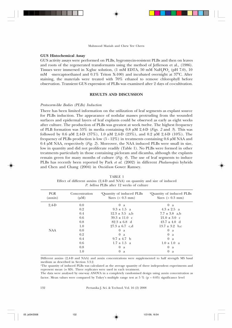
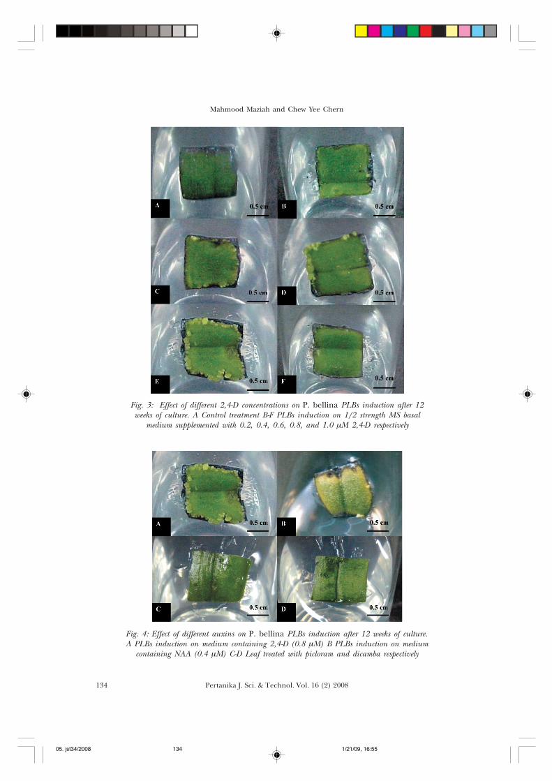
Protocorm-like Bodies (PLBs) InductionThere has been limited information on the utilization of leaf segments as explant sourcefor PLBs induction. The appearance of nodular masses protruding from the woundedsurfaces and epidermal layers of leaf explants could be observed as early as eight weeksafter culture. The production of PLBs was greatest at week twelve. The highest frequencyof PLB formation was 53% in media containing 0.8 µM 2,4-D (Figs. 2 and 3). This wasfollowed by 0.6 µM 2,4-D (37%), 1.0 µM 2,4-D (23%), and 0.2 µM 2,4-D (10%). Thefrequency of PLBs production is low (5 - 12%) in treatments containing 0.6 µM NAA and0.4 µM NAA, respectively (Fig. 2). Moreover, the NAA induced PLBs were small in size,low in quantity and did not proliferate readily (Table 1). No PLBs were formed in othertreatments particularly in those containing picloram and dicamba, although the explantsremain green for many months of culture (Fig. 4). The use of leaf segments to inducePLBs has recently been reported by Park et al. (2002) in different Phalaenopsis hybridsand Chen and Chang (2004) in Oncidium Gower Ramsey.
TABLE 1Effect of different auxins (2,4-D and NAA) on quantity and size of induced
P. bellina PLBs after 12 weeks of culture
PGR Concentration aQuantity of induced PLBs aQuantity of induced PLBs(auxin) (µM) Sizes (< 0.3 mm) Sizes (> 0.3 mm)
2,4-D 0.0 0 a 0 a0.2 9.3 ± 1.5 a 4.3 ± 2.5 a0.4 12.3 ± 3.5 a,b 7.7 ± 3.8 a,b0.6 39.3 ± 11.0 c 21.0 ± 3.0 c0.8 82.3 ± 6.8 d 43.7 ± 4.0 d1.0 27.3 ± 6.7 c,d 13.7 ± 3.2 b,c
NAA 0.0 0 a 0 a0.2 0 a 0 a0.4 9.7 ± 4.7 b 0 a0.6 1.7 ± 1.5 a 1.0 ± 1.0 a0.8 0 a 0 a1.0 0 a 0 a
Different auxins (2,4-D and NAA) and auxin concentrations were supplemented to half strength MS basalmedium as described in Section 3.3.2.aThe quantity of induced PLBs was calculated as the average quantity of three independent experiments andrepresent mean (± SD). Three replicates were used in each treatment.The data were analysed by one-way ANOVA in a completely randomised design using auxin concentration asfactor. Mean values were compared by Tukey’s multiple range test at 5 % (p = 0.05) significance level
05. jst34/2008 1/21/09, 16:54132

133Pertanika J. Sci. & Technol. Vol. 16 (2) 2008
Agrobacterium-mediated Genetic Transformation of Phalaenopsis bellina
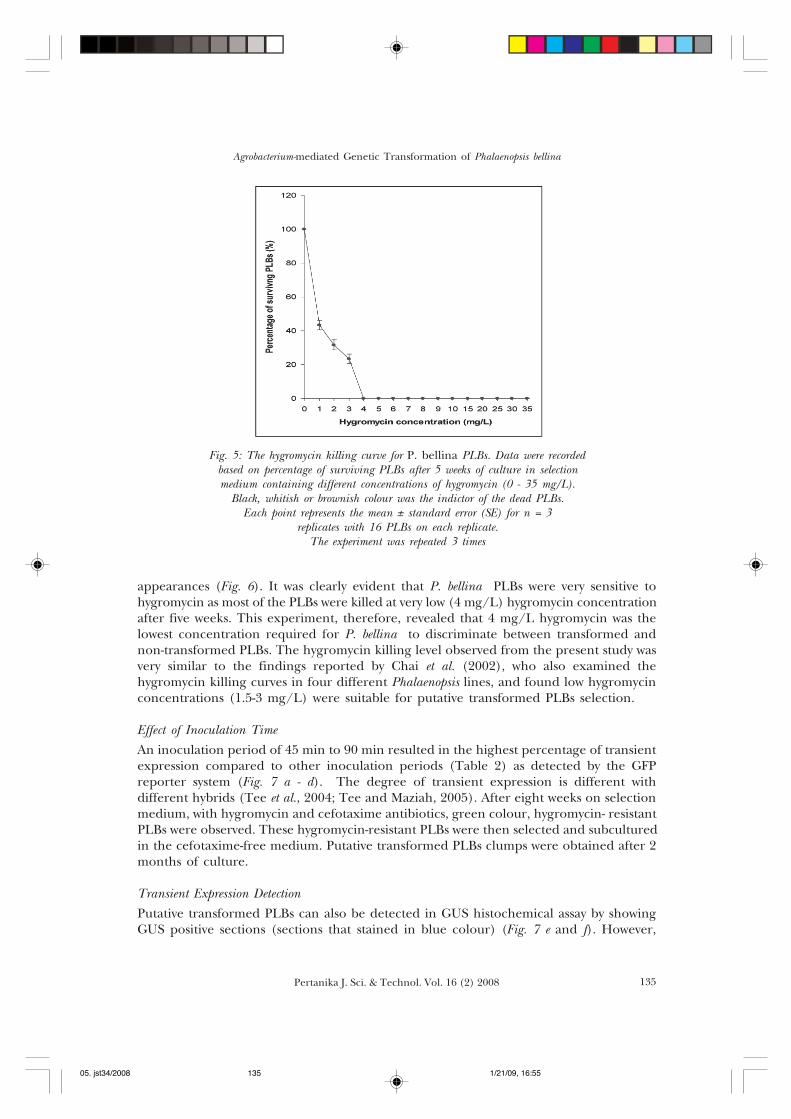
Determination of Hygromycin Killing CurveIn this experiment, 16 two-month-old PLBs of P. bellina were placed on 1/2 strength MSbasal medium containing various concentrations of hygromycin (0, 1, 2, 3, 4, 5, 6, 7, 8,9, 10, 15, 20, 25, 30, and 35 mg/L) for five weeks, and the number of surviving PLBs wasrecorded weekly. Black, whitish or brownish colour was an indicator of dead PLBs. Anearly inhibitory effect of hygromycin at 10 mg/L on PLBs was initially observed duringthe first week of culture. Increased hygromycin concentration reduced the growthfrequency from 100% at hygromycin 0 mg/L to 43% at hygromycin 1 mg/L, to 32% athygromycin 2 mg/L, to 23% at hygromycin 3 mg/L and to 0% at hygromycin 4 - 35 mg/L (Fig. 5). All of the PLBs multiplied healthily and remained greenish on hygromycin-free medium, whereas, PLBs cultured on medium containing " 4 mg/L hygromycin, for5 weeks resulted in complete fatality, giving entirely black, brown or white necrotic PLBs
Fig. 2: Effect of different auxins concentrations on PLBs induction from leaf segments of P. bellina.All results were scored after 12 weeks of culture. The results indicate the mean standard error
(± SE) of 3 independent experiments with 10 replicates for each treatment concentration.The experiment was repeated thrice. The data were analysed by one-way ANOVA in acompletely randomised design using auxin concentration as factor. Mean values were
compared by Tukey’s multiple range test at 5 % (p = 0.05) significance level
Perc
enta
ge o
f PL
Bs
indu
ced
(%)
a,b
a,b a,b
c,d
d
b,c
Auxin types
b
05. jst34/2008 1/21/09, 16:54133
Mahmood Maziah and Chew Yee Chern
Pertanika J. Sci. & Technol. Vol. 16 (2) 2008134
Fig. 3: Effect of different 2,4-D concentrations on P. bellina PLBs induction after 12weeks of culture. A Control treatment B-F PLBs induction on 1/2 strength MS basal
medium supplemented with 0.2, 0.4, 0.6, 0.8, and 1.0 µM 2,4-D respectively
Fig. 4: Effect of different auxins on P. bellina PLBs induction after 12 weeks of culture.A PLBs induction on medium containing 2,4-D (0.8 µM) B PLBs induction on medium
containing NAA (0.4 µM) C-D Leaf treated with picloram and dicamba respectively
05. jst34/2008 1/21/09, 16:55134
135Pertanika J. Sci. & Technol. Vol. 16 (2) 2008
Agrobacterium-mediated Genetic Transformation of Phalaenopsis bellina
appearances (Fig. 6). It was clearly evident that P. bellina PLBs were very sensitive tohygromycin as most of the PLBs were killed at very low (4 mg/L) hygromycin concentrationafter five weeks. This experiment, therefore, revealed that 4 mg/L hygromycin was thelowest concentration required for P. bellina to discriminate between transformed andnon-transformed PLBs. The hygromycin killing level observed from the present study wasvery similar to the findings reported by Chai et al. (2002), who also examined thehygromycin killing curves in four different Phalaenopsis lines, and found low hygromycinconcentrations (1.5-3 mg/L) were suitable for putative transformed PLBs selection.
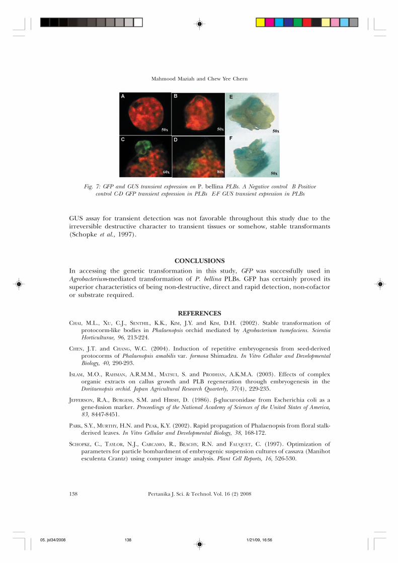
Effect of Inoculation TimeAn inoculation period of 45 min to 90 min resulted in the highest percentage of transientexpression compared to other inoculation periods (Table 2) as detected by the GFPreporter system (Fig. 7 a - d). The degree of transient expression is different withdifferent hybrids (Tee et al., 2004; Tee and Maziah, 2005). After eight weeks on selectionmedium, with hygromycin and cefotaxime antibiotics, green colour, hygromycin- resistantPLBs were observed. These hygromycin-resistant PLBs were then selected and subculturedin the cefotaxime-free medium. Putative transformed PLBs clumps were obtained after 2months of culture.
Transient Expression DetectionPutative transformed PLBs can also be detected in GUS histochemical assay by showingGUS positive sections (sections that stained in blue colour) (Fig. 7 e and f). However,
Fig. 5: The hygromycin killing curve for P. bellina PLBs. Data were recordedbased on percentage of surviving PLBs after 5 weeks of culture in selectionmedium containing different concentrations of hygromycin (0 - 35 mg/L).
Black, whitish or brownish colour was the indictor of the dead PLBs.Each point represents the mean ± standard error (SE) for n = 3
replicates with 16 PLBs on each replicate.The experiment was repeated 3 times
05. jst34/2008 1/21/09, 16:55135
Mahmood Maziah and Chew Yee Chern
Pertanika J. Sci. & Technol. Vol. 16 (2) 2008136
05. jst34/2008 1/21/09, 16:55136
137Pertanika J. Sci. & Technol. Vol. 16 (2) 2008
Agrobacterium-mediated Genetic Transformation of Phalaenopsis bellina
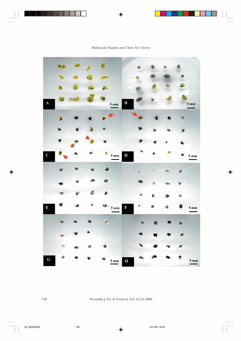
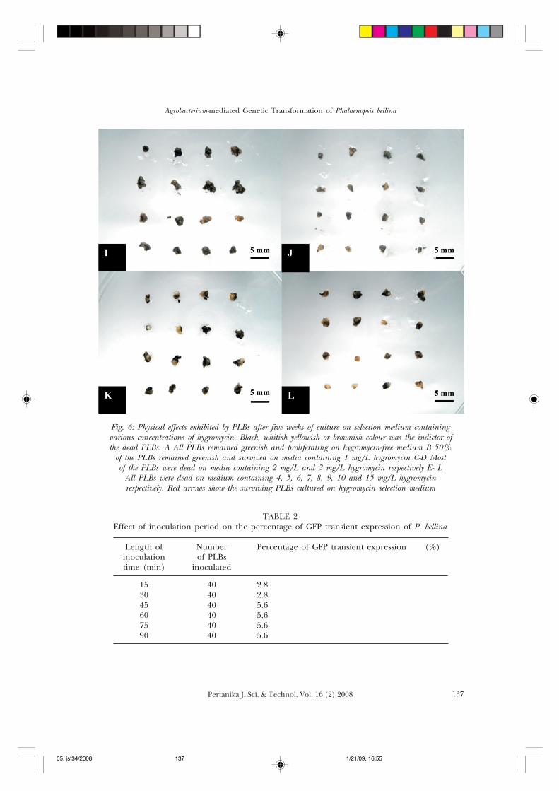
Fig. 6: Physical effects exhibited by PLBs after five weeks of culture on selection medium containingvarious concentrations of hygromycin. Black, whitish yellowish or brownish colour was the indictor ofthe dead PLBs. A All PLBs remained greenish and proliferating on hygromycin-free medium B 50%
of the PLBs remained greenish and survived on media containing 1 mg/L hygromycin C-D Mostof the PLBs were dead on media containing 2 mg/L and 3 mg/L hygromycin respectively E- L
All PLBs were dead on medium containing 4, 5, 6, 7, 8, 9, 10 and 15 mg/L hygromycinrespectively. Red arrows show the surviving PLBs cultured on hygromycin selection medium
TABLE 2Effect of inoculation period on the percentage of GFP transient expression of P. bellina
Length of Number Percentage of GFP transient expression (%)inoculation of PLBstime (min) inoculated
15 40 2.830 40 2.845 40 5.660 40 5.675 40 5.690 40 5.6
05. jst34/2008 1/21/09, 16:55137
Mahmood Maziah and Chew Yee Chern
Pertanika J. Sci. & Technol. Vol. 16 (2) 2008138
GUS assay for transient detection was not favorable throughout this study due to theirreversible destructive character to transient tissues or somehow, stable transformants(Schopke et al., 1997).
CONCLUSIONSIn accessing the genetic transformation in this study, GFP was successfully used inAgrobacterium-mediated transformation of P. bellina PLBs. GFP has certainly proved itssuperior characteristics of being non-destructive, direct and rapid detection, non-cofactoror substrate required.
REFERENCESCHAI, M.L., XU, C.J., SENTHIL, K.K., KIM, J.Y. and KIM, D.H. (2002). Stable transformation of
protocorm-like bodies in Phalaenopsis orchid mediated by Agrobacterium tumefaciens. ScientiaHorticulturae, 96, 213-224.
CHEN, J.T. and CHANG, W.C. (2004). Induction of repetitive embryogenesis from seed-derivedprotocorms of Phalaenopsis amabilis var. formosa Shimadzu. In Vitro Cellular and DevelopmentalBiology, 40, 290-293.
ISLAM, M.O., RAHMAN, A.R.M.M., MATSUI, S. and PRODHAN, A.K.M.A. (2003). Effects of complexorganic extracts on callus growth and PLB regeneration through embryogenesis in theDoritaenopsis orchid. Japan Agricultural Research Quarterly, 37(4), 229-235.
JEFFERSON, R.A., BURGESS, S.M. and HIRSH, D. (1986). !-glucuronidase from Escherichia coli as agene-fusion marker. Proceedings of the National Academy of Sciences of the United States of America,83, 8447-8451.
PARK, S.Y., MURTHY, H.N. and PEAK, K.Y. (2002). Rapid propagation of Phalaenopsis from floral stalk-derived leaves. In Vitro Cellular and Developmental Biology, 38, 168-172.
SCHOPKE, C., TAYLOR, N.J., CARCAMO, R., BEACHY, R.N. and FAUQUET, C. (1997). Optimization ofparameters for particle bombardment of embryogenic suspension cultures of cassava (Manihotesculenta Crantz) using computer image analysis. Plant Cell Reports, 16, 526-530.
Fig. 7: GFP and GUS transient expression on P. bellina PLBs. A Negative control B Positivecontrol C-D GFP transient expression in PLBs E-F GUS transient expression in PLBs
05. jst34/2008 1/21/09, 16:56138
139Pertanika J. Sci. & Technol. Vol. 16 (2) 2008
Agrobacterium-mediated Genetic Transformation of Phalaenopsis bellina
TEE, C.S., MAZIAH, M., TAN, C.S. and ABDULLAH, M.P. (2003). Evaluation of different promotersderiving the GFP reporter gene and selected target tissues for particle bombardment ofDendrobium Sonia 17. Plant Cell Reports, 21, 452-458.
TEE, C.S. and MAZIAH, M. (2005). Optimization of biolistic bombardment parameters for DendrobiumSonia 17 callus using GFP and GUS as the reporter system. Plant Cell, Tissue and Organ Culture,80, 77-89.
TOKUHARA, K. and MII, M. (2003). Highly-efficient somatic embryogenesis from cell suspensioncultures of Phalaenopsis orchids by adjusting carbohydrate sources. In Vitro Cellular andDevelopmental Biology, 39, 635-639.
05. jst34/2008 1/21/09, 16:56139
