airway management and smoke inhalation injuryintheburnpatient · airway management and smoke...
TRANSCRIPT
Airway Managementand Smoke InhalationInjury in the Burn PatientLeopoldo C. Cancio, MD, FACS
Plastic surgeons frequently provide care topatients who have burn injuries and concomitantsmoke inhalation injury (II). About 10% of patientsadmitted to burn centers have II, which greatlyincreases their risk for postburn pneumonia andmortality, especially at the midranges of age andburn size.1–3 This article reviews the essentialdiagnostic and therapeutic interventions in thetreatment of these patients.
An understanding of II and what to do about ithas only developed over the last 50 years.Consider the scene at Massachusetts GeneralHospital on the evening of November 28, 1942,following one of the largest indoor fire disastersin U.S. history, at the Cocoanut Grove nightclub.Of the approximately 1000 occupants, 114 weretaken to Massachusetts General Hospital within2 hours, of whom 39 lived to be admitted:
As the patients from the scene of the disasterwere crowded into the hospital it becameapparent early that they were divided sharplyinto two groups: the living and the dead ornear dead. None in the former group died inthe first 12 hours; none in the latter grouplived more than a few minutes after arrival.4
It is not entirely clear which process—carbonmonoxide poisoning, hypoxia, upper-airwayobstruction, or a combination—was responsiblefor these early deaths:
The first clue to the high incidence of pulmo-nary burns was afforded by the number who
died within the first few minutes after reachingthe hospital. They were cyanotic, comatose,or restless, and had severe upper respiratorydamage.some were cherry-red in color,suggesting carbon monoxide inhalation.4
Of those who were admitted, five developedprogressive dyspnea and pulmonary edema overthe next several hours that required ‘‘radicaltherapy’’ (ie, endotracheal intubation, immediatetracheostomy, and delivery of oxygen by tent ortranstracheal catheter). In the ‘‘final stage’’ of theinjury, they developed diffuse bronchiolitis,mucous plugging, peripheral airway obstruction,and lobular collapse. Uncharacteristically, pneu-monia was not observed.4
Although it is incomplete from a current-daystandpoint with respect to answers, the CocoanutGrove monograph poses many of the same ques-tions that burn specialists, faced with a patientwho has severe II, must address today:
� What are the indications for endotrachealintubation?� What is the ideal timing for tracheostomy?� What diagnostic procedures should be
performed for patients who are suspectedof having II?� Which method of gentle mechanical ventila-
tion should be used for these patients?� Are there any special fluid resuscitation
requirements?� Which drugs may improve outcome?
The opinions or assertions contained herein are the private views of the author, and are not to be construed asofficial or as reflecting the views of the Department of the Army or Department of Defense.U.S. Army Institute of Surgical Research, 3400 Rawley E. Chambers Avenue, Fort Sam Houston, TX 78234-6315,USAE-mail address: [email protected]
KEYWORDS� Smoke inhalation injury � Burns � Burns inhalation� Carbon monoxide poisoning � Hydrogen cyanide� High-frequency ventilation
Clin Plastic Surg 36 (2009) 555–567doi:10.1016/j.cps.2009.05.0130094-1298/09/$ – see front matter. Published by Elsevier Inc. pl
asti
csur
gery
.thec
lini
cs.c
om
Report Documentation Page Form ApprovedOMB No. 0704-0188
Public reporting burden for the collection of information is estimated to average 1 hour per response, including the time for reviewing instructions, searching existing data sources, gathering andmaintaining the data needed, and completing and reviewing the collection of information. Send comments regarding this burden estimate or any other aspect of this collection of information,including suggestions for reducing this burden, to Washington Headquarters Services, Directorate for Information Operations and Reports, 1215 Jefferson Davis Highway, Suite 1204, ArlingtonVA 22202-4302. Respondents should be aware that notwithstanding any other provision of law, no person shall be subject to a penalty for failing to comply with a collection of information if itdoes not display a currently valid OMB control number.
1. REPORT DATE 01 OCT 2009
2. REPORT TYPE N/A
3. DATES COVERED -
4. TITLE AND SUBTITLE Airway management and smoke inhalation injury in the burn patient
5a. CONTRACT NUMBER
5b. GRANT NUMBER
5c. PROGRAM ELEMENT NUMBER
6. AUTHOR(S) Cancio L. C.,
5d. PROJECT NUMBER
5e. TASK NUMBER
5f. WORK UNIT NUMBER
7. PERFORMING ORGANIZATION NAME(S) AND ADDRESS(ES) United States Army Institute of Surgical Research, JBSA Fort Samhouston, TX 78234
8. PERFORMING ORGANIZATIONREPORT NUMBER
9. SPONSORING/MONITORING AGENCY NAME(S) AND ADDRESS(ES) 10. SPONSOR/MONITOR’S ACRONYM(S)
11. SPONSOR/MONITOR’S REPORT NUMBER(S)
12. DISTRIBUTION/AVAILABILITY STATEMENT Approved for public release, distribution unlimited
13. SUPPLEMENTARY NOTES
14. ABSTRACT
15. SUBJECT TERMS
16. SECURITY CLASSIFICATION OF: 17. LIMITATION OF ABSTRACT
UU
18. NUMBEROF PAGES
13
19a. NAME OFRESPONSIBLE PERSON
a. REPORT unclassified
b. ABSTRACT unclassified
c. THIS PAGE unclassified
Standard Form 298 (Rev. 8-98) Prescribed by ANSI Std Z39-18
� How should carbon monoxide andhydrogen cyanide (HCN) poisoning inpatients who have II be treated?� Should patients who have II be transferred
to a burn center?� Are there any life-threatening, long-term
sequelae of II?
The pathophysiology of II is complex, but it canbe classified into three types, based on anatomiclocation. The first type includes upper-airwayinjuries caused primarily by thermal injury to themouth, oropharynx, and larynx. The second typeincludes lower airway and parenchymal injuries(eg, tracheal, bronchial, and alveolar injuries)caused by chemical and particulate constituentsof smoke. Unless otherwise specified, the term‘‘inhalation injury’’ usually means injuries of thistype. The third type includes metabolic asphyxia-tion, which is the process by which certain smokeconstituents (most commonly carbon monoxide orHCN) impair oxygen delivery to, or consumptionby, the tissues. All three types of II may coexistin a given patient, whose care may be furthercomplicated by cutaneous burns or mechanicaltrauma.
AIRWAYMANAGEMENT
The indications for endotracheal intubation inpatients who have II include decreased mental
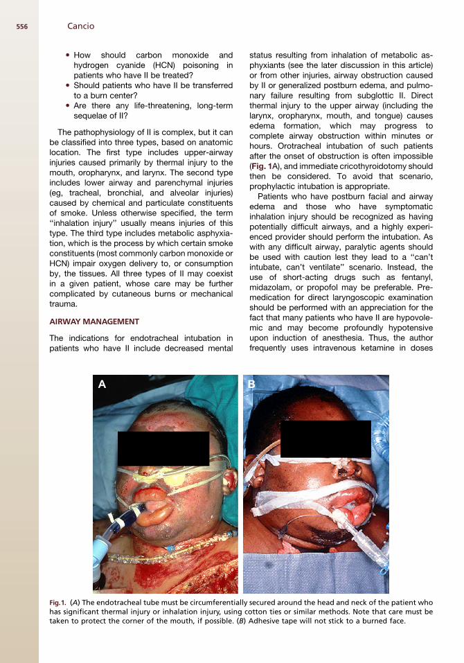
status resulting from inhalation of metabolic as-phyxiants (see the later discussion in this article)or from other injuries, airway obstruction causedby II or generalized postburn edema, and pulmo-nary failure resulting from subglottic II. Directthermal injury to the upper airway (including thelarynx, oropharynx, mouth, and tongue) causesedema formation, which may progress tocomplete airway obstruction within minutes orhours. Orotracheal intubation of such patientsafter the onset of obstruction is often impossible(Fig. 1A), and immediate cricothyroidotomy shouldthen be considered. To avoid that scenario,prophylactic intubation is appropriate.
Patients who have postburn facial and airwayedema and those who have symptomaticinhalation injury should be recognized as havingpotentially difficult airways, and a highly experi-enced provider should perform the intubation. Aswith any difficult airway, paralytic agents shouldbe used with caution lest they lead to a ‘‘can’tintubate, can’t ventilate’’ scenario. Instead, theuse of short-acting drugs such as fentanyl,midazolam, or propofol may be preferable. Pre-medication for direct laryngoscopic examinationshould be performed with an appreciation for thefact that many patients who have II are hypovole-mic and may become profoundly hypotensiveupon induction of anesthesia. Thus, the authorfrequently uses intravenous ketamine in doses
Fig.1. (A) The endotracheal tube must be circumferentially secured around the head and neck of the patient whohas significant thermal injury or inhalation injury, using cotton ties or similar methods. Note that care must betaken to protect the corner of the mouth, if possible. (B) Adhesive tape will not stick to a burned face.
Cancio556
one quarter to one half of the full anesthetic dosefor this purpose (ie, 0.25–0.5 mg/kg, instead of1 mg/kg). In a patient who is awake and in situa-tions that are not true emergencies, intubationusing a transnasally inserted fiberoptic broncho-scope with topical anesthesia is another excellentapproach. The primary risk associated withprophylactic intubation in such patients iscatastrophic loss of the airway, especially duringtransport. Thus, cotton ties (1/2-in umbilical ties),rather than adhesive tape, are used to secure theendotracheal tube circumferentially around thepatient’s neck (see Fig. 1A, B). Also, the tubemay become obstructed in patients who havecopious mucous production. This may beprevented by frequent (hourly or more often)suctioning (Fig. 2A, B).
Although II directly damages the airway, cuta-neous thermal injury causes generalized edemathroughout the body, including the airway. Somechildren who have scald injuries and no IIwhatsoever require endotracheal intubation, inparticular when they are younger than 2.8 yearsold and the burns cover more than 19% of the totalbody surface area (TBSA).5 In adults, the authorrecommends prophylactic endotracheal intuba-tion for patients who have burns over more than40% of the TBSA until the resuscitation period iscomplete (first 48 hours), even when II is absent.
Not all patients who have smoke exposurerequire endotracheal intubation.6 Awake trans-nasal fiberoptic laryngoscopic examination can
be used to determine whether a patient who hasmild symptoms also has laryngeal edema andrequires intubation.7,8 The author usesa bronchoscope for this purpose because itpermits evaluation of the subglottic airway (seethe later discussion in this article).
As with patients who are mechanically ventilatedfor other reasons, every effort should be made toliberate the patient who has II from the ventilatoras soon as possible. To this end, the authorperforms daily sedation breaks and reevaluationsfor extubation. He also uses aggressive physicaltherapy, including tilt-table exercises, standing,and even ambulation, in selected patients despitethe presence of an endotracheal or tracheostomytube. Contraindications to extubation, aside fromthose common to all patients, include upper-airway edema so severe that the patient cannotbreathe around an occluded endotracheal tubewith the cuff deflated, worsening edema (due toresuscitation during the first 48 hours postburn),and significant problems with pulmonary toilet.Note that the airways do not have to be completelyhealed because natural coughing is effective atclearing moderate amounts of plugs, secretions,and other matter.
Whether and when to perform tracheostomy forpatients who have II continues to be debated. Inboth adults and children, the route of intubationseems less important than avoidance of highpeak inspiratory pressures and high cuffpressures.9–11 The author’s practice is to performtracheostomy at 14 days for those patients whoremain ventilator dependent.
Earlier tracheostomy may be necessary to facili-tate pulmonary toilet, which may be lifesaving inpatients who have severe II when they begin toslough the airway mucosa, bleed into the airway,and form obstructing clots and casts. This maybegin within a few days after the injury (Fig. 3A, B).Performing a percutaneous tracheostomy may bemore challenging in patients who are bleeding intothe airway, and an open tracheostomy may beadvantageous in that setting.
DIAGNOSIS OF INHALATION INJURY
Before transferring a patient to a burn center, it issufficient to identify the patient’s risk for airwayand breathing problems and to protect the airway,and it is not usually necessary to make a definitivediagnosis as to the presence or absence of II.For this purpose, fiberoptic laryngoscopicexamination (see the previous discussion in thisarticle), patient history and physical examination,and carboxyhemoglobin levels (if available) areused. The mechanism of injury, signs, symptoms,
Fig. 2. (A) Endotracheal tube completely blocked byinspissated mucus and debris. (B) Endotracheal tubecompletely blocked by mucus and carbonaceoussputum. In both cases, emergency extubation andreintubation were required.
Airway Management and Smoke Inhalation Injury 557
and physical examination provide clues to thepresence of II, but not diagnostic certainty. Shiraniand colleagues1 found that patients who havea history of injury in a closed space, facial burns,large burn sizes, or advanced age are more likelyto have II. Other clues to diagnosis include thepatient’s loss of consciousness at the fire sceneand the presence of noxious fumes at the fire.Clark and colleagues12 retrospectively reviewedthe presenting symptoms of 805 patients whohad II. In 108, complete data were available(Table 1). From these data, it can be deducedthat the absence of classic signs of airwayobstruction (eg, stridor, voice change, dyspnea)should not reassure one that II is absent.
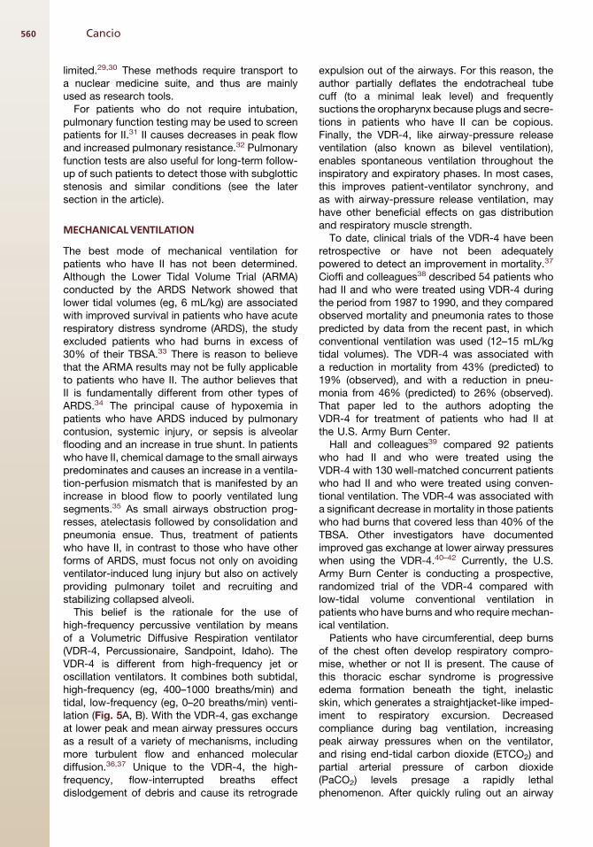
Fiberoptic bronchoscopy (FOB) provides whathas been called a ‘‘gold standard’’ for the diag-nosis of II.13 Several authors have developedgrading schemes for the severity of injury basedon data from FOB.14–16 One such system wasprospectively evaluated and is provided in Table 2.In addition, patients may have varying amounts ofcarbonaceous material (soot) in the airways, mayhave copious or no secretions, may progressfrom necrosis to sloughing of the airway, andmay present with areas of pallor rather thanhyperemia (Fig. 4A, B). Finally, FOB may be falselynegative if an FOB examination is performedimmediately after injury in patients who haveburn shock. A repeat FOB examination 24 to 48hours later may be more revealing.17 Effortsto grade the severity of II by the macroscopic
appearance of the airways using FOB examinationhave been inconsistent and subjective. When thediagnosis is uncertain by FOB, biopsy may behelpful but is not widely used.18,19
Most patients who have II have a normal chestradiograph on initial presentation. Thus, a normalchest radiograph cannot be used to rule out
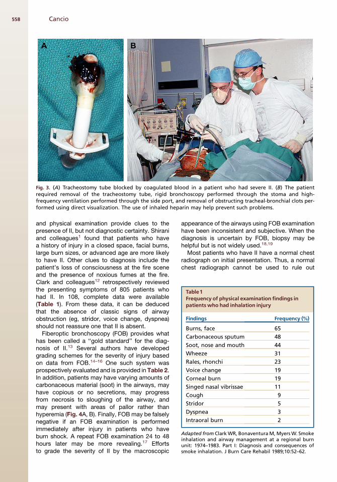
Fig. 3. (A) Tracheostomy tube blocked by coagulated blood in a patient who had severe II. (B) The patientrequired removal of the tracheostomy tube, rigid bronchoscopy performed through the stoma and high-frequency ventilation performed through the side port, and removal of obstructing tracheal-bronchial clots per-formed using direct visualization. The use of inhaled heparin may help prevent such problems.
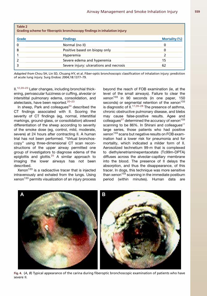
Table1Frequency of physical examination findings inpatients who had inhalation injury
Findings Frequency (%)
Burns, face 65
Carbonaceous sputum 48
Soot, nose and mouth 44
Wheeze 31
Rales, rhonchi 23
Voice change 19
Corneal burn 19
Singed nasal vibrissae 11
Cough 9
Stridor 5
Dyspnea 3
Intraoral burn 2
Adapted from Clark WR, Bonaventura M, Myers W. Smokeinhalation and airway management at a regional burnunit: 1974–1983. Part I: Diagnosis and consequences ofsmoke inhalation. J Burn Care Rehabil 1989;10:52–62.
Cancio558
II.12,20–23 Later changes, including bronchial thick-ening, perivascular fuzziness or cuffing, alveolar orintersitital pulmonary edema, consolidation, andatelectasis, have been reported.20–23
In sheep, Park and colleagues24 described theCT findings associated with II. Scoring theseverity of CT findings (eg, normal, interstitialmarkings, ground-glass, or consolidation) alloweddifferentiation of the sheep according to severityof the smoke dose (eg, control, mild, moderate,severe) at 24 hours after contracting II. A humantrial has not been performed. ‘‘Virtual bronchos-copy’’ using three-dimensional CT scan recon-structions of the upper airway permitted onegroup of investigators to diagnose edema of theepiglottis and glottis.25 A similar approach toimaging the lower airways has not beendescribed.
Xenon133 is a radioactive tracer that is injectedintravenously and exhaled from the lungs. Usingxenon133 permits visualization of an injury process
beyond the reach of FOB examination (ie, at thelevel of the small airways). Failure to clear thexenon133 in 90 seconds (in one paper, 150seconds) or segmental retention of the xenon133
is diagnostic of II.17,26–28 The presence of asthma,chronic obstructive pulmonary disease, and blebsmay cause false-positive results. Agee andcolleagues27 determined the accuracy of xenon133
scanning to be 86%. In Shirani and colleagues’1
large series, those patients who had positivexenon133 scans but negative results on FOB exam-ination had a lower risk for pneumonia and formortality, which indicated a milder form of II.Aerosolized technetium 99-m that is complexedto diethylenetriaminepentacetate (Tc99m-DPTA)diffuses across the alveolar-capillary membraneinto the blood. The presence of II delays theabsorption, and thus the disappearance, of thistracer. In dogs, this technique was more sensitivethan xenon133 scanning in the immediate postburnperiod (within minutes). Human data are
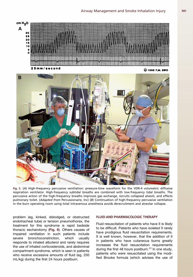
Table 2Grading scheme for fiberoptic bronchoscopy findings in inhalation injury
Grade Findings Mortality (%)
0 Normal (no II) 0
B Positive based on biopsy only 0
1 Hyperemia 2
2 Severe edema and hyperemia 15
3 Severe injury: ulcerations and necrosis 62
Adapted from Chou SH, Lin SD, Chuang HY, et al. Fiber-optic bronchoscopic classification of inhalation injury: predictionof acute lung injury. Surg Endosc 2004;18:1377–79.
Fig. 4. (A, B) Typical appearance of the carina during fiberoptic bronchoscopic examination of patients who havesevere II.
Airway Management and Smoke Inhalation Injury 559
limited.29,30 These methods require transport toa nuclear medicine suite, and thus are mainlyused as research tools.
For patients who do not require intubation,pulmonary function testing may be used to screenpatients for II.31 II causes decreases in peak flowand increased pulmonary resistance.32 Pulmonaryfunction tests are also useful for long-term follow-up of such patients to detect those with subglotticstenosis and similar conditions (see the latersection in the article).
MECHANICALVENTILATION
The best mode of mechanical ventilation forpatients who have II has not been determined.Although the Lower Tidal Volume Trial (ARMA)conducted by the ARDS Network showed thatlower tidal volumes (eg, 6 mL/kg) are associatedwith improved survival in patients who have acuterespiratory distress syndrome (ARDS), the studyexcluded patients who had burns in excess of30% of their TBSA.33 There is reason to believethat the ARMA results may not be fully applicableto patients who have II. The author believes thatII is fundamentally different from other types ofARDS.34 The principal cause of hypoxemia inpatients who have ARDS induced by pulmonarycontusion, systemic injury, or sepsis is alveolarflooding and an increase in true shunt. In patientswho have II, chemical damage to the small airwayspredominates and causes an increase in a ventila-tion-perfusion mismatch that is manifested by anincrease in blood flow to poorly ventilated lungsegments.35 As small airways obstruction prog-resses, atelectasis followed by consolidation andpneumonia ensue. Thus, treatment of patientswho have II, in contrast to those who have otherforms of ARDS, must focus not only on avoidingventilator-induced lung injury but also on activelyproviding pulmonary toilet and recruiting andstabilizing collapsed alveoli.
This belief is the rationale for the use ofhigh-frequency percussive ventilation by meansof a Volumetric Diffusive Respiration ventilator(VDR-4, Percussionaire, Sandpoint, Idaho). TheVDR-4 is different from high-frequency jet oroscillation ventilators. It combines both subtidal,high-frequency (eg, 400–1000 breaths/min) andtidal, low-frequency (eg, 0–20 breaths/min) venti-lation (Fig. 5A, B). With the VDR-4, gas exchangeat lower peak and mean airway pressures occursas a result of a variety of mechanisms, includingmore turbulent flow and enhanced moleculardiffusion.36,37 Unique to the VDR-4, the high-frequency, flow-interrupted breaths effectdislodgement of debris and cause its retrograde
expulsion out of the airways. For this reason, theauthor partially deflates the endotracheal tubecuff (to a minimal leak level) and frequentlysuctions the oropharynx because plugs and secre-tions in patients who have II can be copious.Finally, the VDR-4, like airway-pressure releaseventilation (also known as bilevel ventilation),enables spontaneous ventilation throughout theinspiratory and expiratory phases. In most cases,this improves patient-ventilator synchrony, andas with airway-pressure release ventilation, mayhave other beneficial effects on gas distributionand respiratory muscle strength.
To date, clinical trials of the VDR-4 have beenretrospective or have not been adequatelypowered to detect an improvement in mortality.37
Cioffi and colleagues38 described 54 patients whohad II and who were treated using VDR-4 duringthe period from 1987 to 1990, and they comparedobserved mortality and pneumonia rates to thosepredicted by data from the recent past, in whichconventional ventilation was used (12–15 mL/kgtidal volumes). The VDR-4 was associated witha reduction in mortality from 43% (predicted) to19% (observed), and with a reduction in pneu-monia from 46% (predicted) to 26% (observed).That paper led to the authors adopting theVDR-4 for treatment of patients who had II atthe U.S. Army Burn Center.
Hall and colleagues39 compared 92 patientswho had II and who were treated using theVDR-4 with 130 well-matched concurrent patientswho had II and who were treated using conven-tional ventilation. The VDR-4 was associated witha significant decrease in mortality in those patientswho had burns that covered less than 40% of theTBSA. Other investigators have documentedimproved gas exchange at lower airway pressureswhen using the VDR-4.40–42 Currently, the U.S.Army Burn Center is conducting a prospective,randomized trial of the VDR-4 compared withlow-tidal volume conventional ventilation inpatients who have burns and who require mechan-ical ventilation.
Patients who have circumferential, deep burnsof the chest often develop respiratory compro-mise, whether or not II is present. The cause ofthis thoracic eschar syndrome is progressiveedema formation beneath the tight, inelasticskin, which generates a straightjacket-like imped-iment to respiratory excursion. Decreasedcompliance during bag ventilation, increasingpeak airway pressures when on the ventilator,and rising end-tidal carbon dioxide (ETCO2) andpartial arterial pressure of carbon dioxide(PaCO2) levels presage a rapidly lethalphenomenon. After quickly ruling out an airway
Cancio560
problem (eg, kinked, dislodged, or obstructedendotracheal tube) or tension pneumothorax, thetreatment for this syndrome is rapid bedsidethoracic escharotomy (Fig. 6). Others causes ofimpaired ventilation in such patients includesevere bronchoconstriction, which usuallyresponds to inhaled albuterol and rarely requiresthe use of inhaled corticosteroids, and abdominalcompartment syndrome, which is seen in patientswho receive excessive amounts of fluid (eg, 250mL/kg) during the first 24 hours postburn.
FLUID AND PHARMACOLOGIC THERAPY
Fluid resuscitation of patients who have II is likelyto be difficult. Patients who have isolated II rarelyhave prodigious fluid resuscitation requirements.It is well known, however, that the addition of IIin patients who have cutaneous burns greatlyincreases the fluid resuscitation requirementsduring the first 48 hours postburn.43 In one study,patients who were resuscitated using the modi-fied Brooke formula (which advises the use of
Fig. 5. (A) High-frequency percussive ventilation: pressure-time waveform for the VDR-4 volumetric diffusiverespiration ventilator. High-frequency subtidal breaths are combined with low-frequency tidal breaths. Thepercussive action of the high-frequency breaths improves gas exchange, recruits collapsed alveoli, and effectspulmonary toilet. (Adapted from Percussionaire, Inc) (B) Continuation of high-frequency percussive ventilationin the burn operating room using total intravenous anesthesia avoids derecruitment and alveolar collapse.
Airway Management and Smoke Inhalation Injury 561
2 mL/kg/TBSA burned as the lactated Ringer’sdose for the first 24 hours) actually receivedmore than 5 mL/kg/TBSA burned.44 Efforts toanticipate this response by starting patients onhigher infusion rates are likely to result in increasedcomplications of volume overload.45 On the otherhand, fluid restriction does not protect the lungsor improve outcome. For example, Herndon andcolleagues46 demonstrated an increase in lunglymph flow (indicating increased microvascularpermeability) in fluid-restricted sheep withcombined II and burns. Thus, resuscitation ofpatients who have combined II and burns shouldbe conducted with close attention to providingneither too much nor too little fluid, with hourlyattention to endpoints such as achieving urineoutput of 30 to 50 mL/h in adults or 1 to 1.5 mL/kg/h in children who weigh less than 30 kg.
Despite research that has greatly improved theunderstanding of the pathophysiology of inhalationinjury,47 pharmacologic options for treatment of IIremain limited. Nevertheless, inhaled heparin isan important addition. In a retrospective study,Desai and colleagues48 reported a reduction in re-intubation rates and mortality in burned childrenwho were treated using inhaled heparin andN-acetylcystine. On the other hand, Holt andcolleagues49 reviewed their experience with theuse of inhaled heparin and N-acetylcystine inadults who had II. There was no difference in thenumber of days on a ventilator or in mortalitybetween those who received this treatment andthose who did not. The divergent results of the
two studies may be due to the fact that children,who have smaller airways and endotracheal tubes,are more vulnerable to airway obstruction.5
Because obstructing clots and casts area common life-threatening problem during theacute phase for people who have II, and becausethis therapy is inexpensive and does not causesystemic anticoagulation, the author routinelyprovides nebulized heparin to all patients whohave II, beginning on admission and continuingas long as the patients are intubated and theirairways remain friable.
Pneumonia, most often secondary to invasivegram-negative rods (such as Pseudomonas aeru-ginosa or Klebsiella pneumoniae) or to Staphyloc-cus aureus, remains a dreaded complication inpatients who have II or extensive thermalinjury.1,50,51 Unfortunately, prophylactic antibioticshave not been shown to prevent infection inpatients who have II or burns. Especially whenhospitalized for weeks to months, such patientsare at risk for colonization and infection withmultiple drug-resistant organisms; this riskincreases with indiscriminant antibiotic exposure.Compounding the problem is the fact that burninjury alone causes hyperdynamic systemicinflammatory response syndrome, which is char-acterized by many of the same signs and symp-toms as sepsis. Thus, elevated temperature orwhite blood cell count do not correlate well withsystemic infection,52 so other clinical indicators(eg, hyperglycemia, tachypnea, tube-feeding intol-erance) must be sought. Early institution of broad-spectrum antibiotics, an aggressive diagnosticapproach that includes bronchoalveolar lavage,and rapid tailoring of the regimen to matchorganism sensitivities are crucial.
METABOLIC ASPHYXIANTS
Along with smoke, patients can inhale compoundsthat impair oxygen delivery to, or use by, thetissues. Chief among these is carbon monoxide,which is produced by the partial combustion ofcarbon-containing compounds such as cellulosics(eg, wood, paper, coal, charcoal), natural gases(eg, methane, butane, propane), and petroleumproducts. Carbon monoxide poisoning isa common cause of death at fire scenes53,54 andis also a leading cause of non–fire-related deathsin the United States.55 In addition to combiningwith hemoglobin to form carboxyhemoglobin(COHb), where in the CO has an affinity for hemo-globin which is 200 times that of oxygen, carbonmonoxide also impairs mitochondrial functionand COHb causes brain injury as the result ofoxidative stress, inflammation, and excitatory
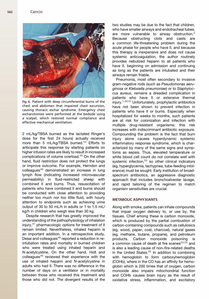
Fig. 6. Patient with deep circumferential burns of thechest and abdomen that impaired chest excursion,causing thoracic eschar syndrome. Emergency chestescharotomies were performed at the bedside usinga scalpel, which restored normal compliance andeffective mechanical ventilation.
Cancio562
amino acids.56 The organs most vulnerable tocarbon monoxide poisoning are those mostaffected by oxygen deprivation, namely, thecardiovascular system and the brain.
Currently, the diagnosis of carbon monoxidepoisoning requires measurement of arterialCOHb levels using a co-oximeter; the PaO2 levelin such patients is frequently normal or high, anda standard 2-wavelength pulse oximeter willfalsely provide a high peripheral saturation ofoxygen (SpO2) reading, even when COHb levelsare in the lethal range (R50%), because it cannotdiscriminate between COHb and oxygenatedhemoglobin.57 Only an arterial saturation ofoxygen (SaO2) reading derived from an arterialblood gas sample and analyzed using a co-oxi-meter will show depressed hemoglobin oxygensaturation. The half-life of COHb may be variable;in one study, the half-life of COHb in patientstreated using 100% oxygen was 74 min � 25SD, but ranged from 26 to 148 minutes.58
The mainstay of treatment is 100% oxygenadministered by nonrebreather mask or endotra-cheal tube until the COHb level is less than 5%59
or for 6 hours.60 Controversy surrounds the useof hyperbaric oxygen therapy (HBOT) to treatsuch patients. Although HBOT accelerates theclearance of carbon monoxide beyond thatachieved using 100% oxygen at 1 atmosphere,the main rationale for its use is prevention of de-layed neurocognitive syndrome. This syndromeproduces memory loss and other cognitivedefects, with onset from 2 to 28 days after expo-sure.60,61 The Cochrane group reviewed 6randomized controlled trials (RCTs) of HBOT forprevention of neurologic sequelae. Four studiesshowed no benefit, two studies did show benefit,and the pooled analysis showed no benefit. Theinvestigators concluded that the efficacy of
HBOT in this setting is uncertain.62 In the specialcase of a patient who has burns, II, and carbonmonoxide poisoning, there is almost no evidenceconcerning the use of HBOT. Grube andcolleagues63 described a case series of 10 suchpatients who were treated using HBOT. Severalsignificant problems complicated the use ofHBOT, including aspiration, seizures, and progres-sive hypovolemia. That experience pointed to thedifficulty inherent in transporting hemodynamicallyunstable patients who have burn shock and II to anHBOT chamber and providing care in thechamber. Furthermore, the data suggest that theuse of HBOT for prevention of delayed neurocog-nitive syndrome need not begin until the twenty-third hour after exposure. The author does notroutinely provide HBOT to patients during resusci-tation from burn shock.
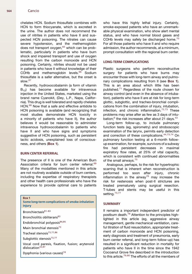
HCN is produced by the combustion of mate-rials such as plastics, foam, paints, wool, andsilk. It impairs the cellular use of oxygen by bindingto the terminal cytochrome on the electron trans-port chain, causing lactic acidosis and, potentially,elevated mixed venous oxygen saturation. Thehalf-life of HCN in the human body is about 1hour. Thus, HCN may be a significant factor ina variable percentage of patients who have II.53,64
The diagnosis of HCN poisoning is difficultbecause a rapid assay is not available. HCN andcarbon monoxide poisoning share many features,including signs and symptoms related to thecentral nervous and cardiovascular systems. Alist of features that can be observed andcompared is provided in Table 3. Three types ofantidote are available for HCN poisoning. A HCNantidote kit in the United States contains amylnitrite for inhalation and sodium nitrite and sodiumthiosulfate for intravenous injection. The nitritesoxidize hemoglobin to methemoglobin, which
Table 3Carbonmonoxide and HCN poisoning comparison
Features CarbonMonoxide HCN
Loss of consciousness May be transitory Usually sustained
Dilated pupils Rare Common
Seizure Uncommon Common
Hypotension Uncommon Common (after initial ‘‘catecholaminerush’’)
Breathing Tachypnea Tachypnea, then bradypnea/centralapnea
Lactate (correlation with levels oftoxin)
Variable Strong
Adapted from Baud FJ. Cyanide: critical issues in diagnosis and treatment. Hum Exp Toxicol 2007;26:191–201.
Airway Management and Smoke Inhalation Injury 563
chelates HCN. Sodium thiosulfate combines withHCN to form thiocyanate, which is excreted inthe urine. The author does not recommend theuse of nitrites in patients who have II and sus-pected HCN poisoning. The nitrates can causesevere hypotension, and the methemoglobindoes not transport oxygen,65 which can be prob-lematic, particularly in patients who have burnshock and impaired transport and use of oxygenresulting from the carbon monoxide and HCNpoisoning. Certainly, nitrites should not be usedin patients who have II without knowledge of theirCOHb and methemoglobin levels.66 Sodiumthiosulfate is a safer alternative, but the onset isslow.67
Recently, hydroxocobalamin (a form of vitaminB12) has become available for intravenousinjection in the United States, marketed using thebrand name Cyanokit, (Dey, L.P., Napa, Califor-nia). This drug is well tolerated and rapidly chelatesHCN.68 Now that a safe and effective antidote toHCN poisoning is available (and recognizing thatmost studies demonstrate HCN toxicity ina minority of patients who have II), the authorbelieves it would be reasonable to administerintravenous hydroxocobalamin to patients whohave II and who have signs and symptomssuggestive of HCN poisoning, such as persistentlactic acidosis, unexplained loss of conscious-ness, and others (Box 1).
BURN CENTER REFERRAL
The presence of II is one of the American BurnAssociation criteria for burn center referral.69
Many of the modalities mentioned in this articleare not routinely available outside of burn centers,including the expertise of respiratory therapistsand other health care professionals who have theexperience to provide optimal care to patients
who have this highly lethal injury. Certainly,smoke-exposed patients who have an unremark-able physical examination, who show alert mentalstatus, and who have normal blood gases andCOHb levels may safely be discharged home.70
For all those patients who have II and who requireadmission, the author recommends, at a minimum,prompt consultation with the regional burn center.
LONG-TERM COMPLICATIONS
Plastic surgeons who perform reconstructivesurgery for patients who have burns mayencounter those with long-term airway and pulmo-nary complications resulting from II (see Box 1).This is an area about which little has beenpublished.71 Regardless of the route chosen forairway control (and even in the absence of intuba-tion), patients who have II are at risk for long-termglottic, subglottic, and tracheo-bronchial compli-cations from the combination of injury, intubation,infection, and chronic inflammation.72,73 Theseproblems may arise after as few as 3 days of intu-bation;6 the risk increases after about 21 days.43
Long-term follow-up, to include pulmonaryfunction tests (eg, spirometry) and stroboscopicexamination of the larynx, permits early detectionand correction of these complications.44,73,74 Onpulmonary function testing at a 6-month follow-up examination, for example, survivors of a subwayfire had persistent decreases in maximalexpiratory flow rates, at 25% of vital capacity,which is consistent with continued abnormalitiesof the small airways.75
Analogous, perhaps, to the risk for hypertrophicscarring that is inherent when reconstruction isperformed too soon after injury, chronicinflammation in the airway76 may increase therisk for restenosis when post-II strictures aretreated prematurely using surgical resection.T-tubes and stents may be useful in thissetting.73,77
SUMMARY
II remains a important independent predictor ofpostburn death.78 Attention to the principles high-lighted in this article (eg, aggressive airwaymanagement, gentle mechanical ventilation, care-ful titration of fluid resuscitation, appropriate treat-ment of carbon monoxide and HCN poisoning,early diagnosis and treatment of pneumonia, earlyburn center referral, and long-term follow-up) hasresulted in a significant reduction in mortality forpatients who have II in the time since the 1942Cocoanut Grove fire described in the introductionto this article.79,80 The efforts of all the members of
Box1Some long-term complications of smoke-inhalationinjury
Bronchiectasis81–83
Bronchiolitis obliterans83
Endobronchial polyposis84,85
Main bronchial stenosis73
Tracheal stenosis73,77,86
Subglottic stenosis73,77
Vocal cord paresis, fixation, fusion; arytenoiddislocation33,87
Dysphonia (various causes)74
Cancio564
the multidisciplinary burn team, including nurses,physicians, respiratory therapists, and basicscientists, among others, have contributed andwill continue to contribute to progress for patientswho have this difficult injury.
ACKNOWLEDGMENTS
The author gratefully acknowledges the assis-tance of Ms. Gerri Trumbo, Ms. Helen Wessel,and Dr. Corina Moraru in conducting this review.
REFERENCES
1. Shirani KZ, Pruitt BA Jr, Mason AD Jr. The influence
of inhalation injury and pneumonia on burn mortality.
Ann Surg 1987;205:82–7.
2. Tredget EE, Shankowsky HA, Taerum TV, et al. The
role of inhalation injury in burn trauma. A Canadian
experience. Ann Surg 1990;212:720–7.
3. Thompson PB, Herndon DN, Traber DL, et al. Effect
on mortality of inhalation injury. J Trauma 1986;26:
163–5.
4. Aub JC, Beecher HK, Cannon B, et al. Management of
the Cocoanut Grove burns at the Massachusetts
General Hospital. Philadelphia: J.B. Lippincott; 1943.
5. Zak AL, Harrington DT, Barillo DJ, et al. Acute respi-
ratory failure that complicates the resuscitation of
pediatric patients with scald injuries. J Burn Care
Rehabil 1999;20:391–9.
6. Cha SI, Kim CH, Lee JH, et al. Isolated smoke inha-
lation injuries: acute respiratory dysfunction, clinical
outcomes, and short-term evolution of pulmonary
functions with the effects of steroids. Burns 2007;
33:200–8.
7. Madnani DD, Steele NP, de Vries E. Factors that
predict the need for intubation in patients with
smoke inhalation injury. Ear Nose Throat J 2006;85:
278–80.
8. Goh SH, Tiah L, Lim HC, et al. Disaster prepared-
ness: experience from a smoke inhalation mass
casualty incident. Eur J Emerg Med 2006;13:330–4.
9. Saffle JR, Morris SE, Endelman L. Early tracheos-
tomy does not improve outcome in burn patients.
J Burn Care Rehabil 2002;23:431–8.
10. Kadilak PR, Vanasse S, Sheridan RL. Favorable
short- and long-term outcomes of prolonged trans-
laryngeal intubation in critically ill children. J Burn
Care Rehabil 2004;25:262–5.
11. Palmieri TL, Jackson W, Greenhalgh DG. Benefits of
early tracheostomy in severely burned children. Crit
Care Med 2002;30:922–4.
12. Clark WR, Bonaventura M, Myers W. Smoke inhala-
tion and airway management at a regional burn
unit: 1974–1983. Part I: diagnosis and conse-
quences of smoke inhalation. J Burn Care Rehabil
1989;10:52–62.
13. Anonymous. Evidence-based surgery. J Am Coll
Surg 2003;196:306–12.
14. Endorf FW, Gamelli RL. Inhalation injury, pulmonary
perturbations, and fluid resuscitation. J Burn Care
Res 2007;28:80–3.
15. Chou SH, Lin SD, Chuang HY, et al. Fiber-optic bron-
choscopic classification of inhalation injury: predic-
tion of acute lung injury. Surg Endosc 2004;18:
1377–9.
16. Bingham HG, Gallagher TJ, Powell MD. Early bron-
choscopy as a predictor of ventilatory support for
burned patients. J Trauma 1987;27:1286–8.
17. HuntJL,AgeeRN,PruittBAJr. Fiberopticbronchoscopy
in acute inhalation injury. J Trauma 1975;15:641–9.
18. Masanes MJ, Legendre C, Lioret N, et al. Fiberoptic
bronchoscopy for the early diagnosis of subglottal
inhalation injury: comparative value in the assess-
ment of prognosis. J Trauma 1994;36:59–67.
19. Masanes MJ, Legendre C, Lioret N, et al. Using
bronchoscopy and biopsy to diagnose early inhala-
tion injury. Macroscopic and histologic findings.
Chest 1995;107:1365–9.
20. Hantson P, Butera R, Clemessy JL, et al. Early
complications and value of initial clinical and para-
clinical observations in victims of smoke inhalation
without burns. Chest 1997;111:671–5.
21. Teixidor HS, Rubin E, Novick GS, et al. Smoke inha-
lation: radiologic manifestations. Radiology 1983;
149:383–7.
22. Wittram C, Kenny JB. The admission chest radio-
graph after acute inhalation injury and burns. Br J
Radiol 1994;67:751–4.
23. Lee MJ, O’Connell DJ. The plain chest radiograph
after acute smoke inhalation. Clin Radiol 1988;39:
33–7.
24. Park MS, Cancio LC, Batahinsky AI, et al. Assess-
ment of severity of ovine smoke inhalation injury by
analysis of computed tomographic scans. J Trauma
2003;55:417–29.
25. Gore MA, Joshi AR, Nagarajan G, et al. Virtual bron-
choscopy for diagnosis of inhalation injury in burnt
patients. Burns 2004;30:165–8.
26. Moylan JA Jr, Wilmore DW, Mouton DE, et al. Early
diagnosis of inhalation injury using 133 xenon lung
scan. Ann Surg 1972;176:477–84.
27. Agee RN, Long JM III, Hunt JL, et al. Use of133xenon in early diagnosis of inhalation injury.
J Trauma 1976;16:218–24.
28. Schall GL, McDonald HD, Carr LB, et al. Xenon
ventilation-perfusion lung scans. The early diagnosis
of inhalation injury. JAMA 1978;240:2441–5.
29. Clark WR, Grossman ZD, Ritter-Hrncirik C, et al.
Clearance of aerosolized 99mTc-diethylenetriamine-
pentacetate before and after smoke inhalation.
Chest 1988;94:22–7.
30. Lin WY, Kao CH, Wang SJ. Detection of acute
inhalation injury in fire victims by means of
Airway Management and Smoke Inhalation Injury 565
technetium-99m DTPA radioaerosol inhalation lung
scintigraphy. Eur J Nucl Med 1997;24:125–9.
31. Whitener DR, Whitener LM, Robertson KJ, et al.
Pulmonary function measurements in patients with
thermal injury and smoke inhalation. Am Rev Respir
Dis 1980;122:731–9.
32. Petroff PA, Hander EW, Clayton WH, et al. Pulmonary
function studies after smoke inhalation. Am J Surg
1976;132:346–51.
33. Anonymous. Ventilation with lower tidal volumes as
compared with traditional tidal volumes for acute
lung injury and the acute respiratory distress
syndrome. The Acute Respiratory Distress Syndrome
Network. N Engl J Med 2000;342:1301–8.
34. Cancio LC, Batchinsky AI, Dubick MA, et al. Inhalation
injury: pathophysiology and clinical care. Proceed-
ings of a symposium conducted at the Trauma
Institute of San Antonio, San Antonio, TX, USA on 28
March 2006. Burns 2007;33:681–92.
35. Shimazu T, Yukioka T, Ikeuchi H, et al. Ventilation-
perfusion alterations after smoke inhalation injury in
an ovine model. J Appl Phys 1996;81:2250–9.
36. Krishnan JA, Brower RG. High-frequency ventilation
for acute lung injury and ARDS. Chest 2000;118:
795–807.
37. Salim A, Martin M. High-frequency percussive venti-
lation. Crit Care Med 2005;33:S241–5.
38. Cioffi WG Jr, Rue LW 3rd, Graves TA, et al. Prophy-
lactic use of high-frequency percussive ventilation in
patients with inhalation injury. Ann Surg 1991;213:
575–82.
39. Hall JJ, Hunt JL, Arnoldo BD, et al. Use of high-
frequency percussive ventilation in inhalation
injuries. J Burn Care Res 2007;28:396–400.
40. Rodeberg DA, Housinger TA, Greenhalgh DG, et al.
Improved ventilatory function in burn patients using
volumetric diffusive respiration. J Am Coll Surg
1994;179:518–22.
41. Reper P, Wibaux O, Van Laeke P, et al. High
frequency percussive ventilation and conventional
ventilation after smoke inhalation: a randomised
study. Burns 2002;28:503–8.
42. Carman B, Cahill T, Warden G, et al. A prospective,
randomized comparison of the Volume Diffusive
Respirator vs conventional ventilation for ventilation
of burned children. 2001 ABA paper. J Burn Care
Rehabil 2002;23:444–8.
43. Navar PD, Saffle JR, Warden GD. Effect of inhalation
injury on fluid resuscitation requirements after
thermal injury. Am J Surg 1985;150:716–20.
44. Lund T, Goodwin CW, McManus WF, et al. Upper
airway sequelae in burn patients requiring endotra-
cheal intubation or tracheostomy. Ann Surg 1985;
201:374–82.
45. Pruitt BA Jr. Protection from excessive resuscitation:
‘‘pushing the pendulum back’’. J Trauma 2000;49:
567–8.
46. Herndon DN, Traber DL, Traber LD. The effect of
resuscitation on inhalation injury. Surgery 1986;100:
248–51.
47. Cancio LC. Current concepts in the pathophysiology
and treatment of inhalation injury. Trauma 2005;7:
19–35.
48. Desai MH, Mlcak R, Richardson J, et al. Reduction in
mortality in pediatric patients with inhalation injury
with aerosolized heparin/N-acetylcystine therapy.
J Burn Care Rehabil 1998;19:210–2.
49. Holt J, Saffle JR, Morris SE, et al. Use of inhaled
heparin/N-acetylcystine in inhalation injury: does it
help? J Burn Care Res 2008;29:192–5.
50. Howard PA, Cancio LC, McManus AT, et al. What’s
new in burn-associated infections? Curr Surg 1999;
56:397–405.
51. Edelman DA, Khan N, Kempf K, et al. Pneumonia after
inhalation injury. J Burn Care Res 2007;28:241–6.
52. Murray CK, Hoffmaster RM, Schmit DR, et al. Evalu-
ation of white blood cell count, neutrophil
percentage, and elevated temperature as predictors
of bloodstream infection in burn patients. Arch Surg
2007;142:639–42.
53. Barillo DJ, Goode R, Esch V. Cyanide poisoning in
victims of fire: analysis of 364 cases and review of
the literature. J Burn Care Rehabil 1994;15:46–57.
54. McGwin G Jr, Chapman V, Rousculp M, et al. The
epidemiology of fire-related deaths in Alabama,
1992–1997. J Burn Care Rehabil 2000;21:75–83.
55. Anonymous. Carbon monoxide–related deaths—
United States, 1999–2004. MMWR Morb Mortal
Wkly Rep 2007;56:1309–12.
56. Thom SR. Hyperbaric-oxygen therapy for acute
carbon monoxide poisoning. N Engl J Med 2002;
347:1105–6.
57. Hampson NB. Pulse oximetry in severe carbon
monoxide poisoning. Chest 1998;114:1036–41.
58. Weaver LK, Howe S, Hopkins R, et al. Carboxyhe-
moglobin half-life in carbon monoxide–poisoned
patients treated with 100% oxygen at atmospheric
pressure. Chest 2000;117:801–8.
59. Ilano AL, Raffin TA. Management of carbon
monoxide poisoning. Chest 1990;97:165–9.
60. Piantadosi CA. Carbon monoxide poisoning. N Engl
J Med 2002;347:1054–5.
61. Piantadosi CA, Sylvia AL, Jobsis-Vandervliet FF.
Differences in brain cytochrome responses to
carbon monoxide and cyanide in vivo. J Appl Phys
1987;62:1277–84.
62. Juurlink DN, Buckley NA, Stanbrook MB, et al.
Hyperbaric oxygen for carbon monoxide poisoning.
Cochrane Database Syst Rev 2005;1. CD002041.
Doi:10.1002/14651858.pub2.
63. Grube BJ, Marvin JA, Heimbach DM. Therapeutic
hyperbaric oxygen: help or hindrance in burn
patients with carbon monoxide poisoning? J Burn
Care Rehabil 1988;9:249–52.
Cancio566
64. Baud FJ, Barriot P, Toffis V, et al. Elevated blood
cyanide concentrations in victims of smoke inhala-
tion. N Engl J Med 1991;325:1761–6.
65. Hall AH, Kulig KW, Rumack BH. Suspected cyanide
poisoning in smoke inhalation: complications of
sodium nitrite therapy. J Toxicol Clin Exp 1989;9:3–9.
66. Kirk MA, Gerace R, Kulig KW. Cyanide and methemo-
globin kinetics in smoke inhalation victims treated
with the cyanide antidote kit. Ann Emerg Med 1993;
22:1413–8.
67. Dart RC. Hydroxocobalamin for acute cyanide
poisoning: new data from preclinical and clinical
studies; new results from the prehospital emergency
setting. Clin Toxicol 2006;44:1–3.
68. Borron SW, Baud FJ, Barriot P, et al. Prospective
study of hydroxocobalamin for acute cyanide
poisoning in smoke inhalation. Ann Emerg Med
2007;49:794–801.
69. Anonymous. Guidelines for the operation of burn
centers. In: Resources for optimal care of the injured
patient. Chicago: Committee on Trauma, American
College of Surgeons; 2006. p. 79–86.
70. Mushtaq F, Graham CA. Discharge from the acci-
dent and emergency department after smoke inha-
lation: influence of clinical factors and emergency
investigations. Eur J Emerg Med;11:141–4.
71. Palmieri TL. Inhalation injury: research progress and
needs. J Burn Care Res 2007;28:549–54.
72. Jones WG, Madden M, Finkelstein J, et al. Trache-
ostomies in burn patients. Ann Surg 1989;209:
471–4.
73. Gaissert HA, Lofgren RH, Grillo HC. Upper airway
compromise after inhalation injury. Complex stric-
tures of the larynx and trachea and their manage-
ment. Ann Surg 1993;218:672–8.
74. Casper JK, Clark WR, Kelley RT, et al. Laryngeal and
phonatory status after burn/inhalation injury: a long
term follow-up study. J Burn Care Rehabil 2002;23:
235–43.
75. Fogarty PW, George PJ, Solomon M, et al. Long term
effects of smoke inhalation in survivors of the King’s
Cross underground station fire. Thorax 1991;46:
914–8.
76. Arakawa A, Fukamizu H, Hashizume I, et al.
Macroscopic and histological findings in the heal-
ing process of inhalation injury. Burns 2007;33:
855–9.
77. Timon CI, McShane D, McGovern E, et al. Treatment
of combined subglottic and critically low tracheal
stenoses secondary to burn inhalation injury. J Lar-
yngol Otol 1989;103:1083–6.
78. Cancio LC, Galvez E Jr, Turner CE, et al. Base deficit
and alveolar-arterial gradient during resuscitation
contribute independently but modestly to the
prediction of mortality after burn injury. J Burn Care
Res 2006;27:289–96.
79. Rue 3rd LW, Cioffi WG, Mason AD, et al. Improved
survival of burned patients with inhalation injury.
Arch Surg 1993;128:772–80.
80. Baud FJ. Cyanide: critical issues in diagnosis and
treatment. Hum Exp Toxicol 2007;26:191–201.
81. Mahut B, Delacourt C, de Blic J, et al. Bronchiec-
tasis in a child after acrolein inhalation. Chest
1993;104:1286–7.
82. Slutzker AD, Kinn R, Said SI. Bronchiectasis and
progressive respiratory failure following smoke inha-
lation. Chest 1989;95:1349–50.
83. Tasaka S, Kanazawa M, Mori M, et al. Long-term
course of bronchiectasis and bronchiolitis obliterans
as late complications of smoke inhalation. Respira-
tion 1995;62:40–2.
84. Adams C, Moisan T, Chandrasekhar AJ, et al. Endo-
bronchial polyposis secondary to thermal inhala-
tional injury. Chest 1979;75:643–5.
85. Williams DO, Vanecko RM, Glassroth J. Endobron-
chial polyposis following smoke inhalation. Chest
1983;84:774–6.
86. Yang JY, Yang WG, Chang LY, et al. Symptomatic
tracheal stenosis in burns. Burns 1999;25:72–80.
87. Cobley TD, Hart WJ, Baldwin DL, et al. Complete
fusion of the vocal cords: an unusual case. Burns
1999;25:361–3.
Airway Management and Smoke Inhalation Injury 567