airway secretory micrornaome changes during rhinovirus
TRANSCRIPT

RESEARCHARTICLE
Airway Secretory microRNAome Changesduring Rhinovirus Infection in EarlyChildhoodMaria J. Gutierrez1, Jose L. Gomez7, Geovanny F. Perez3,4,5,6, KrishnaPancham7,StephanieVal8, DineshK. Pillai3,4,5,6, MamtaGiri6, Sarah Ferrante6, Robert Freishtat4,5,6,9,MaryC. Rose3,4,5,6, Diego Preciado8, Gustavo Nino2,3,4,5,6*
1 Division of Pediatric Allergy Immunology, Johns Hopkins University School of Medicine, Baltimore,Maryland,United States of America,2 Division of Pulmonary, Critical Care and SleepMedicine, YaleUniversity School of Medicine, New Haven, Connecticut, United States of America,3 Division of Pulmonaryand SleepMedicine, Children’s National Medical Center, Washington, DC, United States of America,4 Department of Pediatrics, GeorgeWashington University School of Medicine and Health Sciences,Washington, DC, United States of America, 5 Department of Integrative SystemsBiology and Center forGeneticMedicineResearch,GeorgeWashington University, Washington, DC, United States of America,6 Center for GeneticMedicineResearch, Children’s NationalMedical Center, Washington, DC, UnitedStates of America,7 Division of Pediatric Pulmonology, University of Kentucky, Lexington, Kentucky, UnitedStates of America,8 Division of Pediatric Otolaryngology-Head and Neck Surgery, Children’s NationalMedical Center, Washington, DC, United States of America,9 Division of Emergency Medicine, Children’sNational Medical Center, Washington, DC, United States of America
Abstract
BackgroundInnate immune responses are fine-tuned by small noncoding RNAmolecules termedmicro-
RNAs (miRs) that modify gene expression in response to the environment. During acute
infections, miRs can be secreted in extracellular vesicles (EV) to facilitate cell-to-cell genetic
communication. The purpose of this study was to characterize the baseline population of
miRs secreted in EVs in the airways of young children (airway secretorymicroRNAome)
and examine the changes during rhinovirus (RV) infection, the most common cause of
asthma exacerbations and the most importantearly risk factor for the development of
asthma beyond childhood.
MethodsNasal airway secretions were obtained from children (�3 yrs. old) during PCR-confirmed
RV infections (n = 10) and age-matched controls (n = 10). Nasal EVs were isolated with
polymer-based precipitation and global miR profiles generated using NanoStringmicroar-
rays. We validated our in vivo airway secretorymiR data in an in vitro airway epitheliummodel using apical secretions from primaryhuman bronchial epithelial cells (HBEC) differ-
entiated at air-liquid interface (ALI). Bioinformatics tools were used to determine the unified
(nasal and bronchial) signature airway secretorymiRNAome and changes duringRV infec-
tion in children.
PLOSONE | DOI:10.1371/journal.pone.0162244 September 19, 2016 1 / 20
a11111
OPENACCESS
Citation:Gutierrez MJ, Gomez JL, Perez GF,PanchamK, Val S, Pillai DK, et al. (2016) AirwaySecretorymicroRNAomeChanges during RhinovirusInfection in Early Childhood. PLoS ONE 11(9):e0162244. doi:10.1371/journal.pone.0162244
Editor: Alexander Larcombe, Telethon Institute forChild Health Research, AUSTRALIA
Received:March 30, 2016
Accepted:August 21, 2016
Published:September 19, 2016
Copyright:© 2016 Gutierrez et al. This is an openaccess article distributed under the terms of theCreative Commons Attribution License, which permitsunrestricteduse, distribution, and reproduction in anymedium, provided the original author and source arecredited.
Data Availability Statement:All relevant data arewithin the paper and its Supporting Information files.
Funding: This work was supported by grants: GN—National Heart, Lung and Blood Institute,NHLBI/HL090020 (K12 Genomics of Lung), http://www.nhlbi.nih.gov; JLG—National Heart, Lung and BloodInstitute,NHLBI/1K01HL125474-01, http://www.nhlbi.nih.gov; GN—National Institute for Child Health andHuman Development, NICHC/HD001399 (K12 ChildHealth ResearchCareer DevelopmentAward),https://www.nichd.nih.gov; GN—National Center forAdvancing Translational Sciences, KL2TR000076,https://www.nichd.nih.gov; GN—National Center for

ResultsMultiscale analysis identified four signaturemiRs comprising the baseline airway secretory
miRNAome: hsa-miR-630, hsa-miR-302d-3p,hsa- miR-320e, hsa-miR-612. We identified
hsa-miR-155 as the main change in the baseline miRNAome duringRV infection in young
children.We investigated the potential biological relevance of the airway secretion of hsa-mir-155 using in silicomodels derived from gene datasets of experimental in vivo humanRV infection. These analyses confirmed that hsa-miR-155 targetome is an overrepresentedpathway in the upper airways of individuals infected with RV.
ConclusionsComparative analysis of the airway secretorymicroRNAome in children indicates that RV
infection is associated with airway secretion of EVs containingmiR-155, which is predicted
in silico to regulate antiviral immunity. Further characterization of the airway secretorymicroRNAomeduring health and disease may lead to completely new strategies to treat
and monitor respiratory conditions in all ages.
IntroductionImmune responses are fine-tuned by small RNA molecules termedmicroRNAs (miRs) thatmodify gene expression in response to the environment. miRs comprise a large family of highlyconserved, short, non-codingRNAs that regulate post-transcriptional gene-silencing throughinhibition of translation or promotion of mRNA degradation.[1]miRs regulate approximately60% of protein encoding genes.[2] They exist in body fluids, including saliva, nasal secretions,sputum, urine, breast milk, and blood.[3] To maintain their stability in extracellular body fluids,they are released in membrane-bound extracellular vesicles (EVs). miR-containing EVs aredeemed important for genetic exchange and communication between cells.[4] Specifically, extra-cellular miRs are known to regulate key steps in cell proliferation, differentiation and migrationand to play an important role in immune responses to infections, autoimmunity and cancer.[4]
Respiratory immune responses are fine-tuned by miRs. Resident and migrating lungimmune cells such as macrophages, dendritic cells (DC), lymphocytes and airway epitheliumand smooth muscle cells undergo post-translational regulation of immune-related genes viamiRs.[5] Numerous miRs have been reported to have physiological roles in maintaining tissuehomeostasis and normal development in the airways and the lung.[6, 7] There is compellingevidence demonstrating that several miRs also play pivotal roles in fine-tuning important path-ogenic pathways including the regulation of the effector function of T helper (Th) 2 cells inallergic asthma,[8] the regulation of host defense immune responses,[9] and the repair andremodeling of the airways.[5] Despite the importance of miRs in the genetic regulation of therespiratory system, there is paucity of data describing the baseline population of miRs secretedin EV in the human airways (airway secretorymiRNAome). The importance of investigatingthe airway secretorymiRNAome is that it may have a powerful role in regulating cell-to-cellgenetic communication through the entire respiratory system (from the nose to the small air-ways), particularly given the stability and mobility of EVs (and its miR cargo) in extracellularbody fluids.[3, 10] Moreover, the dynamic regulation of the airway secretorymiRNAomemaybe a key mechanism during environmental exposures and acute infections, instances inwhich cell-to-cell communication is crucial to synchronize host immune defense and inflam-matory signaling pathways.
Airway SecretorymicroRNAome Changes duringRhinovirus Infection in Early Childhood
PLOSONE | DOI:10.1371/journal.pone.0162244 September 19, 2016 2 / 20
Advancing Translational Sciences, UL1TR000075,https://www.nichd.nih.gov; JLG—FAMRI: YoungClinical Scientist Award, 113393, http://www.famri.org/. The funders had no role in study design, datacollection and analysis, decision to publish, orpreparationof the manuscript.
Competing Interests: The authors have declaredthat no competing interests exist.

The purpose of this study was to characterize the baseline population of miRs secreted inEVs in the airways of young children (airway secretorymicroRNAome) and examine thechanges during rhinovirus (RV) infection. RV is the most common cause of asthma exacerba-tion in children and adults[11] and RV-induced wheezing illnesses during the first 3 years oflife are the strongest risk factor (10 times increased odds) for the development of asthmabeyond childhood.[12]Our central hypothesis was that RV infection in young children elicitsdistinctive signatures in the airway secretorymicroRNAome that may modulate the balancebetweenTh1 antiviral immunity and Th2 pro-asthmatic responses during early life. Theimpact of our research is that it highlights the untapped potential of investigating the humanairway secretorymiRNAome during health and disease and it provides new insights into thepotential immune regulatory role of virally induced miR secretion, which may ultimatelyenhance our knowledge on the early origins of asthma and may identify new strategies to treatand monitor a myriad of respiratory disorders in all ages.
Materials andMethods
Nasal washing collection and study subjectsNasal airway secretions were collected in patients�3 yr. of age with PCR-confirmed RV infec-tion (n = 10). All subjects were enrolled during the hospital admission for RV infection. Con-trols were age-matched children (n = 10) with non-detectable virus by PCR testing. Clinicaland demographic variables were obtained by reviewing electronic medical records and pre-sented in S1 Table. Sample was obtained while they were undergoing diagnostic nasal lavage(respiratory virus detection by PCR) at Children's National Medical Center.[13] RV positive(RV-infected group) or negative virus status (control group) was confirmedby a viral multiplexPCR panel for 12 targets (rhinovirus, RSV A, RSV B, HMPV, parainfluenza 1–3, influenza Aand B, H1N1, H1N3, Adenovirus) used for clinical purposes (Luminex, TX, USA). We used astandard nasal lavage technique consisting of gently washing the nasal cavity with 3–4 mL ster-ile normal saline as previously described.[13]The Institutional ReviewBoard (IRB) of Chil-dren’s National Medical Center, Washington D.C. approved the study and granted a waiver ofinformed consent given that this research involved materials (data, documents, records, orspecimens) collected solely for non-research purposes (clinical indications).
Extracellular vesicles isolation and characterizationNasal exosomes were isolated with a polymer-based precipitation method (ExoQuick—SystemBiosciences,Mountain View, CA) according to manufacturer’s protocol.[14] Isolated exosomeswere characterized by Western Blot (WB) using the harbor transpanin CD63 as exosomalmarker. Anti-CD63 WB primary antibodies (System Biosciences,Mountain View, CA) wereused at a 1: 1,000 dilutions and the HRP secondary antibody at 1: 20,000 dilutions. Exosomalquantification was performedwith a commercially available kit (ExoCET method—SystemBiosciences,Mountain View, CA) that directly measures.[15] Exosomal particle size analysiswas performedwith a Dynamic Light Scattering (DLS) instrument (Zetasizer, Malvern Instru-ments, UK) and Nanoparticle Tracking Analysis (NTA) software (Malvern Instruments, UK)using the Stokes Einstein equation to calculate exosomal hydrodynamic diameters.[16]
ExtracellularmicroRNA profilingThe RNA contained in extracellular vesicles/exosomes was isolated and purified using a phe-nol-free lysis buffer and rapid spin columns (SeraMir kit System Biosciences,Mountain View,CA). We performedRNA separation, detection and quantitation with the Agilent Small RNA
Airway SecretorymicroRNAome Changes duringRhinovirus Infection in Early Childhood
PLOSONE | DOI:10.1371/journal.pone.0162244 September 19, 2016 3 / 20
Kit and a Bioanalyzer instrument (2100 Bioanalyzer, Agilent Technologies, Santa Clara, CA).The global microRNAs (miRs) profile was obtained using NanoString human microarrays(human V2 miRNA array>800 probes, Nanostring Technologies, Seattle, WA). To accountfor differences in hybridization and purification, data were normalized to the average countsfor all control spikes in each sample using a proprietary bioinformatics software (nSolver™Analysis Software 2.5, Nanostring Technologies, Seattle, WA). Briefly, we calculated a back-ground level of expression for each sample using the mean level of the negative controls plustwo standard deviations of the mean. MiRNA expressing less than two standard deviationsfrom the mean were set to 0 expression. Those miRNAs that were considered non-zero expres-sion, were normalized using a scaling factor based on the top 100 expressing miRNAs across allsamples. For each sample, the average of the geometricmeans of the top 100 expressing miR-NAs across all samples was divided by the geometricmean of each sample.[17]
Isolation of secreted extracellular vesicles from air-liquid interfacedifferentiated human bronchial epithelial cellsNasal miRs data were contrasted with normal airway epithelial secretions obtained in vitrofrom the apical side of air-liquid interface (ALI)-differentiated human bronchial epithelium.[18] Human bronchial epithelial cells (HBEC) were purchased from Lonza, Walkersville, MD(Catalog number CC-2540, Lonza Inc., Switzerland). HBEC were amplified on collagen-coatedT-75 flasks as previously described,[19] then plated apically on type IV collagen coated 12 welltranswell plates (Fisher Scientific, Pittsburgh, PA), grown submerged for 7–10 days until 100%confluence. Apical media was removed and cells differentiated at air-liquid interface (ALI).After 20 days at ALI, cells were gently washed 4 times with PBS apically and baso-laterally andprotein-free BEBM was added to the basal side. Apical secretions were removed and extracellu-lar vesicles isolated and characterized as described above. Small RNA separation, detection andquantitation was performedwith the Agilent Small RNA Kit chip (2100 Bioanalyzer, AgilentTechnologies, Santa Clara, CA) and miRs profiled using NanoString human microarrays (800probes) (Nanostring Technologies, Seattle, WA).
Bioinformatics and Statistical analysisBiological network analysis was conducted using the identified baseline airway extracellularmicroRNAs (baseline airway miRNAome: hsa-miR-630, hsa-miR-302d-3p, hsa- miR-320e, hsa-miR-612.) and the hsa-mir-155 targets overlapping the GSE11348 dataset (describedbelow)with the use of QIAGEN’s Ingenuity Pathway Analysis (IPA, QIAGEN RedwoodCity, CA,www.qiagen.com/ingenuity). Overrepresented pathways were defined as those containingmore targets than expected by chance, as calculated by the right-tailed Fisher’s exact test. Dif-ferences between groups were analyzed using unpaired T or Mann-Whitney U tests. A p-value<0.05 was considered significant.
hsa-miR-630,hsa-miR-302d-3p,hsa- miR-320e, hsa-miR-612 and hsa-mir-155 TargetsThe miRTarBase release 4.5 (http://mirtarbase.mbc.nctu.edu.tw/index.php) was used to iden-tify predicted targets of hsa-miR-630, hsa-miR-302d-3p, hsa- miR-320e, hsa-miR-612 and hsa-mir-155. Briefly, miRTarBase is a database of miRNA-target interactions (MTIs). This databaseis manually curated and enriched for MTIs validated experimentally by reporter assay, westernblot, microarray and next-generation sequencing experiments.[20] The selection included tar-gets for Homo sapiens -5p sequences. This list was used to identify hsa-miR-630, hsa-miR-
Airway SecretorymicroRNAome Changes duringRhinovirus Infection in Early Childhood
PLOSONE | DOI:10.1371/journal.pone.0162244 September 19, 2016 4 / 20

302d-3p, hsa- miR-320e, hsa-miR-612 for biological network analysis as described above. Inaddition, hsa-mir-155 targets were used in combination with gene expression validation data-sets to model the effect of RV infection in vivo as described below.
Microarray Analysis of Gene ExpressionOmnibus GSE11348To evaluate the effect of rhinovirus infection in human airway epithelium, the GSE11348 data-set was retrieved from the GEO database (http://www.ncbi.nlm.nih.gov/geo/). The GSE11348is a study of gene expression profiles during in vivo human rhinovirus infection.[21]Gene-spring version 12.6 (Agilent Technologies, Santa Clara, CA) was used to analyze the dataset.The.CEL files were normalized using the RMA summarization algorithm with baseline trans-formation to median for all samples. Following microarray data pre-processing a one-wayANOVA test with the Tukey's honestly significance difference test was applied to identify dif-ferentially expressed transcripts between pre-infection, 8 hours and 48 hours post-experimen-tal rhinovirus infection. Results of the ANOVA were corrected for multiple hypothesis testing(Benjamini-Hochberg). Statistical significancewas defined as a false discovery rate (FDR)<5%. Transcripts with�1.2-fold change between conditions were selected for further analysesdescribedbelow.
hsa-mir-155 Targetome in experimental in vivo human rhinovirusinfectionR software (R: A Language and Environment for Statistical Computing. R Foundation for Sta-tistical Computing, Vienna, Austria) was used for data analysis. R stats package version 3.0.1was implemented. A custom script was used to overlap the hsa-mir-155 miRTarBase tran-scripts above with the output following analyses of the GSE11348 dataset using the gene sym-bol according to the genome reference consortium human reference 38. Predicted hsa-mir-155were identified to characterize their temporal behavior following experimental RV infection.
Results
Determinationof the baseline secretoryairway miRNAomeIn order to characterize the effect of RV infection in the airway secretion of extracellularmiRs,we first determined the global profile of miRs secreted in extracellular vesicles/exosomes underbasal conditions, which we refer here as the baseline secretory airway miRNAome. BecausemiRs have extensive regulatory functions in the airway, particularly in the development andfunction of the airway epithelium, [6, 7] we anticipated that a set of extracellular miRs wouldbe secreted at high levels under normal conditions in all subjects studied. The importance ofidentifying the baseline secretory airway miRNAome is that it may serve as homeostatic back-ground to investigate the presence of new extracellularmiRs during pathological conditions. Inour case, we were interested in the newly secreted miRs during RV infection.
Our first step was to isolate extracellularmiRs from the nasal secretions of 10 children with-out acute viral respiratory infection (controls). Fig 1 shows the workflow utilized includingstandard extracellular vesicles/exosome isolation with a polymer-based centrifugation method(Exoquick) and characterization based on particle size (DSL Nanotracking), AChE activity(ExoCET assay) and immune markers (CD 63; Fig 1B) as previously described.[16]We nextisolated small RNA and confirmed the presence of miRs using Agilent Bioanalyzer (Fig 1C).Isolated small RNA was used to profile extracellular miRs with a NanoString panelcontaining> 800 human targets. The top 20 miRs more abundant (and present in all subjects)are presented in Table 1. S2 Table contains the total baseline extracellularmiRs. To visualize
Airway SecretorymicroRNAome Changes duringRhinovirus Infection in Early Childhood
PLOSONE | DOI:10.1371/journal.pone.0162244 September 19, 2016 5 / 20

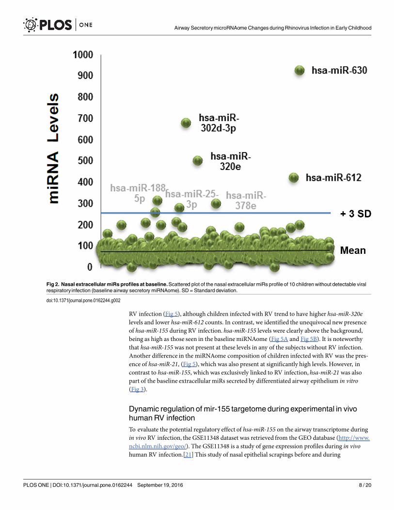
better the baseline airway secretorymiRNAome we built scattered plots that highlight the pop-ulation of extracellularmiRs significantly above background (> 3 SD from mean miR counts;Fig 2). This analysis identified 7 candidate miRs that were clearly above all the other miRs: hsa-miR-630, hsa-miR-302d-3p, hsa- miR-320e, hsa-miR-612, hsa-miR-378e, hsa-miR-25-3p, hsa-miR-188-5p.
Given that in vivo nasal washes reflect a mixed secretion (naso-oropharynx) that it is sus-ceptible to contamination by environmental particles (either in the nose or introduced duringthe collection),we decided to validate our in vivo airway exosomal miRNAome findings usingan in vitro model of the human airway epithelium. Analogous to what we have previouslydescribedwith the airway secretome[19, 22] and directional immune profiling,[18] for theseexperiments we cultured human bronchial epithelial cells (HBEC) differentiated at ALI andcollected apical secretions to obtain a representative “clean” sample of the unified (nasal andbronchial) airway secretorymiRNAome. Next, we profiled extracellular vesicles/exosomes inthe apical secretions of ALI-differentiated bronchial epithelium and overlapped these data withour in vivo findings. As shown in Fig 3, we found astonishing similarities between the in vivoand in vitro airway miRNAome. In fact, the top 4 extracellularmiRs found initially in nasalsecretions (hsa-miR-630, hsa-miR-302d-3p, hsa- miR-320e, hsa-miR-612.) were also found in
Fig 1. Isolationof extracellular vesicles (EV) from nasal secretions. A)Workflow of isolationmethod. B)Dynamic Light Scattering (DLS) Nanoparticle Tracking analysis identified secretedEVmostly in the 50–150 nmrange. C) ExoCET (AChE activity assay) and D) CD63 western blotting of the isolated vesicles indicated that wehad successfully isolated exosomes. E) Representative result from small RNA Bioanalyzer confirming thepresence of miRs in the isolated EVs.
doi:10.1371/journal.pone.0162244.g001
Airway SecretorymicroRNAome Changes duringRhinovirus Infection in Early Childhood
PLOSONE | DOI:10.1371/journal.pone.0162244 September 19, 2016 6 / 20

abundant top levels in our in vitro airway epithelial model (scattered plots and Venn diagramin Fig 3). We concluded that these four miRs are the main signature of the baseline airwaysecretorymiRNAome, which is found constitutively in the nasal secretions of young children.Notably, although this baseline airway secretorymiRNAome was present at high concentra-tions in all subjects, the specific composition varied among individuals (Fig 3) indicating thatthis homeostatic population of miRs has a dynamic range among different subjects and per-haps overtime within individuals. Bioinformatics analysis of the predictive targets of the airwaysecretorymiRNAome (enriched for epithelial expression) showed cellular assembly, organiza-tion, development and repair as top functions (Fig 4A). Moreover, Ingenuity pathway analysis(IPA) of these miRs identified several targets involved in epithelial remodeling and mesenchy-mal differentiation via regulation of protein kinase B (Akt), transforming growth factor beta(TGFβ), mitogen-activated protein kinase (MAPK) signaling. The identified overrepresentednetworks with IPA can be visualized in Fig 4B. Collectively, these results re-enforced the notionthat the baseline extracellular miR secretion may play in role in the homeostasis of the airwaysmodulating key pathways involved in the differentiation, repair and remodeling of the airways.[5–7]
Effect of RV infection in the airway secretorymicroRNAomeWe next examined the airway secretorymiRNAome profiles in children with PCR-confirmedRV infection (n = 10). As expected,we identified abundant levels of hsa-miR-630, hsa-miR-302d-3p, hsa- miR-320e, hsa-miR-612 (baseline miRNAome) in the nasal airway secretions ofall children (Fig 5). S3 Table contains all the airway extracellular miRs identified in the nasalsecretions of children with RV infection. As shown in the summarized scattered plots, we didnot identify significant differences in the relative abundance of the baseline miRNAome during
Table 1. Top 20 baselinenasal airway extracellular miRs (n = 10 children).
Gene Name Target Sequence miRNA counts
Mean SD
hsa-miR-630 AGUAUUCUGUACCAGGGAAGGU 930 98.8
hsa-miR-302d-3p UAAGUGCUUCCAUGUUUGAGUGU 682 292
hsa-miR-320e AAAGCUGGGUUGAGAAGG 502 69.5
hsa-miR-612 GCUGGGCAGGGCUUCUGAGCUCCUU 425 122
hsa-miR-188-5p CAUCCCUUGCAUGGUGGAGGG 315.2 223.4
hsa-miR-378e ACUGGACUUGGAGUCAGGA 303.5 96.5
hsa-miR-25-3p CAUUGCACUUGUCUCGGUCUGA 282.3 91.4
hsa-miR-1827 UGAGGCAGUAGAUUGAAU 260.6 110
hsa-miR-222-3p AGCUACAUCUGGCUACUGGGU 216 122.3
hsa-miR-144-3p UACAGUAUAGAUGAUGUACU 213.2 18.5
hsa-miR-125b-5p UCCCUGAGACCCUAACUUGUGA 203.1 139.8
hsa-miR-631 AGACCUGGCCCAGACCUCAGC 201.8 120
hsa-miR-192-5p CUGACCUAUGAAUUGACAGCC 201.7 108.2
hsa-miR-297 AUGUAUGUGUGCAUGUGCAUG 191.7 117.8
hsa-miR-495 AAACAAACAUGGUGCACUUCUU 189.6 117.2
hsa-miR-601 UGGUCUAGGAUUGUUGGAGGAG 189.2 57.6
hsa-miR-371a-3p AAGUGCCGCCAUCUUUUGAGUGU 175 96.4
hsa-miR-548ad GAAAACGACAAUGACUUUUGCA 168.8 79.3
hsa-miR-570-3p CGAAAACAGCAAUUACCUUUGC 167.9 81.3
hsa-miR-548x-3p UAAAAACUGCAAUUACUUUC 165.7 76
doi:10.1371/journal.pone.0162244.t001
Airway SecretorymicroRNAome Changes duringRhinovirus Infection in Early Childhood
PLOSONE | DOI:10.1371/journal.pone.0162244 September 19, 2016 7 / 20
RV infection (Fig 5), although children infected with RV trend to have higher hsa-miR-320elevels and lower hsa-miR-612 counts. In contrast, we identified the unequivocal new presenceof hsa-miR-155 during RV infection. hsa-miR-155 levels were clearly above the background,being as high as those seen in the baseline miRNAome (Fig 5A and Fig 5B). It is noteworthythat hsa-miR-155 was not present at these levels in any of the subjects without RV infection.Another difference in the miRNAome composition of children infected with RV was the pres-ence of hsa-miR-21, (Fig 5), which was also present at significantly high levels. However, incontrast to hsa-miR-155, which was exclusively linked to RV infection, hsa-miR-21 was alsopart of the baseline extracellularmiRs secreted by differentiated airway epithelium in vitro(Fig 3).
Dynamic regulation of mir-155 targetomeduring experimental in vivohumanRV infectionTo evaluate the potential regulatory effect of hsa-miR-155 on the airway transcriptome duringin vivo RV infection, the GSE11348 dataset was retrieved from the GEO database (http://www.ncbi.nlm.nih.gov/geo/). The GSE11348 is a study of gene expression profiles during in vivohuman RV infection.[21] This study of nasal epithelial scrapings before and during
Fig 2. Nasal extracellular miRs profiles at baseline.Scattered plot of the nasal extracellular miRs profile of 10 childrenwithout detectable viralrespiratory infection (baseline airway secretory miRNAome). SD = Standard deviation.
doi:10.1371/journal.pone.0162244.g002
Airway SecretorymicroRNAome Changes duringRhinovirus Infection in Early Childhood
PLOSONE | DOI:10.1371/journal.pone.0162244 September 19, 2016 8 / 20

Fig 3. Multi-scaleairway secretorymiRs profiling comparing in vivonasalmiRs vs. in vitromiRs isolated from the apical secretions ofALI-differentiatedhuman bronchial epithelial cells (HBEC).Venn diagram identified 4 overlapping extracellular hsa-miR-630, hsa-miR-302d-3p, hsa-miR-320e,hsa-miR-612 (red squares; baseline airway epithelialmiRNAome). Stacked normalizedbars show individual baseline airwayepithelialmiRNAome profiles (n = 10 children)
doi:10.1371/journal.pone.0162244.g003
Airway SecretorymicroRNAome Changes duringRhinovirus Infection in Early Childhood
PLOSONE | DOI:10.1371/journal.pone.0162244 September 19, 2016 9 / 20

experimental RV infectionwas relevant to cross-validate in silico our observations seen onnasal secretions from children infected with RV. We focused on the targetome analysis of hsa-miR-155, given our current observations and recent evidence demonstrating the antiviral effectof hsa-miR-155 against RV in vitro.[23]
The initial hsa-miR-155 miRTarBase list of predicted targets identified a total of 841 recordsfor predicted targets of hsa-miR-155, which represented 723 unique genes. The two most com-mon validation methodologies to validate these targets were proteomics (52% of the targets) andreporter assay (23% of the targets). We overlapped the hsa-miR-155 miRTarBase list of predictedtargets with the filtered output from the analysis of the GSE11348 dataset. A total of 81 geneswere identified as part of the hsa-miR-155 targetome during experimental in vivo human RVinfection (presented in S4 Table). Our in silico analysis demonstrated a potential complex effectof hsa-miR-155 during RV infection, with a dynamic regulation at 8h and 48 h. At 8 hours thelargest changes recorded included the DPP7 gene, which was upregulated 1.1 fold, and theNAMPT, IL-8 and TNFAIP2 genes downregulated 1.2 fold each. At 48 hours NAMPT, IL-8 andTNFAIP2 were upregulated 1.4, 2.1, 1.2 fold, respectively, while the DPP7 gene was downregu-lated 1.4 fold. Interestingly, most of the genes targeted by hsa-miR-155 that are implicated in theregulation of the host immune response to RV underwent early silencing at 8hr but subsequentupregulation at 48h (Fig 6A). The SOCS1 gene had the largest change between the two time peri-ods with a 2.7-fold upregulation at 48 hours from a 1.1-fold downregulation at 8 hours.
To further evaluate the temporal changes in genes targeted by the hsa-mir-155 followingexperimental RV infection in the human upper airway response we performed a pathway
Fig 4. IPA pathway analysis of predictive targets of the baseline airway secretorymiRNAome. IPA analysisof hsa-miR-630, hsa-miR-302d-3p, hsa-miR-320e,hsa-miR-612 identified cellular assembly, organization,development and repair as top functions (A) and overrepresented gene networks for AKT (B) TGF beta, MMP andMAPK signaling (C).
doi:10.1371/journal.pone.0162244.g004
Airway SecretorymicroRNAome Changes duringRhinovirus Infection in Early Childhood
PLOSONE | DOI:10.1371/journal.pone.0162244 September 19, 2016 10 / 20

analysis and identified overrepresented networks with IPA. Importantly, at 8 hours the topupstream regulator for the list was hsa-mir-155 (p = 5.46x10⁻²⁶) with 19 genes included. Thedirection of the regulatory effect exerted by hsa-mir-155 on these molecules at 8 hours was con-cordant with the literature for the downregulation of ANKFY1, BACH1, MYD88, CEBPB,MATR3, IL6, TNFRSF10A,FMNL2, SOCS1,MECP2 and CXCL8. Fig 6B illustrates the changesat 8 and 48 hours post-infection for the hsa-mir-155 pathway in the genes captured on the anal-ysis. At 48 hours the top upstream regulator for the list was still hsa-mir-155 (p = 5.23x10⁻²⁸)with 21 genes included. Two additional pathways activated at 48 hours included TGFB1(p = 1.1x10⁻¹⁵) and response to dsRNA (p = 4.99x10⁻¹⁵). The direction of the regulatory effectexerted by hsa-mir-155 on these molecules at 48 hours was concordant with the literature forthe upregulation of CCL2, CXCL8, HK2 and STAT3.
Fig 5. Nasal extracellular miRs profiles during rhinovirus infection. (A) Scattered plot of nasal airwayextracellular miRs (control vs. rhinovirus) airway show similar baseline miRNAnome (hsa-miR-630, hsa-miR-302d-3p, hsa-miR-320e,hsa-miR-612miR) in rhinovirus (RV)-infected and control uninfected children except for thepresence of hsa-mir155 (red square) and hsa-mir21 in individuals with RV. (B) Boxplots depicting individual levelsof hsa-mir155 and hsa-mir21 (log 2) and 25–75 percentiles. ** p<0.01
doi:10.1371/journal.pone.0162244.g005
Airway SecretorymicroRNAome Changes duringRhinovirus Infection in Early Childhood
PLOSONE | DOI:10.1371/journal.pone.0162244 September 19, 2016 11 / 20

DiscussionThere is compelling evidence demonstrating that microRNAs (miR) modify gene expression inthe airways and lungs[5, 24, 25] and is well-established that they represent a powerfulmecha-nism that regulate normal and pathogenic responses to numerous respiratory environmentalchallenges.[26–28]More recently, it has been increasingly clear that some miRs are selectivelysorted, packaged and exported in protective membrane-bound extracellular vesicles (EV) thatprovide stability and mobility to the miR cargo,[3, 4, 10] allowing genetic communication
Fig 6. In silicomodel of the dynamic regulationofmir-155 targetome during experimental in vivohumanRV infection. (A) Earlydownregulation (8hr) of validated hsa-miR-155 targets implicated in host immune response followed by upregulationat 48h (B). IPA networkanalysis identified overrepresented targets for hsa-mir-155 at 8hr after RV infection.
doi:10.1371/journal.pone.0162244.g006
Airway SecretorymicroRNAome Changes duringRhinovirus Infection in Early Childhood
PLOSONE | DOI:10.1371/journal.pone.0162244 September 19, 2016 12 / 20

between distant cells.[4] EVs containing miRs have been successfully isolated from nasal[29]and pulmonary secretions[30] as well as from resident and migrating individual lung immunecells such as macrophages,[31, 32] dendritic cells (DCs), lymphocytes and airway epithelium.[33, 34] Moreover, the functional transferring of miRs via EVs, and consequent cell-to-cellgenetic reprogramming, has been confirmed in animal models and several cell systems[35, 36]including human airway epithelial cells exposed to cigarette smoke.[31] Collectively, this solidscientific evidence highlights the biological importance and untapped potential of investigatingthe mechanisms and function of the airway secretion of extracellularmiRs during health anddisease. Nonetheless, there is paucity of data characterizing the global population of extracellu-lar miRs in human respiratory secretions, which we refer here as the human airway secretorymicroRNAome. The purpose of this study was to begin addressing this critical gap in the fieldcharacterizing in vivo: 1) the baseline miRs secreted in EVs in the airways of young childrenand; 2) the microRNAome changes during rhinovirus (RV) infection.
Our initial experiments examined the baseline secretory airway miRNAome. After isolationof EVs and purification of small RNA, we determined the global miR profile in the nasal secre-tion of young children (< 3yrs old). We used this age group because long-term airway remod-eling[37] and subsequent asthma risk[12] is largely determined during the first 3 years of life,[12] suggesting that this early developmental window is a critical period for airway genetic re-programming. Our studies identified a baseline population of miRs in the nasal airway secre-tions of all the children included (Fig 2). The top 4 extracellular nasal miRs were also found inabundant top levels in our in vitro airway epithelial model (hsa-miR-630, hsa-miR-302d-3p,hsa- miR-320e, hsa-miR-612), suggesting that these four secreted miRs comprise a signatureairway secretorymiRNAome. The most abundant extracellularmiR was hsa-miR-630, whichhas been reported to control airway epithelial cell death and survival,maintaining a complexregulation of its cell cycle and apoptotic balance.[38] Specifically, in A549 cells, miR-630 inhib-its cell proliferation by targeting cell-cycle kinase 7 (CDC7) kinase, but at the same time inhib-its multiple activators of apoptosis under genotoxic stress.[38] The importance of maintainingsynchronous cell cycle/apoptosis under stress conditions is highlighted by our recent studiesdemonstrating that mitotic asynchrony in repairing tissue promotes chronic inflammation andfibrosis via up-regulation of transforming growth factor beta (TGFβ) signaling.[39] Interest-ingly, when we examined the collective predictive function of the airway secretorymiRNAome,including hsa-miR-630, hsa-miR-302d-3p, hsa- miR-320e, hsa-miR-612 targets, we identifiedagain cell death and survival as top functions, in addition to overrepresented gene pathways forTGFβ signaling and other regulators of airway epithelial remodeling (Fig 4B). These results arein overall agreement with our previous work defining the human airway secretome, in whichwe also found cell death and survival as the top collective functions of the apical secretions ofthe human airway epithelium.[18] Taken together, our results suggest that the baseline airwaysecretorymiRNAome may contribute to maintaining a cell death/survival balance in thehuman epithelial barrier, providing a secretorymiR homeostatic program that might coordi-nate repair and remodeling of the airways under stress conditions.
RV is the most common cause of asthma exacerbations[11] and the most important earlyrisk factor for the development of asthma beyond childhood.[11, 12] Our data indicate thatacute RV infection in young children is associated with airway secretion of EV containing hsa-miR155. As shown in Fig 5, hsa-miR155 clearly emerged from the baseline background miRpopulation at top high levels in young children infected with RV. There are no prior studies invivo showing similar findings. However, in vitro studies have demonstrated that has-miR155transfection suppresses RV replication in the human cell line BEAS-2B (derived from normalhuman bronchial epithelial cells).[23] In this elegant work Bondanese and colleagues alsoobservedviral RNA co-immunoprecipitated with argonaute 2 protein (crucial component of
Airway SecretorymicroRNAome Changes duringRhinovirus Infection in Early Childhood
PLOSONE | DOI:10.1371/journal.pone.0162244 September 19, 2016 13 / 20

the miR silencing complex) confirming the functional direct action of miRs against RV. Inagreement with our current findings, bioinformatics predictions and subsequent experimentsdemonstrated that hsa-miR155 is the key miR orchestrating host immune responses againstRV.[23] Similarly, miR-155 has been previously identified by other groups to be a key player inantiviral responses in the respiratory system[5, 40] as well in other systems,[41–43] being criti-cal for host defense against numerous viruses such as influenza, hepatitis C, herpes and HTLV-1 infections. [9, 44, 45]
Notwithstanding the importance of miR-155 in the regulation of antiviral immunity, thereare two additional features that make this molecule very intriguing and important in humanairway immunology. The first is that miR-155 is selectively sorted, packaged and exported inexosomes during immune responses.[1, 4] In fact, miR-155 is the prototype cargo molecule forexosome-mediated immune regulation in several cell systems[1] and is currently being studiedas top candidate for potential miR-driven immune therapies via exosomes.[46] In this context,it is important to mention that one of the strongest pieces of evidence comes from a recentseminal work from Alexander and colleagues, in which miR-155 released from primary bonemarrow-derived DCs (BMDCs) in exosomes were taken up by recipient BMDCs and subse-quently induced complete target gene repression in vitro and in vivo. [47] A second intriguingfeature of miR-155 is that despite being a robust enhancer of Th1 antiviral responses, it is alsoneeded for the development of allergic Th2 responses.[48] Several studies have identified thatmiR-155 is essential for Th2-mediated eosinophilic inflammation in the lung,[28] whichmaybe due to the fact that miR-155(-/-) DCs have limited Th2 priming capacity[49] and thatCD4 (+) Th2 cells require intrinsic miR-155 expression for type-2 immune polarization.[48]Complementing these animal studies, human based research has shown that miR-155 modu-lates the response of human macrophages to IL-13, a crucial cytokine in the programming ofTh2 responses,[50] and that miR-155 levels are dysregulated in Th2-driven conditions such asasthma and allergic rhinitis.[8] Collectively, these data indicate that miR-155 has a powerfuland unique dual role in airway immunology, fine-tuning Th1 (antiviral) and Th2 (allergic)inflammatory responses. Our current study provides in vivo evidence of the airway secretion ofEV containing hsa-miR155 during natural RV infection in young children. This new knowl-edge proves the relevance of miR-155 for human airway immunobiology and highlights theneed for further studies dissecting the potential role of miR-155 in modulating the balancebetweenTh1 antiviral immunity and Th2 pro-asthmatic responses during RV infections. Eluci-dating this notion may provide novel insights into the mechanisms by which RV inducesasthma exacerbations and increases the risk of asthma beyond childhood.[11, 12]
The target gene(s) that mediate(s) the effect of miR-155 in the airways are not completelyclear. Prior studies have demonstrated that miR-155 acts as a positive feedback regulator inantiviral immune responses by targeting SOCS-1. [9] It can also act as a negative regulator ofSHIP[1], hence enhancing type I interferon (IFN) signaling. Additional targets have beenimplicated in the regulatory effect of miR-155 in Th2 responses including ENTPD,[49] S1PR1[48] and the transcription factor PU.1.[51] However, it is unlikely that the effects of miR-155are mediated by single gene downregulation. Seminal experiments establishing the miR-155induced global proteome changes by LC-MS/MS-basedproteomics [52] identified that hun-dreds of proteins with miR-155 seed sequences tend to be downregulated simultaneously dur-ing miR-155 overexpression. Interestingly, this repression was relatively mild, indicating thatthe widespread changes in protein synthesis induced by miR-155 are the result of numeroussmall/moderate effects rather than a single gene effect.[53] In line with this notion, we designedan in silico study to examine the dynamic changes of the miR-155 targetome (all transcriptswith hsa-miR155 seed sequences previously validated as miR-155 targets; S3 Table) during invivo human RV infection. For this analysis we used publicly available datasets containing nasal
Airway SecretorymicroRNAome Changes duringRhinovirus Infection in Early Childhood
PLOSONE | DOI:10.1371/journal.pone.0162244 September 19, 2016 14 / 20

epithelial transcriptomes before and during experimental RV infection in humans(GSE11348), which were relevant to cross-validate in silico our observations seen on the nasalairway microRNAome. As shown in Fig 6, following experimental RV infectionwe observed awave of small/moderate downregulation of the host immune response genes part of the miR-155 targetome with a peak effect at 8hrs and subsequent normalization or/and upregulation by48 hrs. These results re-enforced the relevance of miR-155 during in vivo human RV infection.Additional work is needed to examine the potential functional role (inhibiting viral replication,amplifying IFN signaling and/or modulating Th2 immune responses) of the airway secretionof extracellular miR-155 during infections caused by RV and other respiratory viruses.
In addition to miR-155 we also identified the presence of miR-21 in the airway secretorymiRNAome during RV infection. It is interesting that the parallel production of miR-155 andmiR-21 has been reported before in several cell systems.[54, 55] Indeed, miR-155 and miR-21are considered to have a synergistic effect on increasing STAT3 activity by targeting SHIP1 andPTEN, respectively. [56] Other groups have reported that the parallel secretion of miR-155 andmiR-21 is important for the regulation of Toll-like receptor 4 (TLR4) signaling via a cross-talkSHIP1 and PDCD4 downregulation.[57–59] In our study the levels of miR-155 and miR-21did not show significant correlation, however, this does not exclude the possibility that miR-21influencesmiR-155 targetome. Interestingly, we found EV containing miR-21 in the apicalsecretions of our in vitro model of bronchial airway epithelium in the absence of RV infection(Fig 3), suggesting that miR-21 secretionmay not be an specific response to RV but rather theresult of secondary airway stress conditions such as hypoxia[60] or widespread exposure topro-inflammatory cytokines.[61]However, our in vitro studies need to be interpreted with cau-tion given that we did not assess miR changes after RV infection and we used HBEC lines thatmay not entirely reflect human airway epithelial responses in vivo.[62] In addition, and as ageneral limitation of the present study, we need to consider the cross-sectional nature our find-ings during RV infection. Indeed, miR-155 and miR-21 airway secretion could be transientand/or related to a specific the stage of the infection (e.g. recent onset vs. resolution phase).Longitudinal data with a larger number of patients may be needed to validate our findings.This type of studies would also be useful to address additional questions, including what cell(s)ultimately produce airway extracellularmiR during RV infection. Although the airway epithe-lium plays a key role in mediating innate airway immune responses against RV, [63, 64] it isunclear whether they are the primary source of EVs containing hsa-miR-155. Indeed, immunecells such as DCs and Innate lymphoid cells (ILCs) are increasingly recognized key players inthe regulation of airway immune responses during RV infection[65, 66]and they belong to ablood cell lineage with remarkable capability of releasing exosomes containing miR-155.[67,68] Nevertheless, regardless of the origin, our findings reporting the airway secretion of EVscontaining hsa-miR155 during RV infection have a significant impact in the field, providing invivo validation for the compelling data showing the pivotal role of miR-155 during RV infec-tion in vitro[23] and the increasing evidence demonstrating that miR-155 may regulate Th1and Th2 immunity directly[44, 48, 49] and via exosomal-mediated cell-to-cell genetic repro-graming in several cell systems.[47]
ConclusionOur study identified four extracellular EV-containing miRs (hsa-miR-630, hsa-miR-302d-3p,hsa- miR-320e, hsa-miR-612) that constitute a signature miRNAome present at high concentra-tions in the airway secretions of all individuals included in this study (n = 20). We also identi-fied hsa-miR-155 as the main change in the baseline airway secretorymiRNAome during RVinfection in young children. Interestingly, miR-155 has major roles in exosome-mediated
Airway SecretorymicroRNAome Changes duringRhinovirus Infection in Early Childhood
PLOSONE | DOI:10.1371/journal.pone.0162244 September 19, 2016 15 / 20

immune regulation[1, 47, 69] and in fine-tuning of both, Th1 (antiviral) and Th2 (allergic)pro-asthmatic inflammatory responses. [9, 28, 48, 49] The approaches and findings of thisstudy indicate that further characterization of the airway secretorymicroRNAome duringhealth and disease states may ultimately lead to completely new strategies to treat and monitorrespiratory conditions in all ages.
Supporting InformationS1 Table. Baseline characteristics for subjects.(DOCX)
S2 Table. Nasal airway extracellularmiRs in control (CT) children (n = 10).(DOCX)
S3 Table. Nasal airway extracellularmiRs in RV infected subjects (n = 10 children).(DOCX)
S4 Table. hsa-mir-155 Targetome in experimental in vivo human rhinovirus infection.(DOCX)
Author Contributions
Conceptualization:GN.
Formal analysis:GN MJG JLG.
Funding acquisition:GN JLG.
Investigation: GP KP MJG DKP JLG GN.
Methodology:GN MG SV SF.
Supervision:GN RF MCR DP DKP.
Writing – original draft:MJG JLG GN.
References1. VigoritoE, Kohlhaas S, Lu D, Leyland R. miR-155: an ancient regulator of the immune system. Immunol
Rev. 2013; 253(1):146–57. doi: 10.1111/imr.12057 PMID: 23550644.
2. FriedmanRC, Farh KK, BurgeCB, BartelDP. MostmammalianmRNAs are conserved targets of micro-RNAs. GenomeRes. 2009; 19(1):92–105. doi: 10.1101/gr.082701.108 PMID: 18955434; PubMedCentral PMCID: PMCPMC2612969.
3. Weber JA, Baxter DH, Zhang S, HuangDY, Huang KH, LeeMJ, et al. ThemicroRNAspectrum in 12body fluids. Clin Chem. 2010; 56(11):1733–41. doi: 10.1373/clinchem.2010.147405 PMID: 20847327.
4. Zhang J, Li S, Li L, Li M, Guo C, Yao J, et al. Exosome and Exosomal MicroRNA: Trafficking, Sorting,and Function.Genomics Proteomics Bioinformatics. 2015. doi: 10.1016/j.gpb.2015.02.001 PMID:25724326.
5. Foster PS, PlankM, Collison A, Tay HL, Kaiko GE, Li J, et al. The emerging role of microRNAs in regu-lating immune and inflammatory responses in the lung. Immunol Rev. 2013; 253(1):198–215. doi: 10.1111/imr.12058 PMID: 23550648.
6. Tomankova T, Petrek M, Kriegova E. Involvement of microRNAs in physiological and pathological pro-cesses in the lung. Respir Res. 2010; 11:159. doi: 10.1186/1465-9921-11-159 PMID: 21092244;PubMedCentral PMCID: PMCPMC3001429.
7. Sessa R, Hata A. Role of microRNAs in lung development and pulmonary diseases. PulmCirc. 2013; 3(2):315–28.doi: 10.4103/2045-8932.114758 PMID: 24015331; PubMedCentral PMCID:PMCPMC3757825.
Airway SecretorymicroRNAome Changes duringRhinovirus Infection in Early Childhood
PLOSONE | DOI:10.1371/journal.pone.0162244 September 19, 2016 16 / 20

8. Lu TX, RothenbergME. Diagnostic, functional, and therapeutic roles of microRNA in allergic diseases.J Allergy Clin Immunol. 2013; 132(1):3–13; quiz 4. doi: 10.1016/j.jaci.2013.04.039 PMID: 23735656;PubMedCentral PMCID: PMCPMC3737592.
9. Wang P, Hou J, Lin L, Wang C, Liu X, Li D, et al. InduciblemicroRNA-155 feedback promotes type IIFN signaling in antiviral innate immunity by targeting suppressor of cytokine signaling 1. J Immunol.2010; 185(10):6226–33. doi: 10.4049/jimmunol.1000491PMID: 20937844.
10. Robbins PD, Morelli AE. Regulation of immune responses by extracellular vesicles. Nat Rev Immunol.2014; 14(3):195–208. doi: 10.1038/nri3622PMID: 24566916.
11. Friedlander SL, BusseWW. The role of rhinovirus in asthma exacerbations. J Allergy Clin Immunol.2005; 116(2):267–73. doi: 10.1016/j.jaci.2005.06.003 PMID: 16083778.
12. Jackson DJ, GangnonRE, Evans MD, Roberg KA, AndersonEL, Pappas TE, et al. Wheezing rhinovi-rus illnesses in early life predict asthma development in high-risk children. Am J Respir Crit CareMed.2008; 178(7):667–72. doi: 10.1164/rccm.200802-309OCPMID: 18565953; PubMedCentral PMCID:PMCPMC2556448.
13. Perez GF, Pancham K, Huseni S, Jain A, Rodriguez-MartinezCE, PreciadoD, et al. Rhinovirus-induced airway cytokines and respiratorymorbidity in severely premature children. Pediatr AllergyImmunol. 2015; 26(2):145–52. doi: 10.1111/pai.12346 PMID: 25640734.
14. Biosciences S. ExoQuick Exosome Precipitation SolutionUser Manual available at https://www.systembio.com/downloads/Manual_ExoQuick_WEB.pdf2013.
15. Peterson MF, Otoc N, Sethi JK, Gupta A, Antes TJ. Integrated systems for exosome investigation.Methods. 2015; 87:31–45. doi: 10.1016/j.ymeth.2015.04.015 PMID: 25916618.
16. Dragovic RA, GardinerC, Brooks AS, Tannetta DS, Ferguson DJ, Hole P, et al. Sizing and phenotypingof cellular vesicles using Nanoparticle Tracking Analysis. Nanomedicine: Nanotechnology, Biology andMedicine. 2011; 7(6):780–8.
17. NanoStringTechnologies I. nCounter Expression Data Analysis Guide. 2012.
18. Pillai DK, Sankoorikal BJ, Johnson E, Seneviratne AN, Zurko J, Brown KJ, et al. Directional secretomesreflect polarity-specific functions in an in vitromodel of human bronchial epithelium.Am J Respir CellMol Biol. 2014; 50(2):292–300. doi: 10.1165/rcmb.2013-0188OCPMID: 24010916; PubMedCentralPMCID: PMCPMC3930950.
19. Nino G, Huseni S, Perez GF, Pancham K, MubeenH, Abbasi A, et al. Directional secretory response ofdouble strandedRNA-induced thymic stromal lymphopoetin (TSLP) and CCL11/eotaxin-1 in humanasthmatic airways. PLoSOne. 2014; 9(12):e115398. doi: 10.1371/journal.pone.0115398PMID:25546419; PubMed Central PMCID: PMCPMC4278901.
20. Hsu S-D, Lin F-M,Wu W-Y, Liang C, HuangW-C, ChanW-L, et al. miRTarBase: a database curatesexperimentally validated microRNA–target interactions. Nucleic acids research. 2010:gkq1107.
21. ProudD, Turner RB, Winther B, Wiehler S, Tiesman JP, Reichling TD, et al. Gene expression profilesduring in vivo human rhinovirus infection: insights into the host response. American journal of respira-tory and critical caremedicine. 2008; 178(9):962–8. doi: 10.1164/rccm.200805-670OCPMID:18658112
22. Brown KJ, Seol H, Pillai DK, Sankoorikal BJ, FormoloCA, Mac J, et al. The human secretomeatlas ini-tiative: implications in health and disease conditions. BiochimBiophys Acta. 2013; 1834(11):2454–61.doi: 10.1016/j.bbapap.2013.04.007 PMID: 23603790; PubMed Central PMCID: PMCPMC3755092.
23. Bondanese VP, Francisco-Garcia A, Bedke N, Davies DE, Sanchez-Elsner T. Identification of hostmiRNAs that may limit human rhinovirus replication. World J Biol Chem. 2014; 5(4):437–56.doi: 10.4331/wjbc.v5.i4.437 PMID: 25426267; PubMed Central PMCID: PMCPMC4243148.
24. Ezzie ME, Crawford M, Cho JH, OrellanaR, Zhang S, GelinasR, et al. Gene expression networks inCOPD:microRNAandmRNA regulation. Thorax. 2012; 67(2):122–31. doi: 10.1136/thoraxjnl-2011-200089 PMID: 21940491.
25. Parker D, PrinceA. Innate immunity in the respiratoryepithelium.Am J Respir Cell Mol Biol. 2011; 45(2):189–201. doi: 10.1165/rcmb.2011-0011RT PMID: 21330463; PubMedCentral PMCID:PMCPMC3175551.
26. Fry RC, Rager JE, Bauer R, Sebastian E, Peden DB, Jaspers I, et al. Air toxics and epigenetic effects:ozone alteredmicroRNAs in the sputum of human subjects. Am J Physiol Lung Cell Mol Physiol. 2014;306(12):L1129–37. doi: 10.1152/ajplung.00348.2013 PMID: 24771714; PubMedCentral PMCID:PMCPMC4060009.
27. Graff JW, Powers LS, Dickson AM, Kim J, Reisetter AC, Hassan IH, et al. Cigarette smoking decreasesglobal microRNAexpression in human alveolar macrophages. PLoSOne. 2012; 7(8):e44066. doi: 10.1371/journal.pone.0044066PMID: 22952876; PubMedCentral PMCID: PMCPMC3430644.
Airway SecretorymicroRNAome Changes duringRhinovirus Infection in Early Childhood
PLOSONE | DOI:10.1371/journal.pone.0162244 September 19, 2016 17 / 20

28. Malmhäll C, Alawieh S, Lu Y, SjöstrandM, Bossios A, Eldh M, et al. MicroRNA-155 is essential for T(H)2-mediated allergen-inducedeosinophilic inflammation in the lung. J Allergy Clin Immunol. 2014; 133(5):1429–38, 38.e1-7. doi: 10.1016/j.jaci.2013.11.008 PMID: 24373357.
29. Wu G, Yang G, ZhangR, Xu G, Zhang L, WenW, et al. AlteredmicroRNAExpression Profiles of Extra-cellular Vesicles in Nasal Mucus From Patients With Allergic Rhinitis. Allergy Asthma Immunol Res.2015; 7(5):449–57. doi: 10.4168/aair.2015.7.5.449 PMID: 26122505; PubMedCentral PMCID:PMCPMC4509657.
30. Levanen B, Bhakta NR, Torregrosa Paredes P, BarbeauR, Hiltbrunner S, Pollack JL, et al. AlteredmicroRNAprofiles in bronchoalveolar lavage fluid exosomes in asthmatic patients. J Allergy Clin Immu-nol. 2013; 131(3):894–903. doi: 10.1016/j.jaci.2012.11.039 PMID: 23333113; PubMedCentral PMCID:PMCPMC4013392.
31. Kim YS, Choi EJ, LeeWH, Choi SJ, Roh TY, Park J, et al. Extracellular vesicles, especially derivedfromGram-negative bacteria, in indoor dust induce neutrophilic pulmonary inflammationassociatedwith both Th1 and Th17 cell responses. Clin Exp Allergy. 2013; 43(4):443–54. doi: 10.1111/cea.12085PMID: 23517040.
32. BhatnagarS, Shinagawa K, Castellino FJ, Schorey JS. Exosomes released frommacrophagesinfected with intracellular pathogens stimulate a proinflammatory response in vitro and in vivo. Blood.2007; 110(9):3234–44. doi: 10.1182/blood-2007-03-079152 PMID: 17666571; PubMedCentralPMCID: PMCPMC2200902.
33. Kesimer M, GuptaR. Physical characterization and profiling of airway epithelial derived exosomesusing light scattering.Methods. 2015; 87:59–63. doi: 10.1016/j.ymeth.2015.03.013 PMID: 25823850;PubMedCentral PMCID: PMCPMC4584172.
34. Kulshreshtha A, Ahmad T, Agrawal A, Ghosh B. Proinflammatory role of epithelial cell-derived exo-somes in allergic airway inflammation. J Allergy Clin Immunol. 2013; 131(4):1194–203, 203 e1-14. doi:10.1016/j.jaci.2012.12.1565 PMID: 23414598.
35. Valadi H, EkstromK, Bossios A, SjostrandM, Lee JJ, Lotvall JO. Exosome-mediated transfer ofmRNAs andmicroRNAs is a novel mechanism of genetic exchange between cells. Nat Cell Biol. 2007;9(6):654–9.doi: 10.1038/ncb1596PMID: 17486113.
36. Wahlgren J, KarlsonTde L, Glader P, Telemo E, Valadi H. Activated human T cells secrete exosomesthat participate in IL-2 mediated immune response signaling. PLoSOne. 2012; 7(11):e49723. doi: 10.1371/journal.pone.0049723PMID: 23166755; PubMedCentral PMCID: PMCPMC3500321.
37. MalmstromK, Malmberg LP, O'Reilly R, Lindahl H, Kajosaari M, Saarinen KM, et al. Lung function, air-way remodeling, and inflammation in infants: outcome at 8 years. Ann Allergy Asthma Immunol. 2015;114(2):90–6. doi: 10.1016/j.anai.2014.09.019 PMID: 25455519.
38. Cao JX, Lu Y, Qi JJ, An GS, Mao ZB, Jia HT, et al. MiR-630 inhibits proliferation by targetingCDC7kinase, but maintains the apoptotic balance by targetingmultiplemodulators in human lung cancerA549 cells. Cell Death Dis. 2014; 5:e1426. doi: 10.1038/cddis.2014.386 PMID: 25255219; PubMedCentral PMCID: PMCPMC4225225.
39. Alcala SE, Benton AS, Watson AM, Kureshi S, Reeves EM, Damsker J, et al. Mitotic asynchronyinduces transforminggrowth factor-beta1 secretion from airway epithelium.Am J Respir Cell Mol Biol.2014; 51(3):363–9. doi: 10.1165/rcmb.2013-0396OCPMID: 24669775; PubMedCentral PMCID:PMCPMC4189490.
40. Inchley CS, SonerudT, Fjaerli HO, Nakstad B. Nasal mucosal microRNAexpression in childrenwithrespiratorysyncytial virus infection. BMC Infect Dis. 2015; 15:150. doi: 10.1186/s12879-015-0878-zPMID: 25884957; PubMedCentral PMCID: PMCPMC4387708.
41. Su C, Hou Z, ZhangC, Tian Z, Zhang J. Ectopic expression of microRNA-155enhances innate antiviralimmunity against HBV infection in human hepatoma cells. Virol J. 2011; 8:354. doi: 10.1186/1743-422X-8-354 PMID: 21762537; PubMed Central PMCID: PMCPMC3169510.
42. ThounaojamMC, Kundu K, Kaushik DK, Swaroop S, Mahadevan A, Shankar SK, et al. MicroRNA155regulates Japanese encephalitis virus-induced inflammatory response by targetingSrc homology 2-containing inositol phosphatase 1. J Virol. 2014; 88(9):4798–810. doi: 10.1128/JVI.02979-13 PMID:24522920; PubMed Central PMCID: PMCPMC3993824.
43. Zawislak CL, Beaulieu AM, Loeb GB, Karo J, Canner D, BezmanNA, et al. Stage-specific regulation ofnatural killer cell homeostasis and response against viral infection by microRNA-155.Proc Natl AcadSci U S A. 2013; 110(17):6967–72. doi: 10.1073/pnas.1304410110PMID: 23572582; PubMedCentralPMCID: PMCPMC3637707.
44. Cheng YQ, Ren JP, Zhao J, Wang JM, Zhou Y, Li GY, et al. MicroRNA-155 regulates interferon-gammaproduction in natural killer cells via Tim-3 signalling in chronic hepatitis C virus infection. Immu-nology. 2015; 145(4):485–97. doi: 10.1111/imm.12463PMID: 25772938; PubMedCentral PMCID:PMCPMC4515129.
Airway SecretorymicroRNAome Changes duringRhinovirus Infection in Early Childhood
PLOSONE | DOI:10.1371/journal.pone.0162244 September 19, 2016 18 / 20

45. Wu Z, Hao R, Li P, Zhang X, Liu N, Qiu S, et al. MicroRNAexpression profile of mouse lung infectedwith 2009 pandemic H1N1 influenza virus. PLoSOne. 2013; 8(9):e74190. doi: 10.1371/journal.pone.0074190PMID: 24066118; PubMedCentral PMCID: PMCPMC3774802.
46. Cheng CJ, Bahal R, Babar IA, Pincus Z, Barrera F, Liu C, et al. MicroRNAsilencing for cancer therapytargeted to the tumourmicroenvironment.Nature. 2015; 518(7537):107–10. doi: 10.1038/nature13905PMID: 25409146; PubMedCentral PMCID: PMCPMC4367962.
47. Alexander M, Hu R, RuntschMC, Kagele DA, MosbrugerTL, Tolmachova T, et al. Exosome-deliveredmicroRNAsmodulate the inflammatory response to endotoxin. Nat Commun. 2015; 6:7321. doi: 10.1038/ncomms8321 PMID: 26084661; PubMedCentral PMCID: PMCPMC4557301.
48. Okoye IS, Czieso S, Ktistaki E, Roderick K, CoomesSM, Pelly VS, et al. Transcriptomics identified acritical role for Th2 cell-intrinsicmiR-155 in mediating allergy and antihelminth immunity. Proc NatlAcad Sci U S A. 2014; 111(30):E3081–90. doi: 10.1073/pnas.1406322111 PMID: 25024218; PubMedCentral PMCID: PMCPMC4121777.
49. Zech A, Ayata CK, Pankratz F, Meyer A, Baudiss K, Cicko S, et al. MicroRNA-155modulatesP2R sig-naling and Th2 primingof dendritic cells during allergic airway inflammation in mice. Allergy. 2015; 70(9):1121–9.doi: 10.1111/all.12643 PMID: 25944053.
50. Martinez-Nunez RT, Louafi F, Sanchez-Elsner T. The interleukin13 (IL-13) pathway in humanmacro-phages is modulated by microRNA-155 via direct targetingof interleukin13 receptor alpha1 (IL13Ral-pha1). J Biol Chem. 2011; 286(3):1786–94. doi: 10.1074/jbc.M110.169367 PMID: 21097505; PubMedCentral PMCID: PMCPMC3023473.
51. Martinez-Nunez RT, Louafi F, Friedmann PS, Sanchez-Elsner T. MicroRNA-155modulates the patho-gen binding ability of dendritic cells (DCs) by down-regulation of DC-specific intercellular adhesionmol-ecule-3 grabbing non-integrin (DC-SIGN). J Biol Chem. 2009; 284(24):16334–42. doi: 10.1074/jbc.M109.011601 PMID: 19386588; PubMedCentral PMCID: PMCPMC2713543.
52. Lossner C, Meier J, Warnken U, RogersMA, Lichter P, Pscherer A, et al. Quantitative proteomics iden-tify novel miR-155 target proteins. PLoSOne. 2011; 6(7):e22146. doi: 10.1371/journal.pone.0022146PMID: 21799781; PubMedCentral PMCID: PMCPMC3143118.
53. SelbachM, Schwanhausser B, Thierfelder N, Fang Z, Khanin R, Rajewsky N.Widespread changes inprotein synthesis induced by microRNAs. Nature. 2008; 455(7209):58–63. doi: 10.1038/nature07228PMID: 18668040.
54. Challagundla KB, Wise PM, Neviani P, Chava H, Murtadha M, Xu T, et al. Exosome-mediated transferof microRNAswithin the tumormicroenvironment and neuroblastoma resistance to chemotherapy. JNatl Cancer Inst. 2015; 107(7). doi: 10.1093/jnci/djv135 PMID: 25972604; PubMedCentral PMCID:PMCPMC4651042.
55. Yang M, Shen H, Qiu C, Ni Y, Wang L, DongW, et al. High expression of miR-21 andmiR-155 predictsrecurrenceand unfavourable survival in non-small cell lung cancer. Eur J Cancer. 2013; 49(3):604–15.doi: 10.1016/j.ejca.2012.09.031 PMID: 23099007.
56. Gracias DT, Stelekati E, Hope JL, Boesteanu AC, DoeringTA, NortonJ, et al. ThemicroRNAmiR-155controls CD8(+) T cell responses by regulating interferon signaling. Nat Immunol. 2013; 14(6):593–602. doi: 10.1038/ni.2576 PMID: 23603793; PubMedCentral PMCID: PMCPMC3664306.
57. O'Neill LA, Sheedy FJ, McCoy CE. MicroRNAs: the fine-tuners of Toll- like receptor signalling. Nat RevImmunol. 2011; 11(3):163–75. doi: 10.1038/nri2957PMID: 21331081.
58. O'Connell RM, Chaudhuri AA, Rao DS, BaltimoreD. Inositol phosphatase SHIP1 is a primary target ofmiR-155. Proc Natl Acad Sci U S A. 2009; 106(17):7113–8. doi: 10.1073/pnas.0902636106PMID:19359473; PubMed Central PMCID: PMCPMC2678424.
59. Sheedy FJ, Palsson-McDermottE, Hennessy EJ, MartinC, O'LearyJJ, RuanQ, et al. Negative regula-tion of TLR4 via targeting of the proinflammatory tumor suppressor PDCD4 by themicroRNAmiR-21.Nat Immunol. 2010; 11(2):141–7.doi: 10.1038/ni.1828 PMID: 19946272.
60. Kulshreshtha R, Ferracin M,Wojcik SE, Garzon R, Alder H, Agosto-Perez FJ, et al. A microRNAsigna-ture of hypoxia. Mol Cell Biol. 2007; 27(5):1859–67. doi: 10.1128/MCB.01395-06 PMID: 17194750;PubMedCentral PMCID: PMCPMC1820461.
61. Kumarswamy R, Volkmann I, ThumT. Regulation and function of miRNA-21 in health and disease.RNA Biol. 2011; 8(5):706–13. doi: 10.4161/rna.8.5.16154 PMID: 21712654; PubMedCentral PMCID:PMCPMC3256347.
62. Kicic A, Sutanto EN, Stevens PT, Knight DA, Stick SM. Intrinsic biochemical and functional differencesin bronchial epithelial cells of childrenwith asthma. Am J Respir Crit CareMed. 2006; 174(10):1110–8.doi: 10.1164/rccm.200603-392OCPMID: 16908868.
63. Wark PA, Johnston SL, Bucchieri F, Powell R, Puddicombe S, Laza-Stanca V, et al. Asthmatic bron-chial epithelial cells have a deficient innate immune response to infection with rhinovirus. J Exp Med.
Airway SecretorymicroRNAome Changes duringRhinovirus Infection in Early Childhood
PLOSONE | DOI:10.1371/journal.pone.0162244 September 19, 2016 19 / 20

2005; 201(6):937–47. doi: 10.1084/jem.20041901PMID: 15781584; PubMedCentral PMCID:PMCPMC2213100.
64. Wang Q, Nagarkar DR, Bowman ER, Schneider D, Gosangi B, Lei J, et al. Role of double-strandedRNA pattern recognition receptors in rhinovirus-induced airway epithelial cell responses. The Journalof Immunology. 2009; 183(11):6989–97. doi: 10.4049/jimmunol.0901386PMID: 19890046
65. Xi Y, Finlayson A,White OJ, Carroll ML, Upham JW. Rhinovirus stimulated IFN-alpha production: howimportantare plasmacytoid DCs, monocytes and endosomal pH? Clin Transl Immunology. 2015; 4(10):e46. doi: 10.1038/cti.2015.27 PMID: 26682054; PubMedCentral PMCID: PMCPMC4673444.
66. Hong JY, Bentley JK, Chung Y, Lei J, Steenrod JM, ChenQ, et al. Neonatal rhinovirus inducesmucousmetaplasia and airways hyperresponsiveness through IL-25 and type 2 innate lymphoid cells. J AllergyClin Immunol. 2014; 134(2):429–39. doi: 10.1016/j.jaci.2014.04.020 PMID: 24910174; PubMedCentralPMCID: PMCPMC4119851.
67. Haasch D, Chen YW, Reilly RM, Chiou XG, Koterski S, SmithML, et al. T cell activation induces a non-coding RNA transcript sensitive to inhibition by immunosuppressant drugs and encoded by the proto-oncogene, BIC. Cell Immunol. 2002; 217(1–2):78–86. PMID: 12426003.
68. Ceppi M, Clavarino G, Gatti E, Schmidt EK, de GassartA, Blankenship D, et al. Ribosomal proteinmRNAs are translationally-regulated during human dendritic cells activation by LPS. Immunome Res.2009; 5:5. doi: 10.1186/1745-7580-5-5 PMID: 19943945; PubMedCentral PMCID: PMCPMC2788525.
69. Faraoni I, Antonetti FR, Cardone J, Bonmassar E. miR-155 gene: a typical multifunctional microRNA.BiochimBiophys Acta. 2009; 1792(6):497–505. doi: 10.1016/j.bbadis.2009.02.013 PMID: 19268705.
Airway SecretorymicroRNAome Changes duringRhinovirus Infection in Early Childhood
PLOSONE | DOI:10.1371/journal.pone.0162244 September 19, 2016 20 / 20