alexander misono harvard medical school year iii...
TRANSCRIPT

Alexander MisonoHarvard Medical School Year IIIGillian Lieberman, MD
October 20, 2008

Patient Presentation
Vertebral Compression Fractures
Vertebroplasty
Alternate Treatment Options
Summary2
Ms. BL is a 84 y.o. woman with hx of osteoporosis & multiple vertebral compression fracturesHPI
Several weeks severe back pain
No inciting event
Worse when standingPE
Kyphosis
Deep pressure over T8 – T9 spinous processes reproduces pain
Neurologist suspected a new VCF and ordered a plain film of the thoracic spine
3

PACS, BIDMC4
Loss of height of T8 and T9 bodies with wedge-shaped deformity
Loss of height of T8 and T9 bodies with wedge-shaped deformity
Focal kyphosis at T8/T9Focal kyphosis at T8/T9
Generalized demineralization characteristic of osteoporosis
Lateral Thoracic Spine XR
****
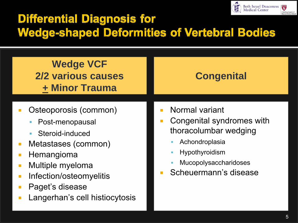
Wedge VCF 2/2 various causes
+ Minor TraumaCongenital
Osteoporosis (common)Post-menopausalSteroid-induced
Metastases (common)HemangiomaMultiple myelomaInfection/osteomyelitisPaget’s diseaseLangerhan’s cell histiocytosis
Normal variantCongenital syndromes with thoracolumbar wedging
AchondroplasiaHypothyroidismMucopolysaccharidoses
Scheuermann’s disease
5

6
Based on identifiers (age/gender), HPI, and PMH, our patient likely
has a wedge VCF 2/2 to osteoporosis + minor trauma

Patient Presentation
Vertebral Compression Fractures
Vertebroplasty
Alternate Treatment Options
Summary7
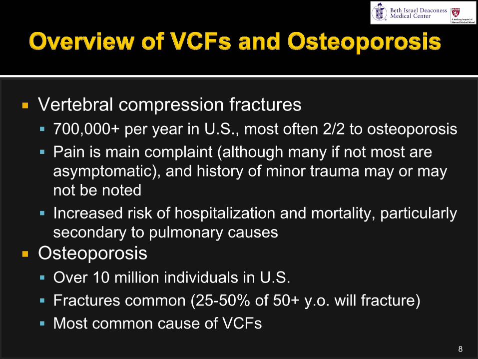
Vertebral compression fractures700,000+ per year in U.S., most often 2/2 to osteoporosisPain is main complaint (although many if not most are asymptomatic), and history of minor trauma may or may not be notedIncreased risk of hospitalization and mortality, particularly secondary to pulmonary causes
OsteoporosisOver 10 million individuals in U.S.Fractures common (25-50% of 50+ y.o. will fracture)Most common cause of VCFs
8

9
Let’s review some relevant anatomy for VCFs

Vertebral Foramen
Transverse Process
Superior articular process
Spinous process
10Netter. Atlas of Human Anatomy.
Vertebral Body
Pedicle
VCF occurs in the vertebral bodyVertebroplasty most often employs a transpedicularapproach. A needle is placed through one or both of the pedicles, avoiding vital surrounding structures

Vertebral Body
Intervertebral Disc
Transverse Process
Spinous Process
Netter. Atlas of Human Anatomy.
Pedicle
11
Spinal nerves exit intervertebral foramina. In vertebroplasty, cement extravasation in this area can lead to adverse events
Intervertebral Foramen

Posterior Internal Venous Plexus
Anterior Internal Venous Plexus
Basivertebral Vein
Netter. Atlas of Human Anatomy. 12
Proximity of venous plexus to intervention can lead to cement emboli in the venous circulation

Netter. Atlas of Human Anatomy.
Supraspinous Ligament
Thoracolumbar Fascia
Erector Spinae Muscles
Skin & Subcutaneous Tissue
Pedicle
13
Trajectory of InterventionSoft tissues traversed include SC fat, fascia, and erector spinaemusculature

Patient Presentation
Vertebral Compression Fractures
Vertebroplasty
Alternate Treatment Options
Summary14

Percutaneous, imaging-guided procedure where radio-opaque cement is injected into compressed vertebra
Needle(s) are strategically placed, providing for a conduit from the outside world to the interior of the vertebral bodyCement is injected through the hollow needle
Aim of procedure is to achieve pain control and to strengthen bone
Note that the procedure does not restore height to the vertebral body, unlike some newer techniques such as kyphoplasty
Performed by interventional radiologists (70%) as well as orthopedic surgeons, neurosurgeons, and anesthesiologists
http://www.ubneurosurgery.com/handler.cfm?event=practice,template&cpid=551015
Vertebroplasty Diagram
*

Vertebroplasty was first performed in 1984 by radiologists in France
Procedure was performed in a middle-aged female patient with a painful hemangioma in her cervical vertebraPain relief was immediate
Vertebroplasty was introduced to U.S. in 1994. Today it used most commonly for osteoporotic VCFs
Tremendous growth in popularityPain relief often immediate and dramatic, such that some patients call it “miraculous”Outpatient procedure with overnight admission at most
http://www.ubneurosurgery.com/handler.cfm?event=practice,template&cpid=551016
Vertebroplasty Diagram

Acute or healing VCF 2/2 osteoporosis, multiple myeloma, metastatic lesionsSevere focal midline pain at fracture levelNo radicular signs/sxAttempted conservative management for 6-12 wks
Unstable fracture (posterior element involvement)Uncontrollable bleedingInfectionNo complaint of pain
Common Indications Contraindications
17

18
Let’s follow Ms. BL’s vertebroplasty experience

Initial imaging studyIdentify structural alteration of vertebral bodyXR
MRI
Nukes
CT
Typically the second imaging studyFluid-sensitive sequences (T2-weighted with fat suppression or short tau inversion recovery (STIR)) identifies bone marrow edema indicative of acute or healing fracture
Bone scintigraphy used when MRI contraindicatedIncreased uptake in acute or healing fracture
Can be used to explore bony structures in complicated fracturesNot regularly employed
19

PACS, BIDMC
XR
Wedge VCFs of T8 & T9
20Lateral Thoracic Spine XR

XR
MRI
PACS, BIDMC
Wedge VCFs in T8 and T9 with:Areas of low signal
corresponding to fracture lines and trabecular impaction
21
Older VCFs in T4 and T12 with no abnormalities signal relative to surrounding vertebrae
T2-Weighted Sagittal MRI of Thoracic Spine
XR
MRI
PACS, BIDMC
22
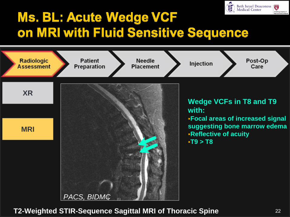
Wedge VCFs in T8 and T9 with:Focal areas of increased signal
suggesting bone marrow edemaReflective of acuityT9 > T8
T2-Weighted STIR-Sequence Sagittal MRI of Thoracic Spine
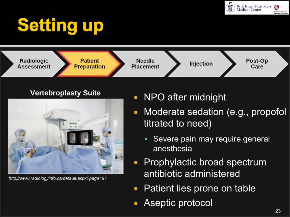
NPO after midnightModerate sedation (e.g., propofoltitrated to need)
Severe pain may require general anesthesia
Prophylactic broad spectrum antibiotic administeredPatient lies prone on tableAseptic protocol
http://www.radiologyinfo.ca/default.aspx?page=87
23
Vertebroplasty Suite

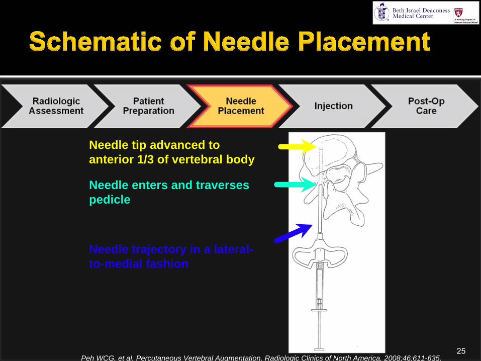
Fluoroscopic guidance employed throughoutThin gauge spinal needles placed for visual guidance along lateral-to-medial trajectorySmall skin incision is made using a scalpelStylet accompanied by large bore needle (11 or 13-gauge) is used to enter soft tissues along the same trajectory as the spinal needles, and then penetrate the pediclesAdvance tip into the anterior 1/3 of vertebral body
24
Peh WCG, et al. Percutaneous Vertebral Augmentation. Radiologic Clinics of North America. 2008;46:611-635.25
Needle trajectory in a lateral- to-medial fashion
Needle enters and traverses pedicle
Needle tip advanced to anterior 1/3 of vertebral body

AP Fluoroscopy Image
Pedicles
PACS, BIDMC
26
Spinal needles
Sternotomy wires from prior procedure
* *

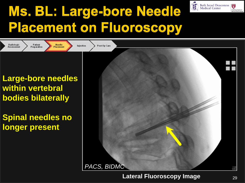
Lateral Fluoroscopy Image
Transverse Process
Spinous Process
Vertebral Body
Pedicle
PACS, BIDMC
27
Spinal needles

AP Fluoroscopy Image
Stylet & Needle
http://hyun.en.ecplaza.net/catalog.asp?DirectoryID =44780&CatalogID=40049
PACS, BIDMC
28
Screw in with clockwise motionHammer
Spinal needle
Lateral Fluoroscopy ImagePACS, BIDMC
29
Large-bore needles within vertebral bodies bilaterally
Spinal needles no longer present
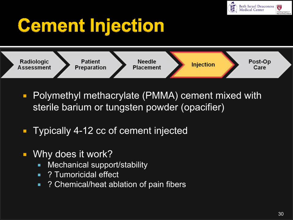
Polymethyl methacrylate (PMMA) cement mixed with sterile barium or tungsten powder (opacifier)
Typically 4-12 cc of cement injected
Why does it work?Mechanical support/stability? Tumoricidal effect? Chemical/heat ablation of pain fibers
30

Lateral Fluoroscopy ImagePACS, BIDMC
Large-bore needles within vertebral body bilaterally
Injection of cement into vertebral body

32
Does anything strike you as abnormal?

Lateral Fluoroscopy ImagePACS, BIDMC

34
Although cement is being injected into the T8 vertebral
body, we see cement in the T9 vertebral body as well

Lateral Fluoroscopy ImagePACS, BIDMC
In this patient, cement extravasated from the vertebral body of T8 and entered the vertebral body of T9 via an anterior communication
This is a rather unusual findingT8
T9

36
Large-bore needles within T8 vertebral body
Cement within T9 vertebral body
In conjunction with the lateral view, we can prove that the cement is within (not outside) the vertebral body
This confirms the anterior communication between T8 and T9
AP Fluoroscopy ImagePACS, BIDMC

37
Where should the cement be? Let’s look at another patient.

38
Large-bore needles within T10 vertebral body
Cement should stay within the vertebral body, as in this companion patient
Lateral Fluoroscopy ImageImage courtesy of Erica Gupta, MD

39
Large-bore needles within T10 vertebral body
Cement confirmed to be within vertebral body on AP view
This is where the cement should be
AP Fluoroscopy Image
Image courtesy of Erica Gupta, MD

40
Back to our patient Ms. BL

Lateral Fluoroscopy ImagePACS, BIDMC
41
Large bore needle being extracted from vertebral body
Cement within vertebral bodies of T8 and T9
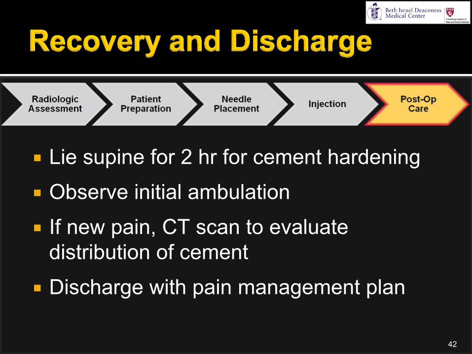
Lie supine for 2 hr for cement hardening
Observe initial ambulation
If new pain, CT scan to evaluate distribution of cement
Discharge with pain management plan
42
43
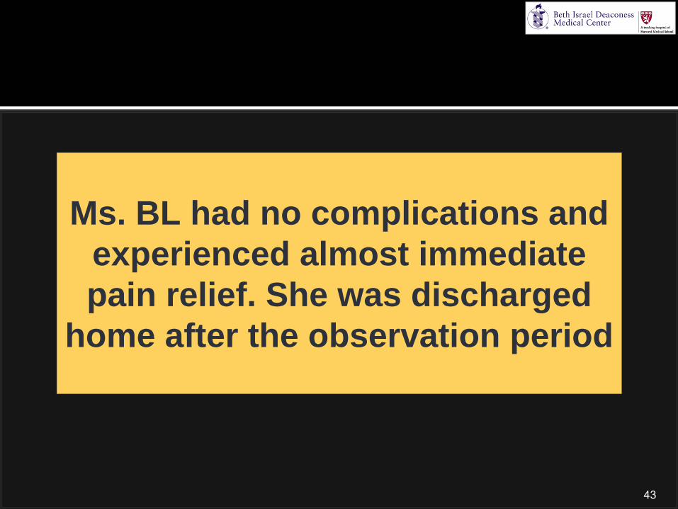
Ms. BL had no complications and experienced almost immediate pain relief. She was discharged
home after the observation period

Cement extravasationposteriorly into spinal canal, neuroforamen, or other surrounding tissues
30-93% of cases
Usually clinically insignificant
Cement emboli via venous plexus
Allergic reaction to cement
Possible increased risk of new VCFs s/p vertebroplasty
Acute Chronic
44

Patient Presentation
Vertebral Compression Fractures
Vertebroplasty
Alternate Treatment Options
Summary45

Activity modificationExternal back-bracingAssistive devicesNarcotic analgesicsPhysical therapyTherapy targeted at underlying pathology (e.g., osteoporosis medication)
46

Despite being used for 10+ years, there is a dearth of reliable efficacy data vs. medical management
Only 1 RCT▪
Showed short-term superiority of vertebroplasty
to medical management▪
Trial only examined out to 2 weeks
Long-term studies show no difference in pain/mobility at 1 or 2 yrsDifficult to assess true efficacy of vertebroplasty due to lack of rigorous studies
Based on limited data, vertebroplasty likely creates most value because of early improvement
Fast pain reliefShort time to functional recoveryLittle analgesic medicationMinimal to no hospital stay 47

KyphoplastyAngioplasty-like balloon inflated in vertebral body before cement injectionRestores vertebral height in addition to pain relief
SKyphoplasty™Coiled plastic tube opens up like popcorn to create cavity before cement injectionNew technique and device (SKy Bone Expander™ from Kyphon)
Role of each of these will be determined going forward…48
http://www.neurosurgery.pitt.edu/spine/conditions/verte bral_fractures.html
Liu J, et al. Preliminary Results for the Treatment of a Pain- Causing Osteoporotic Vertebral Compression Fracture with a Sky Bone Expander . Korean Journal of Radiology. 2008;9(5):420-425

Patient Presentation
Vertebral Compression Fractures
Vertebroplasty
Alternate Treatment Options
Summary49

Osteoporotic vertebral compression fractures are common and are accompanied by morbidity/mortality
Imaging begins with plain film, but MRI is employed to identify target vertebra(e) for intervention
In vertebroplasty, injected cement stabilizes the fracture, thus relieving pain
Vertebroplasty is effective in the right patient population, but further trials are needed
50

Gillian Lieberman, MDErica Gupta, MDJim Wu, MDMike Powell, MDMaria LevantakisLarry BarbarasMy ClassmatesBIDMC Radiology Team
51

1.
Carrino
JA, et al. Vertebral Augmentation: Vertebroplasty
& Kyphoplasty. Seminars in Roentgenology. 2004;39(1):68-84.
2.
Felson’s
Gamuts in Radiology.3.
Kim DH, et al. Osteoporotic Compression Fractures of the Spine: Current Options and Considerations for Treatment. The Spine Journal. 2006;6:479-487.
4.
Lambert RGW. Vertebroplasty
for Osteoporotic Vertebral Fracture. BMJ. 2008;336:1261-1262.5.
Liu J, et al. Preliminary Results for the Treatment of a Pain-Causing Osteoporotic Vertebral Compression Fracture with a Sky Bone Expander . Korean Journal of Radiology.
2008;9(5):420-425.6.
Netter. Atlas of Human Anatomy.7.
Michael SS, et al. Vertebroplasty
and Kyphoplasty. Bulletin of the NYU Hospital for Joint Diseases. 2006;64(3&4):106-113.
8.
Mudano
AS, et al. Vertebroplasty
and Kyphoplasty
are Associated with an Increased Risk of Secondary Vertebral Compression Fractures. Osteoporosis International. 2008; e-publication ahead of print.
9.
Peh
WCG, et al. Percutaneous
Vertebral Augmentation. Radiologic Clinics of North America. 2008;46:611-
635.
10.
Ploeg
WT, et al. Percutaneous
Vertebroplasty
as a Treatment for Osteoporotic Vertebral Compression Fractures: A Systematic Review. European Spine Journal. 2006;15:1749-1758.
11.
Predey
TA, et al. Percutaneous
Vertebroplasty. American Family Physician. 2002;66(4):611-615.12.
Resnick
D, et al. Vertebroplasty and Kyphoplasty.13.
Satre
TJ, et al. Who Should Receive Vertebroplasty? Journal of Family Practice. 2006;55(7):637-638.14.
Schweitzer ME, et al. New Techniques in Interventional Musculoskeletal Radiology.15.
Sheon
RP, et al. Clinical Manifestations and Treatment of Osteoporotic Thoracolumbar
Vertebral Compression Fractures. UpToDate. Accessed 10/11/2008.
52