alkalemic conditions result in blood mass in the circuit...
TRANSCRIPT
Alkalemic conditions result in blood clotting in the circuit soon
after initiating cardiopulmonary bypass
1) Health Care Education and Research Center, Tenri Health Care
University, Nara, Japan
2) Division of Neonatal Intensive Care, Nara Medical Universi ty
Hospital, Nara, Japan
3) Department of Clinical Laboratory, Tenri Hospital, Nara, Japan
4) Research Section 3, Central Research Laboratory, JMS Co., Ltd,
Hiroshima, Japan
Tomonori Soyama 1 , 2 ), Yukihiro Takahashi 2 ), Hideto Yoshida 3 ), Tohru
Sakoda 4 ), Tomokazu Niitsuma 4 ), Toshiya Nishikubo 2 )
Corresponding author:
Tomonori Soyama, Health Care Education and Research Center, Tenri
Health Care University, 80-1, Bessho-cho, Tenri city, Nara, 632 -0018,
Japan
E-mail: [email protected]
Tel: +81-743-63-2002
Abstract
Purpose: This study investigated the relationship between blood clotting in the
circuit soon after initiating cardiopulmonary bypass (CPB) and echinocytes that
appear with alkalemia, using a recirculation circuit filled with heparinized bovine
blood.
Methods: Alkalemic conditions in the recirculation circuit were prepared by adding
various concentrations of NaHCO3 to the priming fluid. Albumin was also added to
confirm its inhibitory effect on blood clotting. Blood pH, hold-up, the pressure
gradient, and red blood cell (RBC) reduction rate were monitored. Blood clots were
examined microscopically.
Results: Although blood pH was elevated under all experimental conditions, clotting
in the circuit increased with increased concentrations of HCO3-. Albumin inhibited
the clotting under the same alkalemic conditions. Microscopic findings revealed
echinocytes in the blood clots.
Conclusions: The shape of echinocytes was transformed by a reduction in the Donnan

equilibrium ratio because of changes in pH inside and outside the RBC membrane. Blood
clotting in the circuit soon after initiating CPB may be caused by echinocytes that
appear under alkalemic conditions. This was inhibited by albumin, suggesting that
the addition of albumin to the priming fluid may prevent such clotting in the circuit
after initiating CPB.
Keywords: alkalemic conditions, blood clotting, echinocyte, Donnan equilibrium ratio,
albumin
INTRODUCTION
Blood clotting has occasionally been reported in the cardiopulmonary bypass
(CPB) circuit soon after initiating CPB despite adequate anticoagulation
management with heparin.1–7) This can cause an obstruction in the venous reservoir
filter and membrane oxygenator, and replacement of these parts may become
necessary during surgery. Two theories have been proposed to explain this
clotting in the circuit: platelet aggregation/thrombus formation2–5) and
agglutination of the echinocytes that appear with alkalemia.6,7) However, the
underlying mechanisms have yet to be elucidated.
Prior to the initiation of CPB, priming fluid is re-circulated to evacuate any
air from the recirculation circuit and to prevent a reduction in temperature.
However, oxygen insufflation and the addition of sodium bicarbonate to the
priming fluid may result in extreme alkalization.
It has been postulated that blood clotting in the circuit soon after initiating
CPB is caused by echinocytes that form as a result of contact between the
alkalized priming fluid and blood. We have previously investigated in vitro the
frequency of echinocytes, platelet aggregation, and blood coagulation abilities
under alkalemic conditions,8,9) demonstrating that the frequency of echinocytes
increased under such conditions.8) In contrast, platelet aggregation and blood
coagulation were not enhanced under these conditions.9) We also confirmed that
the presence of albumin under alkalemic conditions inhibited the appearance of
echinocytes. This suggested that blood clotting in the circuit soon after
initiating CPB could be prevented with albumin.8)
The aim of the present study was to confirm these previous findings by
investigating the relationship between various alkalemic conditions and the

appearance of clotting in the circuit during CPB, using a recirculation circuit
filled with heparinized bovine blood.
MATERIALS AND METHODS
An experimental recirculation circuit was prepared with a venous reservoir
(OXIA RE40; JMS, Hiroshima, Japan), a roller pump (Caps Roller Pump; Stockert
GmbH, Freiburg, Germany), and a membrane oxygenator (OXIA LP; JMS, Hiroshima,
Japan) (Fig.1).
The priming fluid was prepared with a 7% NaHCO3 solution (MEYLON Injection 7%,
Otsuka Pharmaceuticals, Tokyo, Japan), 25% albumin solution (Alb; Cohn Fraction
V, pH 7.0; Wako, Osaka, Japan; dissolved in saline and the final concentration
adjusted to 25%), and lactated Ringer’s solution (Lactec Injection, Otsuka
Pharmaceutical, Tokyo, Japan). The experiment was performed under six different
alkaline conditions involving different amounts of the priming liquid in a total
volume of 1,000 mL were examined (Table 1).
To confirm whether any increase of blood clotting in the circuit was because
of the appearance of echinocytes under alkalemic conditions, and to reduce the
clotting, albumin was added under alkalemic conditions. Three sets involved
different volumes of NaHCO3: no addition (control), 100 mL (NaHCO3 100), and 200
mL (NaHCO3 200). Three further sets included different volumes of albumin (50
mL (Alb 50), 100 mL (Alb 100), and 200 mL (Alb 200)) after the addition of 100
mL NaHCO3, resulting in final albumin concentrations of 1.6, 2.6, and 4.1 g/dL,
respectively. These were combined with lactated Ringer’s solution to adjust
the priming fluid volume to a total of 1,000 mL.
For each of the six conditions, the priming liquid was added into the circuit,
Fig .1
Table .1

which was then stabilized at a blood flow of 2 L/min at a 37°C solution
temperature for 5 min. Subsequently, 400 mL of heparinized bovine blood (with
the heparin isolated from the intestinal mucosa of pig at a concentration of
15,000 IU/L), stored overnight at 4°C after blood collection, was circulated
under each condition. At the same time, oxygen insufflation of the membrane
oxygenator was started at 1 L/min.
After initiating CPB, the following items were measured for a 120-min
recirculation period (or for 60 min in the experiment with NaHCO3 200).
1. CPB Parameters
1) Blood pH
Because blood pH changes with the addition of NaHCO3 to the priming liquid and
oxygen insufflation of the membrane oxygenator, it was measured every 30 min
using a pH meter (F-55; HORIBA, Kyoto, Japan).
2) Hold up and pressure gradient
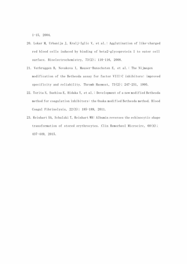
To evaluate the grade of blood clotting in the circuit, the difference in the
liquid level (the “hold up,” HU) (Fig.2) between inside and outside of the
venous reservoir filter was measured every 15 min (Fig.1), as was the pressure
gradient (PG) across the membrane oxygenator. HU was measured in mm using a ruler.
PG was measured using a digital pressure sensor (AP-34A; KEYENCE, Osaka, Japan),
and the units were converted from kPa to mmHg. The PG (mmHg/L) per 1 L of flow
was also calculated.
3) Red blood cell reduction rate
Fig .2

To confirm the protective effects of albumin on red blood cells (RBCs), the
RBCs were counted every 30 min, and the percentage reduction from the initial
count was calculated (RBC reduction rate; RRR). The RBC count was measured using
an automated multichannel blood cell analyzer (XT-1800i, Sysmex, Kobe, Japan).
2. Albumin concentration
To confirm that albumin reduced the grade of blood clotting in the circuit under
alkalemic conditions, albumin concentrations (g/dL) were measured at the
initiation of CPB using a microplate reader (Infinite M200; Tecan Austria GmbH,
Grödig, Austria).
3. Prothrombin time, activated partial thromboplastin time, and soluble fibrin
monomer complex
To investigate whether the blood clot in the circuit was caused by echinocytes,
the prothrombin time (PT; s), activated partial thromboplastin time (APTT; s),
and soluble fibrin monomer complex (SFMC; μg/mL) were measured at the
initiation and completion of circulation using an automated blood coagulation
analyzer (Coapresta 2000, SEKISUI MEDICAL, Tokyo, Japan).
4. Electron microscopic analysis
The membrane oxygenator surface that resulted in a PG because of a blood clot
in the circuit under alkalemic conditions (100 mL NaHCO3, 400 mL lactated
Ringer’s solution, and 200 mL heparinized bovine blood) was analyzed using
scanning electron photomicrography. The membrane oxygenator was fixed with 2.5%
glutaraldehyde for 1 h after washing with saline and the sample was analyzed
using a SEMEDX Type N scanning electron microscope (Hitachi, Tokyo, Japan).
5. Statistical analysis
The aim of this study was to confirm the in vitro findings of a previous study.
Accordingly, this was a single-run experiment, and the effects of the control,
NaHCO3 100, NaHCO3 200, Alb 50, Alb 100, and Alb 200 conditions on blood pH, HU,
PG, and RRR were analyzed using comparisons of two regression slopes. These
analyses were performed using Microsoft Excel, and a P value of <0.05 was
considered statistically significant.10,11)
RESULTS
The results for blood pH, HU, PG, and RRR are shown in Fig.3. Because of the
alkaline conditions, the blood pH slope increased with time across all six
conditions (control, NaHCO3 100, NaHCO3 200, Alb 50, Alb 100, and Alb 200), but
there were no significant differences in their slopes (P > 0.05). The blood pH
of the control condition only increased to 8.29, but the pH increased above 9.0
in the other five conditions (Fig.3a).
There were significant differences between the HU slopes of all six conditions
(P < 0.05) (Fig.3b). Notably, increasing albumin concentration reduced the HU.
In the NaHCO3 200 experiment, a circulation of 2 L/min could not be maintained
due to the increase in HU, and the experiment was terminated after only 60 min.
PG did not differ significantly between the three albumin concentrations (Alb
50, Alb 100, and Alb 200), but there were significant differences with the other
three conditions (P < 0.05) (Fig.3c).
The RRR results for both NaHCO3 100 and NaHCO3 200 differed significantly from
Fig .3
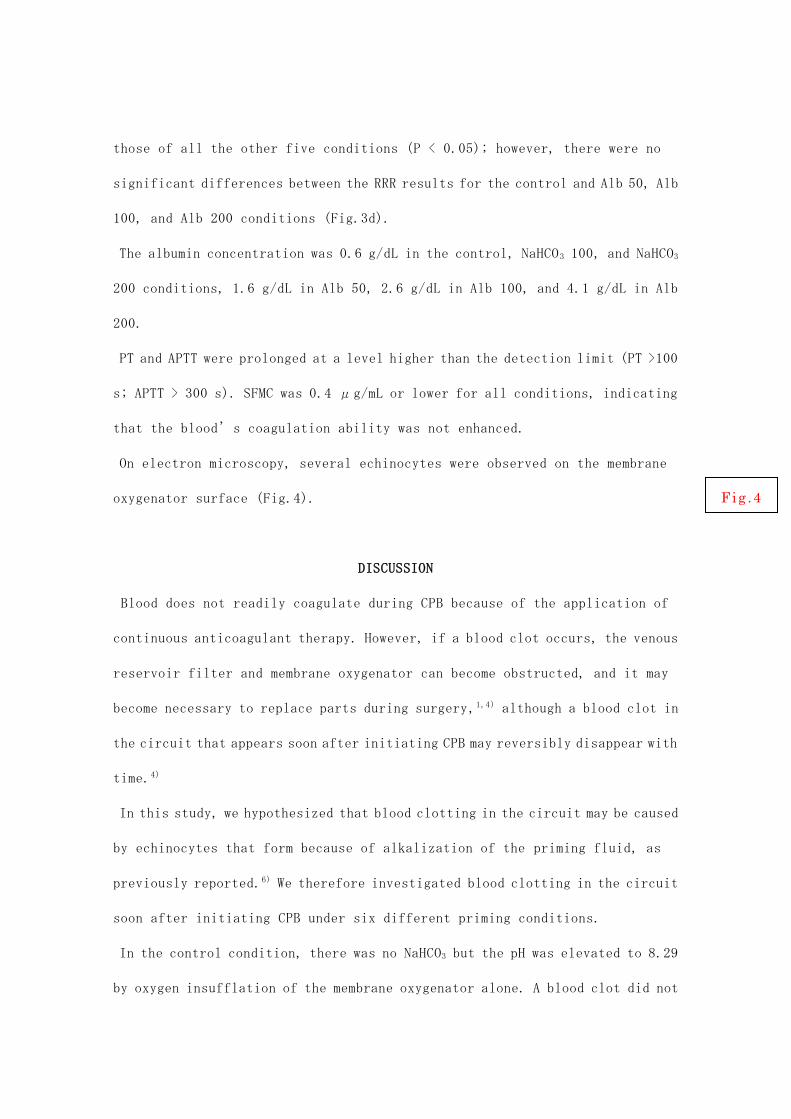
those of all the other five conditions (P < 0.05); however, there were no
significant differences between the RRR results for the control and Alb 50, Alb
100, and Alb 200 conditions (Fig.3d).
The albumin concentration was 0.6 g/dL in the control, NaHCO3 100, and NaHCO3
200 conditions, 1.6 g/dL in Alb 50, 2.6 g/dL in Alb 100, and 4.1 g/dL in Alb
200.
PT and APTT were prolonged at a level higher than the detection limit (PT >100
s; APTT > 300 s). SFMC was 0.4 μg/mL or lower for all conditions, indicating
that the blood’s coagulation ability was not enhanced.
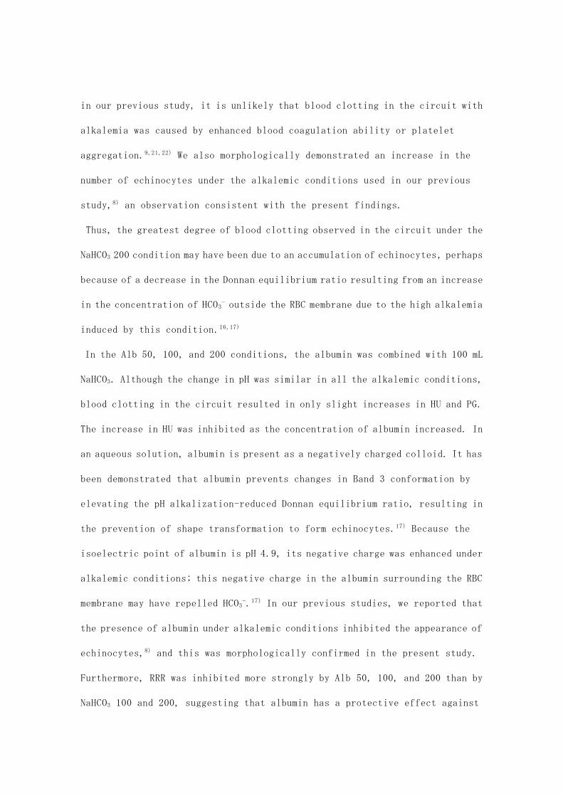
On electron microscopy, several echinocytes were observed on the membrane
oxygenator surface (Fig.4).
DISCUSSION
Blood does not readily coagulate during CPB because of the application of
continuous anticoagulant therapy. However, if a blood clot occurs, the venous
reservoir filter and membrane oxygenator can become obstructed, and it may
become necessary to replace parts during surgery,1,4) although a blood clot in
the circuit that appears soon after initiating CPB may reversibly disappear with
time.4)
In this study, we hypothesized that blood clotting in the circuit may be caused
by echinocytes that form because of alkalization of the priming fluid, as
previously reported.6) We therefore investigated blood clotting in the circuit
soon after initiating CPB under six different priming conditions.
In the control condition, there was no NaHCO3 but the pH was elevated to 8.29
by oxygen insufflation of the membrane oxygenator alone. A blood clot did not
Fig .4

form in the circuit in this condition. This suggests that, without NaHCO3 in
the priming fluid, oxygen insufflation of the membrane oxygenator alone may not
induce echinocyte-forming alkalemic conditions. In contrast, a blood clot did
appear in the circuit in both the NaHCO3 100 and the NaHCO3 200 conditions.
Although the pH change was similar under these two conditions, the blood clot
in the circuit was more prominent in the NaHCO3 200 condition.
The cytoskeletal lining of the RBC membrane maintains a wide extension in a
planar form through the binding of component proteins (e.g., spectrin and
actin); this lines the cytoplasmic side of the lipid bilayer by coupling with
the RBC transmembrane Band 3 protein (Band 3).12,13) The Band 3 conformation
(inward- or outward-facing) depends on the Donnan equilibrium ratio of Cl−, HCO3−,
OH−, and H+ concentrations between the inside (i) and outside (o) of the RBC
membrane, as represented by the following equation: [Cl−]i/[Cl−]o =
[HCO3−]i/[HCO3
−]o = [OH−]i/[OH−]o = [H+]o/[H+]i.14–16) Previous studies have reported
reductions in this ratio with increased pH, and that higher concentrations of
HCO3 − outside the RBC membrane increase the amount of Band 3 with an inward-facing
conformation.16,17) This conformation induces a contraction of the membrane
cytoskeleton and induces the echinocyte shape.16,17) Echinocytes have previously
been shown to be less deformable than discocytes,18,19) and so the obstruction
of pores in the venous reservoir filter and membrane oxygenator may occur because
of echinocytes. However, it is also possible that a change in the electrostatic
attraction on the surface of the echinocyte membrane could potentially result
in a blood clot in the circuit.20) Considering that PT and APTT were markedly
prolonged in the NaHCO3 100 and 200 conditions with a very low SFMC, and that
platelet aggregation ability was not enhanced under the alkalemic conditions
in our previous study, it is unlikely that blood clotting in the circuit with
alkalemia was caused by enhanced blood coagulation ability or platelet
aggregation.9,21,22) We also morphologically demonstrated an increase in the
number of echinocytes under the alkalemic conditions used in our previous
study,8) an observation consistent with the present findings.
Thus, the greatest degree of blood clotting observed in the circuit under the
NaHCO3 200 condition may have been due to an accumulation of echinocytes, perhaps
because of a decrease in the Donnan equilibrium ratio resulting from an increase
in the concentration of HCO3− outside the RBC membrane due to the high alkalemia
induced by this condition.16,17)
In the Alb 50, 100, and 200 conditions, the albumin was combined with 100 mL
NaHCO3. Although the change in pH was similar in all the alkalemic conditions,
blood clotting in the circuit resulted in only slight increases in HU and PG.
The increase in HU was inhibited as the concentration of albumin increased. In
an aqueous solution, albumin is present as a negatively charged colloid. It has
been demonstrated that albumin prevents changes in Band 3 conformation by
elevating the pH alkalization-reduced Donnan equilibrium ratio, resulting in
the prevention of shape transformation to form echinocytes.17) Because the
isoelectric point of albumin is pH 4.9, its negative charge was enhanced under
alkalemic conditions; this negative charge in the albumin surrounding the RBC
membrane may have repelled HCO3−.17) In our previous studies, we reported that
the presence of albumin under alkalemic conditions inhibited the appearance of
echinocytes,8) and this was morphologically confirmed in the present study.
Furthermore, RRR was inhibited more strongly by Alb 50, 100, and 200 than by
NaHCO3 100 and 200, suggesting that albumin has a protective effect against
hemolysis or pore obstruction of the venous reservoir by echinocytic
transformation. These findings therefore confirmed the previous reports that
the addition of albumin can inhibit the appearance of echinocytes in the circuit,
perhaps due to an increase in the Donnan equilibrium ratio between the inside
and outside of the RBC membrane.8,16,17,23)
CONCLUSION
Blood clotting in the circuit soon after initiating CPB may be caused by
echinocytes that appear under alkalemic conditions. The addition of albumin to
the priming fluid may inhibit this echinocyte-induced clotting.

CONFLICTS OF INTEREST
This was a cooperative study in which three institutions participated: Nara
Medical University, JMS Co., Ltd., and Tenri Hospital. The first author
performed the study at the research facility of JMS Co., Ltd., using materials
and instruments provided by the facility. All remaining authors have declared
no conflicts of interest.
REFERENCES
1. Fisher AR: The incidence and cause of emergency oxygenator changeovers.
Perfusion, 14(3); 207-212, 1999.
2. Blombäck M, Kronlund P, Carlsson K, et al.: Pathologic fibrin formation and
cold-induced clotting of membrane oxygenators during cardiopulmonary bypass.
J Cardiothorac Vasc Anesth, 9(1); 34-43, 1995.
3. Wahba A, Philipp A, Birnbaum DE, et al.: Heparin-coated equipment reduces
the risk of oxygenator failure. Ann Thorac Surg, 65(5); 1310-1312, 1998.
4. Fisher AR, Baker M, Whitehorne M, et al.: Normal and abnormal trans-oxygenator
pressure gradients during cardiopulmonary bypass. Perfusion, 18(1); 25-30,
2003.
5. Ryugo M, Mouri N, Tagaya M, et al.: Rapid rise of trans-oxygenator pressure
gradient accompanied by rapid decrease of platelet counts during open-heart
surgery. Jpn J Thorac Surg, 68(6); 439-441, 2015.
6. Soejima K, Nakae S, Sasaki A, et al.: Alkalosis and blood mass formation.
Jpn J Artif Organs, 17(3); 1417-1421, 1988. [Japanese]
7. Murakami A, Moriyasu K, Ii Y, et al.: Clinical and experimental study of the
membrane oxygenator-erythrocyte deformability and blood mass formation on
the surface of membranes. Jpn J Artif Organs, 19(1); 506-509, 1990.
[Japanese]
8. Soyama T, Yoshida H, Takahashi Y, et al.: Study of echinocytes appearing under
alkalemia during cardiopulmonary bypass control. Jpn J Extra-Corporeal
Technol, 41(2); 139-143, 2014. [Japanese]
9. Soyama T, Yoshida H, Takahashi Y, et al.: Is blood accumulation under
alkalemia related to platelet aggregation or thrombus formation during

cardiopulmonary bypass surgery?. Jpn J Extra-Corporeal Technol, 40(1); 1-6,
2013. [Japanese]
10. Andrade JM, Estévez-Pérez MG: Statistical comparison of the slopes of two
regression lines: A tutorial. Anal Chim Acta, 838; 1-12, 2014.
11. Johnson RA, Bhattacharyya GK: Statistics- Principles and Methods. 6th ed.
Hoboken, Wiley. 2011. p441-471.
12. Mohandas N, Chasis JA: Red blood cell deformability, membrane material
properties, and shape: regulation by transmembrane, skeletal and cytosolic
proteins and lipids. Semin Hematol, 30(3); 171-192, 1993.
13. Takakuwa Y, Manno S: Structure of erythrocyte membrane skeleton. Jpn J Clin
Med, 54(9); 2341-2347, 1996. [Japanese]
14. Hamasaki N: Band 3 protein, anion exchange protein of the erythrocyte
membrane. Jpn J Clin Med, 50(9); 2069-2076, 1992. [Japanese]
15. Wong P: Mechanism of control of erythrocyte shape: a possible relationship
to band 3. J Theor Biol, 171(2); 197-205, 1994.
16. Wong P: A basis of echinocytosis and stomatocytosis in the disc-sphere
transformations of the erythrocyte. J Theor Biol, 196(3); 343-361, 1999.
17. Wong P: A hypothesis of the disc-sphere transformation of the erythrocytes
between glass surfaces and of related observations. J Theor Biol, 233(1);
127-135, 2005.
18. Reinhart WH, Chien S: Red cell rheology in stomatocyte-echinocyte
transformation: roles of cell geometry and cell shape. Blood, 67(4);
1110-1118, 1986.
19. Kuzman D, Svetina S, Zeks B, et al.: Elastic properties of the red blood
cell membrane that determine echinocyte deformability. Eur Biophys J, 33(1);
1-15, 2004.
20. Lokar M, Urbanija J, Kralj-Iglic V, et al.: Agglutination of like-charged
red blood cells induced by binding of beta2-glycoprotein I to outer cell
surface. Bioelectrochemistry, 73(2); 110-116, 2008.
21. Verbruggen B, Novakova I, Mauser-Bunschoten E, et al.: The Nijmegen
modification of the Bethesda assay for factor VIII:C inhibitors: improved
specificity and reliability. Thromb Haemost, 73(2); 247-251, 1995.
22. Torita S, Suehisa E, Hidaka Y, et al.: Development of a new modified Bethesda
method for coagulation inhibitors: the Osaka modified Bethesda method. Blood
Coagul Fibrinolysis, 22(3); 185-189, 2011.
23. Reinhart SA, Schulzki T, Reinhart WH: Albumin reverses the echinocytic shape
transformation of stored erythrocytes. Clin Hemorheol Microcirc, 60(4);
437-449, 2015.

FIGURE LEGENDS
Fig. 1 The experimental recirculation circuit comprised a venous reservoir,
roller pump, and membrane oxygenator.
Fig. 2 Blood clotting in the circuit results in a difference in the liquid level
(the “hold up,” HU) between the inside and outside of the venous reservoir
filter.
Fig. 3 The results for blood pH, hold up (HU), the pressure gradient (PG), and
red blood cell reduction rate (RRR). Increases in blood pH were similar under
all the conditions tested (excluding the control condition), regardless of the
volumes of NaHCO3 and albumin added. However, HU and PG increased, and RRR was
greatest with NaHCO3 200. The addition of albumin inhibited these changes to
a greater extent than did NaHCO3 100. The increase in HU was inhibited as the
albumin concentration increased.
Fig. 4 A representative scanning electron photomicrograph of the membrane
oxygenator surface resulting in a pressure gradient due to a blood clot in the
circuit under alkalemic conditions. Many echinocytes can be seen on the membrane
oxygenator surface.

Figure 1 Experimental recirculation circuit
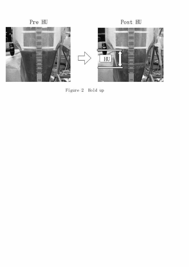
Figure 2 Hold up

a Blood pH Control NaHCO3 100 NaHCO3 200 Alb 50 Alb 100 Alb 200
Control n.s. n.s. n.s. n.s. n.s.
NaHCO3 100 n.s. n.s. n.s. n.s.
NaHCO3 200 n.s. n.s. n.s.
Alb 50 n.s. n.s.
Alb 100 n.s.
Alb 200
b HU Control NaHCO3 100 NaHCO3 200 Alb 50 Alb 100 Alb 200
Control p < 0.05 p < 0.05 p < 0.05 p < 0.05 p < 0.05
NaHCO3 100 p < 0.05 p < 0.05 p < 0.05 p < 0.05
NaHCO3 200 p < 0.05 p < 0.05 p < 0.05
Alb 50 p < 0.05 p < 0.05
Alb 100 p < 0.05
Alb 200
c PG Control NaHCO3 100 NaHCO3 200 Alb 50 Alb 100 Alb 200
Control p < 0.05 p < 0.05 p < 0.05 p < 0.05 p < 0.05
NaHCO3 100 p < 0.05 p < 0.05 p < 0.05 p < 0.05
NaHCO3 200 p < 0.05 p < 0.05 p < 0.05
Alb 50 n.s. n.s.
Alb 100 n.s.
Alb 200
d RRR Control NaHCO3 100 NaHCO3 200 Alb 50 Alb 100 Alb 200
Control p < 0.05 p < 0.05 n.s. n.s. n.s.
NaHCO3 100 p < 0.05 p < 0.05 p < 0.05 p < 0.05
NaHCO3 200 p < 0.05 p < 0.05 p < 0.05
Alb 50 n.s. n.s.
Alb 100 n.s.
Alb 200
Figure 3 Change with time of six different alkaline conditions
6.5
7
7.5
8
8.5
9
9.5
10
0 15 30 45 60 75 90 105 120
Blood pH
Time (min)
Blood pH
6.577.588.59
9.510
0 15 30 45 60 75 90 105 120
Time(min)pH
Control
NaHCO 100
NaHCO 200
Alb 50
Alb 100
Alb 200
0
20
40
60
80
100
120
140
160
0 15 30 45 60 75 90 105 120
HU (mm)
Time (min)
Hold up
6.577.588.59
9.510
0 15 30 45 60 75 90 105 120
Time(min)
pHControl
NaHCO 100
NaHCO 200
Alb 50
Alb 100
Alb 200
0
20
40
60
80
100
120
0 15 30 45 60 75 90 105 120
PG (m
mHg/L)
Time (min)
Pressure gradient
6.577.588.59
9.510
0 15 30 45 60 75 90 105 120
Time(min)
pH
Control
NaHCO 100
NaHCO 200
Alb 50
Alb 100
Alb 200
-25
-20
-15
-10
-5
0
0 15 30 45 60 75 90 105 120
RBC reduction rate (%)
Time (min)
RBC reduction rate
6.577.588.59
9.510
0 15 30 45 60 75 90 105 120
Time(min)
pH
Control
NaHCO 100
NaHCO 200
Alb 50
Alb 100
Alb 200
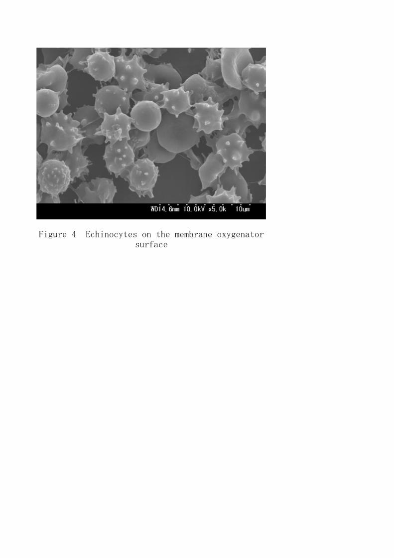
Figure 4 Echinocytes on the membrane oxygenatorsurface

Priming fluid (mL) Control NaHCO3 100 NaHCO3 200 Alb 50 Alb 100 Alb 200
7% NaHCO3 solution 0 100 200 100 100 100
25% Albumin solution 0 0 0 50 100 200
Lactated Ringer's solution 1,000 900 800 850 800 700
Total Volume 1,000 1,000 1,000 1,000 1,000 1,000
NaHCO3: 7% NaHCO3; Alb: 25% albumin solution
Table 1 Priming fluid