alopecia areata part 1
DESCRIPTION
alopeciaTRANSCRIPT
CONTINUING MEDICAL EDUCATION
Alopecia areata update
Part I. Clinical picture, histopathology, and pathogenesis
Abdullah Alkhalifah, MD,a Adel Alsantali, MD,a Eddy Wang, BSc,a Kevin J. McElwee, PhD,a
and Jerry Shapiro, MDa,b
Vancouver, British Columbia, Canada, and New York, New York
From
si
D
Fund
Conf
Jo
Tr
pe
Repr
Sk
C
0190
ª 20
doi:1
Alopecia areata (AA) is an autoimmune disease that presents as nonscarring hair loss, although the exactpathogenesis of the disease remains to be clarified. Disease prevalence rates from 0.1% to 0.2% have beenestimated for the United States. AA can affect any hair-bearing area. It often presents as well demarcatedpatches of nonscarring alopecia on skin of overtly normal appearance. Recently, newer clinical variantshave been described. The presence of AA is associated with a higher frequency of other autoimmunediseases. Controversially, there may also be increased psychiatric morbidity in patients with AA. Althoughsome AA features are known poor prognostic signs, the course of the disease is unpredictable and theresponse to treatment can be variable. Part one of this two-part series on AA describes the clinicalpresentation and the associated histopathologic picture. It also proposes a hypothesis for AA developmentbased on the most recent knowledge of disease pathogenesis. ( J Am Acad Dermatol 2010;62:177-88.)
Learning objectives: After completing this learning activity, participants should be familiar with the mostrecent advances in AA pathogenesis, recognize the rare and recently described variants of AA, and be ableto distinguish between different histopathologic stages of AA.
Key words: alopecia areata; alopecia totalis; alopecia universalis; nonscarring alopecia; pathology;pathogenesis.
Abbreviations used:
AA: alopecia areataAPC: antigen presenting cellAPS: autoimmune polyglandular syndromeAT: alopecia totalisAU: alopecia universalisCD4/8: cluster of differentiation 4/8DEBR: Dundee experimental bald ratHLA: human leukocyte antigenHPA: hypothalamic-pituitary-adrenalMHC: major histocompatibility complexSCID: severe combined immunodeficientTE: telogen effluvium
DEMOGRAPHICSKey point
d Alopecia areata affects all age groups and dif-ferent ethnicities, with equal sex distribution
Alopecia areata (AA) occurs in populations worldwide.It is a common disease encountered by dermatologists,with a frequency ranging from 0.7% to 3.8% of patientsattending dermatology clinics.1,2 In the United States,AAwasestimated tooccur in0.1% to0.2%of thegeneralpopulation,3 with a lifetime risk of 1.7%.4 Overall, AAlikely affects males and females equally.5 Some studiesshow a significant male preponderance in the adult agegroup, although others identify contrasting results.6,7
the Department of Dermatology and Skin Science,a Univer-
ty of British Columbia, Vancouver, and the Department of
ermatology,b New York University.
ing sources: None.
licts of interest: Dr Shapiro is a consultant for Johnson and
hnson Inc. Drs Shapiro and McElwee are cofounders of
ichoScience Innovations Inc. The other authors, editors, and
er reviewers have no relevant financial relationships.
int requests: Jerry Shapiro, MD, University of British Columbia
in Care Center, 835 W 10th Ave, Vancouver, BC, V5Z 4E8,
anada. E-mail: [email protected].
-9622/$36.00
09 by the American Academy of Dermatology, Inc.
0.1016/j.jaad.2009.10.032
Pediatric AA constitutes approximately 20% of AAcases,8 and as many as 60% of patients with AA willpresent with their first patch before 20 years of age.9
One study suggests that 85.5%ofAsianpatientswithAAhave disease onset before 40 years of age.2 The diseaseprevalence peaks between the second and fourthdecades of life.10
CLINICAL PICTURE
Key pointsd AA classically presents as asymptomatic,
well defined patches of nonscarring alopeciawith no overt epidermal changes
177

J AM ACAD DERMATOL
FEBRUARY 2010
178 Alkhalifah et al
d Patches can be mildly reddened or peachy incolor
d Acute diffuse and total alopecia is a newvariant of AA with favorable prognosis
AA can occur on virtually any hair-bearing area,but it affects the scalp in approximately 90% of casesseen in dermatology clinics.5 The disease can be
CAPSULE SUMMARY
d Alopecia areata pathogenesis is not fullyunderstood.
d A new variant of alopecia areata with afavorable prognosis was recentlydescribed.
d The histopathologic picture variesdepending on the stage of disease and,in the absence of classic inflammation, itcan be puzzling to the inexperienceddermatopathologist.
classified based on the extentor pattern of the hair loss.11,12
The hair loss can presentas single delimited patches ofhair loss (most common),multiple patches, or exten-sive hair loss. Based on theextent of hair loss, the dis-ease is clinically classified asfollows: patchy AA, in whichthere is a partial loss of scalphair (Fig 1); alopecia totalis(AT), in which 100% of scalphair is lost (Fig 2); or alopeciauniversalis (AU), in whichthere is a 100% loss of all
scalp and body hair. Approximately 5% of cases willprogress to AT/AU.13The pattern of hair loss observed in AA can varyconsiderably, and less common presentations can beobserved in a minority of patients, including reticularpatches of hair loss; ophiasis type, band-like hair lossin parieto-temporo-occipital area (Fig 3); ophiasisinversus (sisapho), very rare band-like hair loss inthe fronto-parieto-temporal area (Fig 4); and adiffuse thinning over part or all of the scalp.Another variant that should be considered is acutediffuse and total alopecia, which was first describedby Sato-Kawamura et al14 and was reported morerecently by Lew et al15 in a larger series of patientswith similar characteristics. This new variant is char-acterized by its rapid progression and extensiveinvolvement, along with a favorable prognosis.
Classic AA lesions are well demarcated, round oroval, completely bald, smooth-surfaced patches(Fig 5).11 The skin within the patch is usually normalon the first examination; however, it is not uncom-mon to see a slightly peachy11 (Fig 6) or reddened10
color. A characteristic finding that is frequently seenin (or at the border of) the patches is ‘‘exclamationmark hairs.’’5 These are short hairs that are taperedproximally and wider distally (Fig 7). In activedisease, where alopecia patches are expanding, ahair pull test may be positive at the periphery oflesions.11 An interesting feature of AA is its initialsparing of white hairs in patients with graying hair.10
However, eventually white hair is also often lost as
the disease duration becomes chronic. Initial hairregrowth, whether spontaneous or induced by treat-ment, is typically non- or hypopigmented (Fig 8), butthe color usually returns with time.10 The disease isfrequently asymptomatic, although a few patientsreport pruritus, burning sensations, or pain beforehair loss begins.11
The use of videodermo-scopy with a magnification of20 to 70 times may be avaluable, noninvasive toolin equivocal AA cases. Thepresence of numerous yel-low dots and short regrowinghairs is suggested to be acharacteristic feature.16,17
Yellow dots, however, canalso be seen in androgenicalopecia.18 Close examina-tion of the hair shafts at theedge of lesions, particularlyexclamation mark hairs, mayreveal subtle defects in the
structure and cuticle.19
DIFFERENTIAL DIAGNOSISKey pointsd Trichotillomania and tinea capitis are the
most important differential diagnoses inchildren
d Diffuse AA can be easily misdiagnosed astelogen effluvium
In children, the most important entities to rule outare tinea capitis and trichotillomania. Tinea capitiscan be differentiated by the presence of inflammationor at least mild scaling. Trichotillomania may involveirregular or bizarrely shaped lesions. The presence ofbroken hairs with varying lengths gives lesions arough texture, unlike the smooth surface of AA. Thedifferentiation of diffuse AA from telogen effluvium(TE) can be challenging. The patient’s history mayreveal a triggering factor that may point towards adiagnosis of TE. In diffuse AA, the hair pull test mayshow some dystrophic anagen hairs compared to thepure telogen hairs found in TE. Ultimately, a scalpbiopsy may be required to correctly differentiatediffuse AA and TE. Lupus and secondary syphilismay also be considered in the differential diagnosisof AA and may require serology testing or a scalpbiopsy for confirmation. Where a strong family asso-ciation of universal hair loss is observed, the differ-ential diagnosis may include a rare inherited genetichair loss condition called congenital atrichia.20

Fig 4. Ophiasis inversus (sisapho), a rare variant that canmimic male pattern hair loss.
Fig 1. Patchy alopecia areata in the right frontotemporalarea. Note eyebrow involvement.
Fig 3. Ophiasis pattern of alopecia areata.
Fig 2. Alopecia totalis with a 100% loss of scalp hair.
J AM ACAD DERMATOL
VOLUME 62, NUMBER 2
Alkhalifah et al 179
PROGNOSISKey pointsd The extent of AA involvement is probably
the most important prognostic factord In AT/AU, the chance of full recovery is less
than 10%
The course of AA is unpredictable. Up to 50% ofpatients will recover within 1 year even withouttreatment.12 However, most patients will have morethan one episode of hair loss. The most importantfactors indicating a poor prognosis are the extent ofhair loss presentation (extensive AA/AT/AU)21 or anophiasis pattern of hair loss.15 Other factors associ-ated with a poor prognosis include a long duration ofhair loss,15 atopy, a positive family history, the pres-ence of other autoimmune diseases, nail involve-ment, and young age of first onset.11 In children, thedisease may have a tendency towards worsening withtime, even if the initial presentation was mild.21 InAT/AU, the chance of full recovery is less than 10%.22
ASSOCIATED ABNORMALITIESKey pointsd AA can be associated with nail changes in as
many as 66% of patients
d Autoimmune diseases, particularly thyroidi-tis, are the most significant association
d The presence of such abnormalities is one ofthe poor prognostic factors
d Other reported abnormalities include psy-chiatric and asymptomatic ophthalmologicchanges
Nail involvement may be observed in AA, with areported frequency ranging from 7% to 66%.23 Nailpitting is the most common nail abnormality ob-served.23,24 Other abnormalities include trachyo-nychia, Beau lines, onychorhexis, thinning orthickening, onychomadesis, koilonychias, punctuateor transverse leukonychia, and red spotted lunu-lae.11 Nails can be affected before, concurrent with,or after the resolution of hair loss. Several studieshave suggested that nail abnormalities are associatedwith more extensive hair loss.23,24
AA can be found in association with other auto-immune diseases. Thyroid autoimmunity is probablythe main association, with an incidence between 8%and 28%.25 The presence of thyroid autoantibodieshas no clinical correlation with AA severity.26 Vitiligomay be another important association, with a 3% to8% incidence in AA patients compared to a preva-lence of 1% in the US population.27 Atopy is twice ascommon in AA patients compared with the generalpopulation.27 Other diseases and genetic disorders
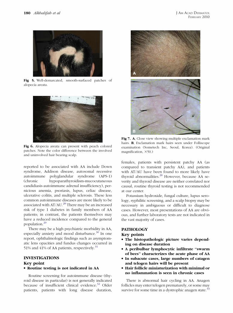
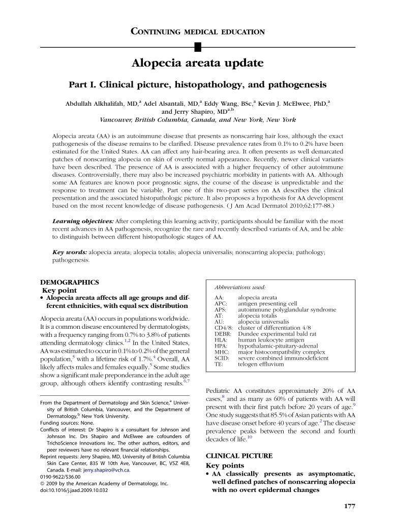
Fig 5. Well-demarcated, smooth-surfaced patches ofalopecia areata.
Fig 6. Alopecia areata can present with peach coloredpatches. Note the color difference between the involvedand uninvolved hair bearing scalp.
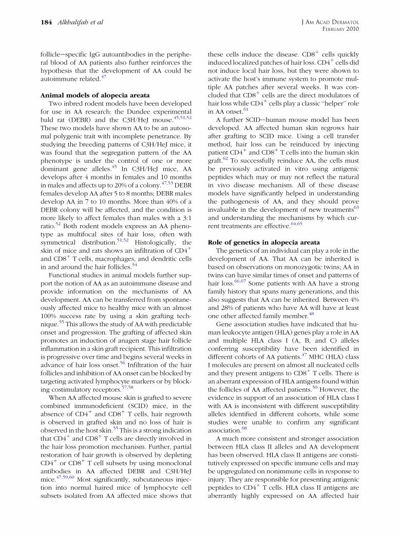
Fig 7. A, Close view showing multiple exclamation markhairs. B, Exclamation mark hairs seen under Folliscopeexamination (Sometech Inc, Seoul, Korea). (Originalmagnification, 350.)
J AM ACAD DERMATOL
FEBRUARY 2010
180 Alkhalifah et al
reported to be associated with AA include Downsyndrome, Addison disease, autosomal recessiveautoimmune polyglandular syndrome (APS-1)(chronic hypoparathyroidism-mucocutaneouscandidiasis-autoimmune adrenal insufficiency), per-nicious anemia, psoriasis, lupus, celiac disease,ulcerative colitis, and multiple sclerosis. These lesscommon autoimmune diseases are more likely to beassociated with AT/AU.28 There may be an increasedrisk of type 1 diabetes in family members of AApatients; in contrast, the patients themselves mayhave a reduced incidence compared to the generalpopulation.29
There may be a high psychiatric morbidity in AA,especially anxiety and mood disturbance.30 In onereport, ophthalmologic findings such as asymptom-atic lens opacities and fundus changes occurred in51% and 41% of AA patients, respectively.31
INVESTIGATIONSKey pointd Routine testing is not indicated in AA
Routine screening for autoimmune disease (thy-roid disease in particular) is not generally indicatedbecause of insufficient clinical evidence.32 Olderpatients, patients with long disease duration,
females, patients with persistent patchy AA (ascompared to transient patchy AA), and patientswith AT/AU have been found to more likely havethyroid abnormalities.28 However, because AA se-verity and thyroid disease are neither correlated norcausal, routine thyroid testing is not recommendedat our center.
Potassium hydroxide, fungal culture, lupus sero-logy, syphilitic screening, and a scalp biopsy may benecessary in ambiguous or difficult to diagnosecases. However, most presentations of AA are obvi-ous, and further laboratory tests are not indicated inthe vast majority of cases.
PATHOLOGYKey pointsd The histopathologic picture varies depend-
ing on disease durationd A peribulbar lymphocytic infiltrate ‘‘swarm
of bees’’ characterizes the acute phase of AAd In subacute cases, large numbers of catagen
and telogen hairs will be presentd Hair follicle miniaturization with minimal or
no inflammation is seen in chronic cases
There is abnormal hair cycling in AA. Anagenfollicles may enter telogen prematurely, or some maysurvive for some time in a dystrophic anagen state.33

Fig 8. White hair regrowth after one session of intrale-sional triamcinolone acetonide.
Fig 9. Classic peribulbar ‘‘swarm of bees’’ inflammation inalopecia areata. Some eosinophils are present within theinfiltrate. (Hematoxylineeosin stain; original magnifica-tion: 320.)
Fig 10. Abnormal hair shaft formation (trichomalacia): asign of active alopecia areata. (Hematoxylineeosin stain;original magnification: 320.)
Fig 11. Catagen transformation in subacute alopecia areata.(Hematoxylineeosin stain; original magnification: 320.)
J AM ACAD DERMATOL
VOLUME 62, NUMBER 2
Alkhalifah et al 181
Consequently, the histopathologic appearance of AAvaries depending on disease duration.34 However,increased numbers of eosinophils can be present inregions of AA affected skin in any stage of AA and area useful diagnostic feature.35,36
In the acute stage, a peribulbar lymphocytic infil-trate ‘‘swarm of bees’’ preferentially targets anagenstage follicles (Fig 9).34 The infiltrate is composed ofbothCD41 andCD81 cellswith theCD41/CD81 ratiobeing higher in clinically active disease.37 As a con-sequence, edema, microvesiculation, apoptosis, ne-crosis, macrophages, and foreign body giant cells canbe seen in and around the affected hair follicles.38
The root sheaths and hair matrix are infiltrated bylymphocytes and there may be hair follicle pigmentincontinence, keratinocyte cell necrosis, and vacuo-lar damage.5,39 Focal matrix cell vacuolization andnecrosis, a relatively uncommon event, is claimed tobe a characteristic feature of AA.40 Ultrastructuralstudies showed that keratinocytic degeneration mayaffect layers ofmatrix cells in AA, unlike the apoptosisof scattered outer root sheath cells in normal cata-gen.41 Anagen arrest, shortly followed by catagen,weakens the hair shaft and causes breakage at thesurface of the skin. As the follicle goes into telogen,
the fractured widened tip will further extrude, result-ing in the typical exclamation point hair.38
Trichomalacia (Fig 10) with marked narrowing ofthe hair shafts (‘‘pencil point hair’’) results in fragilehairs that fall from the scalp in great numbers.40
In the subacute stage, large numbers of catagenhairs, followed by telogen hairs, can be observed.38
The percentage of catagen/telogen is markedlyincreased (Fig 11) and often exceeds 50% of the totalfollicles.40 Some remnant inflammation may persistin or around fibrous streamers as the follicles ascendto telogen level.38
In the chronic stage, there is marked hair follicleminiaturization (Fig 12). The terminal to vellus scalphair follicle ratio is reduced and is likely to be 1:1.38
These miniaturized anagen follicles are situatedslightly deeper than normal vellus follicles.40
Chronic lesions are characterized by the presenceof nanogen follicles (an intermediate stage betweenterminal and vellus anagen; Fig 13).5 Nonscleroticfibrous tracts (streamers) extend along the originalsite of the previous terminal follicles into the sub-cutis.34 The inflammatory infiltrate, if present, islikely to be in the papillary dermis around miniatur-ized follicles.38

Fig 12. Marked miniaturization: a feature of long-standingalopecia areata. (Hematoxylineeosin stain; original mag-nification: 34.)
Fig 13. Nanogen follicle (intermediate stage, betweenterminal and vellus anagen) is very typical of alopeciaareata. (Hematoxylineeosin stain; original magnification:360.)
J AM ACAD DERMATOL
FEBRUARY 2010
182 Alkhalifah et al
In the recovery stage, the terminal to vellus ratioreverts to normal, the percentage of anagen hairsincreases, and there is little or no inflammation.38
The total number of follicles are normal or decreasedin AA compared to normal scalp.40,42
ALOPECIA AREATA PATHOGENESISKey pointsd AA is a lymphocyte cellemediated inflamma-
tory form of hair loss with research evidencesuggesting an underlying autoimmuneetiopathogenesis
d The development of hair loss involves aber-rant modulation of the hair growth cycle,resulting in dystrophic anagen hair folliclesand/or increased frequency of telogen statefollicles
d Genetic susceptibility to the development ofAA involves specific alleles of the HLA regionthough other non-HLA genes are also likelyto be involved
d Susceptibility to the development of AA maybe modified by environmental factors, in-cluding exposure to proinflammatory agentsand possibly other modulators, includingstress and diet
Hair follicle growth cycling modulation inalopecia areata
There are three key phases of the hair cycle: thegrowth phase (anagen), the regression phase (cata-gen), and the resting phase (telogen).43 The cyclingof these phases is finely coordinated by the expres-sion of hormones, cytokines, transcription factors,and their corresponding receptors and is carefullyregulated through endocrine, paracrine, and auto-crine routes. The disruption of these finely tunedpathways can result in the development of hairdiseases. Exogen is a hair follicle cycle event thatdescribes the controlled shedding of club hair fibers.
In healthy individuals, shedding normally occursduring the subsequent anagen growth phase as anew hair fiber is produced. In the development ofalopecias, exogen occurs in advance of renewedanagen growth, leaving a hair follicle devoid ofvisible hair fiber—a state called kenogen.44
In AA, significant disruption of the hair growthcycle clearly occurs, but different perturbations inhair growth occur depending on the pattern, severity,and duration of AA in each patient. There are severalpossible presentations of AA. First, the anagen phaseof a hair follicle can become inflamed and maintainedin a dystrophic anagen state, unable to produce hairfiber of significant size or integrity.45 When there is agreater intensity of inflammation, the hair folliclesmay be forced into a telogen phase and may thencycle through multiple anagenetelogen phases ofbrief duration. Correspondingly, inflammatory cellinfiltration occurs in early anagen follicles withoutmigration to draining lymph nodes as follicles capit-ulate and return to telogen.43,45 Finally, when AA ischronic, the hair follicles tend to persist in a pro-longed telogen phase without an apparent attempt toreturn to an anagen growth phase (Fig 14).38,43,45
Autoimmune activity in alopecia areataThe pathogenesis of AA and the molecular mech-
anisms that lead to hair loss are poorly understood.In the past, AA was believed to be of infectious orneurotrophic origin.46 Recent research studies haveindicated that AA is an inflammation-driven diseaseand is likely an autoimmune disorder.47-49
Circumstantial evidence in support of an autoim-mune mechanism underlying AA comes from severalsources. The association of AA with other autoim-mune diseases has been reported.25 The presence ofinflammatory lymphocytes around and within af-fected hair follicles and the ability to promote hairregrowth with the use of immunosuppressive agents

Fig 14. Hair growth cycle patterns in alopecia areata. A, Hair follicles held in dystrophicanagen by mild inflammatory insult unable to produce significant hair fiber. B, Anagen growthphases truncated by moderate inflammatory insult resulting in rapid cycling and brief hair fibergrowth. C, Hair follicles enter prolonged telogen dormancy with development of chronicalopecia areata.
J AM ACAD DERMATOL
VOLUME 62, NUMBER 2
Alkhalifah et al 183
is consistent with an autoimmune hypothesis.50 Theinfiltration of antigen presenting cells (APCs) such asmacrophages and Langerhans cells both around andwithin the dystrophic hair follicles has also beenobserved.47 This is potentially consistent with aresponse to autoantigens within the hair folliclesand attraction of these APCs.
A major proinflammatory event in AA is suggestedto be the abnormal expression of class I and II majorhistocompatibility complex antigens in hair follicleepithelia.50 There is also an increase in proinflam-matory markers such as intercellular cell adhesionmolecule and endothelial cell selectin in the bloodvessels around the hair follicles. The presence of hair
J AM ACAD DERMATOL
FEBRUARY 2010
184 Alkhalifah et al
follicleespecific IgG autoantibodies in the periphe-ral blood of AA patients also further reinforces thehypothesis that the development of AA could beautoimmune related.47
Animal models of alopecia areataTwo inbred rodent models have been developed
for use in AA research: the Dundee experimentalbald rat (DEBR) and the C3H/HeJ mouse.45,51,52
These two models have shown AA to be an autoso-mal polygenic trait with incomplete penetrance. Bystudying the breeding patterns of C3H/HeJ mice, itwas found that the segregation pattern of the AAphenotype is under the control of one or moredominant gene alleles.45 In C3H/HeJ mice, AAdevelops after 4 months in females and 10 monthsin males and affects up to 20% of a colony.47,53 DEBRfemales develop AA after 5 to 8 months; DEBR malesdevelop AA in 7 to 10 months. More than 40% of aDEBR colony will be affected, and the condition ismore likely to affect females than males with a 3:1ratio.52 Both rodent models express an AA pheno-type as multifocal sites of hair loss, often withsymmetrical distribution.51,52 Histologically, theskin of mice and rats shows an infiltration of CD41
and CD81 T cells, macrophages, and dendritic cellsin and around the hair follicles.54
Functional studies in animal models further sup-port the notion of AA as an autoimmune disease andprovide information on the mechanisms of AAdevelopment. AA can be transferred from spontane-ously affected mice to healthy mice with an almost100% success rate by using a skin grafting tech-nique.55 This allows the study of AA with predictableonset and progression. The grafting of affected skinpromotes an induction of anagen stage hair follicleinflammation in a skin graft recipient. This infiltrationis progressive over time and begins several weeks inadvance of hair loss onset.56 Infiltration of the hairfollicles and inhibition of AA onset can be blocked bytargeting activated lymphocyte markers or by block-ing costimulatory receptors.57,58
When AA affected mouse skin is grafted to severecombined immunodeficient (SCID) mice, in theabsence of CD41 and CD81 T cells, hair regrowthis observed in grafted skin and no loss of hair isobserved in the host skin.55 This is a strong indicationthat CD41 and CD81 T cells are directly involved inthe hair loss promotion mechanism. Further, partialrestoration of hair growth is observed by depletingCD41 or CD81 T cell subsets by using monoclonalantibodies in AA affected DEBR and C3H/HeJmice.47,59,60 Most significantly, subcutaneous injec-tion into normal haired mice of lymphocyte cellsubsets isolated from AA affected mice shows that
these cells induce the disease. CD81 cells quicklyinduced localized patches of hair loss. CD41 cells didnot induce local hair loss, but they were shown toactivate the host’s immune system to promote mul-tiple AA patches after several weeks. It was con-cluded that CD81 cells are the direct modulators ofhair loss while CD41 cells play a classic ‘‘helper’’ rolein AA onset.61
A further SCIDehuman mouse model has beendeveloped. AA affected human skin regrows hairafter grafting to SCID mice. Using a cell transfermethod, hair loss can be reinduced by injectingpatient CD41 and CD81 T cells into the human skingraft.62 To successfully reinduce AA, the cells mustbe previously activated in vitro using antigenicpeptides which may or may not reflect the naturalin vivo disease mechanism. All of these diseasemodels have significantly helped in understandingthe pathogenesis of AA, and they should proveinvaluable in the development of new treatments63
and understanding the mechanisms by which cur-rent treatments are effective.64,65
Role of genetics in alopecia areataThe genetics of an individual can play a role in the
development of AA. That AA can be inherited isbased on observations on monozygotic twins; AA intwins can have similar times of onset and patterns ofhair loss.66,67 Some patients with AA have a strongfamily history that spans many generations, and thisalso suggests that AA can be inherited. Between 4%and 28% of patients who have AA will have at leastone other affected family member.48
Gene association studies have indicated that hu-man leukocyte antigen (HLA) genes play a role in AAand multiple HLA class I (A, B, and C) allelesconferring susceptibility have been identified indifferent cohorts of AA patients.47 MHC (HLA) classI molecules are present on almost all nucleated cellsand they present antigens to CD81 T cells. There isan aberrant expression of HLA antigens found withinthe follicles of AA affected patients.50 However, theevidence in support of an association of HLA class Iwith AA is inconsistent with different susceptibilityalleles identified in different cohorts, while somestudies were unable to confirm any significantassociation.68
A much more consistent and stronger associationbetween HLA class II alleles and AA developmenthas been observed. HLA class II antigens are consti-tutively expressed on specific immune cells and maybe upgregulated on nonimmune cells in response toinjury. They are responsible for presenting antigenicpeptides to CD41 T cells. HLA class II antigens areaberrantly highly expressed on AA affected hair

J AM ACAD DERMATOL
VOLUME 62, NUMBER 2
Alkhalifah et al 185
follicles. Specific alleles, such as DQB1*03 andDRB1*1104, have been reported as markers forsusceptibility to AA.69 These findings suggest theimportance of specific HLA class II alleles in theonset and progression of AA and imply that antigenpresentation to CD41 cells plays a significant role inAA development. Several association studies haveinvestigated non-HLA gene alleles, indicating thatmultiple genes may contribute to AA susceptibility.46
Genome-wide screening for AA susceptibility locihas been challenging. Linkage analysis with the AAmouse model revealed that AA is a complex poly-genic trait.70 Four intervals (Alaa1, 2, 3, and 4) onmouse chromosomes 17, 9, 8, and 15, respectively,were identified as regions of the genome conferringAA susceptibility.70 Mouse gene associations werefound comparable to those in humans. In particular,the equivalent MHC region in mice (Alaa1) wasstrongly associated with AA susceptibility. Studies toidentify the specific susceptibility genes within theseregions are underway.
A genome-wide study was completed on ex-tended human families with multiple AA patients.Intervals on human chromosomes 6, 10, 16, and 18were identified as potential AA susceptibility loci.71
This study further confirmed that the HLA region onhuman chromosome 6 is associated with geneticsusceptibility to AA. However, there was at least onesignificant genetic determinant of AA found at6q.23.3 outside of the HLA gene cluster. Large scale,genome-wide screens using the AA registry and DNAbank72 are nearing completion and should providesignificant new information on the potential geneactivity in AA development beyond the HLA region.
Environmental impact on alopecia areataEnvironmental factors may also contribute to AA
development. Specific gene alleles might provide aninnate degree of susceptibility to AA for an individ-ual, but environmental factors likely cumulativelydetermine the actual onset, hair loss pattern, andseverity of the disease.47,68 However, the exact envi-ronmental stimuli required for AA expression are yetto be determined. Hormonal fluctuation,68 infectiousagents,73 and vaccinations74 have all been cited aspossible triggers for AA. In the mouse model, dietarysoy oil increases resistance to AA development,suggesting that diet might also play a role in AAsusceptibility.56 It is likely that there are manypotential environmental inputs with different factorsinvolved in AA development for different individuals.
Stress is commonly cited as a cause for AA onset,but controlled clinical studies have been inconclu-sive.75-77 Some did not find a significant correlationof hair loss onset with stressful life events,78-80 while
others confirm stressful events in AA patients beforethe onset of disease.30,79,81 In contrast, several stud-ies have shown that individuals with AA are morelikely to exhibit aberrant psychosocial traits, such asincreased anxiety, depression, and aggres-sion.76,78,80,82 Investigations on the skin of miceand humans have indicated increased presence ofmultiple factors associated with stress mediation.83,84
In a mouse model study, the development of AA wasstrongly associated with higher central and periph-eral hypothalamic-pituitary-adrenal (HPA) tone.85
The AA affected mice had a significantly bluntedsystemic HPA response to acute physiologic stressand a decreased habituation response to constantpsychological stress. Taken together, AA may impactstress responses and HPA activity may be signifi-cantly involved in modulating the severity or courseof AA.
Hypothesis for alopecia areata developmentAlthough many hypotheses to explain auto-
immune disease development have been suggestedby immunologists,86-89 most of these scenarios havenot been seriously considered in the context of AA.Currently, AA development hypotheses focus on hairfollicle immune privilege collapse or the inappropri-ate presentation of antigens to the immune systemduring normal hair follicle cycling. Anagen stage hairfollicles retain immune privilege, and a breach inimmune privilege and exposure of unique hairfollicle antigens may result in targeting by the skinimmune system.90-92 This popular hypothesis ishighly ‘‘skin-centric’’ and largely ignores currentimmunologic dogma, although it is attractive in itssimplicity.
An alternative hypothesis is based on the knowl-edge that hair follicle immunoprotection is transient,limited to the anagen growth cycle stage. Regressionof the hair follicle in catagen involves significantapoptosis93 and immune cell infiltration.94-97 Thisnormal hair follicle cycling event may continuouslyexpose the immune system to low levels of hairfollicleederived antigens. Hair follicleespecific au-toantibodies found in humans and animal models inthe absence of AA98-100 may be a consequence of thisconstant low level exposure.
Langerhans cells and dendritic cells are capable ofpresenting cell apoptosisederived antigens to lym-phocytes and stimulating autoimmunity.101-103 Ifcatagen regression became disordered, the associ-ated immune cell infiltrate might inappropriatelycoexpress antigenic peptides and costimulatory mol-ecules and induce an immunologic response.47,56
Mouse studies have revealed that significant proin-flammatory events occur in the lymph nodes in
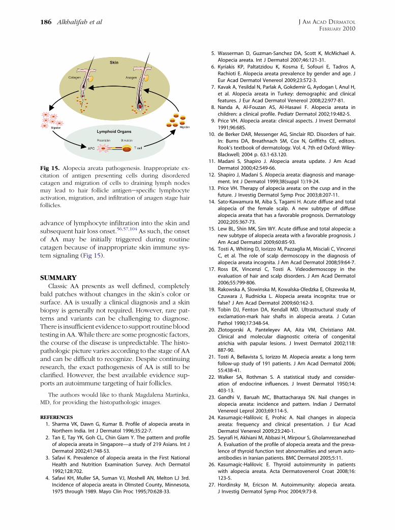
Fig 15. Alopecia areata pathogenesis. Inappropriate ex-citation of antigen presenting cells during disorderedcatagen and migration of cells to draining lymph nodesmay lead to hair follicle antigenespecific lymphocyteactivation, migration, and infiltration of anagen stage hairfollicles.
J AM ACAD DERMATOL
FEBRUARY 2010
186 Alkhalifah et al
advance of lymphocyte infiltration into the skin andsubsequent hair loss onset.56,57,104 As such, the onsetof AA may be initially triggered during routinecatagen because of inappropriate skin immune sys-tem signaling (Fig 15).
SUMMARYClassic AA presents as well defined, completely
bald patches without changes in the skin’s color orsurface. AA is usually a clinical diagnosis and a skinbiopsy is generally not required. However, rare pat-terns and variants can be challenging to diagnose.There is insufficient evidence to support routinebloodtesting in AA. While there are some prognostic factors,the course of the disease is unpredictable. The histo-pathologic picture varies according to the stage of AAand can be difficult to recognize. Despite continuingresearch, the exact pathogenesis of AA is still to beclarified. However, the best available evidence sup-ports an autoimmune targeting of hair follicles.
The authors would like to thank Magdalena Martinka,MD, for providing the histopathologic images.
REFERENCES
1. Sharma VK, Dawn G, Kumar B. Profile of alopecia areata in
Northern India. Int J Dermatol 1996;35:22-7.
2. Tan E, Tay YK, Goh CL, Chin Giam Y. The pattern and profile
of alopecia areata in Singapore—a study of 219 Asians. Int J
Dermatol 2002;41:748-53.
3. Safavi K. Prevalence of alopecia areata in the First National
Health and Nutrition Examination Survey. Arch Dermatol
1992;128:702.
4. Safavi KH, Muller SA, Suman VJ, Moshell AN, Melton LJ 3rd.
Incidence of alopecia areata in Olmsted County, Minnesota,
1975 through 1989. Mayo Clin Proc 1995;70:628-33.
5. Wasserman D, Guzman-Sanchez DA, Scott K, McMichael A.
Alopecia areata. Int J Dermatol 2007;46:121-31.
6. Kyriakis KP, Paltatzidou K, Kosma E, Sofouri E, Tadros A,
Rachioti E. Alopecia areata prevalence by gender and age. J
Eur Acad Dermatol Venereol 2009;23:572-3.
7. Kavak A, Yesildal N, Parlak A, Gokdemir G, Aydogan I, Anul H,
et al. Alopecia areata in Turkey: demographic and clinical
features. J Eur Acad Dermatol Venereol 2008;22:977-81.
8. Nanda A, Al-Fouzan AS, Al-Hasawi F. Alopecia areata in
children: a clinical profile. Pediatr Dermatol 2002;19:482-5.
9. Price VH. Alopecia areata: clinical aspects. J Invest Dermatol
1991;96:68S.
10. de Berker DAR, Messenger AG, Sinclair RD. Disorders of hair.
In: Burns DA, Breathnach SM, Cox N, Griffiths CE, editors.
Rook’s textbook of dermatology. Vol. 4. 7th ed Oxford: Wiley-
Blackwell; 2004 p. 63.1-63.120.
11. Madani S, Shapiro J. Alopecia areata update. J Am Acad
Dermatol 2000;42:549-66.
12. Shapiro J, Madani S. Alopecia areata: diagnosis and manage-
ment. Int J Dermatol 1999;38(suppl 1):19-24.
13. Price VH. Therapy of alopecia areata: on the cusp and in the
future. J Investig Dermatol Symp Proc 2003;8:207-11.
14. Sato-Kawamura M, Aiba S, Tagami H. Acute diffuse and total
alopecia of the female scalp. A new subtype of diffuse
alopecia areata that has a favorable prognosis. Dermatology
2002;205:367-73.
15. Lew BL, Shin MK, Sim WY. Acute diffuse and total alopecia: a
new subtype of alopecia areata with a favorable prognosis. J
Am Acad Dermatol 2009;60:85-93.
16. Tosti A, Whiting D, Iorizzo M, Pazzaglia M, Misciali C, Vincenzi
C, et al. The role of scalp dermoscopy in the diagnosis of
alopecia areata incognita. J Am Acad Dermatol 2008;59:64-7.
17. Ross EK, Vincenzi C, Tosti A. Videodermoscopy in the
evaluation of hair and scalp disorders. J Am Acad Dermatol
2006;55:799-806.
18. Rakowska A, Slowinska M, Kowalska-Oledzka E, Olszewska M,
Czuwara J, Rudnicka L. Alopecia areata incognita: true or
false? J Am Acad Dermatol 2009;60:162-3.
19. Tobin DJ, Fenton DA, Kendall MD. Ultrastructural study of
exclamation-mark hair shafts in alopecia areata. J Cutan
Pathol 1990;17:348-54.
20. Zlotogorski A, Panteleyev AA, Aita VM, Christiano AM.
Clinical and molecular diagnostic criteria of congenital
atrichia with papular lesions. J Invest Dermatol 2002;118:
887-90.
21. Tosti A, Bellavista S, Iorizzo M. Alopecia areata: a long term
follow-up study of 191 patients. J Am Acad Dermatol 2006;
55:438-41.
22. Walker SA, Rothman S. A statistical study and consider-
ation of endocrine influences. J Invest Dermatol 1950;14:
403-13.
23. Gandhi V, Baruah MC, Bhattacharaya SN. Nail changes in
alopecia areata: incidence and pattern. Indian J Dermatol
Venereol Leprol 2003;69:114-5.
24. Kasumagic-Halilovic E, Prohic A. Nail changes in alopecia
areata: frequency and clinical presentation. J Eur Acad
Dermatol Venereol 2009;23:240-1.25. Seyrafi H, Akhiani M, Abbasi H, Mirpour S, Gholamrezanezhad
A. Evaluation of the profile of alopecia areata and the preva-
lence of thyroid function test abnormalities and serum auto-
antibodies in Iranian patients. BMC Dermatol 2005;5:11.26. Kasumagic-Halilovic E. Thyroid autoimmunity in patients
with alopecia areata. Acta Dermatovenerol Croat 2008;16:
123-5.
27. Hordinsky M, Ericson M. Autoimmunity: alopecia areata.
J Investig Dermatol Symp Proc 2004;9:73-8.
J AM ACAD DERMATOL
VOLUME 62, NUMBER 2
Alkhalifah et al 187
28. Goh C, Finkel M, Christos PJ, Sinha AA. Profile of 513 patients
with alopecia areata: associations of disease subtypes with
atopy, autoimmune disease and positive family history. J Eur
Acad Dermatol Venereol 2006;20:1055-60.
29. Wang SJ, Shohat T, Vadheim C, Shellow W, Edwards J, Rotter
JI. Increased risk for type I (insulin-dependent) diabetes in
relatives of patients with alopecia areata (AA). Am J Med
Genet 1994;51:234-9.
30. Ruiz-Doblado S, Carrizosa A, Garcıa-Hernandez MJ. Alopecia
areata: psychiatric comorbidity and adjustment to illness. Int
J Dermatol 2003;42:434-7.
31. Recupero SM, Abdolrahimzadeh S, De Dominicis M, Mollo R,
Carboni I, Rota L, et al. Ocular alterations in alopecia areata.
Eye 1999;13(pt 5):643-6.
32. MacDonald Hull SP, Wood ML, Hutchinson PE, Sladden M,
Messenger AG. British Association of Dermatologists. Guide-
lines for the management of alopecia areata. Br J Dermatol
2003;149:692-9.
33. Messenger AG, Slater DN, Bleehen SS. Alopecia areata:
alterations in the hair growth cycle and correlation with
the follicular pathology. Br J Dermatol 1986;114:337-47.
34. Weedon D. Diseases of cutaneous appendages. In: Weedon
D, editor. Weedon’s skin pathology. London: Churchill Living-
stone; 2002.
35. Elston DM, McCollough ML, Bergfeld WF, Liranzo MO, Heibel
M. Eosinophils in fibrous tracts and near hair bulbs: a helpful
diagnostic feature of alopecia areata. J Am Acad Dermatol
1997;37:101-6.
36. El Darouti M, Marzouk SA, Sharawi E. Eosinophils in fibrous
tracts and near hair bulbs: a helpful diagnostic feature of
alopecia areata. J Am Acad Dermatol 2000;42:305-7.
37. Todes-Taylor N, Turner R, Wood GS, Stratte PT, Morhenn VB. T
cell subpopulations in alopecia areata. J Am Acad Dermatol
1984;11(2 pt 1):216-23.
38. Whiting DA. Histopathologic features of alopecia areata: a
new look. Arch Dermatol 2003;139:1555-9.
39. Tobin DJ. Morphological analysis of hair follicles in alopecia
areata. Microsc Res Tech 1997;38:443-51.
40. Sperling LC. Alopecia areata. In: Sperling LC, editor. An atlas
of hair pathology with clinical correlations. 1st ed New York:
Parthenon Publishing; 2003. pp. 109-38.
41. Tobin DJ, Fenton DA, Kendall MD. Cell degeneration in
alopecia areata. An ultrastructural study. Am J Dermatopa-
thol 1991;13:248-56.
42. Whiting DA. Histopathology of alopecia areata in horizontal
sections of scalp biopsies. J Invest Dermatol 1995;104(suppl 5)
26S-7S.
43. Vogt A, McElwee KJ, Blume-Peytavi U. Biology of the hair
follicle. In: Whiting DA, Blume-Peytavi U, Tosti A, editors. Hair
growth and disorders. Berlin: Springer; 2008. pp. 1-22.
44. McElwee KJ, Sinclair R. Hair physiology and its disor-
ders. Drug Discovery Today: Disease Mechanisms 2008;5:
e163-71.
45. Freyschmidt-Paul P, McElwee KJ, Hoffmann R. Alopecia areata.
In: Whiting DA, Blume-Peytavi U, Tosti A, editors. Hair growth
and disorders. Berlin: Springer; 2008. pp. 311-32.
46. King LE Jr, McElwee KJ, Sundberg JP. Alopecia areata. Curr Dir
Autoimmun 2008;10:280-312.
47. Lu W, Shapiro J, Yu M, Barekatain A, Lo B, Finner A, et al.
Alopecia areata: pathogenesis and potential for therapy.
Expert Rev Mol Med 2006;8:1-19.
48. McDonagh AJ, Tazi-Ahnini R. Epidemiology and genetics of
alopecia areata. Clin Exp Dermatol 2002;27:405-9.
49. Paus R, Slominski A, Czarnetzki BM. Is alopecia areata an
autoimmune-response against melanogenesis-related
proteins, exposed by abnormal MHC class I expression in
the anagen hair bulb? Yale J Biol Med 1993;66:541-54.
50. McElwee KJ, Tobin DJ, Bystryn JC, King LE Jr, Sundberg JP.
Alopecia areata: an autoimmune disease? Exp Dermatol
1999;8:371-9.
51. Sundberg JP, McElwee KJ, Whiting DA, King LE Jr. Alopecia
areata: spontaneous and skin graft-induced mouse models.
In: Chan LS, editor. Animal models of human inflammatory
skin diseases. Vol V. Boca Raton, FL: CRC Press; 2003. pp. 429-
50.
52. McElwee KJ. Alopecia areata: spontaneous rat model of
alopecia areata. In: Chan LS, editor. Animal models of human
inflammatory skin diseases. Vol V. Boca Raton, FL: CRC Press;
2003. pp. 451-68.
53. Sundberg JP, Cordy WR, King LE Jr. Alopecia areata in aging
C3H/HeJ mice. J Invest Dermatol 1994;102:847-56.
54. Zhang JG, Oliver RF. Immunohistological study of the devel-
opment of the cellular infiltrate in the pelage follicles of the
DEBR model for alopecia areata. Br J Dermatol 1994;130:405-
14.
55. McElwee KJ, Boggess D, King LE Jr, Sundberg JP. Experimen-
tal induction of alopecia areata-like hair loss in C3H/HeJ mice
using full-thickness skin grafts. J Invest Dermatol 1998;111:
797-803.
56. McElwee KJ, Silva K, Boggess D, Bechtold L, King LE Jr,
Sundberg JP. Alopecia areata in C3H/HeJ mice involves
leukocyte-mediated root sheath disruption in advance of
overt hair loss. Vet Pathol 2003;40:643-50.
57. Carroll JM, McElwee KJ, King LE Jr, Byrne MC, Sundberg JP.
Gene array profiling and immunomodulation studies define a
cell-mediated immune response underlying the pathogene-
sis of alopecia areata in a mouse model and humans. J Invest
Dermatol 2002;119:392-402.
58. Freyschmidt-Paul P, Seiter S, Zoller M, Konig A, Ziegler A,
Sundberg JP, et al. Treatment with an anti-CD44v10-specific
antibody inhibits the onset of alopecia areata in C3H/HeJ
mice. J Invest Dermatol 2000;115:653-7.
59. McElwee KJ, Spiers EM, Oliver RF. Partial restoration of hair
growth in the DEBR model for alopecia areata after in vivo
depletion of CD41 T cells. Br J Dermatol 1999;140:432-7.
60. McElwee KJ, Spiers EM, Oliver RF. In vivo depletion of CD81 T
cells restores hair growth in the DEBR model for alopecia
areata. Br J Dermatol 1996;135:211-7.
61. McElwee KJ, Freyschmidt-Paul P, Hoffmann R, Kissling S,
Hummel S, Vitacolonna M, et al. Transfer of CD8(1) cells
induces localized hair loss whereas CD4(1)/CD25(-) cells
promote systemic alopecia areata and CD4(1)/CD25(1)
cells blockade disease onset in the C3H/HeJ mouse model.
J Invest Dermatol 2005;124:947-57.
62. Gilhar A, Landau M, Assy B, Shalaginov R, Serafimovich S,
Kalish RS. Mediation of alopecia areata by cooperation
between CD41 and CD81 T lymphocytes: transfer to human
scalp explants on Prkdc(scid) mice. Arch Dermatol 2002;138:
916-22.
63. Sun J, Silva KA, McElwee KJ, King LE Jr, Sundberg JP. The
C3H/HeJ mouse and DEBR rat models for alopecia areata:
review of preclinical drug screening approaches and results.
Exp Dermatol 2008;17:793-805.
64. Zoller M, Gupta P, Marhaba R, Vitacolonna M, Freyschmidt-
Paul P. Anti-CD44-mediated blockade of leukocyte migration
in skin-associated immune diseases. J Leukoc Biol 2007;82:
57-71.
65. Marhaba R, Vitacolonna M, Hildebrand D, Baniyash M,
Freyschmidt-Paul P, Zoller M. The importance of myeloid-
derived suppressor cells in the regulation of autoimmune

J AM ACAD DERMATOL
FEBRUARY 2010
188 Alkhalifah et al
effector cells by a chronic contact eczema. J Immunol 2007;
179:5071-81.
66. Stankler L. Synchronous alopecia areata in two siblings: a
possible viral aetiology. Lancet 1979;1:1303-4.
67. Alsaleh QA, Nanda A, al-Hasawi F, el-Kashlan M. Concurrent
appearance of alopecia areata in siblings. Pediatr Dermatol
1995;12:285-6.
68. McElwee K, Freyschmidt-Paul P, Ziegler A, Happle R, Hoff-
mann R. Genetic susceptibility and severity of alopecia areata
in human and animal models. Eur J Dermatol 2001;11:11-6.
69. Akar A, Arca E, Erbil H, Akay C, Sayal A, Gur AR. Antioxidant
enzymes and lipid peroxidation in the scalp of patients with
alopecia areata. J Dermatol Sci 2002;29:85-90.
70. Sundberg JP, Silva KA, Li R, Cox GA, King LE Jr. Adult-onset
alopecia areata is a complex polygenic trait in the C3H/HeJ
mouse model. J Invest Dermatol 2004;123:294-7.
71. Martinez-Mir A, Zlotogorski A, Gordon D, Petukhova L, Mo J,
Gilliam TC, et al. Genomewide scan for linkage reveals
evidence of several susceptibility loci for alopecia areata.
Am J Hum Genet 2007;80:316-28.
72. Duvic M, Norris D, Christiano A, Hordinsky M, Price V.
Alopecia areata registry: an overview. J Investig Dermatol
Symp Proc 2003;8:219-21.
73. Rodriguez TA, Duvic M. Onset of alopecia areata after
Epstein-Barr virus infectious mononucleosis. J Am Acad
Dermatol 2008;59:137-9.
74. Ikeda T. Produced alopecia areata based on the focal
infection theory and mental motive theory. Dermatologica
1967;134:1-11.
75. Brauner GJ, Goodheart HP. Dermatologic care behind bars. J
Am Acad Dermatol 1988;18:1066-73.
76. Gupta MA, Gupta AK, Watteel GN. Stress and alopecia areata:
a psychodermatologic study. Acta Derm Venereol 1997;77:
296-8.
77. Perini GI, Veller Fornasa C, Cipriani R, Bettin A, Zecchino F,
Peserico A. Life events and alopecia areata. Psychother
Psychosom 1984;41:48-52.
78. Brajac I, Tkalcic M, Dragojevic DM, Gruber F. Roles of stress,
stress perception and trait-anxiety in the onset and course of
alopecia areata. J Dermatol 2003;30:871-8.
79. Gulec AT, Tanriverdi N, Duru C, Saray Y, Akcali C. The role of
psychological factors in alopecia areata and the impact of the
disease on the quality of life. Int J Dermatol 2004;43:352-6.
80. Picardi A, Pasquini P, Cattaruzza MS, Gaetano P, Baliva G,
Melchi CF, et al. Psychosomatic factors in first-onset alopecia
areata. Psychosomatics 2003;44:374-81.
81. Kakourou T, Karachristou K, Chrousos G. A case series of
alopecia areata in children: impact of personal and family
history of stress and autoimmunity. J Eur Acad Dermatol
Venereol 2007;21:356-9.
82. Liakopoulou M, Alifieraki T, Katideniou A, Kakourou T,
Tselalidou E, Tsiantis J, et al. Children with alopecia areata:
psychiatric symptomatology and life events. J Am Acad Child
Adolesc Psychiatry 1997;36:678-84.
83. Siebenhaar F, Sharov AA, Peters EM, Sharova TY, Syska W,
Mardaryev AN, et al. Substance P as an immunomodulatory
neuropeptide in a mouse model for autoimmune hair loss
(alopecia areata). J Invest Dermatol 2007;127:1489-97.
84. Toyoda M, Makino T, Kagoura M, Morohashi M. Expres-
sion of neuropeptide-degrading enzymes in alopecia
areata: an immunohistochemical study. Br J Dermatol
2001;144:46-54.
85. Zhang X, Yu M, Yu W, Weinberg J, Shapiro J, McElwee KJ.
Development of alopecia areata is associated with higher
central and peripheral hypothalamic-pituitary-adrenal tone in
the skin graft induced C3H/HeJ mouse model. J Invest
Dermatol 2009;129:1527-38.
86. Theofilopoulos AN. The basis of autoimmunity: part II.
Genetic predisposition. Immunol Today 1995;16:150-9.
87. Theofilopoulos AN. The basis of autoimmunity: part I. Mecha-
nisms of aberrant self-recognition. Immunol Today 1995;16:90-8.
88. Goodnow CC, Sprent J. Fazekas de St Groth B, Vinuesa CG.
Cellular and genetic mechanisms of self tolerance and
autoimmunity. Nature 2005;435:590-7.
89. Goodnow CC. Multistep pathogenesis of autoimmune dis-
ease. Cell 2007;130:25-35.
90. Paus R, Nickoloff BJ, Ito T. A ‘‘hairy’’ privilege. Trends
Immunol 2005;26:32-40.
91. Ito T, Meyer KC, Ito N, Paus R. Immune privilege and the skin.
Curr Dir Autoimmun 2008;10:27-52.
92. Ito T, Ito N, Bettermann A, Tokura Y, Takigawa M, Paus R.
Collapse and restoration of MHC class-I-dependent immune
privilege: exploiting the human hair follicle as a model. Am J
Pathol 2004;164:623-34.
93. Botchkareva NV, Ahluwalia G, Shander D. Apoptosis in the
hair follicle. J Invest Dermatol 2006;126:258-64.
94. Weedon D, Strutton G. Apoptosis as the mechanism of the
involution of hair follicles in catagen transformation. Acta
Derm Venereol 1981;61:335-9.
95. Lindner G, Botchkarev VA, Botchkareva NV, Ling G, van der
Veen C, Paus R. Analysis of apoptosis during hair follicle
regression (catagen). Am J Pathol 1997;151:1601-17.
96. Eichmuller S, van der Veen C, Moll I, Hermes B, Hofmann U,
Muller-Rover S, et al. Clusters of perifollicular macrophages in
normal murine skin: physiological degeneration of selected
hair follicles by programmed organ deletion. J Histochem
Cytochem 1998;46:361-70.
97. Parakkal PF. Role of macrophages in collagen resorption
during hair growth cycle. J Ultrastruct Res 1969;29:210-7.
98. McElwee KJ, Pickett P, Oliver RF. The DEBR rat, alopecia
areata and autoantibodies to the hair follicle. Br J Dermatol
1996;134:55-63.
99. Tobin DJ, Sundberg JP, King LE Jr, Boggess D, Bystryn JC.
Autoantibodies to hair follicles in C3H/HeJ mice with alope-
cia areata-like hair loss. J Invest Dermatol 1997;109:329-33.
100. Tobin DJ, Orentreich N, Fenton DA, Bystryn JC. Antibodies to
hair follicles in alopecia areata. J Invest Dermatol 1994;102:
721-4.
101. Ueno H, Klechevsky E, Morita R, Aspord C, Cao T, Matsui T,
et al. Dendritic cell subsets in health and disease. Immunol
Rev 2007;219:118-42.
102. Albert ML, Sauter B, Bhardwaj N. Dendritic cells acquire
antigen from apoptotic cells and induce class I-restricted
CTLs. Nature 1998;392:86-9.
103. Mehling A, Beissert S. Dendritic cells under investigation in
autoimmune disease. Crit Rev Biochem Mol Biol 2003;38:1-21.
104. Zoller M, McElwee KJ, Engel P, Hoffmann R. Transient CD44
variant isoform expression and reduction in CD4(1)/CD25(1)
regulatory T cells in C3H/HeJ mice with alopecia areata.
J Invest Dermatol 2002;118:983-92.