altered network properties in c9orf72 repeat expansion
TRANSCRIPT

RESEARCH ARTICLE Open Access
Altered network properties in C9ORF72repeat expansion cortical neurons are dueto synaptic dysfunctionEmma M. Perkins1,2,3†, Karen Burr1,2,4†, Poulomi Banerjee2,4, Arpan R. Mehta1,2,4, Owen Dando3,4,5,Bhuvaneish T. Selvaraj1,2,4, Daumante Suminaite3, Jyoti Nanda1,2,4, Christopher M. Henstridge1,6,Thomas H. Gillingwater1,3, Giles E. Hardingham3,4,5, David J. A. Wyllie3,5,7, Siddharthan Chandran1,2,4,5,7* andMatthew R. Livesey1,3,5,8*
Abstract
Background: Physiological disturbances in cortical network excitability and plasticity are established and widespreadin amyotrophic lateral sclerosis (ALS) and frontotemporal dementia (FTD) patients, including those harbouring theC9ORF72 repeat expansion (C9ORF72RE) mutation – the most common genetic impairment causal to ALS and FTD.Noting that perturbations in cortical function are evidenced pre-symptomatically, and that the cortex is associated withwidespread pathology, cortical dysfunction is thought to be an early driver of neurodegenerative disease progression.However, our understanding of how altered network function manifests at the cellular and molecular level is not clear.
Methods: To address this we have generated cortical neurons from patient-derived iPSCs harbouring C9ORF72RE
mutations, as well as from their isogenic expansion-corrected controls. We have established a model of networkactivity in these neurons using multi-electrode array electrophysiology. We have then mechanistically examined thephysiological processes underpinning network dysfunction using a combination of patch-clamp electrophysiology,immunocytochemistry, pharmacology and transcriptomic profiling.
Results: We find that C9ORF72RE causes elevated network burst activity, associated with enhanced synaptic input, yetlower burst duration, attributable to impaired pre-synaptic vesicle dynamics. We also show that the C9ORF72RE isassociated with impaired synaptic plasticity. Moreover, RNA-seq analysis revealed dysregulated molecular pathwaysimpacting on synaptic function. All molecular, cellular and network deficits are rescued by CRISPR/Cas9 correction ofC9ORF72RE. Our study provides a mechanistic view of the early dysregulated processes that underpin cortical networkdysfunction in ALS-FTD.
(Continued on next page)
© The Author(s). 2021 Open Access This article is licensed under a Creative Commons Attribution 4.0 International License,which permits use, sharing, adaptation, distribution and reproduction in any medium or format, as long as you giveappropriate credit to the original author(s) and the source, provide a link to the Creative Commons licence, and indicate ifchanges were made. The images or other third party material in this article are included in the article's Creative Commonslicence, unless indicated otherwise in a credit line to the material. If material is not included in the article's Creative Commonslicence and your intended use is not permitted by statutory regulation or exceeds the permitted use, you will need to obtainpermission directly from the copyright holder. To view a copy of this licence, visit http://creativecommons.org/licenses/by/4.0/.The Creative Commons Public Domain Dedication waiver (http://creativecommons.org/publicdomain/zero/1.0/) applies to thedata made available in this article, unless otherwise stated in a credit line to the data.
* Correspondence: [email protected];[email protected]†Emma M. Perkins and Karen Burr contributed equally to this work.1Euan MacDonald Centre for MND Research, University of Edinburgh,Edinburgh EH16 4SB, UKFull list of author information is available at the end of the article
Perkins et al. Molecular Neurodegeneration (2021) 16:13 https://doi.org/10.1186/s13024-021-00433-8

(Continued from previous page)
Conclusion: These findings suggest synaptic pathophysiology is widespread in ALS-FTD and has an early andfundamental role in driving altered network function that is thought to contribute to neurodegenerative processes inthese patients. The overall importance is the identification of previously unidentified defects in pre and postsynapticcompartments affecting synaptic plasticity, synaptic vesicle stores, and network propagation, which directly impactupon cortical function.
Keywords: Synaptic, Network, C9ORF72, ALS, FTD, Repeat expansion, Hyperexcitability, Cortical, Neuron, Electrophysiology
BackgroundC9ORF72 hexanucleotide repeat expansion (C9ORF72RE)is the most common mutation found within the ALS-FTDspectrum, giving rise to incurable, rapidly progressive andfatal disease pathologically characterised by degeneration ofcortical neurons and upper and spinal motor neurons.Cortical circuit dysfunction is a consistent and prominentfinding in C9ORF72RE patients [5, 42, 50, 62]. Alteredcortical network excitability is considered to be an earlypathogenic driver of ALS and FTD contributing directly toexcitotoxicity-mediated neurodegeneration of upper motorneurons and cortical neurons [21, 22, 36, 37, 58–60, 62].Furthermore, clinical neurophysiological studies ofC9ORF72RE patients have demonstrated notable impair-ments in cortical network plasticity at the pre-symptomaticstage [5]. For many progressive neurodegenerative diseases,including ALS-FTD, functional impairments in plasticityare thought to manifest early in disease progression, beingrepresentative of altered synaptic homeostasis that precedeand potentially cause neuronal dysfunction and/or loss, andlead to cognitive impairments [38, 55, 56].Our current mechanistic understanding of potential
sources of altered cortical network excitability in ALS-FTD is derived largely from mutant murine models(SOD1 and TDP-43 mutations) of ALS and ALS-FTD[17, 30, 45, 48, 65], but does not extend to provide aphysiological basis for altered network excitability. Similarly,our understanding of the potential synaptic plasticity dysreg-ulation that may occur in ALS-FTD has come from studiesthat use ex vivo brain slice preparations from rodent modelsof rare genetic mutations [24, 54]. Functional impairmentsin synaptic plasticity in ALS-FTD have yet to be examinedin a human-based model system. Identified physiologicaldisturbances in ALS motor neurons [55] also may provideinsights into cortical neuron pathophysiology though thismust remain highly tentative given that diverging potentialpathophysiological mechanisms in C9ORF72RE cortical andspinal neurons are established [52]. Despite its proposedpathogenicity and early prominence, cortical dysfunction inALS-FTD remains poorly defined at both the synaptic andnetwork level.To address this, we have used human induced pluripo-
tent stem cell (iPSC) derived cortical neurons from patientsharbouring C9ORF72RE mutations, combined with gene-
edited isogenic paired lines [52], to interrogate the conse-quence of C9ORF72RE on cortical neuronal physiology. Inview of dysregulation of glutamate homeostasis being amajor hypothesis underlying ALS-FTD [9, 55], we have ex-amined physiological perturbations in iPSC-derived gluta-matergic cortical neurons. We determine that C9ORF72RE
neurons display altered network properties that are under-pinned by synaptic dysfunction, but not altered intrinsiccellular excitability, and display impairments in synapticplasticity. Our transcriptomic analysis highlights dysregu-lated molecular pathways in accordance with physiologicalobservations. Our observations are notably different fromthose previously reported for C9ORF72RE motor neuronsand provide evidence of cortical-specific pathophysiologythat may contribute to cortical dysfunction in ALS-FTD.
MethodsiPSCsDermal fibroblasts from patient and control individualswere obtained under full Ethical/Institutional ReviewBoard approval at the University of Edinburgh. Fibroblastswere reprogrammed to iPSCs by either Sendai virus orretrovirus expressing OCT4, SOX2, C-MYC, and KLF4.iPSCs were maintained in Matrigel (BD Biosciences)-coated plastic dishes in E8 medium (Life Technologies) at37 °C and 5% CO2. Lines were derived from three patientsharbouring repeat expansions in the C9ORF72 gene [52]and a healthy individual with no known association withneurodegenerative disease.
Anterior precursor (aNPC) derivationHuman iPSCs were maintained on Matrigel (Corning),with Advanced DMEM/F12, 20% Knockout SerumReplacement, FGF-2 (10 ng/mL), L-glutamine (1mM), 2-mercaptoethanol (100mM) and 1% penicillin/streptomycin(P/S). All media were obtained from Life Technologies.Human iPSCs were neurally converted in suspensionin chemically defined medium as described [6]. The mediawas changed to base media (Advanced-DMEM/F12, 1% P/S, 1% Glutamax, 1% N-2), 0.5% B-27, FGF-2 (2.5 ng/mL)upon observation of radially organised structures in neuro-spheres (10–21 days) and plated on Laminin (Sigma) coatedtissue culture plates (Nunc) a week later. Neural rosetteswere mechanically isolated, dissociated with Accutase
Perkins et al. Molecular Neurodegeneration (2021) 16:13 Page 2 of 16

(Sigma) and 20-40 k cells were plated in one Laminin-coated well of a 6-well plate in proliferation media (Basemedia, 0.2% B-27, 10 ng/mL FGF-2). aNPCs were grown tohigh density before passaging 1:2 with Accutase on laminincoated plates until passage 5–6 and maintained on 1:100Reduced-growth factor Matrigel-coated plates thereafter orcryopreserved as described [6].
Differentiation of aNPCs into cortical neuronal culturesaNPCs were plated in default media on poly-ornithine(Sigma), laminin (Sigma), fibronectin (Sigma) andMatrigel-coated coverslips in which primary mouse astro-cytes have been propagated. Primary mouse astrocyteswere prepared as previously described [26]. Density ofastrocytes was 100,000 per 13mm coverslip at least 48 hprior to plating aNPCs. Cultures were fed twice a week.Default medium was supplemented with forskolin (10 μM,Tocris) from days 7–21 after aNPC platedown (200,000per coverslip) and with BDNF and GDNF (both 5 ng/mL)from day 28 onwards. Coverslips were then processedfixed and stained as previously described [6]. Multi-electrode arrays were first coated with poly-D-lysine thenthe laminin, fibronectin and Matrigel-coating was appliedto the region containing the electrode arrays (60MEA200/30iR-Ti, Multi Channel Systems). aNPCs were plated tothe coating spot and left for 2 h to adhere. Array wellswere then flooded with default media containing sus-pended DIV14 mouse astrocytes.
ImmunohistochemistryFive–six weeks old cultures on glass coverslips werefixed in 4% PFA at room temperature (RT) for 20 min.They were permeabilised with 0.1% tritonX-100, blockedwith 6% goat serum and stained with primary antibodiesagainst βIII-tubulin (dilution 1:500, Sigma), human nu-clei (dilution 1:200, Millipore), nestin (dilution 1:200,Millipore), GFAP (1:400, Sigma), synapsin-1 (dilution 1:500, Sigma) and PSD-95 (dilution 1:250; Neuromab)sequentially for 2 h at RT. These were then probed withappropriate secondary antibodies and mounted withFluorSave and imaged in Zeiss LSM Z10 confocal micro-scope using 63X objective. For synaptic density analysis,5 fields of 20 μm region across 3 coverslips were analysedfor the co-localised puncta of synapsin-1 and PSD-95using colocalization plugin in ImageJ.
RNA extraction, RNA sequencing and transcriptomicanalysisTotal RNA was extracted from cortical neurons from 2independent isogenic corrected paired cell lines at day35 post platedown using RNeasy Mini kit (Qiagen), ac-cording to the manufacturer’s instructions. RNA sampleswere assessed for concentration (NanoDrop ND-100Spectrometer, NanoDrop Technologies) and quality
(Agilent 2200 Tapestation, Agilent Technologies) beforelibrary preparation. Library preparation and sequencingwere carried out by Edinburgh Genomics (Edinburgh,UK). For each sample, cDNA was converted to a sequen-cing library using the TruSeq stranded mRNA-seqlibrary. Barcoded libraries were pooled and sequencedon an Illumina HiSeq 4000 using 75 base paired-endreads to generate at least 111 million raw reads persample. The reads were mapped to the primary assemblyof the human (hg38) reference genome contained inEnsembl release 90 [12]. Alignment was performed withSTAR, version 2.5.3a [16]. Tables of per-gene readcounts were generated from the mapped reads with fea-tureCounts version 1.5.2 [33]. Differential gene expres-sion analysis, using DESeq2 version 1.18.1, specificallyexamined the intersection in commonly and concor-dantly differentially expressed genes between the twomutant-isogene pairs, using a false discovery rate of 20%,achieved by setting a Benjamini-Hochberg corrected p-value threshold of 0.2 (genes with an average FPKM < 1were disregarded). Gene ontology (GO) analysis wasperformed on all the differentially expressed genes toidentify putatively altered pathways or processes usingtopGO version 2.30.1 [1]. RNA-seq data are availableupon request to the corresponding authors.
MorphologyCortical NPCs were sparsely transduced with lentivirusexpressing GFP in order to label individual cells for ana-lysis (ca. 1 viral particle to 10 cells). Following labellingwith GFP, NPCs were differentiated as mentioned aboveand immunohistochemistry was performed against GFPand β3-tubulin. These were then probed with appropri-ate secondary antibodies and mounted with FluorSaveand imaged in Zeiss LSM Z10 confocal microscopeusing 20X objective. Total neurite length (sum of all theprocesses) in the GFP channel was manually tracedusing ImageJ.
Multi-electrode array (MEA) electrophysiologyExtracellular recordings from 59 channels per array wereacquired at 37 °C in the culture media using aMulti Channel Systems MEA system at a sampling rateof 20 kHz. Data was analysed using the MultichannelSystems software and in-house custom Matlab scripts.
Patch-clamp electrophysiologyFor other electrophysiological experiments, whole-cellpatch-clamp recordings were performed as described [6, 34]using electrodes filled with (in mM): 155 K-gluconate, 2MgCl2, 10 Na-HEPES, 10 Na-PiCreatine, 2Mg2-ATP, and0.3 Na3-GTP, pH 7.3, 300mOsm. For spontaneous actionpotential activity, cells were typically bathed in an extracellu-lar recording comprising (in mM): 152 NaCl, 2.8 KCl, 10
Perkins et al. Molecular Neurodegeneration (2021) 16:13 Page 3 of 16

HEPES, 2 CaCl2, 10 glucose, pH 7.3, 320–330mOsm. FormEPSC recordings, the extracellular solution was supple-mented with TTX (1 nM), picrotoxin (50 μM) and MgCl2(1.5mM). For intrinsic membrane and excitability proper-ties, the extracellular solution was supplemented withCNQX (5 μM) and D-APV (50 μM). Recordings were per-formed at room temperature (20-23 °C). Current and voltagemeasurements were typically low-pass filtered online at 2kHz, digitized at 10 kHz and recorded to computer usingthe WinEDR V2 7.6 Electrophysiology Data Recorder (J.Dempster, Department of Physiology and Pharmacology,University of Strathclyde, UK; www.strath.ac.uk/Departments/PhysPharm/). Series resistance compensationwas applied up to 75%. Recordings were omitted fromanalysis if the series resistance changed by more than 20%during the experiment, or if they exceeded 20MΩ.
Burst analysisBurst detection for both single cells and MEAs were per-formed using custom-written Matlab scripts. For patch-clamp recordings action potentials were identified usingthreshold detection (routinely set at − 10 mV) and burstswere defined as groups of action potentials with a mini-mum inter burst period set as log10 of the intra spikeinterval. For each MEA, 4–10 active channels were se-lected for further analysis. Bursts were identified as ac-tivity 2–5 times the standard deviation of the baseline(as determined by the signal-to-noise ratio) with a mini-mum quiet period set to define separate bursts. This wasroutinely set to 5 s given the lowest observed inter burstperiod was 9.6 s. The spike threshold was variable, butconsistent between each of our isogenic and C9 pairs foreach experiment and optimal to detect as many, butvariable, number of channels with robust activity. On allMEAs there were both active and inactive channels(likely due to some electrodes not having active cellsclose enough) but all the active channels showed thesame pattern of activity with low standard deviation inthe burst start times across channels ranging from 0.10to 0.78 s, indicating a synchronous network across thearea of the MEA electrodes.
mEPSC analysismEPSC recordings were analysed offline using theWinEDR software stated above. A dead time window of10 ms was set and individual mEPSCs were detectedusing an algorithm that selected for mEPSCs below a −4 to – 6 pA amplitude threshold and greater than 1 msin duration. mEPSCs that had a monotonic rising phasewith a 10–90 rise time of lower than 6ms and a Ƭ-decaywith a decay time constant of lower than 25ms were se-lected for analysis. Recordings were then visuallyinspected for validity. For mEPSC analysis (Fig. 2c), datawere obtained from at least 2-min recordings and
neurons that displayed mEPSC frequencies under 0.05Hz were omitted from the analysis. For sucrose experi-ments, baseline mEPSC properties were determinedfrom a 2-min stretch of mEPSC activity of at least 0.05Hz. The transient phase was determined from the onsetof sucrose application to the transition of mEPSC activ-ity to steady-state activity. Steady-state data were deter-mined from at least a 30 s stretch of mEPSC activity incontinued presence of sucrose.
Statistical analysisStatistical analysis was performed using GraphPad Prismsoftware. Data are represented as mean ± s.e.m. *p < 0.05,**p < 0.01, ***p < 0.001. The number of experimental rep-licates (for MEA recordings, this indicates number ofplates; for patch-clamp recordings this indicates numberof cells) is denoted as n and N represents the number ofindependent de novo preparations of batches fromwhich n is obtained. Data were initially determined to beparametric or non-parametric before applying eitherone-way ANOVA with Bonferroni’s multiple comparisonstest or unpaired t-tests or Welch’s t-test, as appropriate.
ResultsC9ORF72RE-derived cortical neurons display networkdysfunctioniPSCs from three patients harbouring C9ORF72RE muta-tions (C9–1,2,3), three paired isogenic control lines (C9-Δ1,2,3) in which the C9ORF72RE mutation had been se-lectively excised by CRISPR/Cas9-mediated gene-editing[52], and an unrelated healthy individual (Con) wereused to generate cultures of excitatory cortical neuronsusing an established protocol [6, 52]. Cortical neuronswere maintained in co-culture with primary mouse as-trocytes in order to promote neuronal maturation [35]and experiments were performed at 4-to-6 weeks postdifferentiation. Cultures efficiently differentiated intoenriched populations of neurons by this time point(Supplementary Figure 1) and presented a marker profileconsistent with a glutamatergic cortical neuron identity(Supplementary Figure 2).To examine network dysfunction in C9ORF72RE excita-
tory cortical neurons we initially used a multi-electrodearray (MEA) recording platform. C9ORF72RE excitatorycortical neurons displayed an increase in the rate of burstfiring (reduced network burst duration and inter-burstlengths versus healthy and isogenic controls) (Fig. 1a). Al-though shorter inter-burst lengths are typically associatedwith increased glutamate-mediated excitatory network ac-tivity, the reduced network burst duration in C9ORF72RE
cortical neurons (Fig. 1a) is inconsistent with this [3, 43].Intra-burst spike frequency was comparable across alllines (Fig. 1a) suggesting that intrinsic excitability isunaffected. We then examined whether spontaneous
Perkins et al. Molecular Neurodegeneration (2021) 16:13 Page 4 of 16
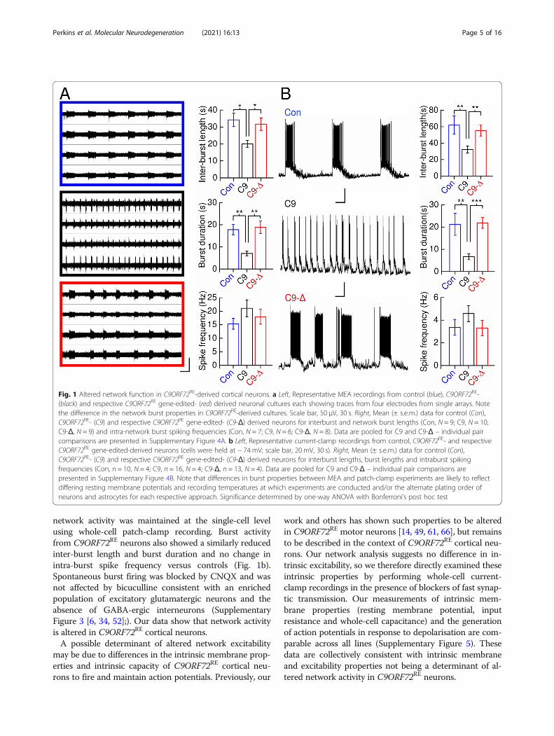
network activity was maintained at the single-cell levelusing whole-cell patch-clamp recording. Burst activityfrom C9ORF72RE neurons also showed a similarly reducedinter-burst length and burst duration and no change inintra-burst spike frequency versus controls (Fig. 1b).Spontaneous burst firing was blocked by CNQX and wasnot affected by bicuculline consistent with an enrichedpopulation of excitatory glutamatergic neurons and theabsence of GABA-ergic interneurons (SupplementaryFigure 3 [6, 34, 52];). Our data show that network activityis altered in C9ORF72RE cortical neurons.A possible determinant of altered network excitability
may be due to differences in the intrinsic membrane prop-erties and intrinsic capacity of C9ORF72RE cortical neu-rons to fire and maintain action potentials. Previously, our
work and others has shown such properties to be alteredin C9ORF72RE motor neurons [14, 49, 61, 66], but remainsto be described in the context of C9ORF72RE cortical neu-rons. Our network analysis suggests no difference in in-trinsic excitability, so we therefore directly examined theseintrinsic properties by performing whole-cell current-clamp recordings in the presence of blockers of fast synap-tic transmission. Our measurements of intrinsic mem-brane properties (resting membrane potential, inputresistance and whole-cell capacitance) and the generationof action potentials in response to depolarisation are com-parable across all lines (Supplementary Figure 5). Thesedata are collectively consistent with intrinsic membraneand excitability properties not being a determinant of al-tered network activity in C9ORF72RE neurons.
Fig. 1 Altered network function in C9ORF72RE-derived cortical neurons. a Left, Representative MEA recordings from control (blue), C9ORF72RE-(black) and respective C9ORF72RE gene-edited- (red) derived neuronal cultures each showing traces from four electrodes from single arrays. Notethe difference in the network burst properties in C9ORF72RE-derived cultures. Scale bar, 50 μV, 30 s. Right, Mean (± s.e.m.) data for control (Con),C9ORF72RE- (C9) and respective C9ORF72RE gene-edited- (C9-Δ) derived neurons for interburst and network burst lengths (Con, N = 9; C9, N = 10;C9-Δ, N = 9) and intra-network burst spiking frequencies (Con, N = 7; C9, N = 6; C9-Δ, N = 8). Data are pooled for C9 and C9-Δ – individual paircomparisons are presented in Supplementary Figure 4A. b Left, Representative current-clamp recordings from control, C9ORF72RE- and respectiveC9ORF72RE gene-edited-derived neurons (cells were held at − 74mV; scale bar, 20 mV, 30 s). Right, Mean (± s.e.m.) data for control (Con),C9ORF72RE- (C9) and respective C9ORF72RE gene-edited- (C9-Δ) derived neurons for interburst lengths, burst lengths and intraburst spikingfrequencies (Con, n = 10, N = 4; C9, n = 16, N = 4; C9-Δ, n = 13, N = 4). Data are pooled for C9 and C9-Δ – individual pair comparisons arepresented in Supplementary Figure 4B. Note that differences in burst properties between MEA and patch-clamp experiments are likely to reflectdiffering resting membrane potentials and recording temperatures at which experiments are conducted and/or the alternate plating order ofneurons and astrocytes for each respective approach. Significance determined by one-way ANOVA with Bonferroni’s post hoc test
Perkins et al. Molecular Neurodegeneration (2021) 16:13 Page 5 of 16

C9ORF72RE-derived cortical neurons have increasedsynaptic inputTo determine whether the altered network activity inC9ORF72RE-derived cortical neurons was of synapticorigin we undertook quantification of the co-localisationof pre- (synapsin-1) and post-synaptic (PSD-95) markersand observed an increase in synaptic densities onC9ORF72RE versus control neurons (Fig. 2a, Supplemen-tary Figure 6A). Increased synaptic density may reflectan increase in neuronal morphology and therefore weexamined the neurite length in our cultures(Supplementary Figure 6B, C). However, consistent withour whole-cell capacitance measurements, our data didnot reveal any morphological changes that could ac-count for the large change in synaptic density inC9ORF72RE neurons. Consistent with these data we alsofound an increase in the frequency of mini excitatorypostsynaptic currents (mEPSCs) in C9ORF72RE neuronscompared to isogenic controls and healthy volunteer-derived neurons (Fig. 2c, d, Supplementary Figure 7).AMPAR-mediated mEPSC amplitudes and kinetics (rise-times and decay-times), and further the expression ofAMPA receptor subunits, were not changed indicatingthat the properties and expression of synaptic AMPAreceptors were not altered (Fig. 2c, d, SupplementaryFigure 7). These findings indicate that C9ORF72RE
cortical neurons display increased synaptic densityresulting in elevated synaptic input.
C9ORF72RE–derived cortical neurons exhibit pre-synapticdysfunctionIncreased pre-synaptic glutamate release could alsounderlie an increased mEPSC frequency and a centraldeterminant of release properties and network burstproperties is the size of the vesicle readily releasable pool(RRP) [10, 32]. The RRP size was functionally estimatedusing hypertonic sucrose, an established method togenerate Ca2+-independent exocytosis of the vesicularRRP [40, 47]. We observed sucrose-application-evokedmEPSC activity that can be classified into an initialtransient phase (initial depletion of the RRP) and asteady-state phase (on-going replenishment of the RRP;Fig. 2e and Supplementary Figure 8A [40];). Given thatconventional measurements of RRP are determined bythe integral of the total evoked current and are directlyproportional to synaptic density we have examined thefold change in mEPSC frequencies because C9ORF72RE
cortical neurons exhibit increased synaptogenesis overcontrol lines. A fold reduction in mEPSC frequency (Fig.2f, Supplementary Figure 8B), but not their amplitude(Supplementary Figure 8C), was observed in C9ORF72RE
neurons compared to control lines for both the transientand steady-state phases. Thus, despite an observed in-crease in mEPSC frequency during baseline recordings,
C9ORF72RE cortical neurons display a functionallyreduced RRP size that is replenished at a slower rate.These data potentially explain the shorter network burstdurations in C9ORF72RE cortical neurons.Physiological vesicular release is Ca2+-dependent and
thus we examined this by measuring fold changes inmESPC frequencies before and after addition of KCl. Nodifferences in the fold change of mEPSC frequencieswere found between C9ORF72RE versus paired isogeniccontrol neurons (Supplementary Figure 9). These datasuggest that overall depolarisation-mediated Ca2+-evokedrelease is equivalent in the pre-synaptic terminals of eachline. Nonetheless our data indicate that the RRP size isimpacted in C9ORF72RE cortical neurons.
Synaptic potentiation in C9ORF72RE cortical neurons isimpairedCortical network plasticity in C9ORF72RE patients isimpaired and suggests that this is due to perturbedactivity-dependent synaptic plasticity [5]. The activity-dependent potentiation of AMPA receptor-mediatedmEPSCs is a central feature of classical models of synap-tic plasticity and therefore we applied a series of de-polarizing voltage steps to our neurons, as previouslydescribed, that leads to a potentiation of the amplitudeof mEPSC in rodent hippocampal neurons [4, 29, 31, 63,64]. Following this depolarisation pulse protocol (DPP),we observed that mEPSCs in control neurons were tran-siently potentiated by around 20% in amplitude from theinitial control period before returning close to controllevels (Supplementary Figure 10). No significant shift inmEPSC amplitude was observed when no stimulationwas applied. Consistent with previous studies on primaryhippocampal neurons [4], we found this mEPSC potenti-ation to be dependent upon the activation of voltage-gated Ca2+ channels and intracellular elevations in Ca2+
as was blocked by voltage-gated Ca2+ channel antagonist,nifedipine, and supplementation of Ca2+ chelatorBAPTA to the patch pipette, respectively (SupplementaryFigure 10E, Supplementary Figure 11A-F). In contrast, weobserved that DPP did not induce any potentiation ofmEPSC amplitudes in C9–1 and C9–2 neurons (Fig. 3a, c,e-g). The respective isogenic C9–1Δ and C9–2Δ neuronsdisplayed a significant mEPSC potentiation post-DPP (Fig.3b, d-g). The mEPSC decay time constant did not changepost-DPP with respect to baseline activity in either con-trol, C9 or C9-Δ neurons suggesting that the compositionof AMPA receptors mediating mEPSCs is unchangedpost-DPP (Fig. 3f; Supplementary Figure 11G). These dataare therefore consistent with a C9ORF72RE-mediatedphysiological disruption of mEPSC potentiation inC9ORF72RE-derived cortical neurons in our model of syn-aptic potentiation.
Perkins et al. Molecular Neurodegeneration (2021) 16:13 Page 6 of 16

Fig. 2 (See legend on next page.)
Perkins et al. Molecular Neurodegeneration (2021) 16:13 Page 7 of 16

RNA sequencing highlights molecular disruption at thesynapse in C9ORF72RE cortical neuronsTo begin to understand the molecular changes thatunderpin the observed physiological dysfunction inC9ORF72RE cortical neurons, we next performed tran-scriptomic analysis of cortical neurons derived from twoindependent C9ORF72RE iPSCs (C9–1 & C9–2) andtheir corresponding isogenic controls (C9-Δ1 & C9-Δ2).Principal component analysis (PCA) of gene expressionshowed segregation of differential gene expressionbetween the two mutant-isogenic pairs, though a highdegree of similarity within a mutant-isogene pair, asexpected (Fig. 4a). We therefore assessed our data set inorder to determine common dysregulated gene expres-sion between the different lines employed (Fig. 4b,Supplementary Table 1). Our biological process geneontology analyses (Fig. 4c) revealed dysregulated expres-sion of genes involved in vesicle regulation (Gopc,Vamp5), cell-cell adhesion (Cbln1, Pcdhgc4), negativeregulation of ion transport (Htr2a) fatty acid metabolismand regulation of DNA-binding transcription factor ac-tivity (Irak1, Sigirr). These novel transcriptomic data re-veal dysregulated multiple pathways in C9ORF72RE
cortical neurons that may contribute to the observedsynaptic dysfunction.
DiscussionIncreased synaptic glutamate transmission within thecortex presents an attractive hypothesis to potentiallyexplain cortical network hyperexcitability present inearly symptomatic C9ORF72RE patients [42, 50, 62] andALS in general [22]. Our data provide a human in vitromechanistic exploration of physiological impairments inC9ORF72RE patient-derived excitatory cortical neuronsthat reveal that perturbed network activity is under-pinned by functional synaptic alterations that impactupon excitability. Furthermore, noting that ALS-FTDpatients exhibit impairments in network plasticity, we
have determined that C9ORF72RE cortical neuronsexhibit impairments in synaptic plasticity. Importantly,the physiological alterations observed in iPSC-derivedC9ORF72RE cortical neurons are disparate from thatpreviously observed in iPSC-derived C9ORF72RE motorneurons where intrinsic excitability appears to be pri-marily affected [55]. Our data reveal that intrinsic excit-ability is not affected in C9ORF72RE cortical neurons.An increase in network burst frequency in C9ORF72RE
patient-derived excitatory cortical neurons is highly con-sistent with a mechanism requiring increased excitatoryinput. Our data demonstrate that C9ORF72RE corticalneurons display an increased synaptic input as a resultof an increased synaptic density. Such findings arebroadly consistent with murine models of ALS, whereincreased synaptic input of excitatory cortical neuronsare observed in pre-symptomatic mutant TDP-43 mice([17]; but see [25]) and SODG93A mice [17, 48, 57].Cortical neurophysiological impairments were not foundin a C9ORF72RE murine model though this model doesnot display classical ALS-FTD pathology or neurodegen-eration [44]. Our transcriptomic approach has revealedpotential causes to this increase in synaptic density.PCDHGC4, a γ-protocadherin, negatively regulates thefunction of neuroligin-1, a post-synaptic molecule thatinteracts with pre-synaptic neurexin to maintain andpromote synapse structures in forebrain neurons [39].Reduced expression of PCDHGC4 in our C9ORF72RE
cortical neurons is therefore compatible with increasedneuroligin-1 function and increased synaptic density.Overexpression of neuroligin-1 has previously beenshown to increase excitatory synaptic activity in in vitrocortical neurons [8]. Equivalently, CBLN1, is a pre-synaptically expressed molecule that interacts with neur-exins and promotes synaptogenesis [51] and is upregu-lated in C9ORF72RE cortical neurons. Contrastingly,CBLN1 has been reported to be downregulated inC9ORF72RE iPSC-derived motor neurons [49]. Together,
(See figure on previous page.)Fig. 2 Synaptic density, mEPSC properties and pre-synaptic dysfunction. a For each line, [control, C9ORF72RE- (C9) and C9ORF72RE gene-edited-(C9-Δ) cortical neurons], low magnification images (scale bar, 20 μm) show immunostaining for βIII-tubulin (yellow, top left), synapsin-1 (red), PSD-95 (green) and a PSD-95/synapsin-1 composite view (lower) from which the region of interest is highlighted. The region of interest is shown withgreater higher magnification image below (scale bar, 10 μm). b Mean ± s.e.m. co-localised PSD-95/synapsin-1 puncta per 20 μm of neurite lengthfor each line type (Control, N = 3; C9, N = 12; C9-Δ, N = 12). Data for C9 and C9-Δ are pooled from three respective C9ORF72RE- and gene-editedpairs – individual pair comparisons are presented in Supplementary Figure 6A. Data from each de novo preparation represents a mean obtainedfrom four coverlips each consisting of five randomly selected image fields. c Sample traces from recordings of mEPSC events from control,C9ORF72RE- and C9ORF72RE gene-edited-cortical neurons. Recordings were made at a holding potential of − 84mV. Scale bar; 20 pA, 10 s. mEPSCsin all lines are mediated by Ca2+-impermeable (NASPM-insensitive) AMPA receptors (CNQX-sensitive). d Mean ± s.e.m. mEPSC frequency andamplitude for each line (Control, n = 14, N = 3; C9, n = 52, N = 12; C9-Δ, n = 42, N = 11). Data are pooled for C9 and C9-Δ – individual paircomparisons are presented in Supplementary Figure 7. e Sample traces from recordings of mEPSC events before and in the presence of sucrosefrom control (Con), C9ORF72RE-(C9) and C9ORF72RE gene-edited (C9-Δ)-cortical neurons. The transient and steady-state phases evoked by sucroseaddition are highlighted. Note the difference in mEPSC frequency in these phases versus baseline. Scale bar, 50 pA, 5 s. f Mean ± s.e.m. foldchange in mEPSC frequency for each line for the transient (Control, n = 14, N = 3; C9, n = 28, N = 10; C9-Δ, n = 30, N = 9) and steady-state phases(Control, n = 8, N = 2; C9, n = 23, N = 8; C9-Δ, n = 23, N = 8). Data are pooled for C9 and C9-Δ – individual pair comparisons are presented inSupplementary Figure 8. Significance of data in the figure determined by one-way ANOVA with Bonferroni’s post hoc test
Perkins et al. Molecular Neurodegeneration (2021) 16:13 Page 8 of 16

Fig. 3 (See legend on next page.)
Perkins et al. Molecular Neurodegeneration (2021) 16:13 Page 9 of 16

these studies indicate that increased cortical glutamate-mediated synaptic input is an early feature of ALS. Futurework will require to determine when increased synapticdensity alongside altered network excitability presents inALS progression in patients.Many ALS-focused studies describing altered gluta-
matergic input have examined synaptic function withoutassessing presynaptic function in detail, nor have theyexamined the consequences for network activity. An in-crease in excitatory synaptic input might be expected toincrease network burst duration in addition to burstfrequency [32]. However, our assessment of networkactivity revealed a decrease in network burst durationand appears to be consistent with a decrease in gluta-matergic synaptic transmission. Consistent with this, ourevaluation of pre-synaptic function revealed an esti-mated reduced size and replenishment of vesicular RRP.Importantly, a reduced RRP size and replenishment ratehas been previously shown to generate early burst ter-mination to shorten burst duration [10, 32]. This pro-vides the most parsimonious explanation of the observedshorter network burst duration in C9ORF72RE corticalneurons. A disruption in the vesicular RRP suggestsmechanisms in which synaptic vesicular trafficking areimpaired. Noting that C9ORF72 is detected in pre-synapticterminals, our data resonate with previous studieshighlighting the role of C9ORF72 protein in vesicular traf-ficking within the trans-Golgi network and endosomal sig-nalling and suggest that C9ORF72 haploinsufficiency mayresult in a reduced RRP [2, 19, 53]. Our transcriptomic dataprovide further evidence of dysregulated genes associatedwith impaired vesicular trans-Golgi network and endoso-mal signalling in ALS, consistent with a growing body ofevidence of impaired vesicular trafficking in ALS that mayimpact on the RRP size [11, 15, 46]. For example, our dataset highlights an upregulation of the GOPC gene, achaperone protein that is expressed across the trans-Golginetwork and endosomes. Amongst many interactions,GOPC is associated with syntaxin-6 that regulates endoso-mal vesicular transport [7]. Furthermore, TDP-43 proteinappears to bind GOPC RNA [41]. Collectively, our data
show a reduction in the RRP that is consistent with impair-ments in vesicular trafficking.Importantly, vesicular release is typically stimulated via
Ca2+-dependent mechanisms. Despite a reduced RRP,our evaluation of depolarisation/Ca2+-dependent vesicu-lar release appears to be equivalent in C9ORF72RE exci-tatory cortical neurons versus isogenic controls. Indeed,our afterhyperpolarisation (AHP) data suggest thatcalcium mediated influx is not impacted to influenceintrinsic excitability properties. However, we mustremain cautious that AHP and exocytosis could be inde-pendently calcium regulated processes in our cells,subject to localised intracellular calcium regulation. Onepotential explanation could be that localised Ca2+-dependent mechanisms controlling vesicular release inC9ORF72RE excitatory cortical neurons are enhancedover control lines to generate the higher release prob-ability required to elevate the fold increase in mEPSCfrequency to comparable levels to the control lines. Dys-regulated cytoplasmic Ca2+ levels in C9ORF72RE-derivedmotor neurons have been previously reported [13, 28]and this elevation in Ca2+ levels may contribute to an in-creased release probability. However, our findings con-trast with those of Jensen et al. [28] who suggest thatKCl-stimulated release is impaired, due to a GA-drivenloss of the protein SV2, in C9ORF72RE patient-derivedcortical neurons.Our data show that synaptic potentiation in
C9ORF72RE excitatory cortical neurons is impaired. Not-ably, transcranical magnetic stimulation-based studiesshow both presymptomatic and post-symptomaticC9ORF72RE patients exhibit an abolishment of activity-dependent cortical network plasticity [5]. Together, thesedata suggest impairments in functional synaptic plasti-city may emerge as an early pathophysiological event inC9ORF72RE–mediated disease progression to impairnetwork plasticity. The pathological determinants of theimpairments in synaptic plasticity remain unknown inC9ORF72RE cortical neurons, though a very recent studyhas shown synaptic plasticity impairments in murineC9orf72 knock out animals [27], suggesting a role for the
(See figure on previous page.)Fig. 3 Synaptic plasticity. a and b Example recordings of mEPSCs prior to and after DPP (10 depolarising pulses of 3 s in duration, every 9 s, from− 84 to + 16 mV) for C9ORF72RE (1. grey and 2. black, respectively; scale bar; 20 pA, 5 s) and C9ORF72RE-Δ (1. light red and 2. red, respectively; scalebar; 10 pA, 5 s) neurons. c, d Individual mEPSC amplitude plots for DPP experiments performed upon C9ORF72RE and C9ORF72RE-Δ neurons,respectively. For C9ORF72RE neurons, mEPSCs before (1) and after DPP (2) are represented in grey and black, respectively. For C9ORF72RE-Δneurons, mEPSCs before (1) and after DPP (2) are represented in light red and red, respectively. Note the lack of potentiation in C9ORF72RE, butnot C9ORF72RE-Δ, neurons. e Cumulative probability plots of mEPSC amplitudes for the initial control period (1) and after DPP (2) of the datashown in C and D. Significance of shift of mEPSC data (Kolmogorov–Smirnov test); C9ORF72RE, p = 0.325; C9ORF72RE-Δ, p < 0.001. f Left, meanmEPSCs for data shown in B (C9ORF72RE) and C (C9ORF72RE-Δ) for initial control period (1) and post DPP (2). Scale bars; 5 pA, 5 ms. Right, meanscaled mEPSCs. g Mean ± s.e.m. fold increase of mEPSC amplitude 10 minutes post-DPP from initial control period for control (n = 17), C9–1 (n =14), C9–1Δ (n = 13), C9–2 (n = 4) and C9–2Δ (n = 10). Example traces and data for the control line are presented in Figure S10. Significancedetermined by unpaired t-tests (C9ORF72RE versus C9ORF72RE-Δ) and one-way ANOVA with Bonferroni’s post hoc test (control versus C9ORF72RE )
Perkins et al. Molecular Neurodegeneration (2021) 16:13 Page 10 of 16

Fig. 4 Transcriptomic analysis of C9ORF72RE cortical neurons. a Principal component analysis of gene expression derived from RNA sequencingfrom C9ORF72RE cortical neurons (C9–1, C9–2; black) and isogenic gene-corrected control cortical neurons (C9–1Δ, C9–2Δ; red). Each data pointrepresents a de novo differentiation of cortical neurons. As highlighted, the isogenic controls cluster accordingly with their respective parentalC9ORF72RE lines. b Scatter plot showing comparison of gene expression (as average Fragments Per Kilobase of transcript per Million mappedreads) from cortical neurons derived from two independent C9ORF72RE iPSC lines and their corresponding isogenic controls. Green and orangedata points denote the overlap of significantly up- and down-regulated genes in both mutant-correction pairs, respectively (false discovery rate,p < 0.2). c Selected gene ontology terms enriched in dysregulated genes
Perkins et al. Molecular Neurodegeneration (2021) 16:13 Page 11 of 16

C9orf72 protein in plasticity mechanisms. In addition toour own data set indicating impact upon synaptic physi-ology, data sets highlight that altered gene expression inC9ORF72RE patient tissue [46] and disrupted cell signallingpathways in iPSC-derived C9ORF72RE neurons [13] are im-plicated in synaptic plasticity. Furthermore, recent tran-scriptomic work has associated the expression of di-peptiderepeat proteins with a reduction in expression of a mediatorof synaptic plasticity [15]. Our data therefore firmly deter-mines synaptic plasticity impairments are present in humanC9ORF72RE cortical neurons.Crucially, C9ORF72RE mutations are causal to both
ALS and FTD. In this regard, we note that the vastmajority of clinical pathophysiological measurementsdescribing hyperexcitability are made from the motorcortex, which is primarily affected in ALS [22]. Nonethe-less cortical hypexcitability is evidenced in rodentmodels of FTD [20]. We acknowledge that our data mayhave preferential relevance for FTD over ALS, or viceversa. This is likely to become more apparent with in-creased pathophysiological characterisation ofC9ORF72RE ALS-FTD patients. Further, early identifica-tion of pre-symptomatic individuals and longitudinalstratification of these observations will allow us to placefurther confidence upon the pathophysiological stagingthat our observations are most likely to mirror. Our datarepresent one in vitro time point, but as discussed,closely align with aspects of pre-symptomatic corticalneuron impairments evidenced in rodent models of ALSand ALS-FTD. Importantly, the network excitability isnot investigated in these models. Our data suggest thatincreases in synaptic transmission may lead to increasednetwork burst frequency, however at the same time, in-dicate that other concurrent processes reduce burst ac-tivity, reflective of multiple changes in the molecularlandscape of synaptic function, potentially in an attemptto homeostatically compensate against other networkimpairments. Indeed, work performed more extensivelyin the context of Alzheimer’s disease has established thathomeostatic network activity adaptation, including im-paired synaptic plasticity, is an early feature and pre-cedes that of pathophysiological network failure likelyresulting in the manifestation of the clinically observednetwork hyperexcitability [18]. In this regard, there areintriguing similarities in our data set to cortical neuronsderived from Alzheimer’s patient iPSCs that display in-creased synaptic activity at the same time point [23]..
ConclusionsIn summary, our study provides physiological evidencesupporting the involvement of widespread glutamatergicsynaptic dysfunction as a potential pathogenic mechan-ism of C9ORF72RE-mediated disease, a disease that has
considerable impact in cortical neurons in addition tomotor neurons. We reveal that pre and post-synaptic de-fects are highly prominent in cortical neurons in ALS-FTD and are suggested to combine to generate early net-work excitability alterations and impaired synaptic plas-ticity. We note these are very different physiologicalperturbations previously reported for motor neurons de-rived from C9ORF72RE patients and therefore this studyshows that the pathophysiological processes in differentbrain regions are likely to show divergence. These earlysynaptic defects are likely linked to core mechanisms ofneurodegenerative disease progression and symptoms inALS-FTD.
Supplementary InformationThe online version contains supplementary material available at https://doi.org/10.1186/s13024-021-00433-8.
Additional file 1: Supplementary Figure 1. Neuronal specification.Example images of immunostaining against neuronal precursor markernestin (A) and human nuclei (B) with neuronal marker βIII-tubulin andastrocyte marker GFAP. Our cultures at week 5 post-differentiation gener-ate dense human nuclei-positive neuronal populations (mean ± sem %human nuclei+ cells with βIII-tubulin; C9–1, 96.8 ± 4.1; C9–1Δ, 97.4 ± 3.0;C9–2, 90.4 ± 3.2; C9–2Δ, 90.2 ± 4.4; C9–3, 98.5 ± 4.9; C9–3Δ, 96.4 ± 7.1; datafrom 3 de novo plate downs) with only negligible detectable levels ofnestin (mean ± sem % nestin; C9–1, 1.1 ± 0.01; C9–1Δ, 2.5 ± 0.1; C9–2,2.3 ± 0.1; C9–2Δ, 3.5 ± 0.1; C9–3, 1.0 ± 0.1; C9–3Δ, 2.0 ± 0.1; data from 3 denovo plate downs). Data are consistent with previous cortical neuron dif-ferentiations with cell lines used in this study and a cortical neuron proto-col that gives rise to a highly efficient neuronal differentiation [6, 34, 52];.Scale bars, 100 μm.
Additional file 2: Supplementary Figure 2. A, B and C. Neuronalspecification. RNA-seq analysis of established neuronal and glia markers(MAPT, NEFL, ALDH1L1, AQ4, MYRF), neuronal markers for anterior/corticaldevelopment (OTX2, PAX6, FOXP4, BCL6), hindbrain development(HOXB2), cortical layers (CUX1, POU3F2, PCP4, FOXP2), plus glutamatergic(CAMK2A, SLC17A6 and SLC17A7) and GABA-ergic neurons (GAD2,SLC32A1, PVALB) in C9 and C9-Δ lines. We note that out analysis ob-tained extensive detection of cortical transcripts from our cultures andare consistent with predominantly glutamatergic neurons. Note that they axis is presented using a logarithmic scale. Data in C also show synapticmarkers DLG4 and SYN1. Data are representative of mean ± sem fromtwo pooled C9 lines (black bars) and their respective isogenic lines (redbars), as further detailed in Fig. 4. Data were derived from 3 plate downsfrom each line.
Additional file 3: Supplementary Figure 3. Pharmacological block ofnetwork activity. A, Example whole cell current-clamp recordings of effectof AMPA receptor blocker, CNQX (30 μM), upon network activity. Scalebar, 50 mV, 20 s. CNQX generated full block of network burst activity. B,As in A though for GABAA receptor blocker, bicuculline (30 μM). Scale bar,50 mV, 5 s. C, Mean (± s.e.m.) percentage shift in burst frequency in pres-ence of either CNQX or bicuculline for each line type (CNQX – Con, n = 5,N = 3; C9, n = 5, N = 2; C9-Δ; n = 5, N = 3 / bicuculline – Con, n = 5, N = 3;C9, n = 5, N = 3; C9-Δ; n = 5, N = 3). Bicuculline did not significantly impactupon network burst activity. Expectedly, data are consistent with anenriched population of excitatory glutamatergic cortical neurons [6, 34,52].
Additional file 4: Supplementary Figure 4. Network burst data. A,Mean (± s.e.m.) MEA-determined burst duration, interburst length andspike frequency within the burst for each respective C9ORF72RE andC9ORF72RE-Δ isogenic pair (C9–1, N = 4; C9–1Δ, N = 6; C9–2, N = 6; C9–2Δ,N = 3). Significance determined by unpaired t-test. B, Mean (± s.e.m.)patch-clamp-determined burst duration, interburst length and spike
Perkins et al. Molecular Neurodegeneration (2021) 16:13 Page 12 of 16

frequency within the burst for each respective C9ORF72RE and C9ORF72RE-Δ isogenic pair (C9–1, n = 8, N = 2; C9–1Δ, n = 8, N = 2; C9–2, n = 8, N = 2;C9–2Δ, n = 5, N = 2). Significance determined by unpaired t-test.
Additional file 5: Supplementary Figure 5. Intrinsic excitability ofC9ORF72RE-derived cortical neurons. A, Mean (± s.e.m.) data for eachControl- (Con), C9ORF72RE- (C9) and respective C9ORF72RE gene-edited-(C9-Δ) derived neurons for passive membrane properties (Con, n = 6, N =2; C9–1, n = 20, N = 3; C9–1Δ, n = 11, N = 3; C9–2, n = 17, N = 3; C9–2Δ,n = 12, N = 3; C9–3, n = 17, N = 3; C9–3Δ, n = 15, N = 3). The data showsinput resistance (RIN), whole-cell capacitance, resting membrane potential(RMP). B, Representative whole-cell current-clamp recordings of evokedresponses to current injection (− 20 pA to + 30 pA, 0.5 s duration, 5 pA in-crements) for a control, C9ORF72RE- and C9ORF72RE neuron. Cells wereheld at − 74 mV. Scale bar (40 mV, 100 ms). C, Mean (± s.e.m.) action po-tential (AP) number-current relationships generated from each pairedC9ORF72RE- and C9ORF72RE gene-edited-derived neurons (C9–1, C9–2, C9–3) including control data. D, Mean (± s.e.m.) action potential parameters(threshold, amplitude, and afterhypolarisation, AHP). AP properties weremeasured from the first evoked AP of the rheobasic current injection. Sig-nificance determined by unpaired t-test. We note that we did find slight,but statistically significant differences for one line in the RMP and AHPdata. However, these are unlikely to be the cause of the altered networkexcitability because they are i) extremely modest and ii) not a conservedfinding across all lines.
Additional file 6: Supplementary Figure 6. Synaptic puncta andneurite length. A, Mean (± s.e.m.) co-localised PSD-95/Synapsin-1 puncta foreach respective C9ORF72RE and C9ORF72RE-Δ isogenic pair (C9–1, N = 4; C9–1Δ, N = 4; C9–2, N = 4; C9–2Δ, N = 4; C9–3, N = 4; C9–3Δ, N = 4). Significancedetermined by unpaired t-test. B. To address neuronal morphology wetransduced cortical NPCs with a low GFP-lentivirus titre in order to be ableto visualise individual neurons. As a measure of morphology, we then mea-sured the neurite lengths (total sum of all processes) for each cell. C. Datashow the mean ± s.e.m. neurite length (in μm) for each respectiveC9ORF72RE and C9ORF72RE-Δ isogenic pair (C9–1, n = 44 cells; C9–1Δ, n = 24;C9–2, n = 39; C9–2Δ, n = 20; C9–3, n = 17; C9–3Δ, n = 27). All data derivedfrom 2 de novo preparations. Significance determined by unpaired t-test.
Additional file 7: Supplementary Figure 7. mEPSC amplitude, rise timeand decay properties. A, Cumulative probability plots of mEPSC inter-eventtime for each C9ORF72RE- and corresponding C9ORF72RE gene-edited neurons.Data was obtained from at least 2-min recordings and neurons that displayedmEPSC frequencies under 0.05 Hz were omitted from the analysis. Significanceof cumulative probability plots determined using Kolmogorov-Smirnov test.Mean ± s.e.m. mEPSC frequency for each line and respective edit are showninset (C9–1, n= 23, N= 5; C9–1Δ, n= 15, N= 4; C9–2, n= 17, N= 4; C9–2Δ,n= 11, N= 3; C9–3, n= 12, N= 3; C9–3Δ, n= 16, N= 4). B, As A though formEPSC amplitude. C, Mean ± s.e.m. mEPSC rise time (10–90%) and τ decayproperties for each line (C9–1, n = 15, N= 4; C9–1Δ, n= 12, N= 3; C9–2, n= 12,N= 3; C9–2Δ, n= 5, N= 2; C9–3, n= 8, N = 3; C9–3Δ, n= 13, N= 3). Signifi-cance determined by Welch’s t-test or unpaired t-test. Other than mEPSC fre-quency (Fig. 2), data are not consistent with altered mEPSC properties. Wenote that we did find slight, but statistically significant differences for one linein the mEPSC amplitude and rise time data. However, these are unlikely to bethe cause of altered network excitability observed in our cultures because theyare i) extremely modest and ii) not a conserved finding across all lines. D.RNA-seq analysis of AMPA receptor subunits (GRIA1–4) in C9 and C9-Δ lines.Note that the y axis is presented using a logarithmic scale. Data are represen-tative of mean ± sem from two pooled C9 lines (black bars) and their respect-ive isogenic lines (red bars), as detailed in Fig. 4. Data were derived from 3plate downs from each line. The data are not consistent with any change inexpression between C9 and C9-Δ lines.
Additional file 8: Supplementary Figure 8. mEPSC amplitude in thepresence and absence of hypertonic sucrose. A, Representative recordingof mEPSC activity before (baseline) and in the presence of sucrose (0.5 M,filled bar). The initial transient and steady-state phases of the sucrose-evoked response are highlighted. Scale bars; 50 pA, 5 s. B, Mean ± s.e.m.fold change in mEPSC frequency for each line for the transient (C9–1,
n = 9, N = 3; C9–1Δ, n = 10, N = 3; C9–2, n = 8, N = 3; C9–2Δ, n = 6, N = 2;C9–3, n = 11, N = 4; C9–3Δ, n = 14, N = 4) and steady state phases (C9–1,n = 7, N = 2; C9–1Δ, n = 6, N = 3; C9–2, n = 8, N = 3; C9–2Δ, n = 6, N = 2;C9–3, n = 8, N = 3; C9–3Δ, n = 11, N = 3). C, As in B, though for mEPSCamplitude for the transient (C9–1, n = 7; C9–1Δ, n = 6; C9–2, n = 8; C9–2Δ, n = 6; C9–3, n = 10, N = 3; C9–3Δ, n = 12, N = 3) and steady-statephases (C9–1, n = 7; C9–1Δ, n = 6; C9–2, n = 8; C9–2Δ, n = 6; C9–3, n = 7,N = 3; C9–3Δ, n = 11, N = 3). Significance determined by two-tailed un-paired t-test. The patch pipette solution was supplemented with BAPTA(1 mM) to prevent potential Ca2+-dependent modulation of post-synapticneuron properties.
Additional file 9: Supplementary Figure 9. KCl-evoked release prop-erties. A, Sample traces from recordings of mEPSC events before and inthe presence of KCl (30 mM) from C9ORF72RE- and C9ORF72RE-Δ-corticalneurons (C9–2 and C9–2Δ). Scale bars; 50 pA, 5 s. B, Mean ± s.e.m. foldchange in mEPSC frequency for each line in the presence of KCl (C9–1,n = 5, N = 2; C9–1Δ, n = 5, N = 2; C9–2, n = 3, N = 2; C9–2Δ, n = 3, N = 2;C9–3, N = 2, n = 7, C9–3Δ, n = 5, N = 2). No statistical difference betweeneach C9ORF72RE and C9ORF72RE-Δ pair was determined (unpaired t-test).mEPSC frequency in the presence of KCl was determined from a stretchof recording at least 1 min in duration. The patch pipette solution wassupplemented with BAPTA (1 mM) to prevent potential Ca2+-dependentmodulation of post-synaptic neuron properties.
Additional file 10: Supplementary Figure 10. Depolarisation-mediated mEPSC amplitude potentiation. A. Example recordings ofmEPSCs prior (1. light blue) to and after (2. blue) the depolarisation pulseprotocol (DPP, 10 depolarising pulses of 3 s in duration, every 9 s, from −84 to + 16 mV). Example post-DPP mEPSCs are sampled from the 8–10min stretch of data. Scale bar; 10 pA, 2.5 s. B. Individual mEPSC amplitudeplot in an example experiment. mEPSCs before (1) and after DPP (2) arerepresented in light blue and blue, respectively. The grey bar indicates thestimulation period. Note the transient increase in mEPSC amplitude post-DPP. C. Cumulative probability plot showing a shift (p < 0.001, Kolmogo-rov–Smirnov test) in mEPSC amplitude data in B from the initial controlperiod (1. light blue) to the 10 min post-DPP period (2. blue) in whichthere is consistent, transient potentiation of mEPSC amplitude. D. Left,mean mEPSCs for data shown in B for initial control period (1. light blue)and 10 min post-DPP (2. blue). Scale bar; 5 pA, 5 ms. Right, mean mEPSCsscaled to amplitude and time base. E. To test whether potentiation wasCa2+-dependent we performed DPP in the presence of nifedipine, ablocker of voltage-gated Ca2+ channels, or BAPTA, a Ca2+ chelator, sup-plemented to the patch pipette. Data shows mean ± s.e.m. fold increaseof mEPSC amplitude 10 min post-DPP from initial control period for thecontrol line (n = 17), + nifedipine (n = 14) and + BAPTA (n = 6). Exampletraces and data presented in Supplementary Figure 11. Significance deter-mined by one-way ANOVA with Bonferroni’s post hoc test.
Additional file 11: Supplementary Figure 11. Ca2+-dependentmEPSC potentiation. A. Example recordings of mEPSCs prior to (1. lightblue) and after (2. blue) depolarisation in the presence of nifedipine(10 μM, applied to the extracellular solution). Scale bar; 20 pA, 2.5 s. B.Individual mEPSC amplitude plots for DPP experiments in the presenceof nifedipine. mEPSCs before (1) and after DPP (2) are represented in lightblue and blue, respectively. C. Cumulative probability plot of mEPSCamplitudes for the initial control period (1. light blue) and after DPP (2.blue) in the presence of nifedipine of the data shown in B. Whilst shift inmEPSC amplitude in the presence of nifedipine is significant (p < 0.01,Kolmogorov–Smirnov test), the shift is a decrease in mEPSC amplitude. D.As in A though in the presence of BAPTA (10 mM, to the intracellularsolution). E. As in B though in the presence of BAPTA. F. Cumulativeprobability plot of mEPSC amplitudes for the initial control period (1. lightblue) and after DPP (2. blue) in the presence of BAPTA for the data shownin E. Shift in mESPC amplitude is not significant (p = 0.937, Kolmogorov–Smirnov test). G. The fold change in mEPSC decay time constant post-DPP with respect to baseline activity (Con, n = 5; C9, n = 5; C9-Δ, n = 5).Fold changes are not significant (one-way ANOVA with Bonferroni’s mul-tiple comparisons test). Data are consistent with the fact that averagescaled mEPSC traces from pre- and post-DPP stages are superimposable.
Additional file 12.
Perkins et al. Molecular Neurodegeneration (2021) 16:13 Page 13 of 16

AbbreviationsALS: Amyotrophic lateral sclerosis; AMPA: (α-amino-3-hydroxy-5-methyl-4-isoxazolepropionic acid); ANOVA: Analysis of variance; aNPC: anterior neuralprecursors; C9ORF72RE: C9ORF72 repeat expansion; CNQX: Cyanquixaline (6-cyano-7-nitroquinoxaline-2,3-dione); DPP: Depolarisation pulse protocol;FTD: Frontotemporal dementia; GABA: Gamma aminobutyric acid; GO: Geneontology; iPSC: induced pluripotent stem cell; MEA: Multi-electrode array;mEPSC: mini excitatory post-synaptic current; RNA: Ribonucleic acid;RRP: Readily releasable pool; RT: Room temperature; s.e.m.: standard error ofthe mean
AcknowledgementsWe are very grateful to members of the Hardingham laboratory for thegeneration of primary mouse astrocytes. We thank Professor S. Chattarji(Centre for Brain Development and Repair, Instem, Bangalore, India) forhelpful comments on the manuscript. We thank David Story of the Chandranlaboratory for technical and administrative assistance.
Authors’ contributionsConceptualization, EMP, KB, SC and MRL; Methodology, EMP, KB, PB, ARM,OD, CMH, BTS, DS, JN, GEH, TGH, DJAW, SC and MRL; Investigation, EMP, KB,PB, ARM, OD, BTS, CMH, TGH, and MRL Writing – Original Draft, EMP, KB,PB, ARM, BTS, CMH, GEH, TGH, DJAW, SC and MRL; Writing – Review &Editing, EMP, KB, ARM, OD, BTS, CMH, TGH, GEH, DJAW, SC andMRL; Funding Acquisition, DJAW, GEH, TGH, SC and MRL; Resources, GEH,TGH, DJAW, SC; Supervision, SC and MRL. The authors read and approvedthe final manuscript.
FundingFunded by MRC, Euan MacDonald Centre, DBT-India, ISSF (Wellcome Trust/University of Edinburgh), RS MacDonald Seedcorn fund, MND Scotland, RoyalSociety of Edinburgh (CRF) and the Wellcome Trust (Grant 092742/Z/10/Z toD.J.A.W., S.C. and G.E.H.). SC and GEH labs are funded by the UK DementiaResearch Institute(DRI), which receives its funding from UK DRI Ltd, fundedby the MRC, Alzheimer’s Society and Alzheimer’s Research UK. A.R.M. is aLady Edith Wolfson Clinical Fellow and is jointly funded by the Medical ResearchCouncil and the Motor Neurone Disease Association (MR/R001162/1). BTS is aRowling - DRI fellow at the University of Edinburgh.
Availability of data and materialsThe datasets used and/or analysed during the current study are availablefrom the corresponding author on reasonable request.
Ethics approval and consent to participateDermal fibroblasts from patient and control individuals were obtained underfull Ethical/Institutional Review Board approval at the University ofEdinburgh.
Consent for publicationNot applicable.
Competing interestsNone declared.
Author details1Euan MacDonald Centre for MND Research, University of Edinburgh,Edinburgh EH16 4SB, UK. 2Centre for Clinical Brain Sciences, University ofEdinburgh, Edinburgh EH16 4SB, UK. 3Centre for Discovery Brain Sciences,University of Edinburgh, Edinburgh EH8 9XD, UK. 4UK Dementia ResearchInstitute at the University of Edinburgh, Edinburgh EH16 4SB, UK. 5SimonsInitiative for the Developing Brain, University of Edinburgh, Edinburgh EH89XD, UK. 6Division of Systems Medicine, School of Medicine, University ofDundee, Dundee DD1 9SY, UK. 7Centre for Brain Development and Repair,inStem, Bangalore 560065, India. 8Sheffield Institute for TranslationalNeuroscience, University of Sheffield, Sheffield S10 2HQ, UK.
Received: 17 July 2020 Accepted: 14 February 2021
References1. Alexa A, Rahnenführer J, Lengauer T. Improved scoring of functional
groups from gene expression data by decorrelating GO graphstructure. Bioinformatics. 2006;22:1600–7. https://doi.org/10.1093/bioinformatics/btl140.
2. Aoki Y, Manzano R, Lee Y, Dafinca R, Aoki M, Douglas AGL, Varela MA,Sathyaprakash C, Scaber J, Barbagallo P, Vader P, Mäger I, Ezzat K, TurnerMR, Ito N, Gasco S, Ohbayashi N, El Andaloussi S, Takeda S, Fukuda M,Talbot K, Wood MJA. C9orf72 and RAB7L1 regulate vesicle trafficking inamyotrophic lateral sclerosis and frontotemporal dementia. Brain. 2017;140:887–97. https://doi.org/10.1093/brain/awx024.
3. Arnold FJ, Hofmann F, Bengtson CP, Wittmann M, Vanhoutte P, Bading H.Microelectrode array recordings of cultured hippocampal networks reveal asimple model for transcription and protein synthesis-dependent plasticity. JPhysiol. 2005;564:3–19. https://doi.org/10.1113/jphysiol.2004.077446.
4. Baxter AW, Wyllie DJ. Phosphatidylinositol 3 kinase activation and AMPAreceptor subunit trafficking underlie the potentiation of miniature EPSCamplitudes triggered by the activation of L-type calcium channels. JNeurosci. 2006;26:5456–69. https://doi.org/10.1523/JNEUROSCI.4101-05.2006.
5. Benussi A, Cosseddu M, Filareto I, Dell’Era V, Archetti S, Sofia Cotelli M,Micheli A, Padovani A, Borroni B. Impaired long-term potentiation-likecortical plasticity in presymptomatic genetic frontotemporal dementia. AnnNeurol. 2016;80:472–6. https://doi.org/10.1002/ana.24731.
6. Bilican B, Livesey MR, Haghi G, Qiu J, Burr K, Siller R, Hardingham GE, Wyllie DJ,Chandran S. Physiological normoxia and absence of EGF is required for thelong-term propagation of anterior neural precursors from human pluripotentcells. PLoS One. 2014;9:e85932. https://doi.org/10.1371/journal.pone.0085932.
7. Charest A, Lane K, McMahon K, Housman DE.J Biol Chem. Association of anovel PDZ domain-containing peripheral Golgi protein with the Q-SNARE(Q-soluble N-ethylmaleimide-sensitive fusion protein (NSF) attachmentprotein receptor) protein syntaxin 6. 2001;276:29456–65. https://doi.org/10.1074/jbc.M104137200.
8. Chubykin AA, Atasoy D, Etherton MR, Brose N, Kavalali ET, Gibson JR, SüdhofTC. Neuron. Activity-dependent validation of excitatory versus inhibitorysynapses by neuroligin-1 versus neuroligin-2. 2007;54:919–31. https://doi.org/10.1016/j.neuron.2007.05.029.
9. Cleveland DW, Rothstein JD. From Charcot to Lou Gehrig: decipheringselective motor neuron death in ALS. Nat Rev Neurosci. 2001;2:806–19.https://doi.org/10.1038/35097565.
10. Cohen D, Segal M. Network bursts in hippocampal microcultures areterminated by exhaustion of vesicle pools. J Neurophysiol. 2011;106:2314–21. https://doi.org/10.1152/jn.00969.2010.
11. Coyne AN, Lorenzini I, Chou CC, Torvund M, Rogers RS, Starr A, Zaepfel BL,Levy J, Johannesmeyer J, Schwartz JC, Nishimune H, Zinsmaier K, Rossoll W,Sattler R, Zarnescu DC. Post-transcriptional inhibition of Hsc70-4/HSPA8expression leads to synaptic vesicle cycling defects in multiple models ofALS. Cell Rep. 2017;21:110–25. https://doi.org/10.1016/j.celrep.2017.09.028.
12. Cunningham F, Achuthan P, Akanni W, Allen J, Amode MR, Armean IM,Bennett R, Bhai J, Billis K, Boddu S, Cummins C, Davidson C, Dodiya KJ, GallA, Girón CG, Gil L, Grego T, Haggerty L, Haskell E, Hourlier T, Izuogu OG,Janacek SH, Juettemann T, Kay M, Laird MR, Lavidas I, Liu Z, Loveland JE,Marugán JC, Maurel T, McMahon AC, Moore B, Morales J, Mudge JM, NuhnM, Ogeh D, Parker A, Parton A, Patricio M, Abdul Salam AI, Schmitt BM,Schuilenburg H, Sheppard D, Sparrow H, Stapleton E, Szuba M, Taylor K,Threadgold G, Thormann A, Vullo A, Walts B, Winterbottom A, Zadissa A,Chakiachvili M, Frankish A, Hunt SE, Kostadima M, Langridge N, Martin FJ,Muffato M, Perry E, Ruffier M, Staines DM, Trevanion SJ, Aken BL, Yates AD,Zerbino DR, Flicek P. Ensembl 2019. Nucleic Acids Res. 2019;47:D745–51.https://doi.org/10.1093/nar/gky1113.
13. Dafinca R, Scaber J, Ababneh N, Lalic T, Weir G, Christian H, Vowles J,Douglas AG, Fletcher-Jones A, Browne C, Nakanishi M, Turner MR, Wade-Martins R, Cowley SA, Talbot K. C9orf72 hexanucleotide expansions areassociated with altered endoplasmic reticulum calcium homeostasis andstress granule formation in induced pluripotent stem cell-derived neuronsfrom patients with amyotrophic lateral sclerosis and frontotemporaldementia. Stem Cells. 2016;34:2063–78. https://doi.org/10.1002/stem.2388.
14. Devlin AC, Burr K, Borooah S, Foster JD, Cleary EM, Geti I, Vallier L, Shaw CE,Chandran S, Miles GB. Human iPSC-derived motoneurons harbouring
Perkins et al. Molecular Neurodegeneration (2021) 16:13 Page 14 of 16

TARDBP or C9ORF72 ALS mutations are dysfunctional despite maintainingviability. Nat Commun. 2015;6:5999. https://doi.org/10.1038/ncomms6999.
15. Dickson DW, Baker MC, Jackson JL, DeJesus-Hernandez M, Finch NA, Tian S,Heckman MG, Pottier C, Gendron TF, Murray ME, Ren Y, Reddy JS, Graff-Radford NR, Boeve BF, Petersen RC, Knopman DS, Josephs KA, Petrucelli L,Oskarsson B, Sheppard JW, Asmann YW, Rademakers R, van Blitterswijk M.Extensive transcriptomic study emphasizes importance of vesicular transportin C9orf72 expansion carriers. Acta Neuropathol Commun. 2019;7:150.https://doi.org/10.1186/s40478-019-0797-0.
16. Dobin A, Davis CA, Schlesinger F, Drenkow J, Zaleski C, Jha S, Batut P,Chaisson M, Gingeras TR. STAR: ultrafast universal RNA-seq aligner.Bioinformatics. 2013;29:15–21. https://doi.org/10.1093/bioinformatics/bts635.
17. Fogarty MJ, Klenowski PM, Lee JD, Drieberg-Thompson JR, Bartlett SE, NgoST, Hilliard MA, Bellingham MC, Noakes PG. Cortical synaptic and dendriticspine abnormalities in a presymptomatic TDP-43 model of amyotrophiclateral sclerosis. Sci Rep. 2016;6:37968. https://doi.org/10.1038/srep37968.
18. Frere S, Slutsky I. Alzheimer’s disease: from firing instability to homeostasisnetwork collapse. Neuron. 2018;97:32–58. https://doi.org/10.1016/j.neuron.2017.11.028.
19. Frick P, Sellier C, Mackenzie IRA, Cheng CY, Tahraoui-Bories J, Martinat C,Pasterkamp RJ, Prudlo J, Edbauer D, Oulad-Abdelghani M, Feederle R,Charlet-Berguerand N, Neumann M. Novel antibodies reveal presynapticlocalization of C9orf72 protein and reduced protein levels in C9orf72mutation carriers. Acta Neuropathol Commun. 2018;6:72. https://doi.org/10.1186/s40478-018-0579-0.
20. García-Cabrero AM, Guerrero-López R, Giráldez BG, Llorens-Martín M, Avila J,Serratosa JM, Sánchez MP. Hyperexcitability and epileptic seizures in amodel of frontotemporal dementia. Neurobiol Dis. 2013;58:200–8. https://doi.org/10.1016/j.nbd.2013.06.005.
21. Geevasinga N, Menon P, Nicholson GA, Ng K, Howells J, Kril JJ, Yiannikas C,Kiernan MC, Vucic S. Cortical function in asymptomatic carriers and patientswith C9orf72 amyotrophic lateral sclerosis. JAMA Neurol. 2015;72:1268–74.https://doi.org/10.1001/jamaneurol.2015.1872.
22. Geevasinga N, Menon P, Özdinler PH, Kiernan MC, Vucic S. Pathophysiologicaland diagnostic implications of cortical dysfunction in ALS. Nat Rev Neurol.2016;12:651–61. https://doi.org/10.1038/nrneurol.2016.140.
23. Ghatak S, Dolatabadi N, Trudler D, Zhang X, Wu Y, Mohata M, AmbasudhanR, Talantova M, Lipton SA. Mechanisms of hyperexcitability in Alzheimer’sdisease hiPSC-derived neurons and cerebral organoids vs isogenic controls.Elife. 2019;8:e50333. https://doi.org/10.7554/eLife.50333.
24. Gorrie GH, Fecto F, Radzicki D, Weiss C, Shi Y, Dong H, Zhai H, Fu R, Liu E, LiS, Arrat H, Bigio EH, Disterhoft JF, Martina M, Mugnaini E, Siddique T, DengHX. Dendritic spinopathy in transgenic mice expressing ALS/dementia-linked mutant UBQLN2. Proc Natl Acad Sci U S A. 2014;111:14524–9. https://doi.org/10.1073/pnas.1405741111.
25. Handley EE, Pitman KA, Dawkins E, Young KM, Clark RM, Jiang TC, Turner BJ,Dickson TC, Blizzard CA. Synapse dysfunction of layer V pyramidal neuronsprecedes Neurodegeneration in a mouse model of TDP-43 Proteinopathies.Cereb Cortex. 2017;27:3630–47. https://doi.org/10.1093/cercor/bhw185.
26. Hasel P, Dando O, Jiwaji Z, Baxter P, Todd AC, Heron S, Márkus NM,McQueen J, Hampton DW, Torvell M, Tiwari SS, McKay S, Eraso-Pichot A,Zorzano A, Masgrau R, Galea E, Chandran S, Wyllie DJA, Simpson TI,Hardingham GE. Neurons and neuronal activity control gene expression inastrocytes to regulate their development and metabolism. Nat Commun.2017;8:15132. https://doi.org/10.1038/ncomms15132.
27. Ho WY, Navakkode S, Liu F, Soong TW, Ling SC. Deregulated expression of alongevity gene, Klotho, in the C9orf72 deletion mice with impaired synapticplasticity and adult hippocampal neurogenesis. Acta Neuropathol Commun.2020;8:155. https://doi.org/10.1186/s40478-020-01030-4.
28. Jensen BK, Schuldi MH, McAvoy K, Russell KA, Boehringer A, Curran BM,Krishnamurthy K, Wen X, Westergard T, Ma L, Haeusler AR, Edbauer D,Pasinelli P, Trotti D. Synaptic dysfunction induced by glycine-alaninedipeptides in C9orf72-ALS/FTD is rescued by SV2 replenishment. EMBO MolMed. 2020:e10722. https://doi.org/10.15252/emmm.201910722.
29. Kato HK, Watabe AM, Manabe T. Non-Hebbian synaptic plasticity inducedby repetitive postsynaptic action potentials. J Neurosci. 2009;29:11153–60.https://doi.org/10.1523/JNEUROSCI.5881-08.2009.
30. Kim J, Hughes EG, Shetty AS, Arlotta P, Goff LA, Bergles DE, Brown SP.Changes in the excitability of neocortical neurons in a mouse model ofamyotrophic lateral sclerosis are not specific to Corticospinal neurons and
are modulated by advancing disease. J Neurosci. 2017;37:9037–53. https://doi.org/10.1523/JNEUROSCI.0811-17.2017.
31. Kullmann DM, Perkel DJ, Manabe T, Nicoll RA. Ca2+ entry via postsynapticvoltage-sensitive Ca2+ channels can transiently potentiate excitatorysynaptic transmission in the hippocampus. Neuron. 1992;9:1175–83.
32. Lavi A, Perez O, Ashery U. Shaping neuronal network activity by presynapticmechanisms. PLoS Comput Biol. 2015;11:e1004438. https://doi.org/10.1371/journal.pcbi.1004438.
33. Liao Y, Smyth GK, Shi W. featureCounts: an efficient general purposeprogram for assigning sequence reads to genomic features. Bioinformatics.2014;30:923–30. https://doi.org/10.1093/bioinformatics/btt656.
34. Livesey MR, Bilican B, Qiu J, Rzechorzek NM, Haghi G, Burr K, HardinghamGE, Chandran S, Wyllie DJ. Maturation of AMPAR composition and theGABAAR reversal potential in hPSC-derived cortical neurons. J Neurosci.2014;34:4070–5. https://doi.org/10.1523/JNEUROSCI.5410-13.2014.
35. Livesey MR, Magnani D, Hardingham GE, Chandran S, Wyllie DJ. Functionalproperties of in vitro excitatory cortical neurons derived from humanpluripotent stem cells. J Physiol. 2016;594:6573–82. https://doi.org/10.1113/JP270660.
36. Menon P, Kiernan MC, Vucic S. Cortical hyperexcitability precedes lowermotor neuron dysfunction in ALS. Clin Neurophysiol. 2014;126:803–9.https://doi.org/10.1016/j.clinph.2014.04.023.
37. Menon P, Geevasinga N, van den Bos M, Yiannikas C, Kiernan MC, Vucic S.Cortical hyperexcitability and disease spread in amyotrophic lateral sclerosis.Eur J Neurol. 2017;24:816–24. https://doi.org/10.1111/ene.13295.
38. Milnerwood AJ, Raymond LA. Early synaptic pathophysiology inneurodegeneration: insights from Huntington's disease. Trends Neurosci.2010;33:513–23. https://doi.org/10.1016/j.tins.2010.08.002.
39. Molumby MJ, Anderson RM, Newbold DJ, Koblesky NK, Garrett AM,Schreiner D, Radley JJ, Weiner JA. γ-Protocadherins Interact with Neuroligin-1 and negatively regulate dendritic spine morphogenesis. Cell Rep. 2017;18:2702–14. https://doi.org/10.1016/j.celrep.2017.02.060.
40. Moulder KL, Mennerick S. Reluctant vesicles contribute to the total readilyreleasable pool in glutamatergic hippocampal neurons. J Neurosci. 2005;25:3842–50.
41. Narayanan RK, Mangelsdorf M, Panwar A, Butler TJ, Noakes PG, Wallace RH.Identification of RNA bound to the TDP-43 ribonucleoprotein complex inthe adult mouse brain. Amyotroph Lateral Scler Frontotemporal Degener.2013;14:252–60. https://doi.org/10.3109/21678421.2012.734520.
42. Nasseroleslami B, Dukic S, Broderick M, Mohr K, Schuster C, Gavin B,McLaughlin R, Heverin M, Vajda A, Iyer PM, Pender N, Bede P, Lalor EC,Hardiman O. Characteristic increases in EEG connectivity correlate withchanges of structural MRI in amyotrophic lateral sclerosis. Cereb Cortex.2019;29:27–41. https://doi.org/10.1093/cercor/bhx301.
43. Odawara A, Katoh H, Matsuda N, Suzuki I. Physiological maturation anddrug responses of human induced pluripotent stem cell-derived corticalneuronal networks in long-term culture. Sci Rep. 2016;6:26181. https://doi.org/10.1038/srep26181.
44. Peters OM, Cabrera GT, Tran H, Gendron TF, McKeon JE, Metterville J, WeissA, Wightman N, Salameh J, Kim J, Sun H, Boylan KB, Dickson D, Kennedy Z,Lin Z, Zhang YJ, Daughrity L, Jung C, Gao FB, Sapp PC, Horvitz HR, BoscoDA, Brown SP, de Jong P, Petrucelli L, Mueller C, Brown RH Jr. HumanC9ORF72 Hexanucleotide expansion reproduces RNA foci and dipeptiderepeat proteins but not Neurodegeneration in BAC transgenic mice.Neuron. 2015;88:902–9. https://doi.org/10.1016/j.neuron.2015.11.018.
45. Pieri M, Carunchio I, Curcio L, Mercuri NB, Zona C. Increased persistentsodium current determines cortical hyperexcitability in a genetic model ofamyotrophic lateral sclerosis. Exp Neurol. 2009;215:368–79. https://doi.org/10.1016/j.expneurol.2008.11.002.
46. Prudencio M, Belzil VV, Batra R, Ross CA, Gendron TF, Pregent LJ, Murray ME,Overstreet KK, Piazza-Johnston AE, Desaro P, Bieniek KF, DeTure M, Lee WC,Biendarra SM, Davis MD, Baker MC, Perkerson RB, van Blitterswijk M, StetlerCT, Rademakers R, Link CD, Dickson DW, Boylan KB, Li H, Petrucelli L.Distinct brain transcriptome profiles in C9orf72-associated and sporadic ALS.Nat Neurosci. 2015;18:1175–82. https://doi.org/10.1038/nn.4065.
47. Rosenmund C, Stevens CF. Definition of the readily releasable pool ofvesicles at hippocampal synapses. Neuron. 1996;16:1197–207.
48. Saba L, Viscomi MT, Caioli S, Pignataro A, Bisicchia E, Pieri M, Molinari M,Ammassari-Teule M, Zona C. Altered functionality, morphology, andvesicular glutamate transporter expression of cortical motor neurons from a
Perkins et al. Molecular Neurodegeneration (2021) 16:13 Page 15 of 16

Presymptomatic mouse model of amyotrophic lateral sclerosis. CerebCortex. 2016;26:1512–28. https://doi.org/10.1093/cercor/bhu317.
49. Sareen D, O’Rourke JG, Meera P, Muhammad AK, Grant S, Simpkinson M,Bell S, Carmona S, Ornelas L, Sahabian A, Gendron T, Petrucelli L, Baughn M,Ravits J, Harms MB, Rigo F, Bennett CF, Otis TS, Svendsen CN, Baloh RH.Targeting RNA foci in iPSC-derived motor neurons from ALS patients with aC9ORF72 repeat expansion. Sci Transl Med. 2013;5:208ra149.
50. Schanz O, Bageac D, Braun L, Traynor BJ, Lehky TJ, Floeter MK. Corticalhyperexcitability in patients with C9ORF72 mutations: relationship tophenotype. Muscle Nerve. 2016;54:264–9. https://doi.org/10.1002/mus.25047.
51. Seigneur E, Südhof TC. Genetic ablation of all cerebellins reveals synapseorganizer functions in multiple regions throughout the brain. J Neurosci.2018;38:4774–90. https://doi.org/10.1523/JNEUROSCI.0360-18.2018.
52. Selvaraj BT, Livesey MR, Zhao C, Gregory JM, James OT, Cleary EM, ChouhanAK, Gane AB, Perkins EM, Dando O, Lillico SG, Lee YB, Nishimura AL, PoreciU, Thankamony S, Pray M, Vasistha NA, Magnani D, Borooah S, Burr K, StoryD, McCampbell A, Shaw CE, Kind PC, Aitman TJ, Whitelaw CBA, Wilmut I,Smith C, Miles GB, Hardingham GE, Wyllie DJA, Chandran S. C9ORF72 repeatexpansion causes vulnerability of motor neurons to Ca2+−permeable AMPAreceptor-mediated excitotoxicity. Nat Commun. 2018;9:347. https://doi.org/10.1038/s41467-017-02729-0.
53. Snowden JS, Rollinson S, Thompson JC, Harris JM, Stopford CL,Richardson AM, Jones M, Gerhard A, Davidson YS, Robinson A, GibbonsL, Hu Q, DuPlessis D, Neary D, Mann DM, Pickering-Brown SM. Distinctclinical and pathological characteristics of frontotemporal dementiaassociated with C9ORF72 mutations. Brain. 2012;135:693–708. https://doi.org/10.1093/brain/awr355.
54. Spalloni A, Origlia N, Sgobio C, Trabalza A, Nutini M, Berretta N, Bernardi G,Domenici L, Ammassari-Teule M, Longone P. Postsynaptic alteration ofNR2A subunit and defective autophosphorylation of alphaCaMKII atthreonine-286 contribute to abnormal plasticity and morphology of uppermotor neurons in presymptomatic SOD1G93A mice, a murine model foramyotrophic lateral sclerosis. Cereb Cortex. 2011;21:796–805. https://doi.org/10.1093/cercor/bhq152.
55. Starr A, Sattler R. Synaptic dysfunction and altered excitability inC9ORF72 ALS/FTD. Brain Res. 2018;1693:98–108. https://doi.org/10.1016/j.brainres.2018.02.011.
56. Styr B, Slutsky I. Imbalance between firing homeostasis and synapticplasticity drives early-phase Alzheimer’s disease. Nat Neurosci. 2018;21:463–73. https://doi.org/10.1038/s41593-018-0080-x.
57. van Zundert B, Peuscher MH, Hynynen M, Chen A, Neve RL, Brown RH Jr,Constantine-Paton M, Bellingham MC. Neonatal neuronal circuitry showshyperexcitable disturbance in a mouse model of the adult-onsetneurodegenerative disease amyotrophic lateral sclerosis. J Neurosci. 2008;28:10864–74. https://doi.org/10.1523/JNEUROSCI.1340-08.2008.
58. Vucic S, Howells J, Trevillion L, Kiernan MC. Assessment of corticalexcitability using threshold tracking techniques. Muscle Nerve. 2006;33:477–86.
59. Vucic S, Nicholson GA, Kiernan MC. Cortical hyperexcitability may precedethe onset of familial amyotrophic lateral sclerosis. Brain. 2008;131:1540–50.https://doi.org/10.1093/brain/awn071.
60. Wainger BJ, Cudkowicz ME. Cortical Hyperexcitability in amyotrophic lateralsclerosis: C9orf72 repeats. JAMA Neurol. 2015;72:1235–6. https://doi.org/10.1001/jamaneurol.2015.2197.
61. Wainger BJ, Kiskinis E, Mellin C, Wiskow O, Han SS, Sandoe J, Perez NP,Williams LA, Lee S, Boulting G, Berry JD, Brown RH Jr, Cudkowicz ME, BeanBP, Eggan K, Woolf CJ. Intrinsic membrane hyperexcitability of amyotrophiclateral sclerosis patient-derived motor neurons. Cell Rep. 2014;7:1–11.https://doi.org/10.1016/j.celrep.2014.03.019.
62. Williams KL, Fifita JA, Vucic S, Durnall JC, Kiernan MC, Blair IP, NicholsonGA. Pathophysiological insights into ALS with C9ORF72 expansions. JNeurol Neurosurg Psychiatry. 2013;84:931–5. https://doi.org/10.1136/jnnp-2012-304529.
63. Wyllie DJ, Nicoll RA. A role for protein kinases and phosphatases in theCa(2+)-induced enhancement of hippocampal AMPA receptor-mediatedsynaptic responses. Neuron. 1994;13:635–43. https://doi.org/10.1016/0896-6273(94)90031-0.
64. Wyllie DJ, Manabe T, Nicoll RA.Neuron. A rise in postsynaptic Ca2+potentiates miniature excitatory postsynaptic currents and AMPA responsesin hippocampal neurons. 1994;12:127–38. https://doi.org/10.1016/0896-6273(94)90158-9.
65. Zhang W, Zhang L, Liang B, Schroeder D, Zhang ZW, Cox GA, Li Y, Lin DT.Hyperactive somatostatin interneurons contribute to excitotoxicity inneurodegenerative disorders. Nat Neurosci. 2016;19:557–9. https://doi.org/10.1038/nn.4257.
66. Zhao C, Devlin AC, Chouhan AK, Selvaraj BT, Stavrou M, Burr K, Brivio V, HeX, Mehta AR, Story D, Shaw CE, Dando O, Hardingham GE, Miles GB,Chandran S. Mutant C9orf72 human iPSC-derived astrocytes cause non-cellautonomous motor neuron pathophysiology. Glia. 2020;68:1046–64. https://doi.org/10.1002/glia.23761.
Publisher’s NoteSpringer Nature remains neutral with regard to jurisdictional claims inpublished maps and institutional affiliations.
Perkins et al. Molecular Neurodegeneration (2021) 16:13 Page 16 of 16