altered processing of sensory stimuli in patients with ... · altered processing of sensory stimuli...
TRANSCRIPT

Altered processing of sensory stimuli in patients with migraine
Marina de Tommaso, Anna Ambrosini, Filippo Brighina, Gianluca Coppola, Armando Perrotta,
Francesco Pierelli, Giorgio Sandrini, Massimiliano Valeriani, Daniele Marinazzo, Sebastiano
Stramaglia and Jean Schoenen
Abstract
Migraine is a cyclic disorder, in which functional and morphological brain changes fluctuate over
time, culminating periodically in an attack. In the migrainous brain, temporal processing of external
stimuli and sequential recruitment of neuronal networks are malfunctioning. These changes reflect
complex CNS dysfunction patterns. Assessing the altered temporal patterns of the brain’s
electrophysiological activity, using multimodal evoked potentials and nociceptive reflex responses,
can aid in understanding the pathophysiology of migraine. In this Review, we summarize the most
important findings on temporal processing of evoked and reflex responses in migraine. Considering
these data, we propose that thalamocortical dysrhythmia may be responsible for the altered
synchronicity in migraine. To test this hypothesis in future research, electrophysiological recordings
of temporal patterns of sensory processing in patients with migraine should be combined with
neuroimaging studies of the accompanying anatomical and functional changes.
de Tommaso, M. et al. Nat. Rev. Neurol. advance online publication XX Month 2014;
doi:10.1038/
Neurophysiopathology of Pain, Scienze Mediche di Base, Neuroscienze e Organi di Senso (SMBNOS)
Department, Policlinico General Hospital, University of Bari, Via Amendola 207 A, I-70124 Bari, Italy (M. de
Tommaso). Headache Clinic, Istituto di Ricovero e Cura a Carattere Scientifico (IRCCS) Neuromed, Via
Atinense 18, I-86077, Pozzilli, Isernia, Italy (A. Ambrosini, A. Perrotta, F. Pierelli). Department of
Experimental Biomedicine and Clinical Neurosciences (BioNeC), University of Palermo, Via G. La Loggia 1,
I-90129, Palermo, Italy (F. Brighina). G. B. Bietti Foundation Istituto di Ricovero e Cura a Carattere
Scientifico (IRCCS), Departments of Neurophysiology of Vision and Neurophthalmology, Via Livenza 3, I-
00198, Rome, Italy (G. Coppola). Headache Science Centre, Istituto di Ricovero e Cura a Carattere

Scientifico (IRCCS) C. Mondino Institute of Neurology Foundation, Via Mondino 2, I-27100, Pavia, Italy (G.
Sandrini). Headache Centre, Division of Neurology, Ospedale Pediatrico Bambino Gesù, Istituto di Ricovero
e Cura a Carattere Scientifico (IRCCS), Piazza Sant'Onofrio 4, I-00165, Rome, Italy (M. Valeriani).
Department of Data Analysis, Faculty of Psychology and Pedagogical Sciences, Ghent, University of Ghent,
Ghent, Belgium (D. Marinazzo). Physics Department, Aldo Moro University, Istituto Nazionale di Fisica
Nucleare (INFN), Via Orabona 4, I-70126 Bari, Italy (S. Stramaglia). Department of Neurology, Headache
Research Unit, University of Liège, Citadelle Hospital, Boulevard du 12ême
de Ligne 1, B-4000 Liège, Belgium
(J. Schoenen).
Correspondence to:
A. Ambrosini
Competing interests
The authors declare no competing interests.
Key points
Migraine is the most prevalent neurological disorder in the general population and a
considerable societal burden. Its episodic form consists of paroxysmal attacks separated by
remissions of variable duration, but in some patients migraine becomes unremittingly
chronic
Migraine is a functional brain disorder caused by interplay between a genetic
predisposition and hormonal/environmental factors. Electrophysiological studies are able
to identify the abnormal functioning of the migrainous brain between, just before and
during attacks and to monitor the effect of therapeutic interventions
Most electrophysiological studies in migraineurs are characterised between attacks, by
hyperresponsivity to repeated sensory stimuli with abnormal temporal processing of brain
responses, malfunctioning sequential recruitment of neuronal networks and deficit of
habituation
These abnormalities of sensory processing vary over the migraine cycle: they worsen pre-
ictally but tend to disappear during the attack; they differ between episodic and chronic
migraine.
Refined neurophysiological investigations suggest that the cyclic brain dysfunctions in
migraine might be related to an abnormal cross-talk between thalamus and cortex
(thalamocortical dysrhythmia)
Understanding the dysfunction of temporal information processing in migraine paves the
way for novel acute and preventive therapies, including pathophysiology-based
neuromodulatory techniques
Introduction
Migraine is the most prevalent neurological disorder in the general population: its cumulative
lifetime incidence is 43% in women and 18% in men.1 Its episodic form is characterized by
recurrent headache attacks, which are often accompanied by nausea, vomiting photophobia or
phonophobia.2 Some patients develop chronic migraine (at least 15 days of headache per month,
including at least 8 days with typical migraine attacks).2 In about 20% of patients, migraine attacks
are preceded by or associated with an aura composed of transient focal neurological symptoms,
such as scintillating scotomata (blurred areas in the visual field), paraesthesias or language
disturbances. As interictal symptoms and overt brain lesions are absent, migraine is commonly
considered to be a prototypic functional brain disorder.
The common migraine types, migraine with and without aura, are determined by complex
interactions between multiple additive genetic, environmental, hormonal and endogenous
(cognitive and emotional) factors.3 These factors modify dynamic interactions between various

brain areas and components that define the individual’s level of susceptibility to migraine, which
fluctuates and at times becomes sufficiently intense to precipitate a migraine attack. The neural
components involved in susceptibility to migraine include the cerebral cortex, brainstem,
hypothalamus and thalamus, as well as peripheral and central portions of the trigeminovascular
system — the main pain-signalling system of the brain. The relative importance and exact
sequence of activation of these structures during a migraine attack might vary with the migraine
type, and remains a topic for extensive research.3,4
The temporal precision and non-invasiveness of electrophysiological methods is particularly
well suited to study of the cyclic functional brain changes associated with migraine.5 Investigators
using these techniques have demonstrated that the migrainous brain has altered functioning
between migraine attacks, and that this brain dysfunction undergoes cyclic changes up to initiation
of the attack.6 Various electrophysiological parameters have been studied in migraine research:
multimodal evoked potentials, steady-state visual evoked responses, noxious evoked cortical
responses, and nociceptive reflexes. Their results have provided three major sets of observations,
which were consistent across most studies. First, between attacks, a stimulus-frequency-
dependent increase occurs in photic driving and synchronization of EEG alpha (8–13 Hz) and beta
(13–30 Hz) rhythms. Second, the interictal migrainous brain is characterized by a habituation (or
adaptation) deficit of cortical evoked responses to repetitive, non-noxious sensory stimuli, which
normalizes during an attack. Third, noxious evoked responses or reflexes also fail to habituate
interictally, but this abnormality does not reverse during an attack. It should, however, be noted
that not all studies confirmed these results.
In this Review, we describe the data on alterations of neuronal processing in patients with
migraine, affecting habituation, potentiation, summation, sequential dipolar source activation and
synchronization. We provide an overview of these neurophysiological studies and describe the
novel methods used to explore functional brain connectivity in the migrainous brain. We pay
particular attention to the temporal dimension of these abnormalities, which seems crucial to
understanding the functional brain changes in migraine and their clinical correlates.

H1: EEG changes induced by visual stimuli
H2: Increased photic driving
Many studies have focused on steady-state visual evoked potentials (SSVEPs), which are the EEG
response to repetitive visual stimulation. SSVEPs are not generated by amplitude modulation;
instead, they primarily result from phase alignment of the ongoing background EEG activity7 with
the changes in frequency of the repetitive stimulus. This phenomenon, called photic driving,
reflects the tendency of cortical neurons to synchronize their firing with the frequency of the visual
stimuli.
Although normal brain activity is entrained by repetitive low-frequency (±10 Hz) light stimuli,
increased photic driving has been described in response to medium-frequency (±20 Hz) light
stimuli in patients with migraine, and is called the H response.8 SSVEPs to flash stimuli in the
medium-frequency range confirmed increased photic driving in individuals with migraine, without
any relation to migraine severity or duration.9,10 This observation was interpreted as hyper-
responsivity of the brain to visual stimuli. Further analysis showed that in migraine patients SSVEP
amplitude was less stable over time than in controls11. Fluctuation of increased photic driving over
the migraine cycle has also been reported.10,12 Instability, changes over the migraine cycle, and
methodological differences probably explain some of the contradictory results reported in the
literature.12
Another interesting aspect of visually induced changes in EEG recordings is that they might
differ between migraine types. Some SSVEP studies found no differences between migraine with
and without aura,13 but one study showed that interhemispheric SSVEP asymmetry was increased
in about half of patients affected by migraine with aura, whereas in those with migraine without
aura, the amplitude of the second harmonic was increased.14 Another group found an increased
amplitude of the second harmonic in both migraine groups, but an augmented amplitude of the
fourth harmonic at high spatial frequency only in patients who had migraine with aura.15 The
investigators interpreted their results as reflecting increased responsivity of the primary visual
cortex in both types of migraine, albeit with extension of this increased responsivity to include

secondary visual areas in migraine with aura. This hypothesis is being further tested in studies of
EEG mapping during intermittent visual stimulation, as described below.
H2: Increased synchronization
The role of neuronal networks in determining responsivity of the brain to visual stimulation can be
assessed comprehensively by studying the synchronization and causal connections of different
brain areas using non-linear analysis methods. In healthy individuals, the alpha activity is
suppressed during flicker stimulation, possibly as a result of desynchronization.16 By contrast, in
patients who have migraine without aura, the alpha rhythm remains highly synchronized across
different brain areas during visual stimulation.17 This pattern does not depend on the alpha
amplitude, but pertains to the synchrony of temporal activation and dynamic interactions, i.e.
functional connectivity, between brain areas,18 and to their modification by sensory stimuli. Some
researchers have suggested that functional connectivity is determined by both corticocortical and
thalamocortical loops.19 The mechanisms underlying temporal synchrony of EEG rhythms are not
simply a result of the balance between excitatory and inhibitory inputs. Anticonvulsants, for
instance, modulate alpha rhythm synchronization differentially: topiramate, an established
migraine-preventing drug, has no effect on alpha oscillations, whereas levetiracetam, which might
also be effective in migraine prevention, reduces alpha synchronization.20 Low frequency repetitive
transcranial magnetic stimulation (rTMS) that has been tried as preventive treatment in migraine
and is thought to inhibit the underlying occipital cortex, has no effect on alpha phase
synchronization.21
Oscillatory properties of neuronal networks can be accurately assessed by measuring the
directed flow of information between its components, using Granger causality22,23 or dynamic
causal modeling,24 both of which measure effective connectivity. Granger causality detects
connectivity only in linear data; however, a modified version, kernel Granger causality,23can be
used to infer direct dynamic influences from non-linear signals such as EEG data. In a preliminary
study, individuals who had migraine without aura showed increased phase synchronization in the
alpha band during intermittent flash stimulation and reduced connectivity, whereas those who had

migraine with aura displayed clear desynchronization in the beta frequency range and increased
connectivity during visual stimulation (Figure 1).25 Given that brain activation is now described in
terms of increased connectivity of different functional brain networks, visual stimulation seems to
induce a more vigorous cortical activation and spread of information in migraine with aura, than in
migraine without aura (which is characterized by weak interaction between cortical regions),
possibly because of a prevalent resonance of rhythmic activity generated at subcortical and
thalamic levels.22
H1: Evoked brain responses to non-noxious stimuli
H2: Impaired habituation
Habituation—a response decrement as a result of repeated stimulation26—is a multifactorial
process. The properties and characteristics of habituation27 have been revised and refined,28 but
the underlying neural mechanisms are still not completely understood. Habituation has multiple
roles ranging from pruning irrelevant information to protection of the cerebral cortex against
overstimulation. It has been studied to investigate the neuronal substrates of behaviour, learning
processes, and treatment of CNS information in health and disease.29–32
The majority of interictal evoked potential studies in patients with migraine support the
notion that the migrainous brain is characterized by impaired habituation to repetitive stimuli. The
habituation deficit is observed across several sensory modalities, and usually accompanied by a
normal to low amplitude of early responses in averaged data. Lack of habituation is the most
prominent (probably genetically determined) consequence of the functional brain abnormality that
characterises many migraine patients between attacks.33 Of note, the abnormal visual information
processing that occurs in migraine between attacks corresponds neither to sensitization nor to
dishabituation (restoration to full strength of a response previously weakened by habituation). It is
accompanied by initially decreased or normal amplitude of response after a small number of
stimuli, followed by a stable amplitude, or even a transient amplitude increase (potentiation).34–43
The first evidence for altered interictal habituation in patients with migraine came from studies of
contingent negative variation (CNV), a slow-event-related cortical response representing higher

mental functions.44–47 Subsequently, deficient habituation was demonstrated for another event-
related potential, P300, which is elicited in the process of decision-making after visual48 or
auditory49,50 stimulation. Deficient habituation was also subsequently described for several other
modality-specific evoked potentials: pattern-reversal visual evoked potentials (VEP),33–42 visual
evoked magnetoencephalographic (MEG) responses,43 auditory evoked potentials (AEP)51,52 and
somatosensory evoked potentials (SSEP).53–55
However, several other studies were not able to reproduce these results, and found no
habituation deficit in individuals with migraine, possibly because of differences in the methods used
or selection of patients.56-62 The reasons for the discrepant results of habituation studies are not
fully understood. Insufficient blinding of the investigator has been suggested as a possible culprit;63
however, since the same researchers have found the same result (that is, normal habituation) in
individuals with migraine in both blinded and nonblinded studies,57 and lack of habituation has also
been reported in a blinded study, this factor is unlikely to be the sole cause.64 Factors directly
related to the pathophysiology of migraine are probably involved. For instance, the habituation
deficit is not constant in the same patient with migraine. It varies not only over the migraine cycle
(interictal, preictal and ictal), but also within the interictal state, becoming more or less profound
with decreasing time respectively to the next or from the previous attack.65 Moreover, genetic
variants can have an effect on habituation profiles.66,67 Finally, spontaneous clinical worsening or
improving of attack frequency can influence the baseline level of thalamocortical activation,68,69 and
hence the degree of habituation in patients with migraine.55
H2: Variation over the migraine cycle
H3: Episodic migraine
Episodic migraine is by definition a cyclic disorder. The attack itself is not an abrupt event, but the
result of a sequential process that might start several hours before the aura or the headache by the
so-called prodromal or premonitory symptoms. Moreover, attack frequency varies over the
patient’s lifetime. It is thus of major pathophysiological interest to study the changes in brain
responsivity associated with various stages of the migraine cycle. During the days preceding an

attack, CNV and P300 habituation is minimal, and the amplitude of these responses is
maximal.70,71 Within the 12–24 h immediately preceding an attack, and during the attack,
habituation of evoked potentials normalizes. This pattern has been shown for CNV,36,70,71 VEP,57,72
and SSEP61 amplitudes, and for visual P300 latency.73 The R2 component recorded during blink
reflexes evoked by an electrical stimulus delivered with a classic non-nociceptive surface electrode
showed a habituation deficit in patients before a migraine attack,76 although in another study only
slight habituation abnormalities were found interictally.77 In a longitudinal study of brainstem AEP,
habituation of wave IV–V amplitude was deficient in patients with migraine, but did not change over
the migraine cycle.78
To our knowledge, no single satisfactory explanation exists for the cyclic nature of episodic
migraine, except for the one related to the ovarian cycle and variations in sex hormone levels.
Nonetheless, various experimental data suggest some interesting avenues for further research.
For instance, cortical responsivity is cyclic in individuals with migraine,71 and varies in parallel with
changes in platelet serotonin content.73 The periodicity of neurophysiological brain activity might
also be related to psychophysical, genetic66,79 or metabolic factors,80 or to the biorhythms of
hypothalamic activity.81 Migraine periodicity might thus be the result of several interacting biological
cycles. Indeed, the migraine cycle is probably not caused by a single determinant factor, but by a
complex interplay between intrinsic cerebral, hormonal and environmental factors acting on a
genetically predisposed nervous system. Disentangling this interplay is a challenge for future
research, and will be a prerequisite for the development of novel effective therapies.
H3: Chronic migraine
Cortical responsivity is different in episodic and chronic migraine. For instance, the initial amplitude
of visual MEG responses (P100m) was greater in chronic migraine than in interictal episodic
migraine.82 Moreover, these responses show substantial habituation (comparable to that of healthy
controls) to repetitive stimuli,82 which contrasts with the interictal habituation deficit observed in
episodic migraine. Interestingly, the habituation deficit reappears when patients evolve from
chronic to episodic migraine.83 Since the response pattern in chronic migraine is indistinguishable

from that observed during migraine attacks,43,51–62,70–72 our research group has suggested that
patients with chronic migraine are locked in an ictal-like state.84
The most prevalent factor associated with the transition from episodic to chronic migraine is
acute medication overuse. In medication overuse headache (MOH), the cortical response pattern
suggests that the brain is locked in a preictal state, characterized by an increased amplitude of
responses to intermittent stimuli (sensitization) and a consistent deficit of habituation to continuous
or repetitive stimuli.54 This pattern might vary with the class of drug overused. In triptan overusers,
the initial SSEP amplitude is normal, whereas it is increased in overusers of NSAIDs and in those
overusing drugs from both classes.54,85 In both groups of overusers, however, SSEP habituation
was normal.
H2: Possible mechanisms of habituation
Genetic predisposition is likely to influence the brain’s responsivity patterns, although its effects
are variable between patients and migraine types. In migrainous child–parent pairs, habituation of
evoked potentials has a clear familial pattern.66,71 Moreover, in asymptomatic individuals who have
a first-degree relative with migraine, and are thus at risk of developing migraine during their
lifetime, cortical evoked potentials79 and nociceptive blink reflexes86 (nBRs, discussed under
processing of noxious stimuli, below) showed amplitude and habituation abnormalities similar to
those found interictally in people with migraine.
The neural mechanisms underlying habituation and its impairment in patients with migraine
remain poorly understood.87 In theory, habituation deficits could be due to increased excitatory
mechanisms, decreased activity of inhibitory interneurons, or reduced baseline activation of
sensory cortices according to the “ceiling theory”. This theory postulates that an individual has a
similar maximal activation range of sensory cortices, but a variable level of baseline activation.
During repetitive stimulation, the maximum activation level (the ceiling) is reached rapidly and the
response amplitude decreases rapidly (habituates) In subjects with a normal baseline activation
while habituation is delayed or absent when baseline activation is low.88 The first two of these
mechanisms would be expected to give rise to a high initial response amplitude, indicating genuine

hyperexcitability, and a linear decrease of habituation. By contrast, the ceiling theory can also
account for the normal or decreased initial amplitude and the nonlinear and cyclic changes in
habituation. Studies of high-frequency oscillations (HFOs) embedded in evoked cortical responses
have contributed to understanding of the abnormal information processing in migraine. Amplitude
of early HFOs embedded in the common SSEPs, thought to reflect spiking activity in
thalamocortical cholinergic afferents, is decreased interictally in patients with migraine and
normalizes during attacks, whereas that of late HFOs, which probably reflect the activity of
inhibitory cortical interneurons, remain normal89 or decreased90 between attacks. Moreover, a
reduction in amplitude of early HFOs is associated with worsening of the clinical course of
migraine.68 Contrasting with these results, increased amplitudes of early and late HFOs has also
been reported in patients with migraine between attacks.91 These disparate findings may be a
result of differences in recording parameters and selection of patients.
In patients with migraine, activation of the sensorimotor cortex induced by 10 Hz rTMS
increased the amplitude of early and late HFOs in SSEPs, and induced habituation of the
broadband SSEP.55 rTMS significantly increased the amplitude of late HFOs, but had no effect on
either early HFOs or habituation of the broadband SSEP in nonmigrainous controls, probably
because their thalamocortical activity was already maximal at baseline55. These observations
support the hypothesis that the habituation deficit in patients with migraine is due to reduced
thalamic activation, and hence reduced baseline activation of sensory cortices. Concordant data
indicate that the interictal habituation deficit and low initial amplitude of VEPs in individuals with
migraine normalizes after 10Hz rTMS over the visual cortex.36 Further evidence that control of
thalamocortical activity is abnormal in people with migraine between attacks is suggested by the
marked reduction in sensory gating of P50 middle-latency AEPs92 and the significant habituation
deficit in late visual-evoked high frequency activity (oscillations in the gamma range, 20-35 Hz),93 in
comparison to healthy controls. Taken together, these studies indicate a dysfunction in
thalamocortical oscillatory networks, and patients with migraine might, therefore, be considered to
have thalamocortical dysrhythmia (Figure 2).

The thalamocortical dysrhythmia theory postulates that when an anatomical or functional
disconnection of the thalamus from subcortical areas is present, the rhythmic thalamocortical
activity may change to favour low frequency activity (mainly 4–7 Hz theta waves). At the cortical
level, this change will result in reduced firing rates of excitatory pyramidal cells at the beginning of
stimulation, and of fast-spiking inhibitory interneurons during stimulus repetition.94,95 Reduced firing
of fast-spiking interneurons leads to disinhibition of adjacent cortical columns, which is reflected by
a progressive rise in high-frequency gamma band oscillations, the so-called edge effect.95 This
theory could explain both the reduced thalamic and thalamocortical activity observed with HFOs,
and the rise in late visual-evoked gamma band oscillations. Several findings support this
hypothesis. In agreement with the thalamocortical dysrhythmia theory, short-range lateral inhibition
in the visual cortex is more pronounced in migraine patients than in healthy volunteers at the
beginning of the stimulus session.65 Moreover, short-range lateral inhibition in the visual cortex also
increases over successive responses in people with migraine, but remains unchanged in healthy
controls.65 Several quantitative EEG studies in individuals with migraine have shown a widespread
increase in slow (mostly theta) activities, chiefly over temporo-occipital areas,96,97 which similarly
concords with the thalamocortical dysrhythmia theory.
H2: Amplitude–stimulus intensity function
Another time-related modality of stimulus processing that seems to be altered in people with
migraine is the progressive amplitude adaptation of cortical responses to repetitive stimuli of
increasing intensity, which is referred to as the amplitude–stimulus intensity function. When stimuli
are delivered at an increasing intensity, the evoked cortical responses increase in certain
individuals, but decrease in others.98 This so-called augmenting–reducing response has been
widely studied, mainly in the context of auditory stimuli. The intensity dependence of AEP (IDAP) is
expressed by the amplitude–stimulus intensity slope of the cortical N1–P2 wave where N1 is the
greatest negative component between 60 and 150 msec post-stimulus and P1 the greatest
positivity between 120 and 200 msec. Interestingly, IDAP correlates inversely with central
serotonin activity as evaluated indirectly by biochemical and pharmacological methods.99
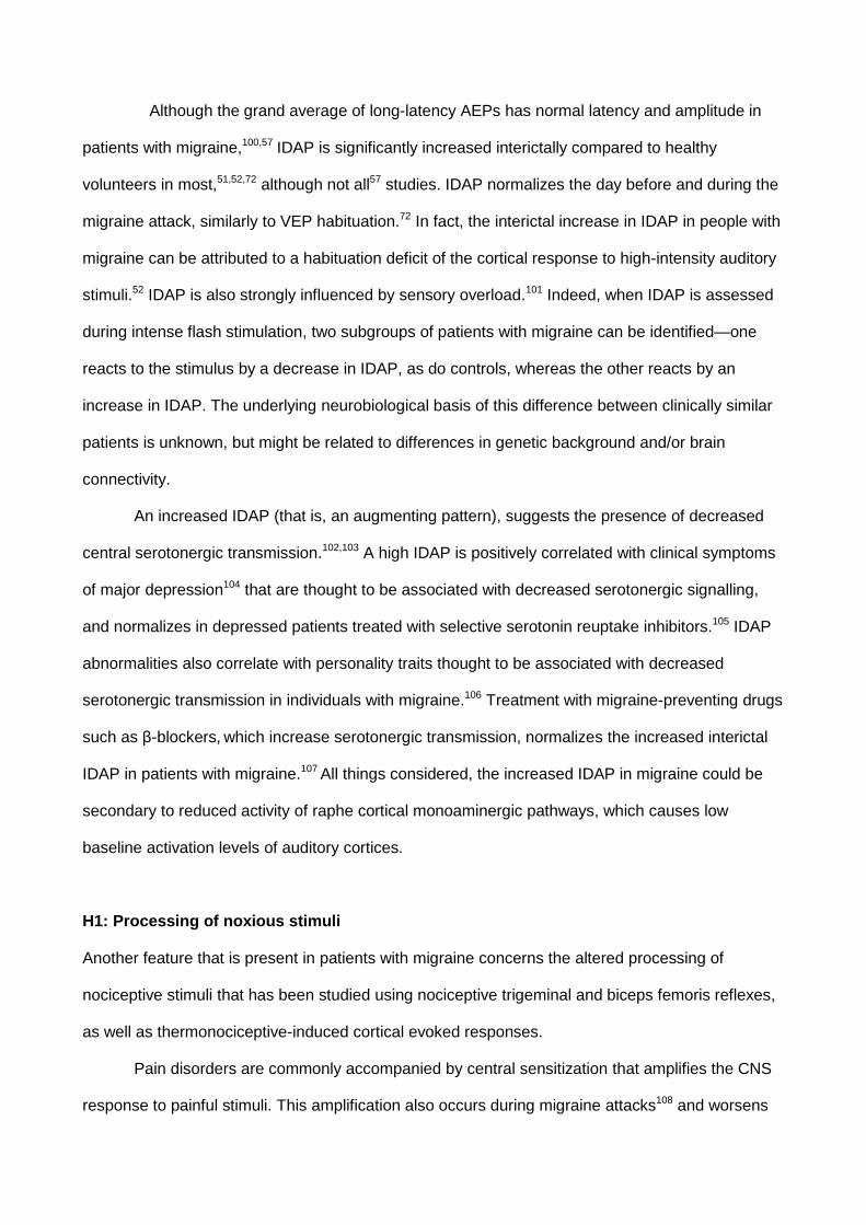
Although the grand average of long-latency AEPs has normal latency and amplitude in
patients with migraine,100,57 IDAP is significantly increased interictally compared to healthy
volunteers in most,51,52,72 although not all57 studies. IDAP normalizes the day before and during the
migraine attack, similarly to VEP habituation.72 In fact, the interictal increase in IDAP in people with
migraine can be attributed to a habituation deficit of the cortical response to high-intensity auditory
stimuli.52 IDAP is also strongly influenced by sensory overload.101 Indeed, when IDAP is assessed
during intense flash stimulation, two subgroups of patients with migraine can be identified—one
reacts to the stimulus by a decrease in IDAP, as do controls, whereas the other reacts by an
increase in IDAP. The underlying neurobiological basis of this difference between clinically similar
patients is unknown, but might be related to differences in genetic background and/or brain
connectivity.
An increased IDAP (that is, an augmenting pattern), suggests the presence of decreased
central serotonergic transmission.102,103 A high IDAP is positively correlated with clinical symptoms
of major depression104 that are thought to be associated with decreased serotonergic signalling,
and normalizes in depressed patients treated with selective serotonin reuptake inhibitors.105 IDAP
abnormalities also correlate with personality traits thought to be associated with decreased
serotonergic transmission in individuals with migraine.106 Treatment with migraine-preventing drugs
such as β-blockers, which increase serotonergic transmission, normalizes the increased interictal
IDAP in patients with migraine.107 All things considered, the increased IDAP in migraine could be
secondary to reduced activity of raphe cortical monoaminergic pathways, which causes low
baseline activation levels of auditory cortices.
H1: Processing of noxious stimuli
Another feature that is present in patients with migraine concerns the altered processing of
nociceptive stimuli that has been studied using nociceptive trigeminal and biceps femoris reflexes,
as well as thermonociceptive-induced cortical evoked responses.
Pain disorders are commonly accompanied by central sensitization that amplifies the CNS
response to painful stimuli. This amplification also occurs during migraine attacks108 and worsens

with increasing attack frequency.109 One mechanism underlying central sensitization is the activity-
dependent change in excitability of central nociceptors, which results in an abnormal amplification
of pain sensation in physiological nociception, a phenomenon referred to as temporal summation
of pain stimuli110 that is equivalent to “wind-up” in animal experiments.111
H2: The nociceptive flexion reflex
The nociceptive flexion or withdrawal reflex (NWR) is a reliable measure of spinal nociception, as
demonstrated by the facts that it requires Aδ fibre activation, that the reflex threshold is related to
the pain perception threshold, and that the reflex magnitude positively correlates with pain intensity
ratings.112,113 Temporal summation of pain develops in parallel with temporal summation of the
NWR of the lower limbs, reflected by a progressive increase in magnitude of the NWR after
constant-intensity electrical stimulation (which activates both Aδ and C fibres,113–115 and is inhibited
by NMDA receptor antagonists).116 Interestingly, descending pain control systems modulate
temporal summation of the NWR,117 and might be dysfunctional in a number of chronic pain
disorders, including migraine. For example, studies of temporal summation of the NWR in people
with migraine show facilitation of temporal pain processing between attacks.118 Administration of a
nitric oxide donor, such as glyceryl trinitrate, that triggers an attack in many migraineurs at delay of
several hours, induces within 120 min a transitory facilitation of temporal summation of the NWR,
in those patients who will develop a migraine attack (Figure 3).119
In individuals with chronic headache, such as MOH evolving from episodic migraine, the
threshold for temporal summation of the NWR is markedly reduced compared to that in controls or
patients with episodic migraine, which indicates a strong facilitation in the temporal processing of
pain.118 In patients with MOH, the suppressing effect of supraspinal diffuse noxious inhibitory
control (DNIC) (Box 2) on temporal summation of the NWR is deficient.118 This effect, which in
humans is termed conditioned pain modulation,120 can be tested by the heterotopic application of a
painful cold stimulus.119 The deficits in conditioned pain modulation or supraspinal diffuse noxious
inhibitory control and facilitation of temporal summation of the NWR normalize after drug
withdrawal, which could be related to the reduction in activity of anandamide hydrolase (also

known as fatty acid amide hydrolase) and hence slowing of the degradation of
endocannabinoids.121
H2: Nociceptive trigeminal evoked responses
The nBR is obtained in orbicularis oculi muscles by stimulating the supraorbital nerve via a
concentric high-density electrode, which activates mainly Aδ afferents. This reflex is mediated via
interneurons of the spinal trigeminal nucleus. Migraine is characterized by an interictal deficit of
nBR habituation during both short74 and long38 series of stimuli. nBR habituation normalizes during
migraine attacks,74 and individuals at risk of developing migraine lack nBR habituation deficits,86
whereas habituation of nociceptive laser-evoked potentials (LEP, discussed further below) remains
deficient.75 Patients with migraine also show temporal summation of the nBR.122
Brief radiant heat pulses generated by CO2 laser stimulation or contact thermode-delivered
stimuli excite Aδ and C-fibre thermonociceptors in superficial skin layers.123 The Aδ fibre input
generates cortical potentials, called respectively LEP or contact-heat evoked potentials (CHEP).
The N2–P2 component of LEP and CHEP is thought to be generated in the posterior part of the
anterior cingulate cortex and in bilateral insula.124
Compared to healthy controls and people with episodic migraine between attacks, the brain
distribution of LEP is shifted rostrally in patients with migraine during an attack125 and in patients
with chronic migraine.126 This anterior shift of activation contrasts with the posterior shift of LEP
observed during capsaicin-induced neuropathic pain in healthy volunteers,127 and with the caudal
displacement of cortical evoked potentials in the cingulate gyrus after intramuscular nociceptive
stimulation of the trapezius muscle in patients with migraine.128 This difference with the data on
LEP can be explained by the different methodology used, which involves stimulation of different
nociceptive afferents.129
Similarly to the cortical evoked potentials elicited by non-noxious stimuli, LEP75,130 and
CHEP131 show habituation deficits in patients with migraine between attacks. However, in contrast
with non-noxious cortical evoked potentials, the lack of habituation of LEP persists during the
attack, and is associated with an increased N2–P2 amplitude.132

Nonlinear analysis of ongoing EEG changes shows subtle changes in the cortical response
to painful laser stimuli in patients with migraine.133 For example, individuals with episodic migraine
have markedly reduced predictability of their EEG rhythms after the laser stimulus compared to
healthy individuals, although their averaged LEP appear normal; however, the averaging technique
used to extract evoked potentials from the background EEG signals might neglect subtle changes
in the processing of pain by the brain. Future studies using analysis of single (non averaged)
nociceptive evoked potentials, refined neurophysiological techniques and the combination of
neurophysiological and imaging methods will help to characterize the pathophysiological features
of central processing of pain in patients with migraine.
In chronic migraine and medication overuse headache, pain-related cortical potentials to
electrical forehead or forearm stimulation were facilitated, but not the nBR.134
H1: Migraine pathogenesis from the neurophysiological perspective
Given the results of the abovedescribed neurophysiological studies, the pathogenesis of
migraine seems to be driven by a complex dysfunction of thalamocortical connectivity and temporal
activation of neuronal networks. Thalamocortical dysrhythmia might also explain the phenomena
observed in patients with migraine treated with transcranial neuromodulation techniques—for
instance, the increased variability of dynamic changes in excitability135 or the paradoxical
homeostatic cerebral plasticity.136–138 In a proof-of-concept study, the plastic cortical changes
induced by rTMS were inversely related to thalamocortical activation 139. This observation suggests
that the paradoxical effects observed after rTMS in patients with migraine might be a consequence
of abnormal thalamocortical drive, which impairs short-term and long-term changes in cortical
synaptic effectiveness, and finally leads to maladaptive responses. Taken together, the
dysfunctions found in the migrainous brain suggest an impairment of thalamocortical control of
temporal activation of different neuronal networks.48
Thalamocortical dysrythmia has been proposed to underlie other functional brain
disorders.140,141 Theta and beta overactivation on the EEG, suggestive of thalamocortical
dysrhythmia, was found in the cortical “pain matrix” of patients with chronic neuropathic pain, and

was attenuated together with the pain in 6 patients treated by central lateral thalamotomy.142 The
anatomical correlates of thalamocortical dysrhythmia in migraine remain to be analysed further with
modern neuroimaging methods. A functional MRI (fMRI) study in people with migraine revealed a
lack of habituation of the BOLD (Box 2) signal during repetitive trigeminal nociceptive stimulation in
areas of the pain matrix (anterior insula and middle cingulate gyrus).143 Interestingly, this difference
with healthy controls was not found for olfactory stimuli, which the researchers attributed to the fact
that olfaction is not relayed in the thalamus.
Our research group has proposed that hypofunctioning serotoninergic projections to the
thalamus and cortex might cause functional disconnection of the thalamus, leading to
thalamocortical dysrhythmia and reduced cortical habituation.40 (Fig. 4)
It has not yet been demonstrated whether the different synchronicity and deficient
habituation characterizing neuronal responses to external stimuli in migraine play a role in the
cortical predisposition to spreading depression, or any other phenomenon able to activate the
trigeminovascular system and induce a migraine attack. It is also not proven (though it would
intuitively seem possible) that the abnormal temporal processing of nociceptive information
predisposes to migraine attacks, central sensitization and possibly chronic migraine. However, the
fact that the interictal cortical hyperresponsivity to sensory stimuli in migraine can be alleviated by
neurostimulation techniques55 (see below) and by preventive anti-migraine drugs,107 both of which
also decrease attack frequency, is indirect evidence that the brain dysfunction between attacks
may predispose patients to recurrent attacks. Considering that the cerebral energy reserve (ATP
content) is significantly lower in migraineurs between attacks compared to healthy subjects80, it is
tempting to speculate that the cortical hyperresponsivity may contribute to disrupt the brain’s
metabolic homeostasis by enhancing energy demands and to start up the biochemical cascade
that leads to the migraine attack.6
H1: Prospects for clinical research
The results of MRI studies suggest that migraine is associated with altered interictal
functional connectivity in subcortical and cortical areas that are devoted to cognitive functions and

pain processing.144,145 Connectivity was stronger between the periaqueductal grey and several
brain areas associated with pain processing, such as the prefrontal cortex, anterior cingulate and
amygdala, areas that are very similar to brain regions implicated in neurophysiological data on
sequential cortical activation during painful stimuli.125,126,144,146 Diffusion-weighted MRI studies
showed that microstructural alterations of white matter, and thus of functional connectivity, are
present across the orbitofrontal cortex, insula, thalamus and dorsal midbrain.147 It was
suggested147 that these alterations might reflect maladaptive plastic changes driven by
dysfunctions in multimodal exogenous and endogenous task processing. In another fMRI study,
thalamic sensitization correlated with widespread mechanical allodynia during the migraine
attack.148 Moreover, in a diffusion tensor MRI study, our research group found dynamic ictal and
interictal microstructural variations in the thalamus that were related to the time from or to the last
migraine attack, and seemed to mimic the cyclic neurophysiological changes described above.149
Collectively, these observations suggest that searching for optimal methods of influencing
the cortical temporal processing of exogenous stimuli that can trigger a migraine attack, or of
modulating endogenous trigeminal noxious inputs that lead to central sensitization and eventually
chronic headache could result in novel interventions for migraine prevention. The modes of action
of anticonvulsants or antidepressants, and of other pharmacological or non-pharmacological
interventions such as neuromodulation methods, should be reconsidered in terms of their ability to
normalize the complex abnormalities of brain connectivity and hyperresponsivity found in patients
with migraine.150 For example, non-invasive cortical neuromodulation techniques such as rTMS
and transcranial direct current stimulation (tDCS) have already been assessed in clinical trials.
Several studies investigated the hypothesis that the cortex in patients with migraine patients is
hyperexcitable between attacks. However, inhibitory low-frequency rTMS over the vertex had no
superior therapeutic effect to sham stimulation,151 and cathodal (i.e.inhibitory) tDCS over the
occipital cortex had no significant preventive effect, although the latter intervention did reduce
attack intensity compared to placebo.152 By contrast, in a pilot trial designed to assess an
alternative hypothesis — that the visual cortex is not hyperexcitable per se, but rather insufficiently
activated at baseline (as described above)87 — anodal (facilitatory) tDCS over the occipital cortex

significantly decreased attack frequency and intensity when used as preventive therapy in patients
with migraine.150
The challenge for future research, therefore, lies in identification of the precise anatomical
structures and functional networks involved in migraine, and determining which pharmacological
and non-pharmacological interventions can optimally modulate the function of these areas and
thereby improve temporal information processing. Such investigations will involve simultaneous
recordings of the above-reported phenomena by neurophysiological and functional neuroimaging
techniques, along with the application of nonlinear algorithms to model brain complexity.153 Novel
therapeutic interventions can then be tested for their capacity to normalize the anatomic and
functional changes associated with migraine and its subtypes.
H1: Conclusions
Most data described here suggest that the cortical processing of non-noxious and noxious
sensory stimuli is different between patients with migraine and healthy controls. The neuronal
networks involved in sensory processing are characterized by different modalities of sequential
recruitment under different environmental or endogenous conditions. The patterns of temporal
activation have been analysed over a range of neuronal activities, from progressive changes of
neuronal recruitment in the habituation or intensity dependence phenomena to facilitation of
noxious stimuli summation, and complex patterns of variability, phase synchronization and
causality that are adapted to describe the properties of a chaotic and nonlinear system. These
intricate processes are different not only between patients with migraine and healthy individuals,
but also vary according to the phases of the migraine cycle in the same patient.
The mechanisms underpinning these complex changes are far from being understood and
how they fit into the puzzle of migraine pathogenesis is still unclear. Owing to their complexity,
however, it is unlikely that the brain dysfunctions can simply be explained by an imbalance
between excitatory and inhibitory circuits.87 We propose that thalamocortical dysrhythmia may be
the culprit for abnormal central processing of non-noxious and noxious sensory stimuli in migraine
patients and that it might be itself caused by a genetically determined, inadequate control of the

thalamus and cortex by monoaminergic (serotonergic) projections originating in the brain stem
(Fig. 4). We further postulate that the cortical hyperresponsivity to sensory stimuli may contribute
causally to migraine attack repetition because it favours an excessive energy expenditure in a
brain with a reduced energy reserve.
In order to reduce discrepancies between studies, more attention should be paid to blinding
of investigators, so that accurate clinical data and headache diaries can be collected before and
during testing. In addition, prospective studies should be conducted to monitor patients’ clinical
fluctuations throughout the migraine cycle. It will also be of utmost importance to gather more data
on the (neurophysiological) phenotype–genotype correlations in patients with the various migraine
types. Finally, improved insight into the nature of the interictal dysfunction of temporal information
processing in individuals with migraine will, we hope, pave the way for novel therapeutic targets
and could herald improved migraine management.
Review criteria
We initially searched the PubMed database to identify articles published up to June 2013. The
search terms used were, “migraine”, “electroencephalography”, “EEG”, “evoked potentials”,
“habituation”, “temporal summation”, “nociceptive withdrawal reflex”, and “blink reflex”, alone and in
combination. The literature search was updated using the additional keywords “migraine”,
“habituation” and “evoked potentials” to identify full-text papers written in English and published in
peer-reviewed journals up to December 2013, using PubMed and Google Scholar databases.
Reviews were considered only when they introduced new concepts or hypotheses.
References
1. Stewart, W.F., Wood, C., Reed, M.L., Roy, J., Lipton, R.B. % AMPP Advisory Group.
Cumulative lifetime migraine incidence in women and men. Cephalalgia 28, 1170-1178
(2008).

2. Headache Classification Subcommittee of the International Headache Society. The
International Classification of Headache Disorders, 3rd Edn. Cephalalgia 33, 629-808
(2013).
3. Goadsby, P.J., Charbit, A.R., Andreou, A.P., Akerman, S. & Holland, P.R. Neurobiology
of migraine. Neuroscience 161, 327–341 (2009).
4. Bernstein, C. & Burstein, R. Sensitization of the trigeminovascular pathway: perspective
and implications to migraine pathophysiology. J. Clin. Neurol. 8, 89-99 (2012).
5. Magis, D., et al. Evaluation and proposal for optimalization of neurophysiological tests in
migraine: part 1--electrophysiological tests. Cephalalgia 27,1323-1338 (2007).
6. Ambrosini, A., Magis, D. & Schoenen, J. Migraine - clinical neurophysiology. Handb.Clin.
Neurol. 97, 275-293 (2010).
7. Moratti, S., Clementz, B.A., Gao, Y., Ortiz, T. & Keil, A. Neural mechanisms of evoked
oscillations: stability and interaction with transient events. Hum. Brain Mapp. 28, 1318-
1333 (2007).
8. Golla, F.L. & Winter, A.L. Analysis of cerebral responses to flicker in patients complaining
of episodic headache. Electroencephalogr. Clin. Neurophysiol. 11, 539-549 (1959).
9. Puca, F.M., de Tommaso, M., Tota, P. & Sciruicchio, V. Photic driving in migraine:
correlations with clinical features. Cephalalgia 16, 246-250 (1996).
10. de Tommaso, M., Sciruicchio, V., Guido, M., Sasanelli, G., Specchio, L.M. & Puca, F.M.
EEG spectral analysis in migraine without aura attacks. Cephalalgia 118, 324-328 (1998).
11. de Tommaso, M. et al. Visually evoked phase synchronization changes of alpha rhythm
in migraine: correlations with clinical features. Int. J. Psychophysiol . 57, 203–210 (2005).
12. Bjørk, M., Hagen, K., Stovner, L.J & Sand, T. Photic EEG-driving responses related to
ictal phases and trigger sensitivity in migraine: a longitudinal, controlled study.
Cephalalgia 31, 444-455 (2011).
13. Genco, S., de Tommaso, M., Prudenzano, A.M., Savarese, M. & Puca, F.M. EEG
features in juvenile migraine: topographic analysis of spontaneous and visual evoked
brain electrical activity: a comparison with adult migraine. Cephalalgia 14, 41-46 (1994).
14. Nyrke, T., Kangasniemi, P. & Lang, A.H. Difference of steady-state visual evoked
potentials in classic and common migraine. Electroencephalogr. Clin. Neurophysiol. 73,
285-294 (1989).
15. Shibata, K., Yamane, K., Otuka, K. & Iwata, M. Abnormal visual processing in migraine
with aura: a study of steady-state visual evoked potentials. J. Neurol. Sci. 271, 119-126
(2008).
16. Birca, A., Carmant, L., Lortie, A. & Lassonde, M. Interaction between the flash evoked
SSVEPs and the spontaneous EEG activity in children and adults. Clin. Neurophysiol.
117, 279-288 (2006).
17. Angelini, L., et al. Steady-state visual evoked potentials and phase synchronization in
migraine patients. Phys. Rev. Lett. 93, 038103 (2004).
18. Friston, K. Functional and effective connectivity: a review. Brain Connectivity 1, 13-36
(2011).
19. Silberstein, R.B. Steady-state visually evoked potentials, brain resonances and cognitive
processes. In Neocortical dynamics and human EEG rhythms (ed. Nunez, P.L.) 272–303
(Oxford University Press, 1995).
20. de Tommaso, M. et al. Effects of levetiracetam vs topiramate and placebo on visually
evoked phase synchronization changes of alpha rhythm in migraine. Clin. Neurophysiol.
118, 2297–2304 (2007).
21. de Tommaso, M. et al. Lack of effects of low frequency repetitive transcranial magnetic
stimulation on alpha rhythm phase synchronization in migraine patients. Neurosci. Lett.
20, 143-147 (2011).
22. Granger, C.W.J. Investigating causal relations by econometric models and crossspectral
methods. Econometrica 37, 424-438 (1969).
23. Marinazzo, D., Pellicoro, M. & Stramaglia, S. Kernel method for nonlinear Granger
causality. Phys. Rev. Lett. 11, 144103 (2008).
24. Friston, K.J., Harrison, L. & Penny, W. Dynamic causal modeling. NeuroImage 19, 1273-
1302 (2003).

25. de Tommaso, M., Stramaglia, S., Marinazzo, D., Trotta, G. & Pellicoro, M. Functional and
effective connectivity in EEG alpha and beta bands during intermittent flash stimulation in
migraine with and without aura. Cephalalgia 33, 938-347 (2013).
26. Harris, J. Habituatory response decrement in the intact organism. Psychol. Bull. 40, 385-
422 (1943).
27. Thompson, R. & Spencer, W. Habituation: a model phenomenon for the study of neuronal
substrates of behavior. Psychol. Rev. 73, 16-43 (1996).
28. Rankin, C.H. et al. Habituation revisited: an updated and revised description of the
behavioral characteristics of habituation. Neurobiol. Learn. Mem. 92, 135-138 (2009).
29. Walpurger, V., Hebing-Lennartz, G., Denecke, H. & Pietrowsky, R. Habituation deficit in
auditory event-related potentials in tinnitus complainers. Hear Res. 181, 57-64 (2003).
30. Schestatsky, P. et al. Neurophysiologic study of central pain in patients with Parkinson
disease. Neurology 69, 2162-2169 (2007).
31. Halberstadt, A.L. & Geyer, M.A. Habituation and sensitization of acoustic startle: opposite
influences of dopamine D1 and D2-family receptors. Neurobiol. Learn. Mem. 92, 243-248
(2009).
32. de Tommaso, M. et al. Laser-evoked potentials habituation in fibromyalgia. J. Pain 12,
116-124 (2011).
33. Schoenen, J., Wang, W., Albert, A. & Delwaide, P. Potentiation instead of habituation
characterizes visual evoked potentials in migraine patients between attacks. Eur. J.
Neurol. 2, 115-122 (1995).
34. Afra, J., Cecchini, A.P., De Pasqua, V., Albert, A. & Schoenen, J. Visual evoked
potentials during long periods of pattern-reversal stimulation in migraine. Brain 121, 233-
241 (1998).
35. Wang, W., Wang, G.P., Ding, X.L. & Wang, Y.H. Personality and response to repeated
visual stimulation in migraine and tension-type headaches. Cephalalgia 19, 718-724
(1999).

36. Bohotin, V. et al. Effects of repetitive transcranial magnetic stimulation on visual evoked
potentials in migraine. Brain 125, 912-922 (2002).
37. Ozkul, Y. & Bozlar, S. Effects of fluoxetine on habituation of pattern reversal visually
evoked potentials in migraine prophylaxis. Headache 42, 582-587 (2002).
38. Di Clemente, L., Coppola, G., Magis, D., Fumal, A., De Pasqua, V. & Schoenen, J.
Nociceptive blink reflex and visual evoked potential habituations are correlated in
migraine. Headache 45, 1388-1393 (2005).
39. Fumal, A. et al. Induction of long-lasting changes of visual cortex excitability by five daily
sessions of repetitive transcranial magnetic stimulation (rTMS) in healthy volunteers and
migraine patients. Cephalalgia 26, 143-149 (2006).
40. Coppola, G. et al. . Interictal abnormalities of gamma band activity in visual evoked
responses in migraine: an indication of thalamocortical dysrhythmia? Cephalalgia 27,
1360-1367 (2007).
41. Coppola, G. et al. Changes in visual-evoked potential habituation induced by
hyperventilation in migraine. J. Headache Pain 11, 497-503 (2010).
42. Coppola, G., Crémers, J., Gérard, P., Pierelli, F. & Schoenen, J. Effects of light
deprivation on visual evoked potentials in migraine without aura. BMC Neurol. 11, 91
(2011).
43. Chen, W., Wang, S., Fuh, J., Lin, C., Ko, Y. & Lin, Y. Peri-ictal normalization of visual
cortex excitability in migraine: an MEG study. Cephalalgia 29, 1202-1211 (2009).
44. Maertens de Noordhout, A., Timsit-Berthier, M., Timsit, M. & Schoenen, J. Contingent
negative variation in headache. Ann. Neurol. 19, 78-80 (1986).
45. Kropp, P. & Gerber, W.D. Contingent negative variation during migraine attack and
interval: evidence for normalization of slow cortical potentials during the attack.
Cephalalgia 15, 123-128 (1995).
46. Schoenen, J. & Timsit-Berthier, M. Contingent negative variation: methods and potential
interest in headache. Cephalalgia 13, 28-32 (1993).

47. Kropp, P. & Gerber, W.D. Contingent negative variation during migraine attack and
interval: evidence for normalization of slow cortical potentials during the attack.
Cephalalgia 15, 123-128 (1995).
48. Evers, S., Bauer, B., Suhr, B., Husstedt, I.W. & Grotemeyer, K.H. Cognitive processing in
primary headache: a study on event-related potentials. Neurology 48, 108-113 (1997).
49. Wang, W. & Schoenen, J. Interictal potentiation of passive "oddball" auditory event-
related potentials in migraine. Cephalalgia 18, 261-265 (1998).
50. Siniatchkin, M., Kropp, P. & Gerber, W.D. What kind of habituation is impaired in migraine
patients? Cephalalgia 23, 511-518 (2003).
51. Wang, W., Timsit-Berthier, M. & Schoenen, J. Intensity dependence of auditory evoked
potentials is pronounced in migraine: an indication of cortical potentiation and low
serotonergic neurotransmission? Neurology 46, 1404-1409 (1996).
52. Ambrosini, A., Rossi, P., De Pasqua, V., Pierelli, F. & Schoenen, J. Lack of habituation
causes high intensity dependence of auditory evoked cortical potentials in migraine. Brain
126, 2009-2015 (2003).
53. Ozkul, Y. & Uckardes, A. Median nerve somatosensory evoked potentials in migraine.
Eur. J. Neurol. 9, 227-232 (2002).
54. Coppola, G. et al. Abnormal cortical responses to somatosensory stimulation in
medication-overuse headache. BMC Neurol. 10, 126 (2010).
55. Coppola, G., De Pasqua, V., Pierelli, F. & Schoenen, J. Effects of repetitive transcranial
magnetic stimulation on somatosensory evoked potentials and high frequency oscillations
in migraine. Cephalalgia 32, 700-709 (2012).
56. Oelkers, R. et al. Visual evoked potentials in migraine patients: alterations depend on
pattern spatial frequency. Brain 122, 1147-1155 (1999).
57. Sand, T. & Vingen, J.V. Visual, long-latency auditory and brainstem auditory evoked
potentials in migraine: relation to pattern size, stimulus intensity, sound and light
discomfort thresholds and pre-attack state. Cephalalgia 20, 804-820 (2000).

58. Lang, E., Kaltenhäuser, M., Neundörfer, B., & Seidler, S. Hyperexcitability of the primary
somatosensory cortex in migraine--a magnetoencephalographic study. Brain 127, 2459-
2469 (2004).
59. Oelkers-Ax, R., Parzer, P., Resch, F. & Weisbrod, M. Maturation of early visual
processing investigated by a pattern-reversal habituation paradigm is altered in migraine.
Cephalalgia 25, 280-289 (2005).
60. Sand, T., Zhitniy, N., White, L.R. & Stovner, L.J. Visual evoked potential latency,
amplitude and habituation in migraine: a longitudinal study. Clin. Neurophysiol. 119,
1020-1027 (2008).
61. Sand, T., White, L., Hagen, K. & Stovner, L. Visual evoked potential and spatial
frequency in migraine: a longitudinal study. Acta Neurol. Scand. Suppl. 189, 33-37
(2009).
62. Demarquay, G., Caclin, A., Brudon, F., Fischer, C. & Morlet, D. Exacerbated attention
orienting to auditory stimulation in migraine patients. Clin. Neurophysiol. 122, 1755-1763
(2011).
63. Omland, P.M., et al. Visual evoked potentials in interictal migraine: no confirmation of
abnormal habituation. Headache 53, 1071-1086.
64. Bednář, M., Kubová, Z. & Kremláček, J. Lack of visual evoked potentials amplitude
decrement during prolonged reversal and motion stimulation in migraineurs. Clin.
Neurophys. DOI: 10.1016/j.clinph.2013.10.050 (2013).
65. Coppola, G., et al. Lateral inhibition in visual cortex of migraine patients between attacks.
J. Headache Pain 14, 20 (2013).
66. Sándor, P.S., Afra, J., Proietti-Cecchini, A., Albert, A. & Schoenen, J. Familial influences
on cortical evoked potentials in migraine. Neuroreport 10, 1235-1238 (1999).
67. Lorenzo, C.D., et al. Cortical response to somatosensory stimulation in medication
overuse headache patients is influenced by angiotensin converting enzyme (ACE) I/D
genetic polymorphism. Cephalalgia 32, 1189-1197 (2012).

68. Restuccia, D., Vollono, C., Del Piero, I., Martucci, L. & Zanini, S. Somatosensory High
Frequency Oscillations reflect clinical fluctuations in migraine. Clin. Neurophysiol. 123,
2050-2056 (2012).
69. Restuccia, D, Vollono, C., Piero, I.D., Martucci, L. & Zanini, S. Different levels of cortical
excitability reflect clinical fluctuations in migraine. Cephalalgia 33, 1035-1047 (2013).
70. Kropp, P. & Gerber, W.D. Prediction of migraine attacks using a slow cortical potential,
the contingent negative variation. Neurosc. Lett. 257, 73-76 (1998).
71. Siniatchkin, M., Kropp, P., Gerber, W.D. & Stephani, U. Migraine in childhood--are
periodically occurring migraine attacks related to dynamic changes of cortical information
processing? Neurosc. Lett. 279, 1-4 (2000).
72. Judit, A., Sándor, P.S. & Schoenen, J. Habituation of visual and intensity dependence of
auditory evoked cortical potentials tends to normalize just before and during the migraine
attack. Cephalalgia 20, 714-719 (2000).
73. Evers, S., Quibeldey, F., Grotemeyer, K.H., Suhr, B. & Husstedt, I.W. Dynamic changes
of cognitive habituation and serotonin metabolism during the migraine interval.
Cephalalgia 19, 485-491 (1999).
74. Katsarava, Z., Giffin, N., Diener, H.C. & Kaube, H. Abnormal habituation of 'nociceptive'
blink reflex in migraine--evidence for increased excitability of trigeminal nociception.
Cephalalgia 23, 814-819 (2003).
75. de Tommaso, M. et al. Habituation of single CO2 laser-evoked responses during interictal
phase of migraine. J. Headache Pain 6, 195-198 (2005).
76. De Marinis, M., Pujia, A., Natale, L., D’arcangelo, E. & Accornero, N. Debreased
habituation of the R2 component of the blink reflex in migraine patients. Clin.
Neurophysiol. 114, 889-893 (2003).
77. de Tommaso, M. et al. Modulation of trigeminal reflex excitability in migraine: effects of
attention and habituation on the blink reflex. Int. J. Psychophysiol. 44, 239-249 (2002).

78. Sand, T., Zhitniy, N., White, L.R. & Stovner, L.J. Brainstem auditory-evoked potential
habituation and intensity-dependence related to serotonin metabolism in migraine: a
longitudinal study. Clin. Neurophysiol. 119, 1190-1200 (2008).
79. Siniatchkin, M., Kropp, P. & Gerber, W.D. Contingent negative variation in subjects at risk
for migraine without aura. Pain 94, 159-167 (2001).
80. Paemeleire, K. & Schoenen, J. (31) P-MRS in migraine: fallen through the cracks.
Headache 53, 676-678 (2013).
81. Maniyar, F.H., Sprenger, T., Monteith, T., Schankin, C. & Goadsby P.J. Brain activations
in the premonitory phase of nitroglycerin-triggered migraine attacks. Brain
http://dx.doi.org/10.1093/brain/awt320.
82. Chen, W., Wang, S., Fuh, J., Lin, C., Ko, Y. & Lin, Y. Persistent ictal-like visual cortical
excitability in chronic migraine. Pain 152, 254-258 (2011).
83. Chen, W.T. et al. Visual cortex excitability and plasticity associated with remission from
chronic to episodic migraine. Cephalalgia 32, 537-543 (2012).
84. Schoenen, J. Is chronic migraine a never-ending migraine attack? Pain 152, 239-240
(2011).
85. Currà, A., et al. Drug-induced changes in cortical inhibition in medication overuse
headache. Cephalalgia 31,1282-1290 (2011).
86. Di Clemente, L. et al. Interictal habituation deficit of the nociceptive blink reflex: an
endophenotypic marker for presymptomatic migraine? Brain 130, 765-770 (2007).
87. Coppola, G., Pierelli, F., & Schoenen, J. Is the cerebral cortex hyperexcitable or
hyperresponsive in migraine? Cephalalgia 27, 1427-1439 (2007).
88. Knott, J.R. & Irwin, D.A. Anxiety, stress and the contingent negative variation. Arch. Gen.
Psychiatry 29, 538-541 (1973).
89. Coppola, G. et al. Somatosensory evoked high-frequency oscillations reflecting thalamo-
cortical activity are decreased in migraine patients between attacks. Brain 128, 98-103
(2005).

90. Sakuma, K., Takeshima, T., Ishizaki, K. & Nakashima, K. Somatosensory evoked high-
frequency oscillations in migraine patients. Clin. Neurophysiol. 115, 1857-1862 (2004).
91. Lai, K.L., Liao, K.K., Fuh, J.L., Wang, S.J. Subcortical hyperexcitability in migraineurs: a
high-frequency oscillation study. Can. J. Neurol. Sci. 38, 309-316 (2011).
92. Ambrosini, A., De Pasqua, V., Afra, J., Sándor, P.S. & Schoenen J. Reduced gating of
middle-latency auditory evoked potentials (P50) in migraine patients: another indication of
abnormal sensory processing? Neurosci. Lett. 22; 132-134 (2001).
93. Coppola, G. et al. Interictal abnormalities of gamma band activity in visual evoked
responses in migraine: an indication of thalamocortical dysrhythmia? Cephalalgia 27,
1360-1367 (2007).
94. Llinás, R.R., Ribary, U., Jeanmonod, D., Kronberg, E. & Mitra, P.P. Thalamocortical
dysrhythmia: a neurological and neuropsychiatric syndrome characterized by
magnetoencephalography. Proc. Natl. Acad. Sci. USA 96, 15222-15227 (1999).
95. Llinás, R.R., Urbano, F.J., Leznik, E., Ramírez, R.R. & van Marle, H.J.F. Rhythmic and
dysrhythmic thalamocortical dynamics: GABA systems and the edge effect. Trends
Neurosci. 28, 325-333 (2005).
96. Bjørk, M.& Sand, T. Quantitative EEG power and asymmetry increase 36 h before a
migraine attack. Cephalalgia 28, 960-968 (2008).
97. Bjørk, M.H., Stovner, L.J., Nilsen, B.M., Stjern, M., Hagen, K. & Sand T. The occipital
alpha rhythm related to the "migraine cycle" and headache burden: a blinded, controlled
longitudinal study. Clin. Neurophysiol. 120, 464-471 (2009).
98. Buchsbaum, M. & Silverman, J. Stimulus intensity control and the cortical evoked
response. Psychosom. Med. 30, 12-22 (1968)
99. Hegerl, U. & Juckel, G. Intensity dependence of auditory evoked potentials as an
indicator of central serotonergic neurotransmission: a new hypothesis. Biol. Psychiatry
33, 173-187 (1993).
100. Drake, M.E., Pakalnis, A. & Padamadan, H. Long-latency auditory event related
potentials in migraine. Headache 29, 239-241 (1989).

101. Ambrosini, A., Coppola, G., Gérardy, P.Y., Pierelli, F. & Schoenen, J. Intensity
dependence of auditory evoked potentials during light interference in migraine. Neurosci.
Lett. 492, 80-83 (2011).
102. Proietti-Cecchini, A., Afra, J. & Schoenen, J. Intensity dependence of the cortical auditory
evoked potentials as a surrogate marker of central nervous system serotonin
transmission in man: demonstration of a central effect for the 5HT1B/1D agonist
zolmitriptan (311C90, Zomig). Cephalalgia 17, 849-854 (1997).
103. Juckel, G., Hegerl, U., Molnár, M., Csépe, V. & Karmos, G. Auditory evoked potentials
reflect serotonergic neuronal activity--a study in behaving cats administered drugs acting
on 5-HT1A autoreceptors in the dorsal raphe nucleus. Neuropsychopharmacology 21,
710-716 (1999).
104. Linka, T., Sartory, G., Gastpar, M., Scherbaum, N. & Müller, B.W. Clinical symptoms of
major depression are associated with the intensity dependence of auditory event-related
potential components. Psychiatry Res. 169, 139-143 (2009).
105. Linka, T., Müller, B.W., Bender, S. & Sartory, G. The intensity dependence of the auditory
evoked N1 component as a predictor of response to Citalopram treatment in patients with
major depression. Neurosc. Lett. 367, 375-378 (2004).
106. Wang. W., Wang, Y.H., Fu, X.M., Sun, Z.M. & Schoenen, J. Auditory evoked potentials
and multiple personality measures in migraine and post-traumatic headaches. Pain 79,
235-242 (1999).
107. Sándor, P.S., Afra, J., Ambrosini, A. & Schoenen, J. Prophylactic treatment of migraine
with beta-blockers and riboflavin: differential effects on the intensity dependence of
auditory evoked cortical potentials. Headache 40, 30-35 (2000).
108. Burstein, R., Cutrer, M.F. & Yarnitsky, D. The development of cutaneous allodynia during
a migraine attack clinical evidence for the sequential recruitment of spinal and
supraspinal nociceptive neurons in migraine. Brain 123,1703-1709 (2000).
109. Buchgreitz, L., Lyngberg, A.C., Bendtsen, L. & Jensen, R. Frequency of headache is
related to sensitization: a population study. Pain 123, 19-27 (2006).

110. Eide, P.K. Wind-up and the NMDA receptor complex from a clinical perspective. Eur. J.
Pain 4, 5-15 (2000).
111. Mendell, L.M. & Wall, P.D. Responses of single dorsal cord cells to peripheral cutaneous
unmyelinated fibres. Nature 206, 97-99 (1965).
112. Rhudy, J.L. et al. Pain catastrophizing is related to temporal summation of pain but not
temporal summation of the nociceptive flexion reflex. Pain 152, 794-801 (2011).
113. Arendt-Nielsen, L., Brennum, J., Sindrup, S. & Bak, P. Electrophysiological and
psychophysical quantification of temporal summation in the human nociceptive system.
Eur. J. Appl. Physiol. Occup. Physiol. 68, 266-273 (1994).
114. Sandrini, G., Serrao, M., Rossi, P., Romaniello, A., Cruccu, G. & Willer, J.C. The lower
limb flexion reflex in humans. Prog. Neurobiol. 77, 353–395 (2005).
115. You, H.J., Dahl, M.C., Chen, J. & Arendt-Nielsen, L. Simultaneous recordings of wind-up
of paired spinal dorsal horn nociceptive neuron and nociceptive flexion reflex in rats.
Brain Res. 960, 235-245 (2003).
116. Arendt-Nielsen, L. et al. The effect of N-methyl-D-aspartate antagonist (ketamine) on
single and repeated nociceptive stimuli: a placebo controlled experimental human study.
Anesth. Analg. 81, 63–68 (1995).
117. Serrao, M. et al. Effects of diffuse noxious inhibitory controls on temporal summation of
the NWR reflex in humans. Pain 112, 353–360 (2004).
118. Perrotta, A. et al. Sensitisation of spinal cord pain processing in medication overuse
headache involves supraspinal pain control. Cephalalgia 30, 272-284 (2010).
119. Perrotta, A. et al. Oral nitric-oxide donor glyceryl-trinitrate induces sensitization in spinal
cord pain processing in migraineurs: a double-blind, placebo-controlled, cross-over study.
Eur J Pain 15, 482-490 (2011).
120. Yarnitsky, D. et al., Recommendations on terminology and practice of psychophysical
DNIC testing. Eur. J. Pain 14, 339 (2010).

121. Perrotta, A. et al. Acute reduction of anandamide-hydrolase (FAAH) activity is coupled
with a reduction of nociceptive pathways facilitation in medication-overuse headache
subjects after withdrawal treatment. Headache 52, 1350-1361 (2012).
122. Giffin, N.J., Katsarava, Z., Pfundstein, A., Ellrich, J. & Kaube, H. The effect of multiple
stimuli on the modulation of the 'nociceptive' blink reflex. Pain 108, 124-128 (2004).
123. Bromm, B & Treede, R.D. Nerve fibre discharges, cerebral potentials and sensations
induced by CO2 laser stimulation. Hum. Neurobiol. 3, 33-40 (1984).
124. Garcia-Larrea L, Frot M, Valeriani M.Brain generators of laser-evoked potentials: from
dipoles to functional significance. Neurophysiol Clin. 33, 279-92 (2003).
125. de Tommaso, M. et al. Topographic and dipolar analysis of laser-evoked potentials during
migraine attack. Headache 44, 947-960 (2004).
126. de Tommaso, M., Losito, L., Difruscolo, O., Libro, G., Guido, M. & Livrea, P. Changes in
cortical processing of pain in chronic migraine. Headache 45, 1208-1218 (2005).
127. Valeriani, M. et al. Short-term plastic changes of the human nociceptive system following
acute pain induced by capsaicin. Clin. Neurophysiol. 114, 1879-1890 (2003).
128. Buchgreitz, L., Egsgaard, L.L., Jensen, R., Arendt-Nielsen, L. & Bendtsen, L. Abnormal
brain processing of pain in migraine without aura: a high-density EEG brain mapping
study. Cephalalgia 30, 191-199 (2010).
129. Valeriani, M., Tonali, P., De Armas, L., Mariani, S., Vigevano, F. & Le Pera, D.,
Nociceptive contribution to the evoked potentials after painful intramuscular electrical
stimulation. Neurosci. Res. 60, 170-175 (2008).
130. Valeriani, M. et al. Reduced habituation to experimental pain in migraine patients: a
CO(2) laser evoked potential study. Pain 105, 57-64 (2003).
131. Lev, R., Granovsky, Y. & Yarnitsky, D. Enhanced Pain Expectation in Migraine: EEG-
Based Evidence for Impaired Prefrontal Function. Headache 53, 1054-1070 (2013).
132. de Tommaso, M. et al. Lack of habituation of nociceptive evoked responses and pain
sensitivity during migraine attack. Clin. Neurophysiol. 116, 1254-1264 (2005).
133. de Tommaso, M., Marinazzo, D. & Stramaglia, S. The measure of randomness by leave-
one-out prediction error in the analysis of EEG after laser painful stimulation in healthy
subjects and migraine patients. Clin. Neurophysiol. 116, 2775-2782 (2005).
134. Ayzenberg, I,. Obermann, M., Nyhuis, P., Gastpar, M., Limmroth, V., Diener, H.C.,
Kaube, H. & Katsarava, Z. Central sensitization of the trigeminal and somatic
nociceptive systems in medication overuse headache mainly involves cerebral
supraspinal structures. Cephalalgia 26, 1106-1114 (2006).
135. Antal, A., Arlt, S., Nitsche, M.A., Chadaide, Z. & Paulus, W. Higher variability of
phosphene thresholds in migraineurs than in controls: a consecutive transcranial
magnetic stimulation study. Cephalalgia 26, 865-870 (2006).
136. Antal, A., Lang, N., Boros, K., Nitsche, M., Siebner, H.R. & Paulus, W. Homeostatic
metaplasticity of the motor cortex is altered during headache-free intervals in migraine
with aura. Cereb. Cortex 18, 2701-2705 (2008).
137. Siniatchkin, M., et al. Abnormal changes of synaptic excitability in migraine with aura.
Cereb. Cortex 22, 2207-2216 (2012).
138. Brighina, F., et al. Abnormal facilitatory mechanisms in motor cortex of migraine with
aura. Eur. J. Pain 15, 928-935 (2011).
139. Pierelli, F., Iacovelli, E., Bracaglia, M., Serrao, M. & Coppola, G. Abnormal sensorimotor
plasticity in migraine without aura patients. Pain 154, 1738-1742 (2013).
140. Adjamian, P., Sereda, M., Zobay, O., Hall, D.A. & Palmer, A.R. Neuromagnetic indicators
of tinnitus and tinnitus masking in patients with and without hearing loss. J. Assoc. Res.
Otolaryngol. 13, 715-731 (2012).
141. Walton, K.D., Dubois, M. & Llinás, R.R. Abnormal thalamocortical activity in patients with
Complex Regional Pain Syndrome (CRPS) type. Pain 150,41-51 (2010).
142. Stern, J., Jeanmonod, D. & Sarnthein, J. Persistent EEG overactivation in the cortical
pain matrix of neurogenic pain patients. Neuroimage 31, 721-731 (2006).
143. Stankewitz, A., Schulz, E. & May, A. Neuronal correlates of impaired habituation in
response to repeated trigemino-nociceptive but not to olfactory input in migraineurs: an
fMRI study. Cephalalgia 33, 256-265 (2013).
144. Mainero, C., Boshyan, J. & Hadjikhani, N. Altered functional magnetic resonance imaging
resting-state connectivity in periaqueductal gray networks in migraine. Ann. Neurol. 70,
838-845 (2011).
145. Russo, A. et al. Pain processing in patients with migraine: an event-related fMRI study
during trigeminal nociceptive stimulation. J. Neurol. 259, 1903-1912 (2012).
146. Tessitore, A. et al. Interictal cortical reorganization in episodic migraine without aura: an
event-related fMRI study during parametric trigeminal nociceptive stimulation. Neurol. Sci.
32, S165-167 (2011).
147. Szabó, N. et al. White matter microstructural alterations in migraine: a diffusion-weighted
MRI study. Pain 153, 651-656 (2012).
148. Burstein, R. et al. Thalamic sensitization transforms localized pain into widespread
allodynia. Ann. Neurol. 68, 81-91 (2010).
149. Coppola, G., et al. Dynamic changes in thalamic microstructure of migraine without aura
patients: a diffusion tensor magnetic resonance imaging study. Eur. J. Neurol.
http://dx.doi.org/10.1111/ene.12296
150. Viganò, A. et al. Transcranial Direct Current Stimulation (tDCS) of the visual cortex: a
proof-of-concept study based on interictal electrophysiological abnormalities in migraine.
J. Headache Pain 14, 23 (2013).
151. Teepker, M. et al. Low-frequency rTMS of the vertex in the prophylactic treatment of
migraine. Cephalalgia 30, 137-144 (2010).
152. Antal, A. et al. Cathodal transcranial direct current stimulation of the visual cortex in the
prophylactic treatment of migraine. Cephalalgia 31, 820-828 (2011).
153. Marinazzo, D., Liao, W., Chen, H. & Stramaglia, S. Nonlinear connectivity by Granger
causality. Neuroimage 15, 330-338 (2011).
Acknowledgements
Drs M. de Tommaso, A. Ambrosini, F. Brighina, G. Coppola, A. Perrotta, F. Pierelli, G.Sandrini
and M. Valeriani participated in writing this Review on behalf of the Italian Group for
Neurophysiology of Migraine, the Italian Society for the Study of Headaches (SISC) and the Italian
Society of Clinical Neurophysiology (SINC).
Author contributions:
All authors researched data for and participated in writing of the article. In addition, A. Ambrosini,
M. de Tommaso, G. Coppola, F. Pierelli and J. Schoenen contributed to discussion of content, and
A. Ambrosini, M. de Tommaso and J. Schoenen contributed to review and/or editing of the
manuscript before submission. M. De Tommaso and A. Ambrosini contributed equally to this
manuscript.
Figure 1
Temporal evolution in effective connectivity (causality-ordinate) as revealed by kernel Granger
causality analysis of averaged EEG data from patients with migraine and healthy controls during a
10-sec flash stimulation (abscissa) at a 21Hz frequency. In the alpha band (a), causal/effective
connections across scalp derivations are reduced in migraine without aura compared to migraine
with aura or healthy controls, while in the beta band (b) migraine with aura patients have increased
causality across cortical areas. This phenomenon may be subtended by an increase of cortical
activation in migraine with aura during visual stimulation.
Data used to generate this Figure were obtained from de Tommaso et al.25
Figure 2
Schematic overview showing amplitude changes in the N20–P25 component of averaged EEG
recordings in patients with migraine and healthy controls. a | HFOs and b | somatosensory evoked
potentials. In healthy controls (panel 1), the N20–P25 component habituates, and early HFOs
(reflecting thalamocortical drive) are greater than late HFOs (generated by intrinsic cortical
activation). In patients with migraine between attacks (panel 2) habituation is absent and early
HFOs are reduced, although late HFOs are normal. During a migraine attack (panel 3), habituation
and early HFOs normalize. After 10Hz HF-rTMS is applied over the somatosensory cortex in
patients with episodic migraine (panel 4), the interictal lack of habituation reverses and both early
and late HFOs increase. Abbreviations: HFO, high frequency oscillation; HF-rTMS, high frequency
repetitive transcranial magnetic stimulation.
Figure 3
Facilitation of the temporal summation threshold of the biceps femoris nociceptive withdrawal reflex
is markedly more facilitated by glyceryltrinitrate administration (versus placebo) in patients with
migraine than in healthy controls. Data for these graphs were obtained from Perrotta et al., 2011.119
Abbreviations: NWR, nociceptive withdrawal reflex; TST, temporal summation threshold, GTN,
glyceryltrinitrate; Plb, placebo.
Figure 4
The migraine headache (1) is due to activation of the trigeminovascular system (TVS), the major
pain-signalling system in the brain. The migraine aura is caused by cortical spreading depression
(CSD) that may or may not activate the trigeminovascular sytem. Genetic channelopathies (2)
predispose to CSD in the rare Familial/Sporadic Hemiplegic forms of migraine (FHM).
The challenge in migraine research is to determine what causes CSD, and what, besides CSD,
can activate the TVS in the more common forms of migraine. Neurophysiological studies described
in this review have contributed to disentangle some of the complex pathomechanisms in migraine.
They have shown that interictal abnormalities of sensory processing might predispose the
migrainous brain to an attack. Interictal thalamocortical dysrhythmia causes hyperresponsivity of
sensory cortices (3) as well as abnormal pain processing (4). The thalamocortical dysrhythmia
itself may be induced by decreased control from brain stem monoaminergic nuclei (5). Cortical

hyperresponsivity combined with a decreased mitochondrial energy reserve favours metabolic
strain and rupture of metabolic homeostasis(6). This may trigger CSD in the cortex and, via
subcortical chemosensitive structures, TVS activation. There is evidence for upper brain stem
activation during migraine attacks. Whether this is due to collateral projections from the trigeminal
nociceptive pathway, to chemosensing of the metabolic dysequilibrium or to input from
hypothalamus and limbic system remains to be determined. Activation of the monoaminergic nuclei
may explain why cortical hyperresponsivity normalizes during an attack. The migraine attack is
associated with sensitization of central nociceptive pathways (7) which can be detected by
abnormalities of noxious evoked cortical and subcortical responses. The latter abnormalities
amplify and persist in chronic migraine (8) where the neurophysiological pattern is that of a «never-
ending attack ».
Common genetic variants set the « migraine threshold » by modulating a number of different
physiological mechanisms: oxidative stress, neuroinflammation, neurotransmission, metabolism..
As described in the text, the neurophysiological abnormalities vary between patients, over the
migraine cycle, under the influence of preventive anti-migraine medications and with migraine
chronification.
Dashed lines indicate connections for which there is little or no experimental evidence yet.

Box 1 Neurophysiological findings associated with migraine
Several abnormalities of sensory processing may be observed in patients with migraine:
Interictal light-induced EEG changes
Stimulus-frequency dependent increase in photic drive
Alpha frequency synchronization and decrease in functional connectivity (in
migraine without aura)
Beta frequency desynchronization and increase in functional connectivity (in
migraine with aura)
Interictal (& ictal) changes in non-noxious sensory evoked potentials
Trend for low amplitude averaged responses to small numbers of repeated stimuli
Deficient habituation during prolonged stimulus repetition (normalizes during attack)
Increased intensity dependence of auditory evoked potentials
Interictal (& ictal) changes in noxious sensory evoked responses
Deficient habituation of cortical evoked responses (persists during attack)
Deficient habituation of nociceptive blink reflexes (normalizes during attack)
Facilitation of temporal summation of the biceps femoris flexion reflex
Changes in chronic migraine
High amplitude averaged cortical responses to small numbers of repeated non-
noxious or noxious stimuli
Normal habituation of non noxious sensory evoked responses
Deficient habituation of noxious sensory evoked responses
Box 2: Glossary
Granger causality/Kernel Granger causality: operative definition of causality for
linear/nonlinear systems

Diffuse noxious inhibitory control (DNIC): pain inhibition by a heterotopic painful
stimulation
BOLD: blood oxygen level dependent
AEP: auditory evoked potentials
IDAP: intensity dependence of auditory evoked potentials
VEP: visual evoked potentials
SSVP: steady state visual evoked potential
SSEP: somato-sensory evoked potential
HFO: high frequency oscillations
MOH: medication overuse headache
NWR: nociceptive withdrawal reflex
nBR: nociceptive blink reflex
LEP: laser evoked potentials
CHEP: contact heat evoked potentials
rTMS: repetitive transcranial magnetic stimulation
tDCS: transcranial direct current stimulation


Figure 1F

Figure 2

Figure 3
Figure 4