amarila malik12*, iman santoso , andi yehuda2,...
TRANSCRIPT

Available online athttp://jurnal.permi.or.id/index.php/mionline
DOI: 10.5454/mi.8.1.3ISSN 1978-3477, eISSN 2087-8575Vol 8, No 1, Maret 2014, p 16-23
*Corresponding author; Phone/Fax: +62-21-7270031/7863433, Email: [email protected]
+62-21-
Recently, we reported on isolates from Tangkuban
Perahu, a volcano in West Java and culture growth in
laboratories in Regensburg, Germany, and Depok,
Indonesia (Handayani 2012). We could
demonstrate growth of (Huber and
Prangishvili 2004) in Regensburg by electron
microscopyand obviously cultured members of the
order (Huber 2002) in
Depok, but the latter could not be characterized
et al.
Sulfolobales
Thermoplasma tales et al.
(Handayani 2012). Now, we focused on the
isolation and culture growth of
et al.
Thermoplamsa
species,e.g. and . These
organisms are cell wall-less thermoacidophilic archaea
with unique tetraetherlipids, which have raised our
interest in biomedical and biotechnical applications
(Freisleben 1999). was
first isolated by Darland . (1970) from sulfuric acid
milieu in self-heated coal refuse piles. Later,
was found in solfataric hot springs
(Segerer 1988). Solfataric environments appear
to be the natural habitat also of (Yasuda
T. acidophilum T. volcanium
Thermoplasma acidophilum
et al
T.
volcanium spp.
et al.
T. acidophilum
Archaea is an organisme with unique feature because of its ability to inhabit an extremophyle conditions. Ourexpeditions to Tangkuban Perahu, West Java aimed to obtain archaealstrains from the solfatara fields located inDomas crater. From the samples, we intended to culture species growing around 55 C below pH2, which until now have not yet been fully characterized. We collected five samples from mud holes withtemperatures from 52 C to 57 C and pH below 2. In serial cultures of up to 8 transfers in Freundt’s medium wegrew tetraetherlipid synthesizing species as documented by phase contrast microscope. Totalmembrane lipid extracts were analysed by thin layer chromatography; the pattern matched total lipid extractsfrom DSM 1728 membranes. For confirmation, 16S rDNA identificationperforming PCR and sequencing were carried-out. Analysis using BLAST showed identities asthe highest similarity of 99%, followed by also with99% similarity (ANKF776908 andANKF776909). This is the first report of culturing cell-wall-less thermoacidophilicarchaea,in particular
species in Indonesian laboratories.
Key words:
Thermoplasma
Thermoplasma
Thermoplasma acidophilumT. acidophilum
T. volcanium,
Thermoplasma
°
° °
archaea, Indonesian volcanoes, Tangkuban Perahu, tetraether lipid,
Archaea merupakan organisme dengan fitur unik karena kemampuannya mendiami kondisi extrem.Ekspedisi ke Tangkuban Perahu, Jawa Barat dilakukan dengan tujuan untuk memperoleh galur archaea dariladang solfatara yang terletak di kawah Domas. Dari sampel yang diperoleh selanjutnya dilakukan pengulturanspesies pada kondisi berkisar 55 C dan di bawah pH 2; pengulturan seperti ini belum dilaporkanada yang melakukan. Sebanyak lima sampel diambil dari lubang lumpur dengan suhu berkisar 52 C sampaidengan 57 C dan pH di bawah 2 telah diperoleh. Dalam kultur seri hingga 8 kali transfer menggunakan mediaFreundt telah berhasil diperoleh kultur spesies pensintesis tetraether lipid yang kemudian didokumentasikan menggunakan mikroskop fase kontras. Ekstrak total lipid membran dianalisis dengankromatografi lapis tipis; pola yang didapat sesuai dengan jumlah pola ekstrak lipid membran
DSM 1728. Untuk konfirmasi, identifikasi menggunakan 16S rDNA juga dilakukan dengan PCRdan kemudian dilanjutkan dengan sequencing DNA. Analisis menggunakan BLAST menunjukkan identitas
dengan kesimilaritasan tertinggi 99 %, dan identitas jugadengan kesimilaritasan tertinggi 99 % (AN KF776908 dan AN KF776909). Penelitian ini merupakanpengulturan archaea termoasidofilik tanpa dinding sel yang pertama di laboratorium di Indonesia, khususnyaspesies .
Kata kunci: archaea, gunung api Indonesia, lipid tetraeter, Tangkuban Perahu,
Thermoplasma
Thermoplasma
Thermoplasma
Thermoplasmaacidophilum
Thermoplasma acidophilum Thermoplasma volcanium
Thermoplasma
Thermoplasma
°
°
°
Characterization of Thermoplasma Species Cultured fromSampling on Tangkuban Perahu, Indonesia
AMARILA MALIK *, IMAN SANTOSO , ANDI YEHUDA , SERUNI K.U. FREISLEBEN ,
SEPTELIA INAWATI WANANDI , HARALD HUBER , ZESSINDA LUTHFA ,
ROSARI SALEH , HANS-JOACHIM FREISLEBEN
1 2 2 2
3 4 2
2 3AND
1 2 3
4
Facultyof Pharmacy, Faculty of Mathematics and Natural Sciences, Faculty of Medicine,Universitas Indonesia, Depok 16424, Indonesia;
Department of Microbiology, Archaea Centre, University of Regensburg, Germany

et al Thermoplasma
et al
. 1995). In Indonesia species,
which have not yet been further characterized, have
been reported from TangkubanPerahu (Huber .
1991), an easily accessible volcano in West Java, South
of Jakarta, near the city of Bandung.Formerly, growth of DSM 1728 was
achieved in fermenters under laboratory conditions at
pH 1.5 to 2.0 and an optimal growth temperature of 59
C (Freisleben . 1994) as a major source of
tetraether lipid, because the organism lacks a cell wall,
and thus the membrane lipids are ‘naked’ and easily
accessible. In nature, members grow
autotrophically metabolizing elemental sulfur, but they
can also grow mixo- and heterotrophically, from
anoxic to oxic conditions (Huber . 1991).
It is intended to optimize growth conditions, to
identify and characterize the archaeal cells and to
extract and purify their tetraether lipids for application
in the biomedical field (e.g. liposomes, archaeosomes
as drug and vaccine delivery systems) and in
nanotechnology (e.g., monomolecular thin film
surface coating) (Freisleben .1995; Bakowsky
. 2000; Patel . 2000; Schiraldi . 2002;
Krishnan and Sprott 2008; Thavasi . 2008). In the
present study, the identification by molecular
techniques performing PCR and sequencing was used.
We applieda pair of primers designed by aligning 16S
rRNA genes specifically targeting the gene encoding
for 16S rRNA, followed by DNA sequence analysis
using BLAST, as well as Clustal W and TreeView X .
On June 11, 2012 (samples KD 1,2) and
January 21, 2013 (samples KD 3,4,5) sampling was
carried out on the Indonesian volcano Tangkuban
Perahulocated in West Java, Bandung. Samples were
taken from solfatara fields in the Domas crater (Kawah
Domas, KD), under similar conditions as described by
Handayani . 2012. In contrast to the former
sampling the temperature range of our new samplings
was quite lower; all samples weretaken from mud holes
between 52 Cand 57 C, because we wanted to
concentrate on and species,
wich are known to grow in this temperature range.
The samples were obtained from acidic mud
holesand warm springs from where a strong smell of
H S was rising. Previous reports regarding archaeal
habitats had shown that the growth temperature of
speciesis around 50 C up to 60 C
(Huber 1991; Yasuda . 1995) which was also
T. acidophilum
et al
Thermoplasma
et al
et al et
al et al et al
et al
et al
T. acidophilum T. volcanium
Thermoplasma
et al. et al
°
° °
° °
R
MATERIALS AND METHODS
Sampling.
2
confirmed in fermentor growth (Freisleben . 1994).
None of samples from five different mud holes and
et al
warm springs had a pH above 2; KD1 and KD5 were
sampled at 57 C; KD2 at 52 C; KD3 at 56 C; and
KD4 at 54 C. Samples were collected into 140 mL
screw-capped glass bottles, filled to the top and firmly
closed.
Freundt’s medium was prepared according to
Freisleben . (1994). All substances were dissolved
to 1 L in aquabidest; the pH was adjusted to 2 with 10%
H SO (v/v). The medium was autoclaved at 121 C for
20 min.
The culture medium was composed of 1 L
Freundt’s medium, 200 mlof a solution containing
glucose (20 g) and Difco yeast extract (DYE, 1 g) and
50 mlinoculum from KD samples. Aliquots of 300 mL
were cultured micro-aerobically in closed 500 mL-
culture bottles at pH 1-2 in anincubator at 55 C with a
shaker at 110 rpm. The culture bottleswere prepared
with a syringe through the rubber top for limited
(“micro-aerobic”) oxygen supply.
After 5 to 7 d the cultures were examined and
documented with an Olympus Phase Contrast
Microscope Model BX41-32000-2. Photos were
takenusing a Digital Microscope Camera Model Dp20
with its manufacturer-provided Camera Software. Cell
counts were accomplished by means of a Neubauer
Chamber.For serial cultures, aliquots of 30 mL were
transferred to new culture bottles containing 300 mL of
Freundt’s medium pre-heated to 55 C.Additional
purification steps, e.g. either by plating, were not
carried out since we considered enrichment by 8 serial
transfers sufficiently selective.
Subsequently, cells from transfers 5 to 8 were
harvested for further examination, such as genetic
characterization and extraction of total membrane
lipids. Aliquots of cells harvested from these cultures
were kept as frozen stocks ( MBFKD-
W2 and MBFKD-B2) in the repository
of the Laboratory of Microbiology and Biotechnology,
Faculty of Pharmacy, Universitas Indonesia.
One gram of cells
(Freisleben . 1994) was extracted three times with
a total amount of 65 mL chloroform/methanol 1:1
(v/v). The cells were centrifuged after each extraction
step and finally the combined extracts centrifuged at 1
500 g for 30 min.
For removal of hydrophilic-
contaminants the lipid extracts were made biphasic by
° ° °
°
°
°
°
×
et al
Thermoplasma
Thermoplasma
et al
2 4
Culture.
Extraction of Total Membrane Lipids from
Cells (Antonopoulos . 2013).
Removal of Hydrophilic Contaminants by Two
Phase Separation.
et al
Volume 7, 2013 Microbiol Indones 17

the addition of chloroform and 0.1 M aqueous NaCl
solution to achieve a ratio of chloroform/methanol/salt
solution of 2:1:0.8 (v/v/v) (Folch . 1957). The
separated chloroform phase was extracted for a second
time with 1/4 of its volume methanol/aqueous NaCl 1:1 (v/v).
The lower phase was filtered by a phase separation paper
(MN 616wa Macherey-Nagel, Düren, Germany) and
evaporated to drynessin a Rotavapor-R (Büchi, Flawil,
Switzerland) with repeated addition of chloroform/
methanol 3:1 in order to readily remove the water.
Thin layer
chromatography (TLC) was carried out on 0.25 mm
layers of silicagel (Merck, Darmstadt, cut to 10 5 cm)
and developed in chloroform/methanol/water 65:25:4
(v/v/v).
Lipids were detected with sulphuric acid/methanol
1:9 (v/v) and heating at 140 C. If heating was
accomplished slowly the isoprenoid-derived lipids
showed characteristic colours of red, brown or yellow
before turning to black.
For comparison, total lipid extracted from
DSM 1728 (Freisleben
. 1994) was chromatographed under the same
condition. Apart from the total lipid extract of strain
DSM 1728, we had a fraction with mild hydrolization
of the phoshoester of MPL (Antonopoulos . 2013).
In TLC the respective band of MPL disappeared and
instead, the MGL band at the front increased
(Antonopoulos . 2013).
Genomic DNA extraction were performed as
described (Herrera and Cockell 2007; Bergmann et al.
2010) modified by the following procedure: After
harvesting by centrifugation at 1 500 g for 30 min the
cells were homogenized and washed twice with 500
and 750 μL STET buffer, respectively (NaCl 100
mM;Triton X-100 5% v/v;EDTA 1 mM; Tris-HCl 10
mM, pH 8.0) by centrifugation for 3 min at 23 C. The
pellet was re-suspended in 557 μL of the same buffer
and incubated with 10 μL lysozyme solution (10 mg
lysozyme from chicken egg white (Sigma, St. Louis,
MO) in 1 mL Tris-HCl 10 mM, pH 8) and 4 μL
proteinaseK(25mgml )inthewaterbathfor1hat37 C.
Subsequently, 65 μL of 5M NaCl and 80 μL of 4%
(w/v) hexadecyltrimethylammonium bromide, CTAB
(Sigma, St. Louis, MO) were added, vortexed,
incubated in the water bath at 65 C and after addition
of 4 μL proteinase K further incubated in the shaking
water bath at 37 C for 1 h. The samples were taken
from the water bath, cooled to RT, and incubated with
RNase in the water bath at 37 C for 15 min. DNA was
et al
Thermoplasma acidophilum et
al
et al
et al
Thin Layer Chromatography.
Molecular Genetic Identification DNAExtrac-
tion.
×
°
× ,
°
°
°
°
°
-1
precipitated by adding 650 μL chloroform-isoamyl
alcohol (24:1, v/v), vortexing for 10 sec and
centrifugation at 1200 for 20 min
Again, 65 μL of 5M NaCland 80 μL CTAB 4%
were added, vortexed and incubated in the water bath at
65 C. Supernatant was carefully removed into new
Eppendorf cups. The pellet was re-suspended in 650 μl
chloroform-isoamyl alcohol, vortexed for 10 sec and
centrifuged at 1 200 for 20 min. The supernatant
was carefully removed into new Eppendorf cups.
Into 500 μL of supernatant, 400 μL of cold
isopropanol were added and shaken very gently until
white threads became visible. Concentrations
measured in our samples were between 29.55 and
54.55 μg mL . The DNA precipitates were dried
in a desiccator for 10 min, then re-suspended in
20 μL TE buffer (10 mM Tris-HCl, 1 mM disodium
EDTA, pH 8.0) and storedat -20 C until used.
For PCR identification of archaeal
isolates, we applied primer design as reported
(Slobodkina . 2004; Baker . 2001), modified to
our condition. First, six species 16S
rDNA sequences ( 2 sequence data, and
4 sequence data) were downloaded from
NCBI database and aligned using Clustal W2
(http://www.ebi.ac.uk/To ols/msa/clustalw2/). Second,
by using the 16RS rRNA gene sequence
(accession number AJ299215.1) as reference, we
designed the primers performing Clone Manager
Suite . The result was further analyzed using the
program available at http://sg.idtdna.com/analyzer/
Applications/OligoAnalyzer/. The forward primer was
5’-GGAGATGGACTCTGAGACAACAG-3’ and the
reverse primer 5’-CTACGGTACGAGCTGACG
ACG-3’. PCR was run in a reaction mixture (50 mL)
containing approximately 0.20 μg genomic DNA, 8
pmol of each primer, 1 μL 10 buffer, 1.6 mM MgSO ,
2 mM of each deoxyribonucleotide triphosphate
(dNTPs), and 1 U of KOD DNA polymerase on a
thermal cycler. The temperature profile was as follows:
initial DNA denaturation for 4 min at 95 C; then 34
cycles of denaturation at 94 C for 30 s and 58 C
primer annealing for 30 s, extension at 70 C for 45 s,
and final extension at 70 C for 4 min.
Amplification
products (2 μL) were visualized in agarose gel
according to standard protocols. For casting the gels,
agarosewas dissolved in 1% Tris-acetate-EDTA (TAE)
buffer (Cytryn . 2000). Ethidiumbromide was
added prior to gel casting with very careful handling.
×
×
g
g
.
°
°
×
°
° °
°
°
1
R
in
vacuo
et al et al
Thermoplasma
T. acidophilum,
T. volcanicum,
T. volcanicum
et al
Identification Using Polymerase Chain
Reaction (PCR).
Visualization of PCR products.
4
Microbiol Indones18 MALIK ET AL.

After running the gels for 30 min on a horizontal gel
electrophoresis device, DNAbands could be visualized
on UV transilluminator connected to a documentation
system. The PCR product was further analyzed by
DNAsequencing (1 BASE, Singapore).
The DNA sequence
information obtained was analyzed by BLASTserver
maintained at the National Center for Biotechnology
Information, Bethesda, MD (http: //www.ncbi.nlm.nih.
gov), i.e. nucleotide BLAST. Furthermore, the partial 16S
rDNAsequences were analyzed by using Clone Manager
Suite and aligned with known 16S rDNA of
species downloaded from database
GenBank (http://http://www.ncbi.nlm.nih.gov/genbank/).
The search for similarity or homology of DNA
sequenceswas done on-line using the BLAST server
maintained at the National Center for Biotechnology
Information (NCBI), Bethesda, MD (http://www.ncbi.
nlm.nih.gov). To check the relationship of our strain to
other existing species, we performed
neighbor-joining method by Clustal W to create a
phylogenetic tree of closely related 16S rDNA
sequences. The tree was edited by means of
TreeViewX software.
The DNA sequences obtained in this study have
been deposited under GenBankAN KF776908 andAN
KF776909 in NCBI database from where they can be
uploaded by employing BankIt .
st
R
R
R
R
Analysis of DNA Sequence Data and Nucleotide
Sequence Accession Numbers.
Thermoplasma
Thermoplasma
RESULTS
Culture Condition.
Comparison of Total Lipid Extract.
From KD samples up to 8
serial cultures were grown under conditions, which are
preferred by species, i .e . ,
microaerophilically at 55 C in Freundt’s medium at
pH below 2. To follow the culture growth, samples
were taken from the cultures at times indicated and
optical density (OD) was read . Fig 1 shows
KD3 culture development of transfers 1, 3, and 5 over a
period of two weeks. We observed a lag phase of 5 days
in culture 1, reduced to 3 days after five transfers with
OD<0.1, then a log/exponential phase to 7-8 d and
OD=0.35. Highest OD of 0.4 was measured in the
culture after 5 transfers on day 10, which was already
considered as late stationary phase, where the OD
already started to decrease. From growth behaviour, it
was decided that cultures should be harvested not later
than on day seven (between 160 and 170 hours). Fig 2
shows serialculture 3 in the phase contrast microscope.
Diameter determination ranged from 0.9 to 1.7μm with
a mean value of 1.2 μm. Counts in the Neubauer
Chamber resulted in 57 10 cells mL .
Cells were harvested on the 7 day of culture and
used for the extraction of total membrane lipids and the
extraction of DNA.
From two
serial cultures total lipid was extracted according to
Thermoplasma
°
×
at λ 578nm
6 -1
th
350300250200150100500
0.45
0.40
0.35
0.30
0.25
0.20
0.15
0.10
0.05
0.00
hours
OD
Fig 1 Serial cultures from KawahDomas KD3 sample to optimize growth conditions for Thermoplasma species.and : KD3 Gen 1, :
Depicted aregenerations 1, 3, 5 monitored by OD at λ 578 nm. KD3 Gen 3, and : KD3 Gen 5.
Volume 7, 2013 Microbiol Indones 19

Antonoploulos . (2013) and compared to total lipid
extract from DSM 1728. The result is
shown in Fig 3. Mild hydrolysis of the main
tetraetherglycophospholipid (MPL) splits the
phosphoester and yields the main tetraetherglycolipid
(MGL). This reaction demonstrates the position of the
two lipids in the chromatogram. The band of MGL at
et al
T. acidophilum
the top of the chromatogram mixes with the apolar dye
present in the total membrane lipid extract from all
cultures. In general, the pattern of the extracts from TP
isolates matches exactly that of DSM
1728 (Antonopoulos . 2013).
T. acidophilum
et al
Primer for PCR and Molecular Identification.
Alignment of and 16ST. acidophilum T. volcanium
Fig 2 Phase contrast image of the third serial culture of sample KD3. Magnificationx200 (scale bar = 5 μm), characteristic cellsof Thermoplasma.
front, apolar
MGL
apolar dye
MPL
start, polar
other polar lipidsPhospholipids &
glycolipidsapolar lipids,
Fig 3 Total membrane lipid extracts from cultures MBFKD-W2 and MBFKD-B2 (left plate, lanes 1 and 2) of sample KD3; Fig.3(b) (right plate, lanes 3 and 4) show the comparison of the total membrane extract fromstrain DSM 1728. Lane 3; the phosphoesters or the tetraetherglycophospholipids of the latter had been mildlyhydrolyzed; the phospholipid bands in the middle part of the chromatogram are not present, especially the thickest bandof the main phospholipid (MPL) disappeared. Instead, the band of the main glycolipid at the top of the chromatogram ismuch thicker than in the other lanes. Lane 4 is total membrane extract from grown underconditions for maximum MPL yield (Freisleben . 1994; Antonopoulos . 2013). MPL, main polar lipid = main(glyco)phosholipid; MGL, main glycolipid.
Thermoplasma acidophilum
Thermoplasma acidophilumet al et al
Microbiol Indones20 MALIK ET AL.

(oxygen) supply was obtained by a syringe through the
tight rubber stopper of the half-litre culture bottles
(empty half-litre aquabidest bottles filled 2/3 volume
with culture medium). Cultures can certainly still be
optimized for faster growth to higher cell concentration
and higher contents of desired lipids, e.g. MPLor MGL.
Our main intention is to obtain special tetraether
lipids from these cells. Hence, we extracted the
membranes with the method described in
Antonopoulos . (2013) and compared the extracts
from cultured Kawah Domas samples with those from
strain DSM 1728. To denote the
position of the main glycophospholipid (MPL) in TLC,
we applied mild hydrolysis to split the phosphoester in
MPL (the only ester in the compound) to yield the main
glycolipid MGL (Antonopoulos . 2013). Hence,
the thick band of MPL in TLC should disappear and
concomitantly, the band of MGL should become
thicker (see TLC, right plate, lane 3). The total
membrane extract contains a contaminating yellow-
brownish apolar dye almost co-migrating with the
solvent front and merging with the high amounts of
MGL in lane 3. Smaller amounts of the latter separate
from the dye (TLC lanes 1 and 2). Summarizing this
part of our study we can state that the TLC pattern of
total membrane extracts from cultured KD isolates
matches exactly the pattern obtained from
DSM 1728 extracts indicating that we
cultured species from the isolates.
In Indonesian biotopes species have
not been further identified except for Huber .
(1991). Hence, it is essential to compare their results
with ours. Starting from the description of isolate
sampling, the location and the habitat conditions were
identical with ours: solfataric mud holes with strongly
acidic pH and moderately hot temperatures around 50
C (in our case pH 2 and 52-57 C). Huber . (1991)
enriched [citation] “cell wall-less highly irregular
coccoid thermo acidophilic archaea” in Darland’s
medium . 1970); we used Freundt’s
medium as published (Freisleben . 1994).
Huber . (1991) cloned the cultured
microorganisms on starch-solidified medium at 60 C
under an air-reduced atmosphere and obtained small
”fried egg”-shaped colonies after 4 d of incubation.
The authors concluded that the Indonesian
strain differed from strains
isolated from other places in the world, but had
alsosome indication of : Cell extracts of
their isolate KD3 DSM 4300 showed serological cross-
reaction with antibodies preparedagainst the histone-
et al
T. acidophilum
et al
T.
acidophilum
Thermoplasma
Thermoplasma
et al
et al
(Darlandet al
et al
et al
T.
volcanium T. volcanium
T. acidophilum
° °
°
rRNA Genes. Several 16S rRNA gene sequences of
Thermoplasma species were aligned by performing
Clustal W. Using T. volcanium (AJ299215) as
reference for primer design the resulting target region
was between nt255 and nt978. The pair of primers was
designed from the regions at nt 255-277 and nt 956-
978, both for forward and reverse primers.
Two PCR products(W2 and B2) were generated
and chosenfor DNA sequencing after gel visualization
as shown in Fig 4, each was obtained from two genomic
DNAsamples extracted from two different cell cultures
but from the same starter culture.
Alignment ofthe first PCR product exerted 99%
similarity with , followed by
with the same percentage (99%).The tree of
T.acidophilum T.
volcanium
phylogenetic relationship (Fig 5) shows the closest NR
028235 and M38637.1, which are both .
Hence, we conclude that our cultures from KD isolates
contain strains belonging to the
species but also contain
strains.
From our sampleswe grew serial enrichment
cultures with up to 8 transfers. As expected, growth
behavior did not change significantly in the serial
cultures. We used the medium and pH according to the
culture conditions of Freisleben . (1994); however,
growth temperature was slightly lower (55 C) than the
optimum laboratory temperature of 59 C. Limited air
T. acidophilum
Thermoplasma
T. acidophilum, T. volcanium
et al
DISCUSSION
°
°
Fig 4 Agarose gel electrophoresis of 16S rDNA PCR ofgenomic DNAs; lane 1 = MBFKD-W2; lane 2 = andMBFKD-B2; lanes M = DNAladder (250 - 1000 kb).
Volume 7, 2013 Microbiol Indones 21

2012). Hence, the identity of “KD3” with the one of
Huber . (1991) is unintentional.
The authors are thankful to the Department of
Biology and the Department of Physics, Faculty of
Mathematics and Natural Sciences, the Department of
Biochemistry and Molecular Biology, Faculty of
Medicine and to the Laboratory of Microbiology and
Biotechnology, Faculty of Pharmacy,Universitas
Indonesia for providing the laboratory facilities. The
authors declare no conflict of interest.
et al
ACKNOWLEDGMENTS
REFERENCES
Antonopoulos E,Freisleben HJ, Krisnamurti DGB,Estuningtyas A, Mulyanto C, Ridwan R, FreislebenSKU. 2013. Fractionation and purification ofmembrane lipids from the archaeon
DSM 1728/10217. Separ Purific Technol.110(7):119-126. doi:10.1016/j.seppur.2013.03.014.
Baker GC, Gaffar S, Cowan DA, Suharto AR. 2001.Bacterial community analysis of Indonesian hotsprings. FEMS Microbiol Lett. 200(1):103-109.10.1111/j.1574-6968.2001.tb10700.x.
Bakowsky U, Rothe U, Antonopoulos E, Martini T, HenkelL, Freisleben HJ. 2000. Monomolecular organization ofthe main tetraether lipid from
at the water-air interface. Chem PhysLipids. 105(1):31-42. doi:10.1016/S0009-3084(99)00131-0.
Bergmann I, Mundt K, Sontag M, Baumstark I, Nettmann E,Klocke M. 2010. Influence of DANN isolation on Q-
Thermoplasmaacidophilum
Thermoplasmaacidophilum
like protein of (DSM 1728).
From our results, we interpret that our samples from
Kawah Domas (TP) and subsequent cultures contain
both, and and we assume
that they can be found together also in other Indonesian
biotopes.This clarifies the question raised by Huber
. (1991) concerning the above cross-reaction.
Furthermore, the authors discuss the differences in GC-
content of 46% in and 38% in
isolated so far from several habitats around
the world,whereas the Indonesian isolate KD3 DSM
4300 exerted a GC-content of 40%. No wonder that
GC-content is in between, since we found both species.
It is our intention to scale-up
cultures sampled from Tangkuban
Perahu in fermenters to obtain sufficient amounts of
tetraether lipid for the production of acid-stable
liposomes or archaeosomes which can be applied to oral
drug and vaccine delivery. Indonesia, a hotspot
ofextremophilicarchaea has been left behind for decades
in this area of research. We hope that our successful
cultivation and identification of will
induce enthusiasm to further investigate extremophilic
forms of life from biotopes in this country.
. It should be mentioned that it was not
intended to have the same isolate ID in our study as
Huber . (1991), i.e., “KD3”. We had various
isolates with varying pH and temperature, numbered
KD1-KD5 in three samplings; our KD3 from the last
sampling on January 21 , 2013 turned out successful in
culturing a species. From other samples,
we managed to culture (Handayani .
T. acidophilum
T. acidophilum T. volcanium
et
al
T. acidophilum T.
volcanium
Thermoplasma
Thermoplasma
et al
Thermoplasm
Sulfolobus et al
Outlook.
Note
st
B2 KD3/Thermoplasma
W2 KD3/Thermoplasma
T. acidophilum M38637.1
T. acidophilum NR 028235.1
T. volcanium AF339746.1
T. volcanium AJ299215.1
T. volcanium NR 028185.1
T. volcanium NR 074223.1
Fig 5 Phylogenetic tree of species with strains MBFKD-W2 and MBFKD-B2 from isolate KD3, Domas Crater,TangkubanPerahu, West Java, Indonesia. The tree was created by alignment using Clustal W2 and by performingneighbor-joining method.
Thermoplasma
Microbiol Indones22 MALIK ET AL.

PCR-based quantification of methanogenic Archaea inbiogas fermenters. System Appl Microbiol. 33(2):78-84. doi:10.1016/j.syapm.2009.11.004.
Cytryn E, Minz D, Oremland RS, CohenY. 2000.Distributionand Diversity of Archaea Corresponding to theLimnological Cycle of a Hypersaline Stratified Lake(Solar Lake, Sinai, Egypt). Appl Environ Microbiol.66(8):3269-3276. doi:10.1128/AEM.66.8.3269-3276.2000
Darland G, Brock TD, Samsonoff W, Conti SF. 1970. Athermophilic, acidophilic mycoplasma isolated from acoal refuse pile. Science 170(3965):1416-1418.doi:10.1126/science.170.3965.1416.
Folch J, Lees M, Stanley GH.1957. A simple method for theisolation and purification of total lipids from animaltissues. J Biol Chem. 226(1):497-509.
Freisleben HJ. 1999. species and ArchealTetraether Lipids. Hayati J Biosci. 6:51-55. doi:10.1007/BF00173339.
Freisleben HJ, Henkel L, Gutermann R, Rudolph P, John G,Sternberg B, Winter S, Ring K. 1994. Fermentorcultivation of for theproduction of cell mass and of the main phospholipidfraction. Appl Microbiol Biotechnol. 40(5):745-752.doi:10.1007/BF00173339.
Freisleben HJ. 2000. Tetraether lipid liposomes. In:ZimmerG(ed) Membrane Structure in Disease and DrugTherapy, M. Dekker, New York. p 127-152.doi:10.1201/9780203910054.ch8.
Freisleben HJ, Zwicker K, Jezek P, John G, Bettin-BogutzkiA, Ring K, Nawroth T. 1995. Reconstitution ofbacteriorhodopsin andATPsynthase from
into liposomes of the purified main tetraetherlipid from : Protonconductance and light-driven ATP synthesis. ChemPhys Lipids. 78(2):137-147. doi:10.1016/0009-3084(95)02491-Z.
Handayani S, Santoso I, Freisleben HJ, Huber H, Andi,Ardiansyah F, Mulyanto C, Luthfa Z, Saleh R,Freisleben SKU, Wanandi SI, Thomm M. 2012Archaeal life on Tangkuban Perahu -sampling andculture growth in Indonesian laboratories. Hayati JBiosci. 19(3):150-154. doi: 10.4308/hjb.19.3.150.
Herrera A, Cockell CS. 2007.Exploring microbial diversityin volcanic environments:Areview of methods in DNAextraction. J Microbiol Methods. 70(1):1-12.doi:10.1016/j.mimet.2007.04.005.
Huber H, Prangishvili D. 2004. Sulfolobales.In: Dworkin Met al (eds) The Prokaryotes: An evolving electronic
Thermoplasma
Thermoplasma acidophilum
Micrococcusluteus
Thermoplasma acidophilum
.
rd
rd
Resource for the microbiological community,3 edn.Springer-Verlag Berlin, Heidelberg. doi:10.1016/S0723-2020(11)80317-6.
Huber H, Stetter KO. 2001.Thermoplasmatales In: DworkinM et al (eds). The Prokaryotes: An evolving electronicresource for the microbiological community,3 edn.Springer-Verlag,Berlin, Heidelberg. p 101-112.
Huber G, Huber R, Jones BE, Laurer A, Neuner A, SegererA, Stetter KO, Degens ET. 1991. HyperthermophilicArchaea and Bacteria Occurring within IndonesianHydrothermal Areas. System Appl Microbiol. 14:397-404.
Krishnan L, Sprott GD. 2008. Archaeosome adjuvants:Immunological capabilities and mechanism(s) ofaction. Vaccine 26(17):2043-2055. doi:10.1016/j.vaccine.2008.02.026.
MacLean D, Jones JDG, Studholme DJ. 2009. Applicationof ‘next-generation’ sequencing technologies tomicrobial genetics. Nature Rev Micro. 7(4):287-296.
Patel GB,Agnew BJ, DesChatelets L, Fleming LP, Sprott GD.2000. In vitro assessment of archaeosome stability fordeveloping oral delivery systems. Int J Pharmaceutics.194(1):39-49.
Schiraldi C, Giuliano M, DeRosa M. 2002. Perspectives onbiotechnological applications of archaea. Archaea1(2):75-86. doi:10.1155/2002/436561.
Segerer A, Langworthy TA, Stetter KO.1988.and
sp. nov. from solfataria fields. Syst ApplMicrobiol. 10(2):161-171. doi:10.1016/S0723-2020(88)80031-6.
Slobodkina GB, Chernyh NA, Slobodkin AI, Subbotina IV.2004. PCR-based identification of hyperthermophilicarchaea of the family Appl EnvironMicrobiol. 70(9):5701-5703. doi:10.1128/AEM.70.9.5701-5703.2004.
Thavasi V, Lazarova T, Filipek S, Kolinski M, Querol E,KumarA, Ramakrishna S, Padrós E, Renugopalakrish-nan V. 2008. Study on the Feasibility of Bacteriorhodop-sin as Bio-Photosensitizer in Excitonic Solar Cell: AFirst Report. J Nanosci Nanotechnol. 8(5):1-9.
.
Thermoplasma acidophilum Thermoplasmavolcanium
Thermococcaceae.
doi:10.1016/S0378-5173(99)00331-2.
Yasuda M, Oyaizu H, Yamagishi A, Oshima T. 1995.Morphological variation of new
isolates from japanese hot springs. ApplEnviron Microbiol 61(9):3482-3485.
Thermoplasmaacidophilum
.
Volume 7, 2013 Microbiol Indones 23

Available online athttp://jurnal.permi.or.id/index.php/mionline
DOI: 10.5454/mi.8.1.1ISSN 1978-3477, eISSN 2087-8575Vol 8, No 1, Maret 2014, p 1-8
*Corresponding author; Phone/Fax: +67-721-781498, Email: [email protected]
Sweet potato flour is generally produced
conventionally by drying of sliced peeled tubers
followed by grinding and sieving to pass different
mesh size. This process has disadvantages such as
limited use in application, primarily in food system,
and low whiteness index. Sweet potato flour has
viscosity profile, rehydration, and solubility that are
less favorable than those of wheat flour. In addition
native flour did not show any thermo pasting behavior
when subjecting to amylograph brabender (Yuliana
and Nurdjanah 2013). The native sweet potatoes flour
has low viscosity that makes it just useful for the food
that require lower viscosity (Aprianita et al. 2009)
In this research we tried to improve or modify
sweet potato flour properties trough spontaneous
fermentation of the fresh tuber, so that the flour is
whiter and has broader application in various food
products. Previous studies showed lactic fermentation
has been implemented in some flour and starch
production, such as in the production of sweet potato
starch (Deng et al. 2013), sour cassava flour (Putri
2011), cocoyam flour (Oke and Bolarinwa 2012) and
rice flour (Lu et al. 2005). Mutwiri (2007) and Kasim
(2012) worked on naturally fermented sweet potato
flour that was prepared by drying a fresh sweet potato
pulp before fermenta sausages filling (Mutwiri 2007).
In African countries, lactic acid fermentation has been
practiced to improve nutritional value and taste. For
examples, amylolytic LAB are mainly distributed in
directly Nigerian fermented meals (Sanni et al. 2002),
and traditional African fermented sorghum (Yousif et
al. 2010).
Lactic acid bacteria as dominant organisms in food
fermentations will convert free sugars to lactic acid,
produce amylolytic enzymes that degrade starch
granules and hydrolyze the short chain amilose and
amylopectin in amorphous region of starch granules.
The degradation of starch granule by lactic acid during
fermentation could change the porosity and surface
et al.
Native sweet potato flour is usually has low whiteness index and limited application to food systems due to its inherent functional properties. Therefore, it needs modification process to improve this property. In this study, sweet potatoes cubes were lactic spontaneously fermented for 120 h before being processed to flour to modify its properties. Selected physico-chemical properties of flour were then determined and compared with the control (without fermentation). The results showed that lactic acid fermentation significantly caused more changes on flour properties. The lactic acid fermentation caused an alteration in the starch granules as evident by Scanning Electron Microscopy. When compared to the control flour, spontaneous fermented flour had lower solubility, higher swelling power, and paste viscosity. The results suggested lactic spontaneous fermentation within 120 h period of time could provide a greater extent of flour modification.
Key words: modified sweet potato flour, spontaneous lactic acid fermentation
Tepung ubi jalar biasanya mempunyai indeks putih yang rendah dan sifat fungsional alami yang menyebabkan keterbatasan aplikasi dalam sistem pangan. Dengan demikian tepung ubi jalar perlu dimodifikasi untuk memperbaiki sifat-sifat fungsionalnya. Pada penelitian ini, potongan ubi jalar difermentasi laktat secara spontan selama 120 hari sebelum diproses menjadi tepung untuk memperbaiki sifat fungsionalnya. Sifat fisiko kimia terpilih kemudian diukur dan dibandingkan dengan tepung hasil perlakuan yang tidak difermentasi (kontrol). Hasil penelitian menunjukkan bahwa fermentasi asam laktat secara nyata menyebabkan perubahan sifat-sifat tepung. Fermentasi menyebabkan perubahan granula pati seperti yang ditunjukkan pada gambar hasil Scanning Electron Microscopy. Jika dibandingkan dengan kontrol, tepung hasil fermentasi mempunyai kelarutan yang lebih rendah, kemampuan pembengkakan granula, dan viskositas pasta yang lebih tinggi. Hasil ini mengindikasikan bahwa fermentasi secara spontan selama 120 jam dapat menghasilkan proses yang berdampak besar terhadap modifikasi tepung.
Kata-kata kunci: fermentasi asam laktat spontan, tepung ubi jalar termodifikasi
Effect of Spontaneous Lactic Acid Fermentation on Physico-Chemical Properties of Sweet Potato Flour
NETI YULIANA*, SITI NURDJANAH, RIBUT SUGIHARTO, AND DEARY AMETHY
Department of Agricultural Product Technology (THP), Agriculture Faculty, Universitas Lampung, Jalan Sumantri Brojonegoro No 1, Bandar Lampung 35145, Indonesia

area of the granule and would modify the properties of
flour. Information about these physicochemical
properties is important to determine the usability of
fermented sweet potatoes flour on various food
products.
Beside amylolitic enzymes, lactic acid bacteria
also produce proteolitic enzymes as well as organic
acids which degrade protein and inactivate polyphenol
oxidase in sweet potatoes. Lower protein content,
inactivation of polyphenol oxidase together with low
free sugar content as a result of the conversion of free
sugar to lactic acid in fermented sweet potatoes flour
may reduce the non enzymatic browning during flour
drying. Thus, it will result in increasing whiteness of
sweet potatoes flour.
MATERIALS AND METHODS
Sweet Potato Fermentation. White-fleshed sweet
potato samples were obtained from Pasar Gintung, a
local market at Bandar Lampung, Province of
Lampung-Indonesia. The sweet potatoes were washed, 3peeled, and cut into cubes (1x1x1 cm ). The potato
cubes (40 g) were put into a 150 mL-fermenting
container, then it was made up to 150 mL volume with
the 3% brine solution. The fermenting containers
containing the samples were pasteurized using a
microwave oven (Sharp) at high level setting for 10 min
to reach temperature around 72-73 °C, and then they
were left at room temperature to cool. The fermentation
was conducted at room temperature (30 °C) for 120 h to
let them reach completely fermentation. This was
indicated by drop in pH to 4.0 and unique acid aroma of
fermented sweet potatoes cube as described in
previously author (Yuliana et al. 2010). Microbial
growth during fermentation was evaluated by
enumerating total of lactic acid bacteria (LAB) using
total plate count method. Appropriate dilutions were
placed on duplicate plates of MRS medium (Oxoid)
with 0.1% (w/v) CaCO . The cultures were incubated at 3
30 °C for 48 h. LAB was identified by the presence of
clear zones around the colonies; The experiment was
designed in three replicates. The control (non
fermented flour) was prepared by drying fresh cubes of
sweet potatoes followed by milling and sieving.
Sweet Potatoes Flour Production. After fermenta-
tion has been completed, the cubes were washed with
running tap water until the water runs clear and then
drained. The fermented cubes of sweet potatoes were
dried in an oven at 65 °C overnight until the moisture
content reaches approximately 12%. These dry
2 YULIANA ET AL. Microbiol Indones
fermented cubes were then ground into flour to pass
through an 80 μm mesh sieve size. The same procedure
was applied to dry cubes from fresh sweet potatoes
without fermentation to make control sweet potatoes
flour. The flour was packed in polythene bags and
stored at ambient temperature of around 25±2 °C for
further analysis.
pH and TA. Sweet potatoes flour samples (5 g)
were weighed in triplicate into a beaker, mixed with 20
ml of distilled water. The resulting suspension stirred
for 5 min and left to settle for 10 min. The pH of the
water phase was measured using a calibrated pH meter
(AACC, 2000). The TA was measured using titration
method with phenolpthalein as indicator of end
titration.
Morphology of Sweet Potatoes Starch Granules.
The morphology of starch granules was evaluated by
scanning electron microscope (JSM-6510LV SEM).
Flour samples were suspended in 95% ethanol and
mounted on circular aluminium stubs with double-
sided sticky tape. The flour granules were evenly
distributed on the surface of the tape, and the ethanol
was allowed to evaporate. The samples were then
coated with 12 nm gold, examined and photographed at
an accelerating voltage of 15 kv with a magnification of
2000×.
Swelling power and solubility. Swelling power
and water solubility index (WSI) determinations were
carried out in the temperature range 60-80 °C at 10 °C
intervals. Briefly, 0.5% flour suspensions were
prepared in 15 mL tubes and heated in a water bath at
60, 70, and 80 °C for 30 min with constant agitation to
avoid sedimentation. This was followed by
centrifugation at 1000× g for 15 min at 20 °C. The
sediment fraction was weighed and its mass related to
the mass of dry starch was expressed as swelling
power (w/w). The solubility was reported as the ratio of
the weight of total soluble starch to the weight of dried
sample.
Pasting properties of flour. The pasting
properties of the flour were evaluated by using a Micro
Visco Amylo Graph (Brabender). Flour suspensions
(10%, w/w) were equilibrated at 30 °C for 1 min, -1heated at 95 °C for 5.5 min, at a rate of 6 °C min , held
at 95 °C for 20 min, cooled down to 50°C at a rate of 6 -1°C min and finally held at 50 °C for 20 min. It was a
programmed heating and cooling cycle. Parameters
recorded were pasting temperature (PT), peak
viscosity (PV), minimum viscosity (MV), or trough
viscosity (TV), final viscosity (FV), and peak time
(PTime). Breakdown viscosity (BV) was calculated as

the difference between PV minus MV, while total
setback viscosity (TSV) was determined as the FV
minus MV. All determinations were performed in
duplicate.
Flour Whiteness. Sweet potatoes flour whiteness
was determined using a Powder Whiteness Tester
Model C 100, Kett Electric Laboratory. A 25 g sample
of flour was weighted and put in the sample container.
The top of container was then closed and container was
inserted into the sample holder in the system. The
values of whiteness were the average from three
measurements.
RESULTS
pH and TA of Flour. Changes in pH and titratable
acidity (TA) of flour are shown in Table 1 and Fig 1.
Fermentation was found to cause a gradual reduction in
a pH from 6.49 to 4.5. Concomitant with the drop in
pH, there was a rise in TA of SP flours throughout the
fermentation process from 0.02 % to 1.68 %.
Flour Whiteness. Fermentation improved the
whiteness of SP flour (Fig 2). Fermented flour was
observed to have higher whiteness index compared to
that in control.
Solubility. Sweet potato flour is difference in
solubilitty. In general, fermented flour has lower
solubility in spite of the temperature over 60 to 80 °C.
Swelling Power. Any significant change in
swelling power of SP flour, as shown in Fig 4. At 60 °C
the swelling power of both treatments was very low,
and this value increased when the heating reached 70
°C. The granules of control sweet potato flour swelled
at a lower temperature (~73 °C) in comparison to those
of spontaneous sweet potato flour, which swelled at
~75 °C. The swelling power of control sweet potatoes
flours increased steadily with a temperature rise from
60 to 80 °C, as opposed to fermented sweet potato flour
with a rapid change of swelling power in this
temperature region. At 80 °C, the swelling power of the
fermented sweet potato flour was greater than that of
control flours.
Pasting Properties. Table 2 demonstrates the
viscosity profile of different flour. The fermented flour
samples had the higher maximum viscosity, break
down and set back than those of control. However, the
control had the higher peak time (10 min), and pasting
temperature (93.87 °C) which may indicate different
structural rigidity in comparison to fermented sweet
potato flour.
Morphology of Granule. Fig 5 shows mild
superficial corrosions on some starch granules by
Volume 7, 2013 Microbiol Indones 3
Control Spontaneous
8
7
6
5
4
3
2
1
0
pH
2
1.8
1.6
1.4
1.2
1
0.8
0.6
0.4
0.2
0
TA
(%
)
Control Spontaneous
Fig 1 pH and titratable acidity (TA) of control (fresh SP) and spontaneous fermented SP flour.
Table 1 pH and titratable acidity (TA) at control (fresh SP) and spontaneously fermented SP flour
Treatment TA (%) pH
Control 0.02±0.005 6.49±0.63
Spontaneous 1.68±0.16 4.5±0.61

Microbiol Indones4 YULIANA ET AL.
Whi
tene
ss (
%)
0
10
20
30
40
50
60
70
80
90
Control Spontaneous
Fig 2 Whiteness (%) of control and spontaneous fermented SP flour.
60 °C 70 °C 80 °C
Temperature
Sol
ubil
ity
(g/g
)
25.00
20.00
15.00
10.00
5.00
0.00
60 °C 70 °C 80 °C
Temperature
14.00
12.00
10.00
8.00
6.00
4.00
2.00
0.0
Sw
elli
ng p
ower
(g/
g)
Fig 3 Solubility of control (fresh) and spontaneous fermented SP flour. : control and : fermented.
Fig 4 Swelling power of control (fresh) and spontaneous fermented SP flour. : control and : fermented.

Volume 7, 2013 Microbiol Indones 5
during fermentation in this research was around 0.619
at day 1 to 7.803 (log 10 CFU) at day 5 (120 h). This
could explain the apparent increase in lactic acid
towards the end of fermentation accompanied by
decrease in pH.
Fermentation was observed to improve the
whiteness index of SP flour (Fig 2). This can be ascribed
to flour purification by spontaneous fermentation and
the decrease of ash, protein and sugar content. It is
generally accepted that the ash content, protein and free
sugar are factor affecting the whiteness of flours such as
those from cassava and rice (Sobowale 2007; Lu et al.
2005). During fermentation, bacteria would produce
proteolityc which degraded protein in sweet potatoes,
convert free sugar to lactic acid thus the content of
protein and free sugar in fermented flour was lower than
those at control. A higher protein and free sugar content
in control flour may cause the non enzymatic browning
during flour drying which result in darker color, thus
fermentation. The spontaneous fermented samples
were much smoother surface angle, etched and had
very shallow pits but the control samples had no pits
when examined using scanning electron microscopy.
DISCUSSION
Fermentation was found to cause a gradual
reduction in a pH. This result is in agreement with
Yuliana et al. (2013) and Adebayo-Oyetoro
(2012) who reported that lactic acid fermentation
causes a rapid drop in pH as in sweet potatoes cube and
cassava tubes fermentation. Spontaneous fermentation
of carbohydrate-rich biomass such as SP, cassava,
sorghum, and caper berries, is mainly lactic acid
fermentation (Yuliana et al. 2013; Kakou et al. 2010;
Yousif et al. 2010; Pulido et al. 2005). The pH of the
fermented SP flour is lowered due to the production of
organic acids by lactic acid bacteria. Total LAB grew
et al.
Table 2 Pasting properties of control and fermented SP flour
A B
Fig 5 Scanning electron micrographs of control and fermented SP flours (15 kV, 2000 ). The arrows show etches on the starch granules. A: control flour and B: spontaneous fermented flour .
Beginning of gelatinization 73.78 ±0.65 75.38±0.83
Maximum viscosity (BU) 215.00±4.16 473.50±89.41
Peak time 10 min 7.5 min
Temperature at max viscosity 93.87±0.69 84.30±2.63
Breakdown (BU) 21.50±10.47 216.00±6.98
Setback (BU) 62.25±5.50 156.50±20.14
Pasting properties Control Fermented

amorphous areas in the starch granule are relatively
susceptible to hydrolytic agents such as various
enzyme and acids . Beside producing enzyme, LAB
also produced organic acids, mainly lactic acid during
fermentation.
The pasting behavior of the control and
spontaneous SP flour was studied by observing
changes in the viscosity of a flour system based on the
rheological principals. During heating in water, starch
of flour began to gelatinize as the granules became
swollen and partially solubilized, contributing to a
viscous starch paste. The control flour samples, had the
higher peak time (Table 2), which may indicate a
greater structural rigidity in comparison to fermented
sweet potato flour. This structural rigidity was also
observed from the lower swelling power as discussed
previously.
The control sweet potato flour had higher (93.87
°C) pasting temperature than the fermented flour
having the lower (84.30 °C). Also control sweet potato
flour had lower peak viscosity as opposed to the
fermented SP flour (Table 2). This observation might
have been influenced by lower rigidity of starch
granules in fermented sweet potato, which in turn
caused instability and consequently disruption upon
the heating and stirring treatment (Leon et al. 2006).
On the other hand, the higher peak viscosity of the
spontaneous fermented flour compared to control flour
samples could be due to the increase of granule size
(Fig 1A), which also led to higher swelling power (Fig
2A) and subsequently higher viscosity. The high
viscosity of fermented SP would make them very
useful in food applications where high thickening
power is required. However, the viscosity of this flour
decreased substantially afterwards. This phenomenon
is probably likely due to lower protein content and free
leaching of amylose and amylopectin from the
granules (Leon et al. 2006).
Fermented flour also showed a retrogradation
tendency, indicated by the rise of viscosity during
cooling period as shown in its setback value. Setback is
a measure of recrystallization of gelatinizatied starch
during cooling or measurement of retrogradation. The
spontaneous fermented SP flour had setback value
higher than control flour. A higher retrogradation
tendency of fermented SP makes it suitable for use in
jelly foods and noodle. A good quality of noodle is
thought to result from the pasted starch that exhibit a
high set back value on cooling (Lii and Chang 1981).
Breakdown viscosity measure of the vulnerability
or susceptibility of the cooked starch to disintegration.
reducing whiteness of flour.
Fermentation was found to make difference in
solubility of sweet potato flour as shown in Fig 3. This
could be caused by starch structural differences, such
as chain length distribution and granular size. Bello-
Perez et al. (2000) reported that the distributions of
chain length in the starches cause differences in
solubility, and Tian et al. (1991) stated that granular
size also affects solubility of the starches where the
smaller the granule size, the higher the starch
solubility. In this study it was revealed that based on
SEM analysis, the granular size of the fermented flour
was bigger than that of control.
Fermentation caused any significant change in
swelling power of SP flour. There was difference start
in rapid swell beetween the granules of control sweet
potato flour and those of spontaneous sweet potato
flour. The starch granules start to swell rapidly only
after the temperature reached the onset of the
gelatinization temperature (Jacguier et al. 2006). The
onset gelatinization temperature of control sweet
potatoes flour was 73.78±0.65 °C, and those of
spontaneous sweet potatoes flour was 75.38±0.83 °C
as determined by micro visco amylograph, correspon-
ded to the start of the rapid increase of swelling power
of these flours.
There was also difference pattern in rapid change
of swelling power between control and fermented SP
flour. The swelling power of flour samples is often
related to their protein and starch contents (Woolfe
1992). During fermentation, bacteria would produce
proteolytic enzymes which degrade protein in sweet
potatoes, thus the content of protein in fermented flour
was lower than those at control. A higher protein
content in control flour may cause the starch granules
to be embedded within a stiff protein matrix, which
subsequently limits the access of the starch to water
and restricts the swelling power. Furthermore, during
fermentation, lactic acid bacteria would produce
amylase that hydrolyzes amylose thus reduce amylose
content. Lower amylose content would increase the
swelling factor of starch (Tester and Morisson 1990).
According to Leach et al. (1959), the major factor that
controls the swelling behavior of a starch is the strength
and character of the micellar network within the
granule. The control flour samples may have a greater
structural rigidity in comparison to fermented sweet
potato flour. LAB is known to produce amylases, and
when starch were treated with amylases there was an
initial attack on the amorphous regions of the starch
granule. French (1984) observed that intercrystalline
Microbiol Indones6 YULIANA ET AL.

Volume 7, 2013 Microbiol Indones 7
Bello-Pérez LA, Contreras-Ramos SM, Jimenez-Aparicio A, Paredes-Lopez O. 2000. Acetylation and characterization of banana (Musa paradisiaca) starch. Acta Cientifica Venezuela 51:143-149.
Deng F-M, Mu T-M,Zhang M, Abegeunde OK. 2013. Composition, structure and physicochemical properties of sweet potato starches isolated by sour liquid processing and centrifugation. Starch/Starke 65(12):162-171.
Leach HW, McCowen LD, Schoch TJ. 1959. Structure of the starch granule. 1. Swelling and solubility patterns of various starches. Cereal Chem. 36:534-544
Lii C-Y, Chang S-M. 1981. Characterization of red bean starch and its noodle quality. J Food Sci. 46(1):78-81. doi:10.1111/j.1365-2621.1981.tb14535.x.
Leon AE, Barrera GN, Perez GT, Ribotta PD, Rosell CM. 2006. Effect of damaged starch levels on flour-thermal behaviour and bread staling. Eur Food Res Technol. 224(2):187-192. doi:10.1007/s00217-006-0297-x.
Lu Z-H, Li L-T, Min W-H, Wang F, Tatsumi E. 2005.The effects of natural fermentation on the physical properties of rice flour and the rheological characteristics of rice noodles. Int J Food Sci Technol. 40(9):985-992. doi:10.1111/j.1365-2621.2005.01032.x.
French, D. 1984. Organization of starch granules in starch:chemistry and technology. Eds RL Whistler, EF Paschal, and JN Bemiller 2rd ed, Academic Press, London:184-247.
Jacguier JC, KaarA, Lyng JG, Morgan DJ, McKenna BM. 2006. Influence of granule size on the flow behaviour of heated rice starch dispersions in excess water. Carbohydrate Polymers 66(4):425-434. doi:10.1016/j.carbpol.2006.03.029.
Kakou CA, Guehi ST, Olo K, Kouame FA, Nevry RK Koussemon CM. 2010. Biochemical and microbial changes during traditional spontaneous lactic acid fermentation process using two varieties of cassava for production of a “Alladjan” starter. Int Food Res J. 17:563-573.
Kassim, SM. July, 2012. Effect of fermentation period on functional and pasting properties of sweet potato flour “Elubo”. Food Sci and Technol, Federal University of Agriculture, Abeokuta.
Mutwiri TW. 2007. Textural characteristics of lactic fermented sweet potato and its performance as sausage. filler http://erepository.uonbi.ac.ke:8080/xmlui/handle/123456789/19104.
Oke MO, Bolarinwa IF. 2012. Effect of Fermentation on physicochemical properties and oxalate content of cocoyam (Colocasia esculenta) flour, ISRN Agronomy Volume 2012:1-4.
Pulido RP, Omar NB, Abriouel H, Lo pez RL,Canamero MM, Galvez A. 2005. Microbiological study of lactic acid fermentation of caper berries by molecular and culture-dependent methods. Applied and Environ Microbiol. 71(12):7872-7879 doi:10.1128/AEM.71.12.7872-7879.2005.
Putri WDR, Haryadi DW, Marseno, Cahyanto MN. 2011.
The higher the breakdown in viscosity, the lower the
ability of the starch sample, to withstand heating and
shear stress during cooking (Adebowale et al. 2005).
Therefore fermented SP flour might be less able to
withstand more heating and shear stress compared to
control flour because of their higher breakdown value.
Other modified technique fermentation was needed to
reduce the breakdown value of this flour. The stability
of fermented flour against heat and mechanical
treatment would also be useful in many other food
applications.
In regard to morphology of granule, there was
presence of the pits and smoother surface angle in the
starch granules seems to indicate some breakdown of
the starch. We presume that they might be caused by the
digestion of some starch by lactic acid bacteria. In
addition, cell wall material on control flour still
attached to the starch granules in control flour, while
those has been released in fermented flour. The size of
starch granules in fermented flour become bigger than
those in control. Fermentation may thus change the
amorphous region of the starch granule, size of the
granule as well as the chemical components and
thereby modify both physical properties of SP flour
and its rheological characteristics.
Based on the systematic analysis of the physical
properties of SP flour and its rheological characteristics,
it can be concluded that lactic spontaneous fermentation
had great effect on starch crystalline and the
amorphous region. The results revealed that
fermentation may change the amorphous region of the
starch granule, size of granules, as well as the chemical
components and it may modify the physical and pasting
properties of SP flour. Therefore, fermented SP flour is
easily swollen and soluble. Further research work is
needed to apply this fermented flour for making product
such as noodle that is suitable from these change
properties.
REFERENCES
AACC, American Association of Cereal Chemists. 2000. Approved methods of the AACC. Methods 02-52
Adebayo-Oyetoro AO, Olatidoye OP, Ogundipe OO, Balogun IO, Apara TO. 2012. Effect of local cassava fermentation methods on functional pasting and sensory properties of lafun. Continental J Agricl Sci. 6(2):1-8.
Aprianita A, Purwandari U, Watson B, Vasiljevic T. 2009. Physico-chemical properties of flours and starches from selected commercial tubers available in Australia. Int Food Res J. 16:507-520

Yousif NMK, Huch M, Schuster T, Cho G- S , Dirar HA, Holzapfel WH, Franz MAP. 2010. Diversity of lactic acid bacteria from Hussuwa, a traditional African fermented sorghum food. Food Microbiol. 27(6):757-768. doi:10.1016/j.fm.2010.03.012.
Yuliana N, Nurdjanah S. 2013. Development of sweet potatoes pickle as modified flour: Effort to balance the need of wheat. Research Report. Universitas Lampung.
Yuliana N, Nurdjanah S. 2009. Sensory properties of spontaneously fermented purple sweet potato pickle at various salt concentration. Jurnal Teknologi dan Industri Hasil Pertanian 14:120-128.
Yuliana N, Nurdjanah S, Ocatrini ZH. 2010. Some biochemical and total lactic acid bacteria changes during natural fermentatioan of the purple sweet potatoes. Proceeding International Seminar on Holticulture to support Food Security: B209-B214.
Yuliana N, Nurdjanah S, Margareta M. 2013.The Effect of a mixed-starter culture of lactic acid bacteria on the characteristics of pickled orange-fleshed sweet potato (Ipomoea batatas L.). Microbiol Indones. 7(1):1-8. doi:10.5454/mi.7.1.1.
Effect of biodegradation by lactic acid bacteria on physical properties of cassava starch. Int Food Res J. 18(3):1149-1154
Sanni A, Morlon-Guyot J, Guyot JP. 2002. New efficient amylase-producing strains of Lactobacillus plantarum and L. fermentum isolated from different Nigerian traditional fermented foods. Int J Food Microbiol. 72(1-2):53-62. doi:10.1016/S0168-1605(01)00607-9.
ShimelisE, Meaza M, Rakishit S. 2006. Physicochemical properties, pasting behaviour and functional characteristics of flours and starches from improved bean (Phaseolus vulgaris L) varieties grown in East African. CIGR E- Journals 8:1-18.
Sobowale AO, Olurin TO, Oyewole OB. 2007. Effect of lactic acid bacteria starter culture fermentation of cassava on chemical and sensory characteristics of fufu flour. Afr J Biotechnol. l 6(16):1954-1958.
Tester RF, Morrison WR. 1990. Swelling and gelatinization of cereal starches. I. Effect of amylopectin, amylose and lipids. Cereal Chem. 67:551-559.
Tian SJ, Rickard JE, Blanshard JM. 1991. Physicochemical properties of sweet potato starch. J Sci Food Agri. 57(4):451-491. doi:10.1002/jsfa.2740570402.
Microbiol Indones8 YULIANA ET AL.

Available online athttp://jurnal.permi.or.id/index.php/mionline
DOI: 10.5454/mi.8.1.2ISSN 1978-3477, eISSN 2087-8575Vol 8, No 1, Maret 2014, p 9-15
*Corresponding author; Phone/Fax: +62-21-5703306/5727615 ext. 450/+62-21-5719060, Email: [email protected]
Tempeh is an indigenous Indonesian fermented
food and has become an important part of the
Indonesian diet for hundreds years. Tempeh is
consumed in relatively large portion and can be found
in a variety of types of cooking and processing
methods. Excellent protein quality has made tempeh
become a meat substitute and becomes popular among
vegetarians (Liem et al.1977).
The process of tempeh making in Indonesia is still
using conventional methods with uncontrolled
condition (Barus et al. 2008). During the fermentation
process, not only fungi involved but also bacteria play
important roles in the formation of flavor and nutrition.
Bacterial growth during tempeh production begins in
the process of soybean soaking. During the
fermentation process, the producers inadvertently
adding bacteria so that the bacteria eventually become
an inseparable part of tempeh, and even has an
important role in determining the quality of tempeh
(Barus et al. 2008; Seumahu et al. 2013).
Several studies have reported the presence of
bacteria in tempeh as Klebsiella pneumoniae and
Citrobacter freundii (Keuth and Bisping 1994), and
also bacteria of the phylum Proteobacteria and
Firmicutes (Seumahu et al. 2012) which are reported as
bacteria producing vitamin B in tempeh. Other 12
contaminants such as Brevibacterium epidermidis and
Micrococcus luteus known to play a role in the
formation of antioxidants in tempeh (Klus and Barz
1995). Furthermore, bacteria in tempeh is also known
as one of the factors that play a role in the formation of a
bitter taste in tempeh (Barus et al. 2008). A diverse
array of lactid acid bacteria and yeasts also play
important role in Indonesian tempeh production
(Efriwati et al. 2013).
K. pneumoniae which is included in family
Enterobacteriaceae is also known both of causing
Tempeh is important traditional Indonesian fermented food made from soybeans employing Rhizopus oligosporus or R. microsporus. During the process of tempeh production, some bacteria from the environment and tempeh starter become an integral part of tempeh, and even have important roles in determining the final quality of tempeh it self. Several studies reported the presence of Klebsiella pneumoniae in tempeh as one of vitamin B12 producing bacteria in tempeh. However, K. pneumoniae also known as opportunistic pathogens causing pneumonia and liver abscess in human. In this study, Enterobacterial Repetitive Intergenic Consensus-Polymerase Chain Reaction (ERIC-PCR) was employed to determine genetic diversity of K. pneumoniae isolated from tempeh and compared them with medical isolates. The result indicated that isolates from tempeh were genetically distinct from those of medical isolates.
Key words: ERIC-PCR, tempeh
Tempe merupakan salah satu makanan utama tradisional Indonesia yang terbuat dari kacang kedelai dengan fermentasi menggunakan cendawan Rhizopus oligosporus atau R. microsporus. Selama proses pengolahan, bakteri yang berasal dari lingkungan dan inokulum awal menjadi bagian yang tidak terpisahkan dari tempe, bahkan memiliki peranan yang penting dalam menentukan kualitas akhir pada tempe. Beberapa penelitian telah melaporkan keberadaan Klebsiella pneumoniae pada tempe sebagai salah satu bakteri penghasil vitamin B12 pada tempe. Akan tetapi, K. pneumoniae juga dikenal sebagai patogen oportunis penyebab penyakit pneumonia dan abses hati pada manusia. Pada penelitian ini, metode Enterobacterial Repetitive Intergenic Consensus-Polymerase Chain Reaction (ERIC-PCR) digunakan untuk membandingkan keragaman genetik K. pneumoniae pada tempe dibandingkan dengan isolat medis. Hasil penelitian ini menunjukkan bahwa secara genetik pneumoniae pada tempe berbeda dengan isolat medis.
Kata kunci: ERIC-PCR, tempe
Klebsiella pneumoniae,
Klebsiella pneumoniae,
Klebsiella pneumoniae from Indonesian Tempeh were Genetically Different from that of Pathogenic Isolates
1 1,2 1EVELINE AYU , ANTONIUS SUWANTO *, AND TATI BARUS
1Department of Biology, Faculty of Biotechnology, Universitas Katolik Atma Jaya,Jalan Jenderal Sudirman 51, Jakarta 12930, Indonesia;
2Department of Biology, Faculty of Mathematics and Natural Sciences, Institut Pertanian Bogor, Darmaga Campus, Bogor 16680, Indonesia

pneumonia disease, an acute infection that attacks the
alveoli (Gori et al. 1996) and liver abscess in human
(Wang et al. 1998). Therefore, the study of genetic
diversity is important for the identification and
characterization of bacterial pathogenicity (Rademaker
and de Bruijn 1997). One of the many molecular
techniques for the study of genetic diversity is
Enterobacterial Repetitive Intergenic Consensus
(ERIC)-PCR. ERIC sequences are short sequences,
which is 126 bp long, with sequences that are conserved
as internal repeat and as non-coding sequences (Lupski
et al. 1992). This technique is often used because it is
simple, rapid, reproducible, and discriminative (Olive
et al. 1999) and has been successfully analyzed the
diversity of different types of bacteria, such as
Mycobacterium tuberculosis (Sechi et al. 1998) and
Vibrio parahaemolyticus (Khan et al. 2002). ERIC-
PCR has also been used to analyzed genetic diversity of
Klebsiella spp. isolated from tempeh (Barus et al.
2013), as well as to study genetic heterogeneity of
many types of Vibrio cholerae (Waturangi et al. 2012).
MATERIALS AND METHODS
This research was conducted in Research
Laboratory, Faculty of Biotechnology, Atma Jaya
Catholic University of Indonesia from May to
December 2013.
Medical isolates of K. pneumoniae. Four medical
isolates ware used for comparison with K. pneumoniae
from tempeh, i.e. K. pneumoniae ATCC BAA-2146, K.
pneumoniae subsp. pneumoniae ATCC 10031, K.
pneumoniae ATCC 35657, and one K. pneumoniae
isolate originated from pneumonia patient, named as
FK isolate, collection of Department of Microbiology,
Faculty of Medicine, Atma Jaya Catholic University.
K. pneumoniae isolates from Tempeh. Tempeh
EMP and WJB was produced in Bogor, West Java,
Indonesia (Barus et al. 2008). A 10 g of fresh tempeh
was placed into 90 mL of sterile physiological salt
0.85% (w/v) NaCl and homogenized in orbitar shaker
(Yih Der) at a speed of 24 x g for one min. Dilution was -1 -6made from 10 until 10 and a 100 µL from dilution of
-4 -5 -610 , 10 , and 10 was spread on Eosin Methylene Blue
(EMB) Agar (Oxoid) and incubated overnight at 37 °C.
A single purple mucoid colony was typical character of
K. pneumoniae. These colonies were further analyzed
by cultivating them on Simmons’ Citrate Agar (SCA)
(Difco), and incubated at 37 °C for 24 h. Tempeh
sampling were conducted twice, i.e. August and
October 2013. Klebsiella sp. 135 isolated from tempeh
10 AYU ET AL. Microbiol Indones
was used for control (Maysella 2010).
Analysis of 16S rRNA Genes. Suspected blue
colonies on Simmons’ Citrate Agar were further
verified using sequencing of genes encoding 16S
rRNA. The whole cells from single colonies on plates
were used directly in PCR reaction as described by
Rademaker and de Bruijn in 1997. The 16S rRNA
gene was amplified employing a PCR machine
(Applied Biosystems, 2720 Thermal Cycler) using
primer 63f (5’- CAGGCCTAACACATGCAAGTC-
3’) and 1387r (5’- GGGCGGWT GTACAAGGC -3’)
(Marchesi et al. 1998). Total PCR volume was 50 µl
containing 2 µL DNA template, 25 µL GoTaq Green®
Master Mix (Promega), 2 µL primer forward dan -1reverse (25 pmol µL ) dan 19 µL nuclease free water.
The PCR protocol was as follows: initial denaturation
at 94 °C for 5 min, denaturation at 92 °C for 30 s,
annealing at 62 °C for 30 s, elongation at 72 °C for 30 s,
and post extention at 72 °C for 7 min. The cycle was
repeated for 30 times. A 5 µL of PCR amplification
products were further verified by electrophoresis in
1% agarose (Bioline) in 1x TAE buffer for 60 min, 80
V. Sequencing of PCR products were performed in
Macrogen Inc., Korea, and were analyzed employing a
program SeqTrace. Sequencing results were compared
to the database with the Basic Local Alignment Search
Tool (BLAST) program which is provided by National
Centre of Biotechnology Information (NCBI).
Genetic Profiling of K. pneumoniae Isolates. 13
bacterial isolates were identified as K. pneumoniae
from EMP tempeh and 10 isolates from WJB tempeh.
A total PCR volume used was 25 µL containing 12.5
µL GoTaq Green® Master Mix (Promega), 1 µL of 25
pmol ERIC1R (5’-ATGTAAGCTCCTGGGGATTCA
C-3’), 1 µL of 25 pmol ERIC2F (5’-AAGTAAGTGAC
TGGGGTGAGCG-3’)Give references for the primers
(Versalovic et al. 1991), 9.5 µL nuclease free water, and
1 µL DNA template which was obtained directly from
the isolates using a sterile toothpick. The PCR protocol
was as follows: initial denaturation at 95 °C for 7 min,
denaturation at 95 °C for 30 s, annealing at 49 °C for 1
min, elongation at 65 °C for 8 min, and post extention at
65 °C for 16 min (Applied Biosystems, 2720 Thermal
Cycler). The PCR cycle was used 30 times. A 5 µL of
PCR products was verified by electrophoresis for 90
min and 70 V, on 1% agarose in 1x TAE buffer. Formed
band profiles were observed under the UV
transilluminator. Formed band profiles were then
compared as biner number and analyzed using FreeTree
(Hampl et al. 2001) and TreeView to construct a
phylogenetics tree (http://taxonomy.zoology.gla.ac.uk/

rod/treeview.html.
RESULTS
Isolation of K. pneumoniae. A total of 58 bacterial
isolates (Table 1) were isolated from EMP and WJB
tempeh. The bacterial colonies were purple in the
center of colony, mucoid, and rounded shape on EMB
medium. The colonies of these isolates changed from
green to blue on Simmons’Citrate medium, which is
specific character of Klebsiella sp.
Fig 1A and 1B showed hypermucoviscocity of K.
pneumoniae ATCC35657 or FK colonies when
touched with inoculating loop. However, colonies of
K. pneumoniae from EMP (Fig 1C) and WJB tempeh
(Fig 1D) did not show hypermucoviscosity. This
character showed significant phenotypic difference
between tempeh and those of medical isolates.
Analysis of 16S rRNA Genes. A total of 18 isolates
from the EMP tempeh and 25 isolates from WJB
tempeh were selected for further amplification of
genes encoding 16S rRNA. Based on 16S rRNA gene
sequence alignments with the NCBI database, K.
pneumoniae were identified in 13 isolates obtained
from EMP tempeh and 10 isolates from WJB tempeh
(Table 1). The other microorganisms were found from
EMP tempeh based on 16S alignment analysis were:
Klebsiella sp., Rhizobium sp., and Enterobacter sp.
while Bacterium sp., Klebsiella sp., and Cronobacter
sakazakii were isolated from WJB tempeh.
Genetic Profiling of K. pneumoniae Isolates. A
total 23 isolates which were identified as K.
pneumoniae were further subjected to ERIC-PCR
analysis in order to compare their genetic diversity
from those of medical isolates. ERIC-PCR of 13
isolates of K. pneumoniae from EMP tempeh showed a
similar pattern (Fig 2). The ERIC-PCR profiles of
medical isolates showed a diverse patterns but
distinctively different from tempeh isolates. This result
indicated that K. pneumoniae presence in tempeh was
not the same as pathogenic K. pneumoniae. Similar
result were also found when we analysed isolates from
WJB tempeh (Fig 3) which showed different ERIC-
PCR profiles from the medical isolates. Although
genetic profiles of K. pneumoniae isolates in WJB
tempeh were more varied than EMP tempeh, we found
noi dentical profiles when compared to those of
medical isolates.
The resulting electrophoresis band profiles were
further converted into binary data matrix. The results
were used to construct a phylogenetic tree using
FreeTree program and TreeView. Fig 4 showed the
genetic relationship of K. pneumoniae isolated from
EMP tempeh. Based on this analysis, the medical
isolates clustered into a separate and distict group.
Tempeh isolates formed two groups, the first one
contains isolates which have similar ERIC profiles,
i.e., I EMP16, I EMP13, I EMP9, I EMP8, I EMP5, I
EMP4, and I EMP. The other group contains of I EMP1,
II EMP5, II EMP2, II EMP4, II EMP1, and II EMP3.
Isolates from WJB tempeh formed a separate group
under one branch except one isolate, I WJB2 (Fig 5).
Again, in this analysis, the medical isolates formed a
separate group outside the branches which contained
isolates from WJB.
DISCUSSION
K. pneumoniae presence in both of tempeh samples
(EMP and WJB) and consistantly exist from the first
Volume 8, 2014 Microbiol Indones 11
Tempeh sample Number of sampling
Number of IsolatesSequencing of
16S rRNA EMB agar SCA
DNA Sequence Alignment
(K. pneumoniae)
EMP I 18 18 10 8
II 8 8 8 5
Total of EMP 26 26 18 13
WJB I 17 10 10 7
II 15 15 15 3
Total of WJB 32 25 25 10
Grand Total
58
51
43
23
Table 1 Klebsiela pneumoniae from fresh tempeh

Microbiol Indones12 AYU ET AL.
and second sampling of each of these tempeh samples.
The presence of K. pneumoniae in tempeh have been
reported before (Keuth et al. 1994, Barus et al. 2008).
Growth of K. pneumoniae on EMB medium showed
distinctive colony morphology, which were dark
purple in the center, mucoid, and rounded shape.
C
D
A B
Fig 1 The phenotype of K. pneumoniae ATCC 35657 (A), FK isolate (B), K. pneumoniae from EMP (C), and WJB tempeh (D).
700 bp
Fig 2 ERIC-PCR from K. pneumoniae from EMP tempeh, ie. (M) Molecular markers, (A) ATCC 10031, (B) ATCC 2014, (C) ATCC 35657, (D) FK, (E) Klebsiella sp. 135, (F) I EMP1, (G) I EMP3, (H) I EMP4, (I) I EMP5, (J) I EMP8, (K) I EMP9, (L) I EMP13, (N) I EMP16, (O) II EMP1, (P) II EMP2, (Q) II EMP3, (R) II EMP4, and (S) II EMP5.

Volume 8, 2014 Microbiol Indones 13
700 bp
Fig 3 ERIC-PCR profiles of K. pneumoniae from WJB tempeh, ie. (M) Molecular markers, (A) ATCC 10031, (B) ATCC 2014, (C) ATCC 35657, (D) FK, (E) Klebsiella sp. 135, (F) I WJB1, (G) I WJB2, (H) I WJB3, (I) I WJB4, (J) I WJB5, (K) I WJB6, (L) I WJB16, (N) II WJB3, (O) II WJB5, and (P) II WJB8.
Medical isolateMedical isolates
Group 2
Group 3
Medical isolates
Group 2
Group 3
Medical isolate
Fig 5 Philogenetic tree generated from ERIC-PCR profiles of K. pneumoniae isolated from WJB tempeh.
Fig 4 Philogenetic tree generated from ERIC-PCR profiles of K. pneumoniae isolated from EMP tempeh.

disease associated with K. pneumoniae in tempeh. On
the other hand, their presence in tempeh might be
beneficial due to their ability to synthesize vitamin B12
and their immunomodulatory effects in human.
To conlude tempeh produced in Indonesia naturally
harbors K. pneumoniae with unique genomic profiles.
K. pneumoniae from tempeh samples in this study
showed that they were genetically different from
isolates known to be pathogenic to human.
ACKNOWLEDGMENT
This work was supported by DANA DIPA IPB
(code : 2013.089.521219). We also acknowlegde the
Department of Microbiology, Faculty of Medicine,
Universitas Katolik Atma Jaya for the FK isolate.
REFERENCES
Barry T, Colleran G, Glennon M, Dunican LK, Gannon F. 1991. The 16s/23s ribosomal spacer region as a target for DNA probes to identify Eubacteria. PCR Methods Appl. 1:51-56. doi:10.1101/gr.1.1.51.
Barus T, Hanjaya I, Sadeli J, Lay BW, Suwanto A, Yulandi A. 2013. Genetic diversity of Klebsiella spp. isolated from tempe based on enterobacterial repetitive intergenic consensus-polymerase chain reaction (ERIC-PCR). HAYATI J Biosci. 20(4):171-176. doi:10.4308/hjb.20.4.171.
Barus T, Suwanto A, Wahyudi AT, Wijaya H. 2008. Role of bacteria in tempeh bitter taste formation: microbiological and molecular biological analysis based on 16S rRNA gene. Microbiol Indones. 2(1):17-21. doi:10.5454/mi.2.1.4.
Efriwati, Suwanto A, Rahayu G, Nuraida L. 2013. Population dynamics of yeasts and lactic acid bacteria (LAB) during tempeh production. HAYATI J Biosci. 20(2):57-64. doi:10.4308/hjb.20.2.57.
Fang CT, Chuang YP, Shun CT, Chang SC, Wang JT. 2004. A novel virulence gene in Klebsiella pneumoniae strains causing primary liver abscess and septic metastatic complications. J Exp Med. 199(5):697-705. doi:10.1084/jem.20030857.
Gori A, Espinasse F, Deplano A, Nonhoff C, Nicolas MH, Struelens MJ. 1996. Comparison of pulsed-field gel electrophoresis and randomly amplified DNA polimorphism analysis for typing extended-spectrum-β-lactamase-producing Klebsiella pneumoniae. J Clin Microbiol. 34(10):2448-2453.
Hagedorn S and Kaphammer B. 1994. Microbial biocatalysis in the generation of flavor and fragrance chemicals. Ann Rev Microbiol. 48:773-800. doi:10.1146/annurev.mi.48.100194.004013.
Hampl V, Pavlicek A, Flegr J. 2001. Construction and bootstrap analysis of DNA fingerprinting-based phylogenetic trees with the freeware program
Fig1showed the phenotype difference tha the
colonies of medical isolates showed more sticky on solid
media than those isolated from tempeh. Mutation in
magA is one of the factors that mutants lost the
hypermucoviscosity phenotype and became susceptible
tophagocytes and a virulent to mice (Fang et al. 2004).
16S rRNA gene sequences are the most common
house keeping genetic marker used for identifying
bacteria in the laboratory (Janda et al. 2007). Although
16S rRNA gene (approximately 1 500 bp ) is large
enough for bioinformatics purposes, but usually it is
not reliable for intra-strain differentiation. For
example, Escherichia coli O104: H4 that is known
pathogenic as well as isolate in recent out breaking
Germany (Mellmann et al. 2011) were confirmed only
as E. coli based on 16S DNA sequence analysis.
We confirmed colonies obtained from tempeh
samples as K. penumoniae based on 16S DNA
sequence analysis before subjected them for ERIC-
PCR analysis to reveal intra-species genetic diversity
(Barus et al. 2013).
In this study, ERIC-PCR method has been
successfully employed to differentiate genetic profiles
of K. pneumoniae isolated from tempeh and those of
medical isolates. The ERIC-PCR profiles of K.
pneumoniae from EMP tempeh (Fig2) and WJB
tempeh (Fig3) were different from those of medical
isolates. Keuth and Bispingin 1994 reported that K.
pneumoniae isolated from Indonesian tempeh were
negative for three known enterotoxins, i.e. Shiga-like
toxin SLTIIA, heat-labil enterotoxin LTIh, and heat-
stable enterotoxin STIh. Therefore, intraspecies
genetic diversity within K. pneumoniae might reflect
different phenotypes which could make the pathogenic
or non-pathogenic.
The phylogenetic analysis showed two separate
clusters representing K. pneumoniae from tempeh and
the medical isolates. The phylogenetic trees generated
from EMP tempeh showed unambiguously that tempeh
isolates were genetically different from pathogenic K.
pneumoniae (Fig4). That figure showed three main
groups were formed from tempeh isolates, while the
medical isolates were clearly separated in a different
group. Similar result was also obtained from isolates
derived fromWJB tempeh (Fig 5)
Our results suggested that K. pneumoniae isolates
inIndonesian tempeh could be a distinctive non-
pathogenic group of this species. This is also in line
with the facts that tempeh production and consumption
have been practiced in Indonesia for centuries and, to
our knowledge, there is no single report on infectious
Microbiol Indones14 AYU ET AL.

Volume 8, 2014 Microbiol Indones 15
Zhang W, McLaughin SF, Henkhaus JK, Leopold B, Brelaszewka M, Prager R, Brzoska PM, Moore RL, Guenther S, Rothberg JM, Karch H. 2011. Prospective genomic characterization of the German Enterohemorr-hagic Escherichia coli O104:H4 outbreak by rapid next generation sequencing technology. PLoS ONE. 6(7):e22751. doi:10.1371/journal.pone.0022751.
Nout MJR and Kiers JL. 2005. Tempe fermentation, innovation and functionality: update into the third mille-nium. J Appl Microbiol. 98(4):789-805. doi:10.1111/j.1365-2672.2004.02471.x.
Olive DM and Bean P. 1999. Principles and applications of methods for DNA-based typing of microbial organisms. J Clin Microbiol. 37(6):1661-1669.
Rademaker JLW and de Bruijn FJ. 1997. Characterization and classification of microbes by REP-PCR genomic fingerprinting and computer assisted pattern analysis, p.151-171. DNA Markers: Protocols, Applications and Overviews. New York: Wiley and Sons.
Sechi LA, Zanetti S, Dupré I, Delogu G, Fadda G. 1998. Enterobacterial repetitive intergenic consensus sequences as molecular targets for typing of Mycobacterium tuberculosis strains. J Clin Microbiol. 36(1):128-132.
Seumahu CA, Suwanto A, Rusmana I, Solihin DD. 2012. Comparison of DNA extraction methods for microbial community analysis in Indonesian tempeh employing amplified ribosomal intergenic spacer analysis. HAYATI J Biosci. 19(2):93-98. doi:10.4308/hjb.19.2.93.
Seumahu CA, Suwanto A, Rusmana I, Solihin DD. 2013. Bacterial and fungal communities in tempeh as reveal by amplified ribosomal intergenic sequence analysis. HAYATI J Biosci. 20(2):65-71. doi:10.4308/hjb.20.2.65.
Versalovic J, Koeuth T, Lupski JR. 1991. Distribution of repetitive DNA sequences in eubacteria and application to fingerprinting of bacterial genomes. Nucleic Acids Res. 19(24):6823-6831. doi:10.1093/nar/19.24.6823.
Wang JH, Liu YC, Lee SS, Yen MY, Chen YS, Wang JH, Wann SR, Lin HH. 1998. Primary liver abscess due to Klebsiella pneumoniae in Taiwan. Clin Infect Dis. 26(6):1434-1438. doi:10.1086/516369.
Waturangi DE, Joanito I, Yogi Y, Thomas S. 2012. Use of REP- and ERIC-CPR to reveal genetic heterogeneity of Vibrio cholerae from edible ice in Jakarta, Indonesia. Gut Pathog. 4:2. doi:10.1186/1757-4749-4-2.
FreeTree: application to trichomonad parasites. Int J Sys Evol Microbiol. 51(3):731-735. doi:10.1099/00207713-51-3-731.
Janda JM and Abbott SL. 2007. 16S rRNA gene sequencing for bacterial identification in the diagnosis library: pluses, perils, and pitfalls. J Clin Microbiol. 45(9):2761-2784. doi:10.1128/JCM.01228-07.
Keuth S and Bisping B. 1994. Vitamin B production by 12
Citrobacter freundii or Klebsiella pneumoniae during tempeh fermentation and proof of enterotoxin absence by PCR. Appl Environ Microbiol. 60(5):1495-1499.
Khan AA, McCarthy S, Wang RF, Cerniglia CE. 2002. Characterization of United States outbreak isolates of Vibrio parahaemolyticus using enterobacterial repetitive intergenic consensus (ERIC) PCR and development of a rapid PCR method for detection of O3:K6 isolates. FEMS Microbiol. 206(2):209-214. doi:10.1111/j.1574-6968.2002.tb11011.x.
Klus K and Barz W. 1995. Formation of polyhydroxylated isoflavones from the soybean seed isoflavones daidzein and glycitein by bacteria isolated from tempeh. Archives Microbiol. 164(6):428-434. doi:10.1007/BF02529741.
Lane DJ, Pace B, Olsen GC, Stahl DA, Sogin ML, Pace NR. 1985. Rapid determination of 16S rRNA sequences for phylogenetic analyses. PNAS 82(20):6955-6959. doi:10.1073/pnas.82.20.6955.
Liem ITH, Steinkraus KH, Cronk TC. 1977. Production of vitamin B in tempeh, a fermented soybean food. Appl 12
Environ Microbiol. 34(6):773-776.
Lupski JR and Weinstock GM. 1992. Short, interspersed repetitive DNA sequences in prokaryotic genomes. J Bacteriol. 174(14):4525-4529.
Marchesi JR, Sato T, Weightman AJ, Martin TA, Fry JC, Hiom SJ, Wade WG. 1998. Design and evaluation of useful bacterium-specific PCR primers that amplify genes coding for bacterial 16S rRNA. Appl Environ Microbiol. 64(2):795-799.
Maysella. 2010. Isolation and genetic diversity analysis of Klebsiella poneumoniae in tempe based on 16S rRNA encoding gene [Thesis]. Jakarta (ID): Faculty of Biotechnology, Atma Jaya Catholic University of Indonesia.
Meacham KJ, Zhang L, Foxman B, Bauer RJ, Marrs CF. 2003. Evaluation of genotyping large numbers of Escherichia coli isolates by enterobacterial repetitive intergenic consensus-PCR. J Clin Microbiol. 41(11):5224-5226. doi:10.1128/JCM.41.11.5224-5226.2003.
Mellmann A, Harmsen D, Cummings CA, Zentz EB, Leopold SR, Rico A, Prior K, Szczepanowski R, Ji Y,

Indonesia has marginal land, such as dryland,
swamp, swampy, tides and peat. However, they are not
utilized optimally (RENSTRA the Ministry of
Agriculture 2009). Based on wide area, ecological
Composition and abundance of culturable bacteria of four soil samples (Ktr50II, D50II, G50II, and A50II)were analyzed. The soil samples were collected from maize rhizosphere that planted in dryland Lombok Island.Each soil sample give different growth performance of maize in greenhouse experiment. This study was toinvestigate the relation of maize growth performance with culturable bacterial community of their rhizosphereand the effect of culture media on number of bacterial isolates recovery. The rhizosphere bacteria were culturedand isolated on commercial media (SEA) and non commercial modification media (NA n, NA n-SE and NA n-RE). rhizosphere bacteria were obtained from four maize rhizophere soil samples. D50II isthe soil sample that caused the better growth performance to the maize, contrary to Ktr50II. D50II hassignificantly highest number of culturable bacterial types, while significantly lowest on Ktr50II. In D50II, atleast 17 bacterial isolates contributed to better growth performance in maize and have relative abundance ofdominant isolate not more than 35 34%. In comparing the rhizosphere bacteria recovered using different culturemedia, bacteria cultivated on SEA have different growth characteristic compared with bacteria cultivated onNA n, NA n SE and NA n-RE. Six bacterial isolates showed antagonistic ability when grew on SEAbut not in allof three media. Compared with commercial media, non commercial modification media can increase totalisolates recovery about 70 6%
culture media, dryland, maize, rhizosphere bacteria
Telah dilakukan kajian komposisi dan kelimpahan bakteri terkultur dari empat sampel tanah (Ktr50II, D50II,G50II, dan A50II). Sampel tanah dikoleksi dari rizosfer tanaman jagung yang tumbuh di lahan kering PulauLombok. Pada penelitian skala rumah kaca diketahui bahwa setiap sampel tanah memberikan performapertumbuhan berbeda pada tanaman jagung. Penelitian ini bertujuan untuk mengetahui hubungan antaraperforma pertumbuhan tanaman jagung dengan komunitas bakteri terkultur penghuni daerah perakarannya sertapengaruh media kultur terhadap perolehan isolat bakteri. Bakteri rizosfer di kultur dan diisolasi menggunakanmedia komersil (SEA) dan media modifikasi non komersil (Naln, NAln-SE and NAln-RE). Sebanyak 34 isolatbakteri diperoleh dari keempat sampel tanah. D50II diketahui memberikan performa pertumbuhan yang lebihbaik pada tanaman jagung, sebaliknya Ktr50II memberikan performa pertumbuhan yang buruk. D50IImempunyai jumlah jenis bakteri terkultur paling banyak dan sebaliknya pada Ktr50II. Pada D50II, sedikitnya 17isolat bakteri terlibat dalam menghasilkan pertumbuhan yang lebih baik pada tanaman jagung, serta denganjumlah kelimpahan relatif isolat dominan tidak lebih dari 35,34%. Bila membandingkan bakteri rizosfer yangdiperoleh dengan media kultur berbeda, terlihat bahwa bakteri yang terkultivasi pada SEA mempunyai karakterpertumbuhan yang berbeda dibandingkan bakteri yang terkultivasi pada NAln, Naln-SE and NAln-RE. Enamisolat bakteri menunjukkan kemampuan antagonis saat tumbuh pada SEA, namun kemampuan tersebut tidakmuncul saat tumbuh pada tiga media lainnya. Dengan media modifikasi non komersil dapat meningkatkanperolehan isolat sekitar 70,6% dibandingkan hanya menggunakan media komersial
bakteri rizosfer, lahan kering, media kultur, tanaman jagung
l l lThirty four strains
.
l l l
. .
Key words:
.
Kata kunci:
Abundance of Culturable Bacteria Isolated from Maize Rhizosphere SoilUsing Four Different Culture Media
ERNIN HIDAYATI , ARIS TRI WAHYUDI *, ANTONIUS SUWANTO ,
RAHAYU WIDYASTUTI
1,3 1 1
2AND
1
2
3
Department of Biology, Faculty of Mathematics and Natural Sciences,Institut Pertanian Bogor, Bogor 16680, Indonesia;
Department of Soil Science and Land Resources, Faculty of Agriculture,Institut Pertanian Bogor, Bogor 16680, Indonesia;
Department of Biology, Faculty of Mathematics and Natural Science,Universitas Mataram, West Lombok 83125. Indonesia
potency, social and economic potency, dryland is
appropriated to optimize. West Nusa Tenggara (NTB)
is one of the provinces in Indonesia with a relatively
wide dryland. From the total area (about 1 673 476.307
ha in Lombok and Sumbawa Islands), only about
626 034.60 ha that can be developed (BAPPEDA NTB
. .
.
ISSN 1978-3477, eISSN 2087-8587Vol 8, No 1, Maret 2014, p 33-40
Available online athttp://jurnal.permi.or.id/index.php/mionline
DOI: 10.5454/mi.8.1.5
*Corresponding author; Phone/Fax: , Email:[email protected]

2003). Now, the dryland is being actively used
primarily for maize plantation
Soil microbes have a significant role in the soil
ecosystem. So that they can be used as a selective
indicator of soil and also as the main index to evaluate
the soil quality and global diversity (Zhang 2013).
Therefore, soil microbe are feasible for fundamental
components that could be considered as one of the
management strategies for sustainable agriculture in
dryland. Soil microbial communities can be influenced
by many factors. Several studies showed that
rhizosphere bacterial communities of maize influenced
by complex interaction, such as soil type (Castellanos
. 2009), plant growth stage (Di Cello . 1997),
cultivar (Dohrmann . 2013) and genotype
(Schmalenberger and Tebbe 2003). Although growing
on the same soil type, same species and age of plant,
sometimes individually of maize has a different growth
performance. This case has not been ever studied yet,
especially in dryland Lombok Island. According to Li
. (2014), rhizosphere bacteria have significant
contribution to crop health, productivity and carbon
sequestration. Thus it is predicted that the difference of
plant growth performances have relation to
rhizosphere bacterial community of that plant.
Rhizosphere bacterial communities have been
analyzed using cultivation technique and metagenome.
Although many molecular techniques used to study
many aspects of environmenal microbe, but this
technique has disadvantage, such as inavailability of
culture isolates. As a result, it is impossible to study
some aspects related to the life and function of the
isolates. Cultivation technique have significantly
contributed to our understanding of living microbes
(Pham and Kim 2012). Until now, cultivation
technique is fundamental tool to provide access to
diverse characteristics and physiology of microbe
(Reid and Buckley 2011). Culture isolates obtained
can be stored for further analysis and application.
Cultivation technique is also not expensive relatively.This
method can be applied by many laboratories, including
laboratories in developing countries. Because of this
technique has been done since long time ago, so that the
results of this technique has been well documented that
could be useful for comparison of metagenome.
The study of microbial communities based on
cultivation techniques highly depend on culture
media. The problem is that soil as complex media for
microbial growth in which they are interacting each
other. Many progress have been obtained from
.
et al.
et al et al
et al
et al
cultivation-based methods, especially related to
modification of nutrient culture media, such as by
reducing nutrient component and concentration of
culture media (Aagot 2001; Connon and
Giovanni 2002; Schoenborn . 2004), adding
growth stimulant (Nichols 2008; D'Onofrio
2010), soil extract amended (Hamaki 2005;
O'Neill 2009; George 2011) plant juice
supplemented (Stamer 1970; Nour 2010)
and adding exudates of artificial root and root exudates
(Baudoin 2003; Kozdroj; Louvel 2011 and
van Elsas 2000). To date, no single media that can bring
satisfaction for microbial cultivation.
Because of the facts that Lombok Island dryland
will be developed for plantation of agricultural crops,
especially maize, it is important to explore many
aspects of culturable bacterial community of the
dryland related to maize plantation. To cover greater
bacterial isolate, this study improved modification of
culture media by supplementing soil extract and root
extract of maize on low nutrient concentration media.
The goal of this study was to explain the contribution of
dryland culturable bacterial community to give good
growth performance of maize. Accordingly, this
investigation examines the relation of maize growth
performance with culturable bacterial community of
their rhizosphere and the effect of culture media on
number of bacterial isolates recovery
Four types of rhizosphere soil
samples used in this study, namely Ktr50II, D50II,
G50II, and A50II. The soil samples were collected
from rhizosphere of two months age of maize (
var BISI 2) that grew in dryland field located at
Lombok Island, West Nusa Tenggara, Indonesia (S
08º13'42.4'', E 116º21'24.4''). Study of soil samples
continued in greenhouse experiment on the growth of
maize for 30 d.As a result, each soil sample application
showed different vegetative growth performance of
maize in greenhouse. D50II gave better growth
performance of maize, A50II and Ktr50II gave worse
growth performance, and G50II gave medium growth
performance. Maize root mass were taken from the
field and placed in plastic bags labeled. The
rhizosphere soil samples were collected as described
by Phillips and Fahey (2006). All soil samples were
placed into sterile plastic bags labeled and stored in the
refrigerator at 6-7 C.
et al.
et al
et al. et al.
et al.
et al. et al.
et al. et al.
et al. et al.
.
Zea
mays
.
MATERIALS AND METHODS
Soil Sampling
0
34 HIDAYATI ET AL. Microbiol Indones

Preparation of Culture Media
Cultivation and Growth Ability of Rhizosphere
Soil Bacteria
. Rhizosphere
bacteria were cultivated using commercial culture
media (Soil Extract Agar) and non commercial
modification culture media (NAln, NAln-SE and
NAln-RE). Soil Extract Agar (SEA, HIMEDIA,
Mumbai, India) consisted of 1 g L glucose,
dipotassium phosphate 0.50 g L , 17.75 g L soil
extract, and 15 g L agar. Nutrient Agar low nutrient
(NAln) consisted of 1% (0.08 g L ) Nutrient Broth
(Criterion, Santa Maria, CA) and 18 g L agar powder.
Nutrient Agar low nutrient supplemented with soil
extract (Naln-SE) consisted of NAln and 50% soil
extract. Nutrient Agar low nutrient supplemented with
root extract of maize (Naln-RE) consisted of NAln
and 25% soil extract. On each medium, of
antifungal nystatin was added.
Firstly, soil extract and root extract prepared. Soil
extract was taken from soil in Lombok Island dryland.
One part of soil sample was mixed with two parts of
sterile water ( ). Soil slurry was sterilized for 1 h
(O'Neill 2009) and then allowed 24 h at room
temperature. The supernatant was filtered with thick
layer sterile cotton. The liquid of soil extract was stored
at 6-7 C. The maize root extract was prepared from
maize root mass ( var BISI 2). One part of the
root mixed with two parts of sterile water ( ). Root
slurry was sterilized for 20 min and then allowed 24 h
at room temperature. The supernatant was filtered with
thick layer sterile cotton. The liquid of soil extract was
stored at 6-7 C.
Ten grams of soil samples were
suspended in 90 mLof sterile saline (0.85% NaCl). The
soil suspension was shaken on a rotary shaker for 15
min. From dilutions 10 , 10 , 10 , 10 , and 10
at 28 C for 7
days. During incubation, total colony forming unit
(CFU), number of bacterial isolates, and CFU of each
isolate were determined. Cultural morphology, colony
texture, colony pigmentation, and cell characteristics
were recorded. The colonies were also streaked on each
medium to verify their characteristic to avoid double
identification. Interesting isolates were identificated
based on size of Terminal Restriction Fragment (TRFs)
of 16S rDNA were digested with the MSpI restriction
enzyme. Purified bacterial isolates were stored in 20%
glycerol at -20 C for use throughout the study.
-1
-1 -1
-1
-1
-1
-1
0
0
-3 -4 -5 -6 -7
0
0
50 μg mL
,100 μL
aliquots of suspensionswerespreadedon the surface of
all of four culture media and incubated
w/v
et al.
Zea mays
w/v
.
Antagonistic Assay
StatisticalAnalysis.
bundance of Bacteria Isolated from Maize
Rhizosphere Soil
. Some previous studies
suggested that bacteria in mix culture could inhibit the
growth of other bacteria by producing antibacterial
substance. In this study, the bacterial isolates were also
examined for their antagonistic ability to inhibit other
isolates. The suspected of antagonist isolates were
streaked on the edge of agar plate of all culture media
and incubated at 28 C. Three days after incubation,
tested isolates were streaked horizontally 5 mm in
opposite to antagonist and further prolonged
incubating for 2 weeks. Inhibition activity of
antagonists were determined as the positive ability
when the colony of testing isolates was retarded.
Each treatment was made in 3
replicate. All result enumerations of isolates and
colony were analyzed using Annova 5% and then
followed by using Turkey Method of minitab 16.
. Thirty-four bacteria isolates were
cultivated from four maize rhizosphere soil (Ktr50II,
D50II, G50II, and A50II ) using four culture media
(SEA, NAln, NAln-SE, and NAln-RE). The number
and isolate types in each rhizosphere soil sample are
showed in Fig 1 A. Number of isolate types were
significantly higher in D50II and significantly lowest
in Ktr50II. At least 17 of the 34 isolates (50%) were
found in D50II. A further 12 of 34 isolates (35.29%)
were found in A50II, nine of the 34 isolates (26.47%)
were found in G50II, and five of the 34 isolates
(14.70%) were found in Ktr50II. Based on bacterial
isolates distribution of each soil sample, 18 of 34
isolates (52.9%) were found in certain soil samples and
the other found in more than one soil samples. Based on
colony forming unit (CFU), the number of CFU was
significantly higher in G50II (13.05x10 ) and then
decreased from D50II (5.07x10 ), A50II (1.87x10 ),
and Ktr50II (1.25x10 ) (Fig 1 B).
Comparison of isolate abundance showed that each
rhizosphere soil sample has different relative
abundance pattern. D50II has the most abundance
isolates and then decreasing from A50II, G50II, and
Ktr50II (Fig 2). Each soil sample was inhabited by
different dominat isolate type. For examples, CDL30 is
the most abundant isolate found in Ktr50II, CDL 38
found abundance in D50II, CDL 6 found abundance in
G50II, and CDL33 found abundance inA50II. Relative
0
7
7 7
7
RESULTS
A
Volume 7, 2013 Microbiol Indones 35

abundance of dominant isolate found in D50II showed
not more than 35.34%, while in the other soil samples
more than 37.60%. CDL 6 was detected as a
predominant isolate was found in all soil samples. CDL
6 is known to having 123 TRF size digested with the
MspI restriction enzyme.
. Fourteen of the 34 isolates
were cultivated on NAln-RE, 12 of 34 isolates were
Ability of Each Culture Media to Cultivate the
Rhizosphere Bacteria
cultivated on NAln, 11 of the 34 isolates were
cultivated on Naln-SE, and 10 of the 34 isolates were
cultivated on SEA (Fig 3). At least 5 isolate types
shared cultivated on NAln, NAl-SE and NAln-RE, 6
isolate types shared on NAln-SE, and NAln-RE, while
7 isolate types shared on NAln, NAln-SE and NAln-
RE.At least 29.4% of bacterial isolates were cultivated
on SEA only while the remaining (70.6%) were
cultivated on NAln, NAln-SE, and NAln-RE.
A B
Fig 1 Comparison of number and isolate type (A) and colony forming unit (B) of maize rhizosphere bacteria cultivated frommaize rhizosphere soil samples cultivated using four culture media (SEA, NAln, NAln-SE, and NAln-RE).
Fig 2 Estimation of relative abundance of maize rizosphere bacteria from Lombok Island dryland related to different growthperformance of maize made by cultivation technique using four different culture media.
36 HIDAYATI ET AL. Microbiol Indones

Growth Differences of Bacterial Isolates on
Each Culture Medium. Nearly all bacterial colonies
developed on SEA have different morphological
appearances compared with NAln, NAln-SE and
NAln-RE (Fig 4). The bacterial colonies were
cultivated on SEA developed during the first day of
incubation. The characteristic of these colonies were as
follows: fast growing, colony diameter average of 2.0
- 2.5 mm with clear differences among each colony
morphology (Fig 4 A). When the dilution higher than
10 , only dominated with growing of certain isolates.5
The growth of these isolates usually followed by
appearance the yellowish color surrounding the
colonies. Bacterial colonies were cultivated on NAln,
NAln-RE, and NAln-SE developed after 3 days of
incubation. The characteristic of the colonies were as
follows: slow growing, colony diameter average of
0.5-2.0 mm, and many of these are very thin and
transparent (Fig 4 B, C, D). Cultivated bacteria from
dilution higher than 10 not showed phenomena such
as on SEA. Physiological differences were also found
only in six isolates such as CDL 12, CDL 19 , CDL 25,
5
Fig 3 Comparison of number and isolate type isolated using four culture media (A) and number of isolate shared amongculture media (B).
5
5
6
7
10
79
6
10
14
11
12
SEA
NAln
NAln-RE
NAln-SE
A B
SEA
NAln
NAln-SE
NAln-RE
C D
A B
Fig 4 Appearance of colony development of maize rhizosphere bacteria on SEA (A), NAln (B), NAln-SE (C), and NAln-RE
(D) at 10 dilution after 6 days of incubation.-3
Volume 7, 2013 Microbiol Indones 37

CDL 26 , CDL 30, and CDL 32, where those isolates
produced yellowish color around the colonies when
grew on SEA. This evidence did not happen when they
grew on NAln, NAln-SE, and NAln-RE.
.
Antagonistic assay was done to six isolates (CDL 12,
CDL 19, CDL 25, CDL 26, CDL 30, and CDL 32) that
produced yellowish color when grew on SEA. This
assay showed that ability to produce yellowish color
is related to antagonistic ability of the isolates in
inhibiting the growth of other isolates (Fig 5). The
antagonistic ability only showed when the antagonist
grew on SEA (Fig 5 A, B, C) and not on NAln, NAln-
SE and NAln-RE (Fig 5 D, E, F). The antagonist
inhibited the growth of certain isolates but not in other
isolates.
The CDL 30 and CDL 32 were detected as the
higher relative abundance in Ktr50II. CDL 30 known
to having 486 TRF size and showed 98% score identity
similar with (NR_114751),
while CDL 32 known to having 485 TRF size and
showed 97% score identity similar with
(NR_037000). In the other soil
samples, relative abundance of the antagonist less than
15%.
his study is the first recorded of the maize
rhizosphere bacterial states in Lombok Island dryland.
Antagonistic Ability in Some Bacterial Isolates
Pseudomonas stutzeri
Pseudomonas
pseudoalcaligenes
DISCUSSION
T
Fig 5 Bacterial antagonist (in vertical streak) isolated from maize rhizosphere soil bacteria. Antagonistic ability developedwhen grew on SEA(A, B, C) but not on NAln (D), NAln-SE (E), and NAln-RE (F).
The results of this experiment showed that based on
total number isolates and colony forming unit (CFU),
D50II has higher number of bacterial isolates
compared with other soil samples. Despite D50II has
lower number of CFU compared with G50II, the
number of CFU about 5.07x10 is more likely
produced better growth.As for the G50II, although that
soil sample has higher of CFU compared with D50II,
less number of isolate likely it was not enough to
produce better growth as D50II. Despite of A50II has
higher number of isolates but less number of CFU may
caused poor growth compared with G50II. In Ktr50II,
lowest number of isolates and CFU may caused the
worse growth. This result explained that the number of
bacterial isolates more important to maize growth than
total CFU. In addition, each kind of bacteria should be
present in a sufficient number to be able to affect the
growth of maize. The results indicated that the maize
growth performance influenced by number and isolate
type on the soil samples.
Although the number of isolate types in A50II
significantly higher than G50II, but the community
seemed to be dominated by only one type of isolate is
CDL33, which the relative abundance about 81.99%
(Fig 2). In addition to number of isolate type and CFU,
the relative abundance pattern that composed each soil
type may contribute to differentiation of the growth
performance of maize. It predicted that more abundant
baceria with balance composition should give better
effect to maize growth.
7
38 HIDAYATI ET AL. Microbiol Indones

More previous study showed many aspects that
known to influence the microbial community (Di Cello
1997; Schmalenberger and Tebbe 2003;
Dohrmann 2013). In this study, when the
influence of the abiotic factors and plant are reduced, it
appeared that the rhizosphere bacterial community
contributed to influence the growth of maize. It is
assumed that abundance of the origin microbial status
in dryland is the initial factor that influence the growth
of maize.
The results also showed that 52.9% of bacterial
isolates were found in certain soil sample and the other
found on more than one soil samples. The similarity of
isolate type was found in the soil samples may it is
because of the soil samples taken from the same soil
type from the same location. While the variation of
isolate type was found in the soil samples may it is
because of each soil sample has different origin
microbial diversity since they are in the land.
Furthermore, the differences of diversity which
contributed to the differences of maize growth
performance.
By doing variation in composition of media such
as low concentration of nutrients (NAln), low
concentration of nutrients supplemented by soil
extract (NAln-SE) and supplemented by maize root
extract (NAln-RE) increased the number of culturable
bacteria obtained from the soil sample compared with
commercial media only. It was suggested that the
different nutrient composition is likely affected the
number and type of bacterial cultivated on each media.
In this experiment, rich nutrient composition of the
SEA may provide conditions that allow for certain
groups of bacteria to grow rapidly and dominate each
other, while the slow growing bacteria will eliminated
with no space to grow. In contrast to the NAln, NAln-
SE and NAln-RE, poor nutrient composition provide
conditions that allow the slow growing and dormant
bacteria to grow. The similar results also found by
other researcher (Brozel and Cloete 1992; Joseph
2003).
The soil extract and root extract were
supplemented to NAln-SE and NAln-RE respectively,
may also contribute as a selection factor of the types of
rhizosphere soil bacteria. According to Hamaki
(2005), soil bacteria and actinomycetes can grow on
soil extract agar media but not on conventional media.
Some experiments also showed that medium of soil
extract amended was suitable for growth of fastidious
soil bacteria (Davis . 2005) and also reduce
et al.
et al.
et al.
et al.
et al
diversity and dominance of fast growing bacteria
(Kozdroj and van Elsas 2000).
According to Tamaki (2005), bacteria in mix
culture can produced antibacterial substances to inhibit
the growth of other bacteria. Dominance also
contributed to the ability of organisms to produce
extracellular antibacterial component (Rao
2005). Refer to Tamaki (2005) and Rao
(2005), it is likely that the antagonistic ability of six
isolates (CDL 12, CDL 19, CDL 25, CDL 26, CDL 30,
and CDL 32) may caused by antibacterial substances
that produced by the isolates. The antagonistic ability
of these rhizosphere soil bacteria not only when they
are in the form of mixed cultures but also explained the
relationship between type of culture media and
antagonistic ability of the bacteria.This antagonistic
mechanism may one of the factor causing fewer
number of cultivated bacteria and lower total CFU of
bacteria on SEA.
et al.
et al.
et al. et al.
REFERENCES
A
.
.
B
agot N, Nybroe O, Nielsen P, Johnsen K. 2001. An altereddiversity is recovered from soil by using
nutrient-poor -selective soil extractmedia. Appl Environ Microbiol. 67(11): 5233-5239.doi:10.1128/AEM.67.11.5233-5239.2001
[BAPPEDA] 2003. Strategic plan for the development ofdryland regions of NTB Province 2003-2007
audoin E, Benizri E, Guckert A. 2003. Impact of artificialroot exudates on the bacterial community structure inbulk soil and maize rhizosphere. Soil Biol Biochem. 35:1183-1192. doi:10.1016/S0038-0717(03)00179-2.
Brozel VS, Cloete TE. 1992. Evaluation of nutrient agarsfor the enumeration of viable aerobic heterotrophs incolling water. Wat Res. 26(8):1111-1117. doi:10.1016/0043-1354(92)90148-W.
Castellanos T, Dohrmann AB, Imfeld G, Baumgarte S,Tebbe CC. 2009. Search of environmental descriptorsto explain the variability of the bacterial diversity frommaize rhizospheres across a regional scale. Eur J SoilBiol. 45:383-393. doi:10.1016/j.ejsobi.2009.07.006
Connon SA, Giovanni SJ. 2002. High-throughput m forculturing microorganisms in very-low-nutrient mediayield diverse new marine isolates. Appl EnvironMicrobiol. 68(8): 3878-3885. doi: 10.1128/AEM.68.8.3878-3885.2002.
Davis KE, Joseph SJ, Jansen PH. 2005. Effects of growthmedium, inoculums size, and incubation time onculturability and isolation of soil bacteria.Appl EnvironMicrobiol. 71 (2): 826-834. doi: 10.1128/AEM.71.2.826-834.2005
Di Cello F, Bevivino A, Chiarini L, Fani R, Paffetti D,Tabacchioni S, Dalmastri C. 1997. Biodiversity of a
Pseudomonaspseudomonas
Volume 7, 2013 Microbiol Indones 39

Burkholderia cepacia
Bt
Acidobacteria
population isolated from themaize rhizosphere at different plant growth stages.Appl Environ Microbiol. 63(11): 4485-93.
D'Onofrio A, Crawford JM, Stewart EJ, Witt K, Gavrish E,Epstein S, Clardy J, Lewis K. 2010. Siderophores fromneighboring organisms promote the growth ofuncultured bacteria. Chem Biol. 17(3): 254-64.doi:10.1016/j.chembiol.2010.02.010.
Dohrmann AB, Küting M, Jünemann S, Jaenicke S, SchlüterA, Tebbe CC. 2013. Importance of rare taxa forbacterial diversity in the rhizosphere of - andconventional maize varietie. The ISME Journal 7: 37-49. doi:10.1038/ismej.2012.77.
George IF, Hartmann M, Liles MR, Agathos SN. 2011.Recovery of as-yet-uncultured soil ondilute soil media. Appl Environ Microbiol. 77(22):8184-8188. doi:10.1128/AEM.05956-11.
Hamaki T, Suzuki M, Fudou R, Jojima Y, Kajiura T, TabuchiA, Sen K, Shibai H. 2005. Isolation of novel bacteriaand actinomycetes using soil-extract agar medium. JBiosci Bioeng. 99(5): 485-492. doi: 10.1263/jbb.99.485.
Joseph SJ, Hugenholtz P, Sangwan P, Osborne CA, JanssenPH. 2003. Laboratory cultivation of widespread andpreviously uncultured sail bacteria. Appl EnvironMicrobiol. 69 (12): 7210-7215. doi: 10.1128/AEM.69.12.7210-7215.2003.
Kozdroj J, van Elsas JD. 2000. Response of the bacterialcommunity to root exudates in soil polluted with heavymetals assessed by molecular and cultural approaches.Soil Biol Biochem. 32: 1405-1417.
Li X, Rui J, Mao Y, Yannarell A, Mackie R. 2014. Dynamicsof the bacterial community structure in the rhizosphereof a maize cultivar. Soil Biol Biochem 68 (2014) 392-401.
Louvel B, Cébron A, Leyval C. 2011. Root exudates affectphenanthrene biodegradation, bacterial communityand functional gene expression in sand microcosms.Int Biodeter Biodegr. 65: 947-953. doi:10.1016/j.ibiod.2011.07.003.
Nichols D, Lewis K, Orjala J, Mo S, Ortenberg R, O'ConnorP, Zhao C, Vouros P, Kaeberlein T, Epstein SS. 2008.
Shor t pept ide induces an “uncul t ivable”microorganism to grow in vitro. Appl EnvironMicrobiol. 74(15): 4889-4897. doi:10.1128/AEM.00393-08.
Nour EH, Hamza MA, Fayez M, Monib M, Ruppel S,Hegazi NA. 2010. The crude plant juices of desertplants as appropriate culture media for the cultivationof rhizospheric microorganisms. J Adv Res. 3: 35-43.doi:10.1016/j.jare.2011.03.002.
O'Neill B, Grossman J, Tsai MT, Gomes JE, Lehmann J,Peterson J, Neves E, Thies JE. 2009. Bacterialcommunity composition in brazilian anthrosol andadjacent soils characterized using culturing andmolecular identification. Microb Ecol. 58(1): 23-35.doi:10.1007/s00248-009-9515-y.
Pham VHT, Kim J. 2012. Cultivation of unculturable soilbacteria. Trends in Biotechnol. 30(9): 475-484.
Phillips RP, Fahey TJ. 2006. Tree species and mycorrhizalassociations influence the magnitude of rhizosphereeffects. Ecology 87(5): 1302-1313.
Rao D, Webb JS, Kjelleberg S. 2005. Competitiveinteractions in mixed-species biofilms containing themarine bacterium ApplEnviron Microbiol. 71(4): 1729-1736. doi:10.1128/AEM.71.4.1729-1736.2005.
Reid A, Buckley M. 2011. The Rare Biosphere: A reportfrom the American Academy of Microbiology.Washington, DC 20036. p 1-28.
[RENSTRA] 2009. Strategic Plan of the Ministry ofAgriculture 2010-2014.
Schmalenberger A, Tebbe CC. 2003. Bacterial diversityin maize rhizospheres: conclusions on the use ofgenetic profiles based on PCR-amplified partial smallsubunit rRNA genes in ecological studies. Mol Ecol.12(1): 51-262. doi: 10.1046/j.1365-294X.2003.01716.x.
Zhang X, Ma L, Gilliam FS, Wanga Q, Li C. 2013. Effectsof raised-bed planting for enhanced summer maizeyield on rhizosphere soil microbial functionalgroups and enzyme activity in Henan Province,China. Field Crops Res. 130: 28-37. doi:10.1016/j.fcr.2012.02.008
Pseudoalteromonas tunicate.
.
40 HIDAYATI ET AL. Microbiol Indones

Currently the use of harmful chemicals as a
preservative of food like tofu, noodles, meatballs,
chicken meat, and fish is prohibited. We must search an
alternatives to the particular safe food and fish
preservatives and for consumption. Bacteriocins from
lactic acid bacteria (LAB), especially their
antibacterial activities, have attracted much attention
and have been the subject of intensive investigation
(Mataragas . 2002). The limited existence of data
regarding bacteriocins from spp. makes this
genus an interesting object to investigate, since it
produces diverse array of antimicrobial peptides
representing several different basic chemical
structures (Adetunji and Olaoye 2011)
The production of bacteriocins or bacteriocin-like
substances has already been described for some
spp., such as , ,
et al
Bacillus
Bacillus Bacillus substilis B. cereus B.
.
Bacillus cereusEscherichia coli
Staphylococcus aureus Salmonella thypi Bacillus subtilis Listeria monocytogenesBacillus cereus
Staphylococcus aureus
Bacillus cereus
Staphylococcus aureus
SS28 isolated from budu, a fermented fish product from West Sumatra, producedantimicrobial compound that had broad spectrum of inhibition against five microorganisms ( ,
, , , and ). The aims of thisresearch are characterization of SS28 antimicrobial activity and observation of its effect to thecellular morphology of with electron microscope. Antimicrobial compound produced by
SS28 was stable at pH range between 2 and 11 and to heating at 121 C for 15 min. Maximumantimicrobial activity was expressed at pH 2-3 and 70°C for 45 min. The activity remained after 15 min exposureto UV light. The main changes observed under SEM and TEM were the alteration ofstructural cell membrane 48 h after exposure to the antimicrobial compound from Bacillus cereus SS28
o
.
Key words:
Kata kunci:
antimicrobial bacteriocin, SS28, budu, characterization, West Sumatera
SS28 diisolasi dari ikan budu, produk fermentasi ikan yang berasal dari Sumatera Baratyang dapat menghasilkan komponen antimikroba bakteriosin dengan spektrum yang luas dan dapatmenghambat lima bakteri ( dan
). Tujuan dari penelitian ini adalah melakukan karakterisasi komponen antimikroba daribakteri SS28 dan mempelajari pengaruhnya terhadap morfologi sel dari bakteri
. Komponen antimikroba yang dihasilkan oleh bakteri SS28 stabil pada perlakuan pH 2-
11 dan pemanasan suhu 121 C selama 15 menit. Aktivitas antimikrobial yang paling tinggi terdapat pada pH 2-3,
suhu 70 C selama 45 menit.Bakteriosin masih stabil setelah terpapar di bawah sinar UV selama15menit.Denganmenggunakan SEM dan TEM terlihat perubahan struktur membran sel bakteri setelahterpapar selama 48 jam oleh komponen antimikroba dari SS28
antimikroba bakteriosin, SS28, budu, karakterisasi, Sumatera Barat
Bacillus cereus
Bacillus cereus
Escherichia coli, Staphylococcus aureus, Salmonella thypi, Bacillus subtilisListeria monocytogenes
Bacillus cereus Staphylococcusaureus Bacillus cereus
Staphylococcus aureusBacillus cereus
Bacillus cereus
o
o
.
Characterization of Antimicrobial Bacteriocin Produced by SS28Isolates from Budu, a Traditionally Fermented Fish Product of West Sumater
Bacillus cereus
a
YUSRA *, FAUZAN AZIMA , NOVELINA , PERIADNADI1 1 1 2
AND
1
2
Departement of Agricultural Processing Technology, Faculty of Agricultural Technology
Departement of Biology, Faculty of Matematics and Natural Sciences,Universitas Andalas, Padang, 25163, Indonesia
stearothemophilus Bacillus
et al et al
Listeria
monocytogenes Streptococcus pyogenes et
al
B. megaterium
B. amyloliquefaciens et al
Bacillus .
et al
et al
Bacillus
Bacillus cereus
and other spp. (Zheng
1999; Cherif . 2001; Stein . 2002). Some
strains produce bacteriocin with broad spectrum of
activity including important pathogens such as
and (Cherif
. 2001). Some produced well characterized
bacteriocins, such as lichenin and megacin produced
by . Bacteriocin had also been isolated
from (Lisboa . 2006).
A number of general physicochemical properties
has been studied to provide information about the
composition and structure of bacteriocins. Various
studies stated that bacteriocins produced by sp
showed resistance to heat treatment and tolerance to
pH, as described by Sharma . (2009), and Khalil
. (2009) about the effects of pH, heating and
exposure to UV light towards sp MTCC 43
bacteriocins.
SS28 isolated from budu showed
very high antimicrobial activity against all tested
ISSN 1978-3477, eISSN 2087-8575Vol 8, No 1, Maret 2014, p 24-32
Available online athttp://jurnal.permi.or.id/index.php/mionline
DOI: 10.5454/mi.8.1.4
*Corresponding author; Phone/Fax:, Email:
+62-751-7051678/+62-751-55475 [email protected]
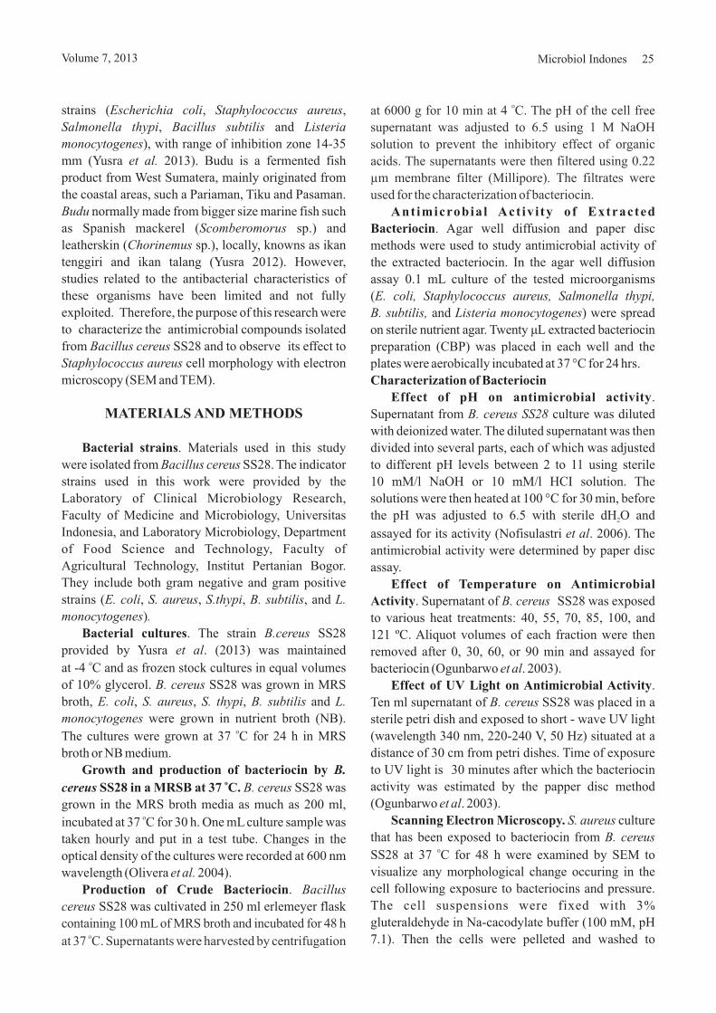
strains ( , ,
, and
), with range of inhibition zone 14-35
mm (Yusra 2013). Budu is a fermented fish
product from West Sumatera, mainly originated from
the coastal areas, such a Pariaman, Tiku and Pasaman.
normally made from bigger size marine fish such
as Spanish mackerel ( sp.) and
leatherskin ( sp.), locally, knowns as ikan
tenggiri and ikan talang (Yusra 2012). However,
studies related to the antibacterial characteristics of
these organisms have been limited and not fully
exploited. Therefore, the purpose of this research were
to characterize the antimicrobial compounds isolated
from SS28 and to observe its effect to
cell morphology with electron
microscopy (SEM and TEM)
. Materials used in this study
were isolated from SS28. The indicator
strains used in this work were provided by the
Laboratory of Clinical Microbiology Research,
Faculty of Medicine and Microbiology, Universitas
Indonesia, and Laboratory Microbiology, Department
of Food Science and Technology, Faculty of
Agricultural Technology, Institut Pertanian Bogor.
They include both gram negative and gram positive
strains ( , , , , and
)
. The strain SS28
provided by Yusra . (2013) was maintained
at -4 C and as frozen stock cultures in equal volumes
of 10% glycerol. SS28 was grown in MRS
broth, , , , and
were grown in nutrient broth (NB).
The cultures were grown at 37 C for 24 h in MRS
broth or NB medium.
SS28 was
grown in the MRS broth media as much as 200 ml,
incubated at 37 C for 30 h. One mL culture sample was
taken hourly and put in a test tube. Changes in the
optical density of the cultures were recorded at 600 nm
wavelength (Olivera 2004).
.
SS28 was cultivated in 250 ml erlemeyer flask
Escherichia coli Staphylococcus aureus
Salmonella thypi Bacillus subtilis Listeria
monocytogenes
et al.
Budu
Scomberomorus
Chorinemus
Bacillus cereus
Staphylococcus aureus
Bacillus cereus
E. coli S. aureus S.thypi B. subtilis L.
monocytogenes .
B.cereus
et al
B. cereus
E. coli S. aureus S. thypi B. subtilis L.
monocytogenes
B. cereus
et al.
Bacillus
cereus
.
MATERIALS AND METHODS
Bacterial strains
Bacterial cultures
Growth and production of bacteriocin by
SS28 in a MRSB at 37 C.
Production of Crude Bacteriocin
o
o
o
B.
cereuso
containing 100 mL of MRS broth and incubated for 48 h
at 37 C. Supernatants were harvested by centrifugationo
at 6000 g for 10 min at 4 C. The pH of the cell free
supernatant was adjusted to 6.5 using 1 M NaOH
solution to prevent the inhibitory effect of organic
acids. The supernatants were then filtered using 0.22
μm membrane filter (Millipore). The filtrates were
used for the characterization of bacteriocin.
o
Antimicrobial Act iv i ty of Extracted
Bacteriocin
Characterization of Bacteriocin
Effect of pH on antimicrobial activity
Effect of Temperature on Antimicrobial
Activity
Effect of UV Light on Antimicrobial Activity
Scanning Electron Microscopy.
. Agar well diffusion and paper disc
methods were used to study antimicrobial activity of
the extracted bacteriocin. In the agar well diffusion
assay 0.1 mL culture of the tested microorganisms
(
and ) were spread
on sterile nutrient agar. Twenty L
placed in each well and the
plates were aerobically incubated at 37 °C for 24 hrs.
.
Supernatant from culture was diluted
with deionized water. The diluted supernatant was then
divided into several parts, each of which was adjusted
to different pH levels between 2 to 11 using sterile
10 mM/l NaOH or 10 mM/l HCI solution. The
solutions were then heated at 100 °C for 30 min, before
the pH was adjusted to 6.5 with sterile dH O and
assayed for its activity (Nofisulastri . 2006). The
antimicrobial activity were determined by paper disc
assay.
. Supernatant of SS28 was exposed
to various heat treatments: 40, 55, 70, 85, 100, and
121 ºC. Aliquot volumes of each fraction were then
removed after 0, 30, 60, or 90 min and assayed for
bacteriocin (Ogunbarwo . 2003).
.
Ten ml supernatant of SS28 was placed in a
sterile petri dish and exposed to short - wave UV light
(wavelength 340 nm, 220-240 V, 50 Hz) situated at a
distance of 30 cm from petri dishes. Time of exposure
to UV light is 30 minutes after which the bacteriocin
activity was estimated by the papper disc method
(Ogunbarwo . 2003).
culture
that has been exposed to bacteriocin from
SS28 at 37 C for 48 h were examined by SEM to
visualize any morphological change occuring in the
cell following exposure to bacteriocins and pressure.
The cell suspensions were fixed with 3%
gluteraldehyde in Na-cacodylate buffer (100 mM, pH
7.1). Then the cells were pelleted and washed to
E. coli, Staphylococcus aureus, Salmonella thypi,
B. subtilis, Listeria monocytogenes
B. cereus SS28
et al
B. cereus
et al
B. cereus
et al
S. aureus
B. cereus
μ extracted bacteriocin
preparation (CBP) was
2
o
Volume 7, 2013 Microbiol Indones 25

remove gluteraldehyde before resuspended in the
same buffer. A drop of each suspension was transferred
to a poly-L-lysine-treated silicon wafer chips that
were kept for 30 min in a hydrated chamber to let the
cells adhere. The attached cells were post fixed by
immersing the chips in 1% osmium tetroxide (OsO ) in
cacodylate buffer for 30 min, then rinsed in the same
buffer and dehydrated in ethanol in ascending
concentrations (%): 50, 70, 95 (2x) and 100 (2x), for 10
min each. The chips were mounted on aluminum
stubs and coated with gold-palladium in a sputter
coater (Emitech K550, Ashford, Kent, England). The
chips were viewed at 3 kV accelerating voltage in a
Hitachi S-4000 field emission scanning electron
microscope (JEM-JEOL JSM-5310LV type) and
secondary electron image of cells for topography
contrast were collected at several magnifications
(Bolshakova . 2004).
The
cell suspensions that has been exposed to bacteriocin
from SS28 at 37 C for 48 h were harvested
by centrifugation and washed twice with 0.1 M
phosphate buffer (pH 7.3). The cells were fixed with
2.5% (v/v) glutaraldehyde, 2.0% (v/v) formaldehyde
in 0.12 M phosphate buffer for 10 days and then
postfixed in 2% (w/v) osmium tetroxide in the same
buffer for 45 min. The samples were dehydrated in a
graded acetone series (30-100%) and embedded in
Araldite-Durcupan for 72 h at 60 °C. Thin sections
4
et al
S. aureus
B.cereus
Transmission electron microscopy.
o
(microtome UPC-20, Leica) were mounted on grids,
covered with collodion film and poststained with 2%
uranyl acetate in Reynold's lead citrate. Its preparation
were observed with transmission electron microscope
tipe JEOL-1010 (Bozzola and Russel 1999, with
modification).
The growth and
bacteriocin production of is
slight increase of cell dry weight was observed for 28 h
of fermentation. During log phase (6 - 22 hours
fermentation), medium pH decreased rapidly. It
occured concurrently with the increase of the cell dry
weigt. The data indicated that the alteration of the
medium pH was inversely proportional with growth of
Ss28.
The effect
of pH on bacteriocin activity was studied. It was
observed that bacteriocin produced by SS28
was stable between pH 2-11 (Fig 2).
The inhibitory activity towards the test isolates was
heat stable (Fig 3). The antimicrobial activity remained
constant after heating at 121 ºC for 15 minutes. The
activity was highest when being heated at 70 ºC for
45 min.
RESULTS
Growth of SS28 and the production of
bacteriocin in MRSB at 37 C.
Effect of pH onAntimicrobialActivity.
Effect of Temperature onAntimicrobialActivity.
B. cereuso
B. cereus Ss28
B. cereus
B. cereus
Fig 1 The growth curve of SS28 isolate on MRS broth mediumBacillus cereus
26 ET AL.YUSRA Microbiol Indones
0
0.05
0.1
0.15
0.2
0.25
0.3
0.35
0.4
0.45
0 5 10 15 20 25 30
Ab
sorb
an(n
m)
Time (h)

Fig 2 Effect of pH on the activitiy of antimictobial compound from SS28 determined based on the size of theinhibition zone (mm)
Bacillus cereus.
Fig 3 Effect of temperature on activity of antimictobial compound from SS28, determined based on the sizeof the inhibition zone (mm)
Bacillus cereus.
Effect of UV Light on antimicrobial activity.
Scanning Electron Microscopy.
Bacteriocin produced by the test isolates was tested
for their sensitivity (loss of activity) to UV light
exposure. The antimicrobial activity was lost or
unstable after exposure to UV-light for 15 and 30 min
(Fig 4).
SEM has been
widely used in microbiology to study the surface
structure of biomaterials and to measure cell
attachment and changes in morphology of bacteria.
The SEM-generated photomicrograph of pathogen
after treatment with antimicrobial
compound from SS28 is presented in
Figure 5.
S. aureus
B. cereus
pH
0
5
10
15
20
25
30
2 3 4 5 6 7 8 9 10 11
Inhib
itio
nZ
one
Siz
e(m
m)
Escherichia coli
Staphylococcus
aureus
Salmonella thypi
Bacillus subtilis
Listeria
monocytogenes
0
5
10
15
20
25
30
35
40 50 70 85 100 121
Inhib
itio
nzo
ne
size
(mm
)
Temperature (°C)
E. coli
S. aureus
S. thypi
B. subtilis
L. monocytogenes
Volume 7, 2013 Microbiol Indones 27

Fig 5 Scanning electron microscopy of cells control (a) (Bajpal . 2009) and after treatment withantimicrobial compound of SS28 (b).
Staphylococcus aureus et alBacillus cereus
Bacteria
0
5
10
15
20In
hib
itio
nzo
ne
size
(mm
)
UV, 15 menit
UV, 30 menit
Fig 4 Effect of uv light on activity of antimictobial compound from SS28, determined based on the size of theinhibition zone (mm)
Bacillus cereus.
A B
Transmission Electron Microscopy. The effect of
antimicrobial compound from SS28 on
bacterial cells was studied using as a
representative of Gram-positive cells. Morphological
investigations were performed using 48-h
culture treated with antimicrobial compound from
SS28 l). Control has exhibited typical
coccus morphology of (Fig 6). Untreated
B. cereus
S. aureus
S. aureus
B.
cereus
S. aureus
(20 μg/m
S.aureus
S. aureus
cells) shows a typically structured nucleus of
and a perfect cell wall (Fig 6A). After 48
hours of exposure to the antimicrobial compound, a
slight alteration can be observed in the cell cytoplasm
(Fig 6B), the cells exhibited notable alteration in
cell cytoplasm. Bacterial cells completely collapsed
48 h after treatment with the antimicrobial compound
(Fig 6).
28 ET AL.YUSRA Microbiol Indones

DISCUSSION
Bacteriocin activity remained stable up to 24 h
fermentation, then the activity started to drop after 28 h
fermentation. Koroleva (1991) stated that most of the
metabolism products resulted in the log phase were in
the form of lactic acids, which causes the decrase in pH
of the medium. This acidic condition will eventually
inhibit growth of the respective bacteria (negative feed
back effect).
Bacteriocin is extracellular secondary metabolite.
The increase in the amount of biomass produced in the
bacterial culture caused the increase in the amount of
the bacteriocin produced. After reaching the stationary
phase, the amount started to decrese (Boe 1996).
Synthesis of bacteriocin by LAB occurred during the
exponential growth phase, usually following the
protein synthesis (Schnell 1998). Torkar and
Matijasic (2003) who did research on the
characterization of bacteriocins produced by
from milk and other dairy products, found that the
production of bacteriocins entered the stationary phase
after 10-16 hours of incubation. The research by
Naclerio . (1993) on the production and activity of
bacteriocins cerein present in the
stationary phase also demonstrated similar result.
The highest antibacterial activity was exhibited at
pH range 2 to 3, while inactivation occurred between
pH 9 to 11. Khalil . (2009) showed that
bacteriocins produced by 22 has
activity antimicrobial against at pH
range 2-8. Naclerio . (1993) who studied the
antimicrobial activity of bacteriocins cerein from
found that the compound's activity was
et al.,
B. cereus
et al
from B. cereus
et al
B. megaterium
S. thypimurium
et al
B. cereus,
Fig 6 Transmission electron microscopy of cells control (A) (Santhana ., (2007) and after treatmentwith antimicrobial compound of SS28 (B).
Staphylococcus aureus et alBacillus cereus
stable between pH 3-12. Growth temperature plays an
important role and is often correlated with bacteriocin
production (Todorov and Dicks 2006).
Similar to the results of Alam . (2011), who
stated that bacteriocin of BS15 retained
activity up to 80 C for 30 min, other bacteriocin
produced by ssp. diacetilactis was reported to
maintain its activity even after boiling for up to 60 min.
On the other hand, Lactacin F was reported to
completely lose the activity when treated at 50 C for 30
min (Kojic . 1991; Kim . 2005). Cleveland
. (2001) suggested several potential advantages of
bacteriocins to serve as biopreservatives, namely: a)
the material is not toxic and susceptible to degradation
by proteolytic enzymes because it is a protein
compound, b) the material does not harm the intestinal
microflora because it is easily digested by
gastrointestinal enzymes, c) the material can reduce the
chemicals as a food preservative, d) flexibility of use,
and e) stability towards sufficiently broad range of pH
and temperature that it is resistant to treatment
processes involving acids and bases, as well as hot and
cold conditions.
Antimicrobial activity of SS28 was the
highest against with inhibition zone diameter
20 mm, after 15 minutes exposure, which decreased to
10 mm after 30 minutes. species and other gram
negative bacteria were sensitive to nisin and other
bacteriocins after exposure to treatments that change
the permeability barrier properties of the outer
membrane (Stevens . 1991). Khalil . (2009)
19 bacteriocin was stable after 15 min
exposure to UV light and was completely destroyed
after 90 min.
et al
B. subtilis
L. lactis
et al et al et
al
B. cereus
S. thypi,
S.
et al et al B.
megaterium
o
o
A B
Volume 7, 2013 Microbiol Indones 29

Bajpai VK, Al-Reza SM, Choi UK, Lee JH, Kang SC. 2009.Chemical composition, antibacterial and antioxidantactivities of leaf essential oil and extracts of
Miki ex Hu. Food and Chem Toxicol.47(8):1876-1883. doi:10.1016/j.fct.2009.04.043.
Bhunia AK, Johnson MC, Ray B. 1987. Direct detection ofan antimicrobial peptide of insodium dodecyl sulfate-polyacrylamide gelelectrophoresis. J Ind Microbiol. 2(5):319-322.doi:10.1007/BF01569434.
Bizani D, Brandelli A. 2002. Characterization of abacteriocin produced by a newly isolated sp.strain 8A. J Appl Microbiol. 93(3):512-519.doi:10.1046/j.1365-2672.2002.01720.x.
Boe, Young J. 1996. Evaluation of optimum for productionfor bacteriocin from sp. JB42 isolationfrom kimichi. J Microbiol Biotechnol. 6(1):63-67.
Bol’shakova AV, Golutvin IA, Nasikan NS, Yaminskii V.2004. Determination of mechanical characteristics ofsurface of block copolymers by atomic force microscopytechniques. Polymer Scie SerA. 46(9):926-932.
Bozzola JJ, Russell LD. 1999. Ultramicrotomy e,
2nd edition. Sudbury, Massachusetts, Jones & Bartlett.
Brotz H, Bierbaum G, Leopold K, Reynolds PE, Sahl HG.1998. The lantibiotic mesarcidin inhibits peptidoglycansynthesis by targeting lipid II. Antimicrob AgentsChemother. 42(1):154-160.
Cherif A, Quazri H, Daffonchio D, Cherif H, Siama BK,Hassen A, Japua S and Boudabous A. 2001. Thurin 7: Anovel bacteriocin produced byBMG 1.7, a new strain isolated from soil. Lett ApplMicrobiol. 32(4):2432-2247. doi:10.1046/j.1472-765X.2001.00898.x.
Cleveland J, Monteville TJ, Nes IF, Chikindas MI. 2001.Bacteriocins: safe, natural antimicrobials for foodpreservation. Int J Food Microbiol. 71(1):1-20.doi:10.1016/S0168-1605(01)00560-8.
Dalmau M, Maier E, Mulet N, Vinas M, Benz R. 2002.Bacterial membrane injuries induced by lactacin F andnisin. Int Microbiol J. 5(2):73-80. doi:10.1007/s10123-002-0063-2.
Diop MB, Dauphin RD, Tine E, Ngom A, Thonart DJ,Philippe T. 2007. Bacteriocin producers fromtraditional food products. Biotechnol Agron SocEnviron. 11(4):275-281.
Hartmann M, Berditsch M, Hawecker J, Ardakani MF,Gerthsen D, UlrichAS. 2010. Damage of the bacterial cellenvelope by antimicrobial peptides gramicidin S andPGLa as levealed by transmission and scanning electronmicroscopy. Antimicrob Agents Chemother. 54(8):3132-3142. doi:10.1128/AAC. 00124-10.
Jack RW, Wan J, Gordon J, Harmark K, Davidson BE,HillierAJ, Wettenhall REH, Hickey MW, Coventry MJ.1996. Characterisation of the chemical andantimicrobial properties of piscicolin 126, a bacteriocinproduced by JG 126. J ApplEnviron Microbiol. 62(8):2897-2903.
Metasequioaglyptostroboides
Pediococcus acidilactici
Bacillus
Lactobacillus
lectronmicroscopy: principles and techniques for biologists
Bacillus thuringenesis
Carnobacterium piscicola
The effect of antimicrobial compound from
supernatant SS28 from wall and cell
membrane was investigated. It could be associated
with the damage in the cell wall and cell membrane and
subsequent lysis and reduction. Immediately after
treatment, 80% of the cell's surface appeared
rough, which is quite different from the normal cells. In
a previous study with ,
which has an inducible autolytic enzyme, bacteriocin
treatment, pressurization or their combination did not
only produce cell death and cell lysis, but also triggered
the autolytic enzyme, which, by hydrolyzing the wall,
disintegrated the cells (Bhunia . 1987;
Kalchayanand . 2002).
Electron microscopy showed cell lysis after
treatment with antimicrobial compound of
SS28. The cell damage caused by antimicrobial
compound resembles that observed with a crude
bacteriocin treatment (Ocana . 1999). Bizani .
(2005) tried to truestigate the effect of cerein 8A
against spore. An approximately 4-5
log reduction was observed when spores were plated
in PCA containing 800 AU ml . As cerein 8A
concentration increased to 1600 AU ml , complete
inhibition of colony development was observed. When
spores were treated with cerein 8A in BHI broth before
plating, similar results were observed. The bactericidal
effect of the antimicrobial compound from
SS28 apparently works by disrupting the
membrane function of target organisms.
To conclude antimicrobial bacteriocin from
was stable over a broad range of pH
(between pH 2 to 11) and to heat-treatment at 121 C for
15 min. The antimicrobial activity was the highest at
being heated at 70 C for 45 min and for 15 min of
exposure to UV light. The main changes observed
under SEM and TEM analyses were structural
disorganization of the cellular membrane
48 h after exposure to the antimicrobial compound of
SS28.
B. cereus
S. aureus
Layconostoc mesenteroides
et al
et al
B. cereus
et al et al
Bacillus cereus
Bacillus
cereus
B.cereus SS28
S. aureus
B. cereus
10
-1
-1
o
o
REFERENCES
Adebayo CO, Aderiye BI. 2010. Antifungal activity ofbacteriocins of lactic acid bacteria from some Nigerianfermented foods. 5(11):1070-1082. doi:10.3923/jm.2010.1070.1082.
lam SI, Kamran M, Sohail M, Ahmad A, Khan SH. 2011.Partial characterization of bacteriocin like inhibitorysubstance from BS15, a local soilisolate. Pakistan J Bot. 43(4):2195-2199.
Res Journal Microbiol.
Bacillus subtilis
A
30 ET AL.YUSRA Microbiol Indones

Kalchayanand N, Frethem C, Dunne P, SikesA, Ray B. 2002.Hydrostatic pressure and bacteriocin triggered cell walllysis of . Innovative FoodScie and Emer Technolo. 3(1):33-40. doi:10.1016/S1466-8564(02)00004-8
Khalil R, Yaser E, Fatima D, Sanaa O. 2009. Isolation andpartial characterization of a bacteriocin produced by anewly isolated 19 strain.Pakistan J of Nutr. 8(3):242-250. doi:10.3923/pjn.2009.242.250.
Kim MH, Kong YJ, Baek H, Hyun HH. 2005. Production,purification, and characterization of micrococcin GO5,a bacteriocin produced by sp. GO5 isolatedfrom kimchi. J of Food Prot. 68(1):157-163
Kojic M, Svircevic J, Banina A and Topisirovic L. 1991.Bacteriocin producing strain ofsubsp. diacitilactis S50. Appl Environ Microbiol.57(6):1835-1837.
Koroleva NS. 1991. Products prepared with lactic acidbacteria and yeasts. R.K. Robinsn (Ed.).Therapeutics Properties of Fermented Milks. ElsevierApplied Science, London and NewYork.
Lisboa MP, Bonatto D, Bizani D, Henriques JAP andBrandelli A. 2006. Characterization of a bacteriocin-like substance produced byisolated from the Brazilian Atlantic forest. IntMicrobiol. 9(2):111-118.
Mataragas M, Melaxopoulous J and Drosinos EA. 2002.Characterization of two bacteriocins produced by
L124 andL442, isolated from dry fermented sausages.
World J Microbiol Biotechnol. 18(9):847-856.doi:10.1023/A:1021239008582.
Moreno I, Lerayer ALS, Baldini VLS, Leitao MFF. 2000.Characterization of bacteriocins produced by
strains. Braz J Microbiol. 31(3):183-191. doi:10.1590/S1517-83822000000300007.
Morgan R. 1989. UV: green. light disinfection. DairyIndustry Int. 54(11):33-35.
Naclerio G, Ricca E, Sacco M, De Felice M. 1993.Antimicrobial activity of a newly identified bacteriocinof . J Appl Environ Microbiol.59(12):4313-4316.
Nofisulastri, Bachruddin Z, Harmayani E. 2006. Productionand extraction of antibacterial bacteriocin from
NWD 015. Indones J Biotechnol.11(2):921-927..
Ocana VS, Aída A, de Ruiz Holgado P and Macías NME.1999. Characterization of a bacteriocin-like substanceproduced by a vaginal train.
65(12):5631-5635.
Ogunbarwo ST, Sanni AI. and Onilude AA. 2003.Characterization of bacteriocin produced by
F1 andOG1.Afr J Biotechnol. 2 (8):219-227.
Olivera FC, Caron GR, Brandelli A. 2004. Bacteriocinproduction by strain P40 in
Leuconostoc mesenteroides
Bacillus meganterium
Micrococus
Lactococcus lactis
In
Bacillus amyloliquefaciens
Leuconostoc mesenterioides Lactobacilluscurvatus
Lactococcus lactis
Bacillus cereus
Pediococcus sp.
Lactobacillus salivarius sAppl Environ Microbiol.
Lactobacillus plantarum Lactobacillus brevis
Bacillus licheniformis
.
cheese whey using response surface methodology.Biochem Eng J. 21(1):53-58. doi:10.1016/j.bej.2004.05.002.
Oscariz JC and Pisabarro AG. 2000. Characterisation andmechanism action of cerein 7, a bacteriocin producedby Bc7. J Appl Microbiol. 89(2):1-10.doi:10.1046/j.1365-2672.2000.01123.x.
Santhana RL, Hing HL, Omar B, Hamidah ZMT, Aida SR,Nor ACP, Vimala B, Paramsarvaran S, Sumarni G andHanjeet K. 2007. Rapid method for transmissionelectron microscope study of Staphylococcus aureusATCC 25923.Annals Microscopy. 7:102-108.
Schnell N, Entian KD, Schneider U, Gots F, Zahner H,Kellner R and Jung G. 1998. Prepeptida sequence ofepidermin, a ribosomally synthesized antibiotic withfour sulphide-ring. Nat London. 333:276-278.doi:10.1038/333276a0.
Shape DV. 2009. Biopreservation of fresh-cut salads usingbacteriocinogenic lactic acid bacteria isolated fromcommercial produce.M.Sc (thesis). Collected in theAAFC, NS and Dalhousie University, Halifax. Canada.
Sharma N, Kapoor G, Gautam N, Neopaney B. 2009.Characterization of a partially purified bacteriocin of
sp MTCC 43 isolated from rhizosphere ofRadish ( ) and its application as apotential food biopreservative. J Sci Industrial Res.68(10):881-886.
Stein T, Brochert S, Conrad B, Feesche J, Hofemeister B andHofemeister J. 2002. Two different lantibiotic-likepeptides originate from the ericin gene cluster of
A1/3. J Bacteriol. 184(6):1703-1711.doi:10.1128/JB.184.6.1703-1711.2002.
Stevens KA, Sheldon BW, Klapes NA, Klaenhammer TR.1991. Nisin treatment for inactivation ofspecies and other gram-negative bacteria. J ApplEnviron Microbiol. 57(12):3613-3615.
Sumner SS, Wallner-Pendleton EA, Froning GW, StetsonLE. 1995. Inhibition of onagar medium and poultry skin by ultraviolet energy. JFood Prot. 59(3):319-321.
Todorov SD and Dicks LMT. 2006. Screening ofbacteriocin-producing lactic acid bacteria from boza, atraditional cereal beverage from Bulgaria. Comparisonof bacteriocins. Proc Biochemi. 41(1):11-19.doi:10.1016/j.procbio.2005.01.026.
Torkar KG, Matijasic BB. 2003. Partial characterisation ofbacteriocins produced by isolates frommilk and milk products. Food Technol Biotechnol.41(2):121-129.
Yang R, Johnson MC, Ray B. 1992. Novel method to extractlarge amounts of bacteriocins from lactic lacticbacteria. JAppl Environ Microbiol. 58(10):3355-3359.
Yusra. 2012. Isolation and Identification of lactic acidbacteria from budu, fish fermented product Spanishmackerel ). FundamentalsGrant Final Report. DP2M Higher Education. BungHatta University, Padang.
Bacillus cereus
BacillusRaphanus sativus
Bacillus subtilis
Salmonella
Salmonella typhimurium
Bacillus cereus
(Scomberomorus guttatus
Volume 7, 2013 Microbiol Indones 31

Zheng GS. 1999. Isolation, partial purification andcharacterization of a bacteriocin produced by a newlyisolated strain. Lett Appl Microbiol.28(5):363-367. doi:10.1046/j.1365-2672.1999.00545.x.
Bacillus subtilis
Yusra, Azima F, Novelina, Periadnadi. 2013. Antimicrobialactivity of lactic acid bacteria isolated fromof West Sumatra to food biopreservatives. PakistanJ of Nutr. 12(7):628-635. doi:10.3923/pjn. 2013.628.635.
budu
32 ET AL.YUSRA Microbiol Indones