american thoracic society documents · 2018-05-02 · the equipment, personnel, competencies, and...
TRANSCRIPT

AMERICAN THORACIC SOCIETYDOCUMENTS
Official American Thoracic Society Technical Standards: FlexibleAirway Endoscopy in ChildrenAlbert Faro, Robert E. Wood, Michael S. Schechter, Albin B. Leong, Eric Wittkugel, Kathy Abode, James F. Chmiel,Cori Daines, Stephanie Davis, Ernst Eber, Charles Huddleston, Todd Kilbaugh, Geoffrey Kurland, Fabio Midulla, David Molter,Gregory S. Montgomery, George Retsch-Bogart, Michael J. Rutter, Gary Visner, Stephen A. Walczak, Thomas W. Ferkol,and Peter H. Michelson; on behalf of the American Thoracic SocietyAd HocCommittee on Flexible Airway Endoscopy in Children
THESE OFFICIAL TECHNICAL STANDARDS OF THE AMERICAN THORACIC SOCIETY (ATS) WERE APPROVED BY THE ATS BOARD OF DIRECTORS, JANUARY 2015
Background: Flexible airway endoscopy (FAE) is an accepted andfrequently performed procedure in the evaluation of children withknown or suspected airway and lung parenchymal disorders.However, published technical standards on how to perform FAE inchildren are lacking.
Methods: The American Thoracic Society (ATS) approved theformation of a multidisciplinary committee to delineate technicalstandards for performing FAE in children. The committee completeda pragmatic synthesis of the evidence and used the evidence synthesisto answer clinically relevant questions.
Results: There is a paucity of randomized controlled trials inpediatric FAE. The committee developed recommendations basedpredominantly on the collective clinical experience of our committeemembers highlighting the importance of FAE-specific airwaymanagement techniques and anesthesia, establishing suggestedcompetencies for the bronchoscopist in training, and defining areasdeserving further investigation.
Conclusions: These ATS-sponsored technical standards describethe equipment, personnel, competencies, and special proceduresassociated with FAE in children.
ContentsOverview
ConclusionsMethodsEquipment and Procedural SettingInfection ControlTrainingCommon Reasons forPerforming Flexible AirwayEndoscopy
Preprocedure EvaluationSedation and MonitoringAirway Management andExamination
The Role of the RigidBronchoscope
BALPerformance
ProcessingDiagnostic Utility of BAL
InfectionBAL in CFBAL in the Diagnosis ofPulmonary Aspiration
BAL in Other DisordersSpecialized ProceduresAtelectasisDocumentationConclusions and Future Directions
Overview
To update the American Thoracic Society(ATS) Official Statement on Flexible
Endoscopy of the Pediatric Airway, the ATSsponsored the development of technicalstandards for the performance of pediatricflexible airway endoscopy (FAE). To completethis effort, an international, multidisciplinarycommittee comprehensively reviewed theliterature and developed this report, includingan online supplement in which we describeselect topics in more detail.
Conclusions
d Equipment and settingB The number and type of
bronchoscopes required at anyindividual institution is determined byeach institution’s understanding of theanticipated number of procedures.
These technical standards were endorsed by the American Academy of Pediatrics, February 2015.
Correspondence and requests for reprints should be addressed to Albert Faro, M.D., Washington University in St. Louis, Campus Box 8116, St. Louis, MO63110. E-mail: [email protected]
This article has an online supplement, which is accessible from this issue’s table of contents at www.atsjournals.org
Am J Respir Crit Care Med Vol 191, Iss 9, pp 1066–1080, May 1, 2015
Copyright © 2015 by the American Thoracic Society
DOI: 10.1164/rccm.201503-0474ST
Internet address: www.atsjournals.org
1066 American Journal of Respiratory and Critical Care Medicine Volume 191 Number 9 | May 1 2015

This is based on the characteristics ofits patient population and typicalindications for FAE. In addition, thisnumber should reflect the facility’sability to clean and disinfect theequipment in a timely fashion.
B The appropriate setting for FAE isdetermined by the patient’s clinicalcondition, facilitates patient safety,allows completion of necessaryprocedures, and provides adequatespace to accommodate equipmentand all necessary personnel.
d Infection controlB The minimal acceptable standard for
reprocessing a flexible bronchoscope ismeticulous manual cleaning followedby high-level disinfection.
B Established guidelines andmanufacturer’s recommendations forinspection, maintenance, storage,cleaning, and manual or automatedreprocessing of flexible bronchoscopesshould be strictly followed.
B Personnel responsible for reprocessingthe flexible bronchoscope shouldreceive appropriate training, includinginitial and annual competency testing.
B Personal protective equipmentshould be used during theprocedure, when handling usedflexible bronchoscopes, andthroughout the cleaning anddisinfection process.
B Institutional protocols shouldinclude the maintenance of aprocedure log as well as a meansfor identifying appropriatelydisinfected bronchoscopes.
d TrainingB Definition of a core set of
demonstrable competencies isrecommended includingsubsequent monitoring anddocumentation of trainee progress.
d Situations where FAE is commonlyperformedB The primary reason for performing
FAE is when, based on the availableclinical data, the need for informationfrom or intervention within the lungsor airways is most safely, effectively,and easily achieved by FAE.
d Preprocedure evaluationB A standardized preprocedural
evaluation should includerecognition of preexistingconditions that may affect theoutcome of FAE.
B All care providers involved in theprocedure should review and agreeupon the procedural plan, theequipment needed, and theappropriate infection controlmeasures required before theprocedure. This may be bestaccomplished in a formalized “TimeOut” process, where individualpatient information is also reviewed.
B Informed consent proceduresshould be followed andappropriately documented in themedical record in accordance withlocal and/or national guidelines.
d Sedation and airway managementB The goals of sedation for FAE depend
on clinical considerations and should(1) provide patient comfort, (2)maintain hemodynamic stability, (3)maintain adequate gas exchange, and(4) provide satisfactory conditions fortherapeutic or diagnostic FAE.
B Collaboration between the endoscopistand the anesthesia or sedationprovider is essential to optimize theinterplay between anesthetic depth,airway management, and accuratediagnosis by FAE.
d Airway examinationB Proximal airway anatomy and airway
dynamics cannot always beaccurately evaluated when FAE isperformed via a laryngeal maskairway or via an endotrachealtube. Nasal passage of the flexibleendoscope is preferred whenupper airway dynamics are to beevaluated.
B Rigid bronchoscopy servesa complementary role with FAE and,depending on the indication, may alsoneed to be performed to adequatelyassess the pediatric airway.
d Bronchoalveolar lavageB Bronchoalveolar lavage (BAL)
is an essential technique toidentify microbiologic or cellularabnormalities of the airway thatmay establish a diagnosis and guideappropriate therapies.
B The optimal manner of performingBAL has not been systematicallystudied and requires furtherinvestigation.
B The interpretation of certainmarkers found in BAL fluidremains uncertain and is an areafor future inquiry.
d Specialized proceduresB Bronchoscopic intubation, biopsies,
airway dilatation, airway stenting,and removal of plugs and clots canall be performed via FAE inchildren in the appropriate settingand for the appropriate indication.
Flexible airway endoscopy (FAE) in youngchildren was first described in 1978 (1),and the technique is now widely usedin the assessment and treatment ofinfants and children with a variety ofpediatric respiratory disease. The flexiblebronchoscope allows for functional andanatomical examination of the upper andlower airways. FAE and specializedprocedures, including bronchoalveolarlavage (BAL), are particularly important inthe diagnosis and treatment of specificrespiratory problems, including congenitalor acquired airway anomalies, persistentor recurrent pulmonary infiltrates,community-acquired or ventilator-associated pneumonia, pulmonaryinfections in immunocompromised hosts,and pulmonary hemorrhage.
The current American Thoracic Society(ATS) guidelines for FAE in children werepublished over 20 years ago (2), andadvances in care have rendered theseguidelines insufficient. The field haswitnessed expansion in the number and typesof procedures performed, and technologicalevolution in available instruments, increaseduse of general anesthesia with concomitantchanges in airway management, and growthin associated applications of FAE haveenhanced the diagnostic and therapeuticcapability of the procedure.
Methods
The ATS Pediatric Planning Committeeappointed a chair and formed a committeeof international experts comprising multipledisciplines to write technical standards.Potential conflicts of interest of ourcommittee members were disclosed, vetted,and strictly managed according to thepolicies of the ATS. The committee wascharged with reviewing the existingliterature on pediatric FAE and presentingrecommendations for technologystandardization or elucidating areas in needof further study. An ATS TechnicalStandards is a document that describes howto perform a procedure. It is not intended tobe a systematic review of the literature.
AMERICAN THORACIC SOCIETY DOCUMENTS
American Thoracic Society Documents 1067
These Technical Standards are not intendedto impose a standard of care but ratherto provide a reasonable approach toperforming FAE in children. Variations,taking into account individualcircumstances, may be appropriate. Theserecommendations should not be viewedas dictates by care providers, third-partypayers, institutional review committees,the court system, or other potentialstakeholders.
Topics were assigned, andsubcommittees were formed. Eachsubcommittee performed a pragmaticevidence synthesis of the literature usingPubMed to search Medline for relevantpublications in the English language from1970 to March 2013. Presentations weremade to the committee during a full-dayworkshop held before the ATS conference inMay 2012 in San Francisco, California. Afterdiscussion, determinations were made bythe committee regarding specific topics to beaddressed in the technical standards, andassignments were made to prepare drafts foreach of the topics. These were submitted tothe chair who then integrated each of thesubsections into a uniform manuscript. Thecommittee reviewed the draft, and furtherdiscussion occurred at a full-day workshopat the 2013 ATS International Conference inPhiladelphia, Pennsylvania and duringa subsequent teleconference.
Equipment and ProceduralSetting
Currently available pediatric flexiblebronchoscopes range in outer diameter(OD) sizes from 2.2 to 6.3 mm, and internalchannel sizes range from 1.2 to 3.2 mm indiameter. The 2.2-mm bronchoscope lacksa suction channel and is therefore limited inits utility. The appropriately sized flexiblebronchoscope must be available to optimizethe examination of any specific patient. Thevast majority of pediatric programs wouldbenefit from possessing a 2.7- or 2.8-mmOD bronchoscope because they are thesmallest with a suction channel. In addition,possession of a 4.4- or 4.9-mm ODbronchoscope provides the bronchoscopistwith the benefit, when appropriate, of thelarger 2.0-mm suction channel. It should benoted that the nominal outer diameter is notalways accurate (3). Institutions mustconsider the age of their patient population,number and types of procedures
performed, and the time needed forbronchoscope cleaning and high-leveldisinfection in determining the appropriatecomplement of instruments to haveavailable.
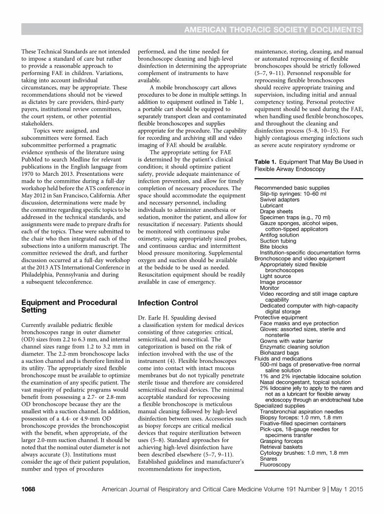
A mobile bronchoscopy cart allowsprocedures to be done in multiple settings. Inaddition to equipment outlined in Table 1,a portable cart should be equipped toseparately transport clean and contaminatedflexible bronchoscopes and suppliesappropriate for the procedure. The capabilityfor recording and archiving still and videoimaging of FAE should be available.
The appropriate setting for FAEis determined by the patient’s clinicalcondition; it should optimize patientsafety, provide adequate maintenance ofinfection prevention, and allow for timelycompletion of necessary procedures. Thespace should accommodate the equipmentand necessary personnel, includingindividuals to administer anesthesia orsedation, monitor the patient, and allow forresuscitation if necessary. Patients shouldbe monitored with continuous pulseoximetry, using appropriately sized probes,and continuous cardiac and intermittentblood pressure monitoring. Supplementaloxygen and suction should be availableat the bedside to be used as needed.Resuscitation equipment should be readilyavailable in case of emergency.
Infection Control
Dr. Earle H. Spaulding deviseda classification system for medical devicesconsisting of three categories: critical,semicritical, and noncritical. Thecategorization is based on the risk ofinfection involved with the use of theinstrument (4). Flexible bronchoscopescome into contact with intact mucousmembranes but do not typically penetratesterile tissue and therefore are consideredsemicritical medical devices. The minimalacceptable standard for reprocessinga flexible bronchoscope is meticulousmanual cleaning followed by high-leveldisinfection between uses. Accessories suchas biopsy forceps are critical medicaldevices that require sterilization betweenuses (5–8). Standard approaches forachieving high-level disinfection havebeen described elsewhere (5–7, 9–11).Established guidelines and manufacturer’srecommendations for inspection,
maintenance, storing, cleaning, and manualor automated reprocessing of flexiblebronchoscopes should be strictly followed(5–7, 9–11). Personnel responsible forreprocessing flexible bronchoscopesshould receive appropriate training andsupervision, including initial and annualcompetency testing. Personal protectiveequipment should be used during the FAE,when handling used flexible bronchoscopes,and throughout the cleaning anddisinfection process (5–8, 10–15). Forhighly contagious emerging infections suchas severe acute respiratory syndrome or
Table 1. Equipment That May Be Used inFlexible Airway Endoscopy
Recommended basic suppliesSlip-tip syringes: 10–60 mlSwivel adaptersLubricantDrape sheetsSpecimen traps (e.g., 70 ml)Gauze sponges, alcohol wipes,cotton-tipped applicators
Antifog solutionSuction tubingBite blocksInstitution-specific documentation forms
Bronchoscope and video equipmentAppropriately sized flexiblebronchoscopes
Light sourceImage processorMonitorVideo recording and still image capturecapability
Dedicated computer with high-capacitydigital storage
Protective equipmentFace masks and eye protectionGloves: assorted sizes, sterile andnonsterile
Gowns with water barrierEnzymatic cleaning solutionBiohazard bags
Fluids and medications500-ml bags of preservative-free normalsaline solution
1% and 2% injectable lidocaine solutionNasal decongestant, topical solution2% lidocaine jelly to apply to the nares andnot as a lubricant for flexible airwayendoscopy through an endotracheal tube
Specialized suppliesTransbronchial aspiration needlesBiopsy forceps: 1.0 mm, 1.8 mmFixative-filled specimen containersPick-ups, 18-gauge needles forspecimens transfer
Grasping forcepsRetrieval basketsCytology brushes: 1.0 mm, 1.8 mmSnaresFluoroscopy
AMERICAN THORACIC SOCIETY DOCUMENTS
1068 American Journal of Respiratory and Critical Care Medicine Volume 191 Number 9 | May 1 2015

avian influenza, a power air-purifyingrespirator hood should be used during theprocedure (11).
Institutional protocols should be inplace to identify appropriately disinfectedflexible bronchoscopes. A record should bemaintained for each procedure to assist inoutbreak investigation. Such a log shouldinclude patient identification information,date of the procedure, and a unique identifierof the flexible bronchoscope used (13, 16).The log can also serve as documentation forregulatory bodies during their site visits.Infection-control professionals shouldensure that institutional policies areconsistent with national guidelines andadvocate for policy compliance (8, 12, 15).
Random bacterial surveillance culturesof processed flexible bronchoscopes or rinsewater have been proposed to ensure effectivedisinfection. However, a correlationbetween bacterial counts from a flexiblebronchoscope and infection after a pediatricFAE has not been established (8, 17–20).
Training
No evidence-based method has beenestablished to assess competency inpediatric FAE. In 2003, the AmericanCollege of Chest Physicians publishedguidelines on interventional proceduresbased solely on expert opinion (21).
Eighty-six percent of United Statespediatric pulmonology training directorssurveyed agreed that a minimum number ofFAEs could be used to define competencyduring training, with the median minimumbeing the completion of 50 supervisedprocedures (22).
In adult pulmonology, multisocietyrecommendations have identified anddefined (23, 24) specific competencies inFAE. Although there are no universallyaccepted tools for assessment ofcompetency in FAE, studies in adultpulmonology have shown that tools suchas core knowledge, including onlinecurriculum and structured simulationtraining with objective assessmentinstruments, may expedite and facilitateFAE training. Suggested core competenciesfor pediatric bronchoscopists areoutlined in Table 2. Rather than using anarbitrary number of procedures to definecompetency, these suggested corecompetencies should form the basis toassess FAE skills.
The roles assumed by pediatric FAEassistants may depend on their professionalbackground; specialized education; clinicalknowledge and experiences; institutional,state, and national scope of practice; andpractice setting. Table 3 details the role ofthe FAE assistant.
Common Reasons forPerforming Flexible AirwayEndoscopy
The reasons for performing diagnostic ortherapeutic FAE vary with clinicalpresentation (Table 4). The primary
indication for FAE is when, based on theavailable clinical data, the information fromor intervention within the lungs or airwaysis most safely, effectively, and easilyobtained by FAE (9).
The only absolute contraindication toFAE is refusal of the parent or guardian toprovide informed consent; even then, ifthe procedure is necessary, the healthcareprovider can seek a court order. The risksand benefits must be considered individuallyfor each patient. When performance ofthe study is necessary, despite coagulopathy,pulmonary hypertension, cardiovascularinstability, and severe hypoxemia orrespiratory failure, appropriate
Table 2. Core Competencies: Pediatric Flexible Airway Endoscopist
1. Understanding indications and contraindications for FAE2. Obtaining appropriate informed consent3. Safetya. Patient safety
i. Monitoring and ability to respond to abnormalitiesb. Provider safety
4. Use the principles of infection control5. Ability to perform FAE through the nasopharynx6. Identify normal upper airway anatomy and function including the nasopharynx7. Identify normal lower airway anatomy and functiona. Identify and enter segmental bronchi
8. Ability to keep the flexible bronchoscope centered and avoid excessive airway wall traumaa. Ability to identify secretions (i.e., clear, frothy, mucoid, purulent, bloody)
9. The ability to recognize abnormalities, includinga. Nasal polypsb. Laryngomalaciac. Laryngeal cleftd. Adenoid and/or tonsillar hypertrophye. Pharyngeal collapsef. Tongue-base obstructiong. Vocal cord paralysis/paresish. Subglottic stenosisi. Subglottic edemaj. Subglottic or supraglottic hemangiomak. Laryngeal or tracheal webl. Laryngeal papillomam. Laryngeal cystn. Tracheal stenosiso. Complete tracheal ringsp. Tracheomalaciaq. Bronchomalaciar. Bronchial stenosiss. Airway compressiont. Mass lesionu. Foreign bodyv. Mucus plugw. Airway granulomax. Tracheoesophageal fistulay. Tracheal bronchus
10. Bronchoalveolar lavage11. Supplementary skills (i.e., therapeutic lavage, bronchoscopic intubation)12. FAE through a laryngeal mask airway and endotracheal tube13. Complications and management14. Anesthesia effectsa. Lidocaine toxicity
15. Cleaning and disinfection
Definition of abbreviation: FAE = flexible airway endoscopy.
AMERICAN THORACIC SOCIETY DOCUMENTS
American Thoracic Society Documents 1069

interventions may be incorporated toallow for the procedure to be safelycompleted (25).
Preprocedure Evaluation
Before FAE, a systematic preoperativeevaluation is essential to reduce the risk ofprocedural complications. Medical historytaking should focus on the nature ofthe suspected underlying pathology.Comorbid conditions that may affect theadministration of anesthesia or sedation,complicate the procedure, or prolongpostoperative recovery should be identifiedand when possible optimized. Particularly incases where dynamic airway abnormalitiesare suspected, the bronchoscopist shoulddetermine the state in which the pathology ismost prominent. This information willdetermine patient positioning, depthof anesthesia, or even the need for anexercise-associated FAE. The history andphysical examination performed by thebronchoscopist should be separate from thatof the anesthesiologist or sedating physician.Implicit in this recommendation is that
when performing FAE in children, theresponsibilities of sedating and monitoringthe patient should be separate from theresponsibility of performing the endoscopy.This division of labor between two trainedindividuals increases the likelihood of a safeoutcome that yields useful information.
Preexisting conditions essential torecognize include hemodynamic instability,severe or uncontrolled pulmonaryhypertension, profound upper or centralairway obstruction (26), immunodeficiency(27), infectious risk to the team (suchas tuberculosis), severe bronchialhyperresponsiveness, and/or uncorrectedbleeding diathesis (28).
Preoperative medications should beconsidered during the preprocedureevaluation. Oral, nasal, or intravenoussedation may be used as adjunctive therapyin select patients with significant anxiety.The bronchoscopist may considersuspending or withholding antibioticsbefore the procedure to maximize thediagnostic yield of BAL. Patients witha history of bronchial hyperresponsivenessmay benefit from an inhaled short-actingb-agonist immediately before the FAE (28).
According to American Heart Associationguidelines, FAE (with or without BAL)does not require routine endocarditisprophylaxis (29). However, prophylaxis inat-risk patients may be considered whentransbronchial or endobronchial biopsyis planned. In general, no routinepreprocedure laboratory assessments areabsolutely required before FAE unlessbiopsy is planned (30). However, review ofavailable pertinent radiographic images ismandatory.
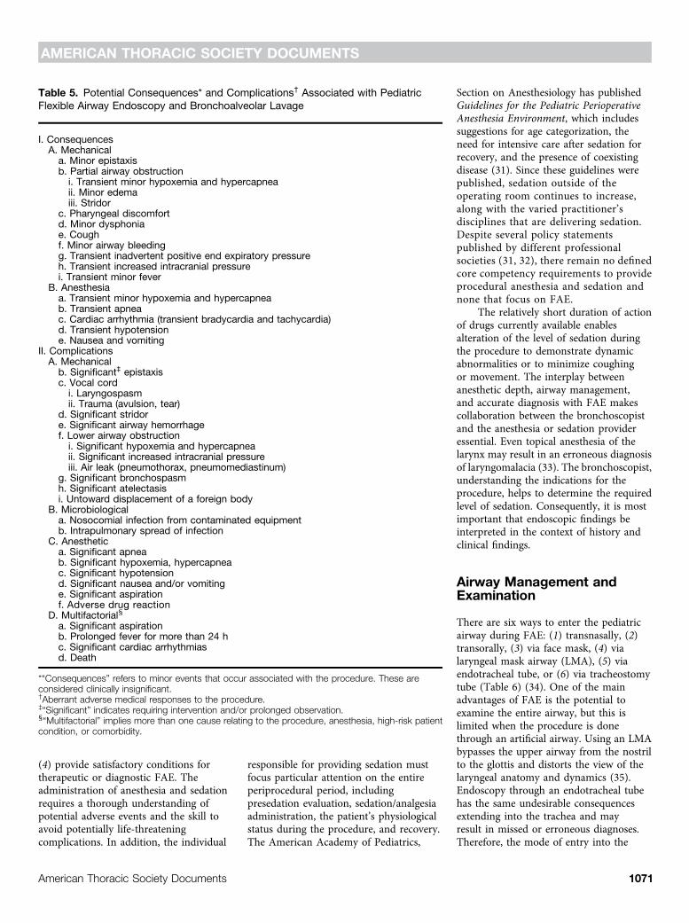
In accord with local and nationalguidelines, appropriate informed consentprocedures should be followed andadequately documented in the medicalrecord. Discussion of potential proceduralrisks should be tailored to the individualpatient and setting (Table 5). Before FAE,all care providers involved in the procedureshould review and agree upon theprocedural plan, equipment required, andindicated infection control measures. Thismay be best accomplished in a formalized“Time Out” process where individualpatient information, such as drug allergies,is also reviewed.
Sedation and Monitoring
The goals of sedation for FAE depend onclinical considerations and include methodsthat (1) provide patient comfort, (2)maintain hemodynamic stability, (3)maintain adequate gas exchange, and
Table 3. The Role of the Assistant(s) in Pediatric FAE
Patient cared Systematically assessing the health status of patients and documenting related healthdata
d Assisting with patient preparation and positioning during sedation or anesthesiainduction
d Administering medications, depending on the expertise and scope of practice of theindividual assistant, and evaluating pharmacological and other therapies mandated bythe particular situation and evidence-based practice
d Responding appropriately to emergency situations to promote optimal patient outcomesby recognizing changes in the patient’s health status
d Documenting patient-related data to ensure continuity in the provision and coordinationof patient care
d Collaborating with other health care professionals to ensure quality and continuity ofcare
Equipment/suppliesd Maintaining familiarity with operation of each FB and all equipment and suppliesd Assessing FAE equipment to maintain good working order, cleanliness, and safetyd Maintaining supplies at levels adequate to safely and effectively perform FAEd Establishing effective communication with equipment manufacturers’ trained technicalpersonnel to facilitate rapid access when troubleshooting problems
d Using training and annual competency checklists to assess proficiency in FBreprocessing
d Using a log or tracking system to match the FB to the patient, bronchoscopist, andmethod (manual or automated system) used to reprocess the FB
Specimensd Assisting with obtaining BAL, brush, and biopsy specimens as directed by thebronchoscopist
d Handling, labeling, and transferring specimens with appropriate documentation todesignated laboratories to ensure appropriate diagnostic testing
Definition of abbreviations: BAL = bronchoalveolar lavage; FAE = flexible airway endoscopy; FB =flexible bronchoscope.The role of the assistant(s) may include these items but is not limited to them.
Table 4. Common Reasons for FlexibleAirway Endoscopy in Children
IndicationDiagnosticUnexplained stridorUnexplained wheezeChronic coughRecurrent pneumoniaMicrobiologic samplingSuspected aspirationSuspected structural anomaliesSuspected endobronchial lesionObstructive sleep apneaRadiographic abnormalityHemoptysis and pulmonary hemorrhageMonitoring of lung allograftMonitoring of the artificial airway
TherapeuticTreatment of persistent atelectasisControl hemorrhageBronchoscopic intubationDilation of a stenotic airway
AMERICAN THORACIC SOCIETY DOCUMENTS
1070 American Journal of Respiratory and Critical Care Medicine Volume 191 Number 9 | May 1 2015
(4) provide satisfactory conditions fortherapeutic or diagnostic FAE. Theadministration of anesthesia and sedationrequires a thorough understanding ofpotential adverse events and the skill toavoid potentially life-threateningcomplications. In addition, the individual
responsible for providing sedation mustfocus particular attention on the entireperiprocedural period, includingpresedation evaluation, sedation/analgesiaadministration, the patient’s physiologicalstatus during the procedure, and recovery.The American Academy of Pediatrics,
Section on Anesthesiology has publishedGuidelines for the Pediatric PerioperativeAnesthesia Environment, which includessuggestions for age categorization, theneed for intensive care after sedation forrecovery, and the presence of coexistingdisease (31). Since these guidelines werepublished, sedation outside of theoperating room continues to increase,along with the varied practitioner’sdisciplines that are delivering sedation.Despite several policy statementspublished by different professionalsocieties (31, 32), there remain no definedcore competency requirements to provideprocedural anesthesia and sedation andnone that focus on FAE.
The relatively short duration of actionof drugs currently available enablesalteration of the level of sedation duringthe procedure to demonstrate dynamicabnormalities or to minimize coughingor movement. The interplay betweenanesthetic depth, airway management,and accurate diagnosis with FAE makescollaboration between the bronchoscopistand the anesthesia or sedation provideressential. Even topical anesthesia of thelarynx may result in an erroneous diagnosisof laryngomalacia (33). The bronchoscopist,understanding the indications for theprocedure, helps to determine the requiredlevel of sedation. Consequently, it is mostimportant that endoscopic findings beinterpreted in the context of history andclinical findings.
Airway Management andExamination
There are six ways to enter the pediatricairway during FAE: (1) transnasally, (2)transorally, (3) via face mask, (4) vialaryngeal mask airway (LMA), (5) viaendotracheal tube, or (6) via tracheostomytube (Table 6) (34). One of the mainadvantages of FAE is the potential toexamine the entire airway, but this islimited when the procedure is donethrough an artificial airway. Using an LMAbypasses the upper airway from the nostrilto the glottis and distorts the view of thelaryngeal anatomy and dynamics (35).Endoscopy through an endotracheal tubehas the same undesirable consequencesextending into the trachea and mayresult in missed or erroneous diagnoses.Therefore, the mode of entry into the
Table 5. Potential Consequences* and Complications† Associated with PediatricFlexible Airway Endoscopy and Bronchoalveolar Lavage
I. ConsequencesA. Mechanical
a. Minor epistaxisb. Partial airway obstructioni. Transient minor hypoxemia and hypercapneaii. Minor edemaiii. Stridor
c. Pharyngeal discomfortd. Minor dysphoniae. Coughf. Minor airway bleedingg. Transient inadvertent positive end expiratory pressureh. Transient increased intracranial pressurei. Transient minor fever
B. Anesthesiaa. Transient minor hypoxemia and hypercapneab. Transient apneac. Cardiac arrhythmia (transient bradycardia and tachycardia)d. Transient hypotensione. Nausea and vomiting
II. ComplicationsA. Mechanical
b. Significant‡ epistaxisc. Vocal cordi. Laryngospasmii. Trauma (avulsion, tear)
d. Significant stridore. Significant airway hemorrhagef. Lower airway obstructioni. Significant hypoxemia and hypercapneaii. Significant increased intracranial pressureiii. Air leak (pneumothorax, pneumomediastinum)
g. Significant bronchospasmh. Significant atelectasisi. Untoward displacement of a foreign body
B. Microbiologicala. Nosocomial infection from contaminated equipmentb. Intrapulmonary spread of infection
C. Anesthetica. Significant apneab. Significant hypoxemia, hypercapneac. Significant hypotensiond. Significant nausea and/or vomitinge. Significant aspirationf. Adverse drug reaction
D. Multifactorialx
a. Significant aspirationb. Prolonged fever for more than 24 hc. Significant cardiac arrhythmiasd. Death
*“Consequences” refers to minor events that occur associated with the procedure. These areconsidered clinically insignificant.†Aberrant adverse medical responses to the procedure.‡“Significant” indicates requiring intervention and/or prolonged observation.x“Multifactorial” implies more than one cause relating to the procedure, anesthesia, high-risk patientcondition, or comorbidity.
AMERICAN THORACIC SOCIETY DOCUMENTS
American Thoracic Society Documents 1071

airway should take into account the clinicalcontext and the reason for the procedure.If the reason for FAE is to assess airwayanatomy or dynamics, then it should beperformed via the nasopharynx. Onthe other hand, if evaluation of airwaydynamics and anatomy is not indicatedfor the procedure, performing FAE ina manner that provides a stable airway isappropriate. Other exceptions to proceedingthrough the nasopharynx include situationswhen (1) the patient is mechanicallyventilated or (2) the upper airway shouldbe avoided to minimize the potential forcontamination of the BAL fluid.
Another advantage of FAE is that thecharacteristics of the flexible bronchoscopepermit the procedure to be done with onlyminimal mechanical distortion of the airwayanatomy and dynamics (36). Positionalmodifications or airway maneuvers,such as placement of an oral airway,nasopharyngeal airway, chin lift, positivepressure, or shoulder roll placement, may
alter the appearance and dynamics of theairway. Therefore, if structural or functionalanomalies are suspected, the patient shouldbe allowed to breathe spontaneously witha natural airway at least for the initial partof the airway evaluation. Once the upperairway and lower airway dynamics areevaluated, an airway adjunct such as a LMAmay be placed if desired. Alternatively, theexamination of airway dynamics may bedone after the diagnostic specimens havebeen obtained, reducing the level ofsedation as necessary.
The Role of the RigidBronchoscope
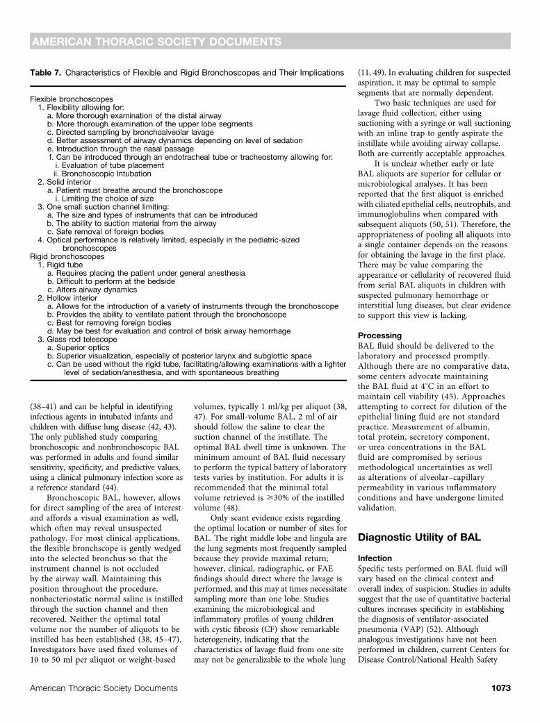
The primary focus of airway endoscopy isoften an accurate, comprehensive anatomicevaluation. There will be times whenpatients will benefit from both rigid andflexible bronchoscopy to more completelyassess the airway or for therapeutic
interventions (Table 7). Adequateevaluation of the posterior aspects of thelarynx, and to a lesser degree the cervicaltrachea, may be difficult with flexiblebronchoscopy. In general, rigid instrumentsare superior for detailed anatomicassessment of the larynx and cervicaltrachea and for operative manipulation,principally foreign body extraction (37). Ingeneral, flexible instruments are superiorfor evaluation of airway dynamics at alllevels, for examination of smaller airways,for BAL, and for removal of mucus plugs inperipheral airways.
BAL
PerformanceBAL can be performed either bronchoscopicallyor nonbronchoscopically. The latter isperformed by blindly inserting anappropriately sized suction catheter (4–8French) through an endotracheal tube
Table 6. Advantages and Disadvantages of Different Airway Entry Techniques
Technique Advantages Disadvantages
Natural airway (transnasal or transoral approach):insufflation of oxygen6 anesthetic gas using nasalprongs, nasal airway, or oral airway
Inspect entire airway More difficult to monitorventilation, airway patencyAssess airway dynamics/malacia
LaryngospasmMay allow for the use of a larger FB comparedwith FAE via an artificial airway Anesthetic waste gas into OR
environmentAllows for airway evaluation with rigid scope
Facemask Inspect entire airway More challenging for theanesthetist than LMA orendotracheal tube
Assess airway dynamics/malacia
LaryngospasmDoes not limit size of FB
May limit movement of scope
Laryngeal mask airway Easy to place Cannot assess upper airwayRelatively secure airway Cannot accurately assess vocal
cord movementCan assist ventilation with positive pressureMay limit size of FBAperture bars may limit/hinderpassage of FB
Requires deeper sedation/anesthesia than natural airwayor facemask approach
Can mask lower airwaydynamics
Endotracheal or tracheostomy tube Easy, fast, stable access to lower airways Cannot assess upper airwaySecure airway, even with deeper levels ofanesthesia
Cannot assess vocal cordmotion
Easy and quick to reinsert FB if needed Cannot assess airway dynamics/malaciaEnables positive pressure ventilation during the
procedure (may be especially useful whenextensive suctioning is required)
May limit size of FB
Potentially avoids contamination of lower airwayspecimens by upper airway secretions
Requires deeper level ofanesthesia
Definition of abbreviations: FAE = flexible airway endoscopy; FB = flexible bronchoscope; LMA = laryngeal mask airway; OR = operating room.
AMERICAN THORACIC SOCIETY DOCUMENTS
1072 American Journal of Respiratory and Critical Care Medicine Volume 191 Number 9 | May 1 2015
(38–41) and can be helpful in identifyinginfectious agents in intubated infants andchildren with diffuse lung disease (42, 43).The only published study comparingbronchoscopic and nonbronchoscopic BALwas performed in adults and found similarsensitivity, specificity, and predictive values,using a clinical pulmonary infection score asa reference standard (44).
Bronchoscopic BAL, however, allowsfor direct sampling of the area of interestand affords a visual examination as well,which often may reveal unsuspectedpathology. For most clinical applications,the flexible bronchscope is gently wedgedinto the selected bronchus so that theinstrument channel is not occludedby the airway wall. Maintaining thisposition throughout the procedure,nonbacteriostatic normal saline is instilledthrough the suction channel and thenrecovered. Neither the optimal totalvolume nor the number of aliquots to beinstilled has been established (38, 45–47).Investigators have used fixed volumes of10 to 50 ml per aliquot or weight-based
volumes, typically 1 ml/kg per aliquot (38,47). For small-volume BAL, 2 ml of airshould follow the saline to clear thesuction channel of the instillate. Theoptimal BAL dwell time is unknown. Theminimum amount of BAL fluid necessaryto perform the typical battery of laboratorytests varies by institution. For adults it isrecommended that the minimal totalvolume retrieved is >30% of the instilledvolume (48).
Only scant evidence exists regardingthe optimal location or number of sites forBAL. The right middle lobe and lingula arethe lung segments most frequently sampledbecause they provide maximal return;however, clinical, radiographic, or FAEfindings should direct where the lavage isperformed, and this may at times necessitatesampling more than one lobe. Studiesexamining the microbiological andinflammatory profiles of young childrenwith cystic fibrosis (CF) show remarkableheterogeneity, indicating that thecharacteristics of lavage fluid from one sitemay not be generalizable to the whole lung
(11, 49). In evaluating children for suspectedaspiration, it may be optimal to samplesegments that are normally dependent.
Two basic techniques are used forlavage fluid collection, either usingsuctioning with a syringe or wall suctioningwith an inline trap to gently aspirate theinstillate while avoiding airway collapse.Both are currently acceptable approaches.
It is unclear whether early or lateBAL aliquots are superior for cellular ormicrobiological analyses. It has beenreported that the first aliquot is enrichedwith ciliated epithelial cells, neutrophils, andimmunoglobulins when compared withsubsequent aliquots (50, 51). Therefore, theappropriateness of pooling all aliquots intoa single container depends on the reasonsfor obtaining the lavage in the first place.There may be value comparing theappearance or cellularity of recovered fluidfrom serial BAL aliquots in children withsuspected pulmonary hemorrhage orinterstitial lung diseases, but clear evidenceto support this view is lacking.
ProcessingBAL fluid should be delivered to thelaboratory and processed promptly.Although there are no comparative data,some centers advocate maintainingthe BAL fluid at 48C in an effort tomaintain cell viability (45). Approachesattempting to correct for dilution of theepithelial lining fluid are not standardpractice. Measurement of albumin,total protein, secretory component,or urea concentrations in the BALfluid are compromised by seriousmethodological uncertainties as wellas alterations of alveolar–capillarypermeability in various inflammatoryconditions and have undergone limitedvalidation.
Diagnostic Utility of BAL
InfectionSpecific tests performed on BAL fluid willvary based on the clinical context andoverall index of suspicion. Studies in adultssuggest that the use of quantitative bacterialcultures increases specificity in establishingthe diagnosis of ventilator-associatedpneumonia (VAP) (52). Althoughanalogous investigations have not beenperformed in children, current Centers forDisease Control/National Health Safety
Table 7. Characteristics of Flexible and Rigid Bronchoscopes and Their Implications
Flexible bronchoscopes1. Flexibility allowing for:
a. More thorough examination of the distal airwayb. More thorough examination of the upper lobe segmentsc. Directed sampling by bronchoalveolar lavaged. Better assessment of airway dynamics depending on level of sedatione. Introduction through the nasal passagef. Can be introduced through an endotracheal tube or tracheostomy allowing for:i. Evaluation of tube placementii. Bronchoscopic intubation
2. Solid interiora. Patient must breathe around the bronchoscope
i. Limiting the choice of size3. One small suction channel limiting:
a. The size and types of instruments that can be introducedb. The ability to suction material from the airwayc. Safe removal of foreign bodies
4. Optical performance is relatively limited, especially in the pediatric-sizedbronchoscopes
Rigid bronchoscopes1. Rigid tube
a. Requires placing the patient under general anesthesiab. Difficult to perform at the bedsidec. Alters airway dynamics
2. Hollow interiora. Allows for the introduction of a variety of instruments through the bronchoscopeb. Provides the ability to ventilate patient through the bronchoscopec. Best for removing foreign bodiesd. May be best for evaluation and control of brisk airway hemorrhage
3. Glass rod telescopea. Superior opticsb. Superior visualization, especially of posterior larynx and subglottic spacec. Can be used without the rigid tube, facilitating/allowing examinations with a lighter
level of sedation/anesthesia, and with spontaneous breathing
AMERICAN THORACIC SOCIETY DOCUMENTS
American Thoracic Society Documents 1073
Network criteria for VAP with commonbacterial organisms specifies diagnosticthreshold values as >104 colony-formingunits (cfu)/ml from BAL and >103 cfu/mlfrom protected specimen brushing (53).In immunocompromised children,the detection of fewer organisms onquantitative culture may suggest activeinfection. Additional testing may includeviral cultures; polymerase chain reactionassays for viruses, Chlamydia, andMycoplasma; antigen detection for virusesand other pathogens; or galactomannantesting for Aspergillus.
Although there are published referencedata for normal total and differential cellcounts (41, 51, 54–58), their derivationfrom children undergoing procedures fora variety of clinical indications may
weaken applicability to other populations.Nonetheless, cellular differential counts canbe useful (47, 51, 59, 60). Microbiologicaldata should be correlated with cytologicdata. The presence of bacteria without aninflammatory reaction is much less likely torepresent infection than contamination.The finding of >5% BAL-obtained cellscontaining intracellular bacteria on directmicroscopic exam (e.g., Gram stain) is anadditional Centers for Disease Control/National Health Safety Network diagnosticcriterion for VAP (53).
BAL in CFAlthough the findings from culture ofexpectorated sputum in adults with CF seemto correlate well with findings from FAEwith BAL or protected brush sampling,
discordance between bacterial culturestaken from the oropharynx with those takenfrom the lower airway by BAL has long beenrecognized (61–63).
Surveillance BAL has proven to beuseful in identifying new organisms inotherwise asymptomatic, newly diagnosedyoung children with CF (64, 65). However,the usefulness of BAL culture results tospecifically direct antimicrobial treatmentof CF lung disease remains controversial(66, 67).
Because of the inhomogeneity of CFlung disease, BAL sampling from a singlelobe may not detect the presence and truediversity of the underlying pathogenicairway organisms. Even when BALspecimens are taken from two lobes,infections may be missed (49, 68). However,
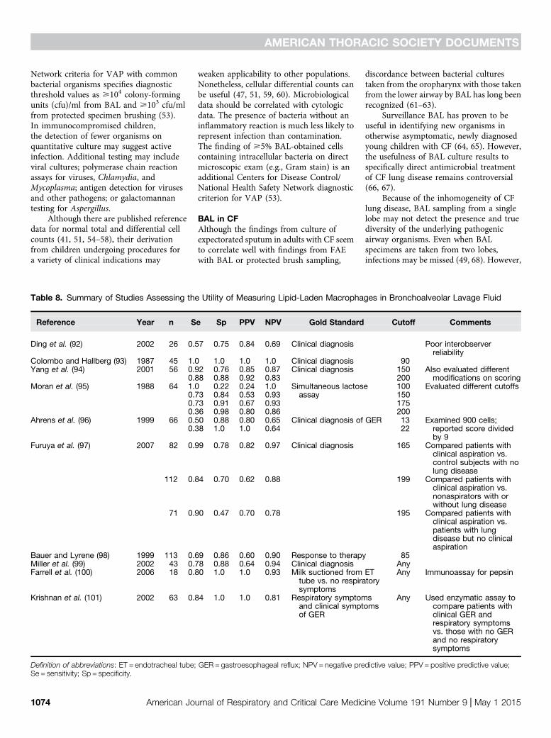
Table 8. Summary of Studies Assessing the Utility of Measuring Lipid-Laden Macrophages in Bronchoalveolar Lavage Fluid
Reference Year n Se Sp PPV NPV Gold Standard Cutoff Comments
Ding et al. (92) 2002 26 0.57 0.75 0.84 0.69 Clinical diagnosis Poor interobserverreliability
Colombo and Hallberg (93) 1987 45 1.0 1.0 1.0 1.0 Clinical diagnosis 90Yang et al. (94) 2001 56 0.92 0.76 0.85 0.87 Clinical diagnosis 150 Also evaluated different
modifications on scoring0.88 0.88 0.92 0.83 200Moran et al. (95) 1988 64 1.0 0.22 0.24 1.0 Simultaneous lactose
assay100 Evaluated different cutoffs
0.73 0.84 0.53 0.93 1500.73 0.91 0.67 0.93 1750.36 0.98 0.80 0.86 200
Ahrens et al. (96) 1999 66 0.50 0.88 0.80 0.65 Clinical diagnosis of GER 13 Examined 900 cells;reported score dividedby 9
0.38 1.0 1.0 0.64 22
Furuya et al. (97) 2007 82 0.99 0.78 0.82 0.97 Clinical diagnosis 165 Compared patients withclinical aspiration vs.control subjects with nolung disease
112 0.84 0.70 0.62 0.88 199 Compared patients withclinical aspiration vs.nonaspirators with orwithout lung disease
71 0.90 0.47 0.70 0.78 195 Compared patients withclinical aspiration vs.patients with lungdisease but no clinicalaspiration
Bauer and Lyrene (98) 1999 113 0.69 0.86 0.60 0.90 Response to therapy 85Miller et al. (99) 2002 43 0.78 0.88 0.64 0.94 Clinical diagnosis AnyFarrell et al. (100) 2006 18 0.80 1.0 1.0 0.93 Milk suctioned from ET
tube vs. no respiratorysymptoms
Any Immunoassay for pepsin
Krishnan et al. (101) 2002 63 0.84 1.0 1.0 0.81 Respiratory symptomsand clinical symptomsof GER
Any Used enzymatic assay tocompare patients withclinical GER andrespiratory symptomsvs. those with no GERand no respiratorysymptoms
Definition of abbreviations: ET = endotracheal tube; GER = gastroesophageal reflux; NPV = negative predictive value; PPV = positive predictive value;Se = sensitivity; Sp = specificity.
AMERICAN THORACIC SOCIETY DOCUMENTS
1074 American Journal of Respiratory and Critical Care Medicine Volume 191 Number 9 | May 1 2015

the sampling of multiple lobes may increasethe risk for complications.
BAL has provided valuable outcomemeasurements in important CF researchstudies (69). Future identification andanalysis of novel protein biomarkers inBAL fluid may provide new means to assessdisease severity and/or response to therapyin patients with CF (70, 71). Additionally,more advanced culture-independentevaluations of BAL fluid using ribosomalRNA sequencing have revealed thepotential to identify nontraditional bacterialspecies in the progression of CF lungdisease (65).
BAL in the Diagnosis of PulmonaryAspirationAlthough there is no question that chronicaspiration can cause respiratory disease,there is disagreement regarding howcommonly this is seen and in particularwhether gastroesophageal reflux leads toaspiration. The controversy is exacerbatedby the lack of a diagnostic “gold standard”for aspiration, significantly complicatingcomparison of various diagnostic tests thatpurport to ascertain the presence ofaspiration.
No unequivocal marker forexogenous aspiration is available forclinical use; BAL findings are influencedby many variables, including what isaspirated, how much is aspirated, andwhen the lung is sampled after theaspiration event.
Lipid-laden alveolar macrophagesmay be found in the BAL of all children,especially those with lung disease, becausethe source of the lipid may be endogenousas well as exogenous. The utility ofa quantitative scoring system such asthe lipid-laden macrophage index iscontroversial because published studies haveused different diagnostic cutoff scoresand different gold standards to diagnoseaspiration (Table 8) (72–77). The test resultmay be a useful adjunct to diagnosis as longas it is interpreted appropriately within theclinical context.
BAL in Other DisordersLymphocyte subpopulation and CD4:CD8 ratios may be useful in diagnosingsome interstitial lung diseases, suchas sarcoidosis and hypersensitivitypneumonitis (78). The role of BAL indiagnosing adult forms of interstitial
lung disease has recently beenreviewed (78). BAL findings in otherconditions, such as alveolar proteinosis,pulmonary hemorrhage, pulmonaryLangerhans cell histiocytosis, chroniclipoid pneumonia, and pulmonaryalveolar microlithiasis (79), are outlinedin Table 9.
Specialized Procedures
In the on-line supplement we discussprinciples for performing the followingprocedures in children: bronchoscopicintubation, transbronchial andendobronchial biopsy, bronchoscopicdilatation, and airway stent placement.
Table 9. Potential Tests on Bronchoalveolar Lavage Fluid Based on SuspectedDiagnosis or Underlying Condition
AspirationLipid-laden macrophages(quantitative score)
Variable reports of PPV and NPV, needs to beinterpreted in clinical context (72–75, 92, 97, 102,103)
Milk proteins (histologic stain) Not available outside the research setting (104, 105)Pepsin (enzymatic or immunologicassays)
Early reports of good NPV and excellent PPV, butfurther validation is needed (106)
Immunosuppressed andventilator-associatedpneumonia
Culture The identification of pathogens not normally found inthe lung is diagnostic; sensitivity has not beenstudied. Quantitative cultures to confirm thesignificance of commonly recovered organisms arethought to be helpful but also not well studied (53,107–109).
Cytology Fair to good PPV and NPV for Pneumocystis jirovecii(110)
PCR Good predictive value for P. jirovecii, Aspergillus(110–112)
Galactomannin Good predictive value for Aspergillus (111–113)Cell counts and differential Utility not well characterized (46, 55, 114)
Cystic fibrosisCulture Yield improved with sampling from multiple lobes (49,
68)Nonculture techniques (e.g.,genome sequencing)
Significantly greater yield of organisms than culture;primarily a research tool (65, 115)
Inflammatory biomarkers Primarily a research tool (70, 71)
Alveolar proteinosis (79, 116)Gross appearance Milky, sediment is often visibleCytology PAS-positive, diastase-resistant amorphic materialElectron microscopy Abundant extracellular multilamellated bodies and
tubular myelin structures; alveolar macrophageswith enlarged foamy cytoplasm
Alveolar hemorrhage (79, 117)Gross appearance Bloody, increasing with each sequential sampleCytology Hemosiderin stained macrophages
Langerhans cell histiocytosisCytology Immunostaining for S-100, CD1a, langerin
(79, 117–119)
Chronic lipoid pneumoniaCytology Oil Red O staining with scoring for lipid-laden
macrophages (79, 120)
Pulmonary alveolar microlithiasisCytology Microlith staining with PAS or von Kossa stain
(79, 121)
Definition of abbreviations: NPV = negative predictive value; PAS = periodic acid–Schiff; PCR =polymerase chain reaction; PPV = positive predictive value.
AMERICAN THORACIC SOCIETY DOCUMENTS
American Thoracic Society Documents 1075
Atelectasis
FAE can safely be performed in infants andchildren with atelectasis in a variety ofsettings (80–84). When atelectasis is notresolving and is problematic, FAE shouldbe considered. Reports suggest FAE utilityin the diagnosis of the etiology of atelectasisin 62 to 100% of cases (80, 82, 83, 85).Therapeutically, FAE has been used torelieve atelectasis in a variety of diseases,including hyaline membrane disease,pneumonia, CF, and plastic bronchitis, withvariable success (81, 86–89). The use ofsterile saline washes is standard practice.Case reports using bronchoscopicallyinstilled mucolytics (84, 90) as well asother agents (91) exist, but the efficacyof these techniques over standardapproaches has not been demonstrated.Flexible bronchoscopy can be performedfor atelectasis when it is persistent,recurrent, and physiologically importantor when the etiology of the atelectasis isin question.
Documentation
The procedure note should systematicallydescribe the FAE procedure to present therationale for management, communicatefindings to others, and provide trackinginformation for internal auditing andquality assurance as well as data formedical research. The documentshould contain patient and provider
information, specific indication(s) forthe procedure, sedation and anesthesiaused, a detailed description of theprocedure and all specimens collected,images, and all complications or adversereactions. The note also serves as thebasis of procedure coding, billing,and reimbursement. In addition,a summary of specific FAE findings andrecommendations should be included inthe document. Based on the committee’scollective clinical observations, it isrecommended that video images fromFAE procedures be recorded and savedwhen the need for future review isa consideration. Still images ofabnormal findings should be storedin the medical record for futurecomparison.
Conclusions and FutureDirections
The technical standards described in thisdocument are limited by the lack ofcontrolled studies in the field and aretherefore based mostly on observationalstudies and expert opinion. Despite theselimitations, the document serves tohighlight some of the dramatic changes inthe technique that have occurred since theATS guidelines for pediatric flexiblebronchoscopy were published. Ofparticular relevance is the evolution ofairway management techniques andanesthesia for FAE. However, the
underlying purpose for performing FAEin a child must remain the priority andfocus.
Establishing clear, documentablecompetencies for the bronchoscopist intraining to achieve is a mandate that must beundertaken in a responsible, thoughtfulmanner. This manuscript underlinesskills that trainees should acquire anddemonstrate before performing FAEindependently.
Although BAL is routinelyperformed, there remain manyunanswered questions regarding the bestmanner to collect BAL fluid. The optimalnumber of aliquots to instill, the volume ofeach aliquot, the total volume instilled, thesite to sample, the number of sites tosample, and many other technique-relatedmatters have not been subjected to anycontrolled trial. In many institutions,other diagnostic or therapeutic proceduresthat can be performed with a flexiblebronchoscope are mostly relegated tobronchoscopists caring for adult patients.This is often due to the small size of thepediatric airway, but in the future, astechnology continues to improve, some ofthese techniques may become availableto the pediatric bronchoscopistequipped with the appropriate trainingand skills.
It is our hope that this document,while providing a framework for how toperform FAE in children, will stimulatefurther discussion, development, andstudy in the field. n
These official technical standards were prepared by an ad hoc subcommittee of the ATS Pediatrics Assembly.
Members of the ad hoc subcommittee:
ALBERT FARO, M.D.ROBERT E. WOOD, Ph.D., M.D.MICHAEL S. SCHECHTER, M.D.ALBIN B. LEONG, M.D.ERIC WITTKUGEL, M.D.KATHY ABODE, R.N., M.P.H.JAMES F. CHMIEL, M.D.CORI DAINES, M.D.STEPHANIE DAVIS, M.D.ERNST EBER, M.D.CHARLES HUDDLESTON, M.D.TODD KILBAUGH, M.D.GEOFFREY KURLAND, M.D.
FABIO MIDULLA, M.D.DAVID MOLTER, M.D.GREGORY S. MONTGOMERY, M.D.GEORGE RETSCH-BOGART, M.D.MICHAEL J. RUTTER, M.D.GARY VISNER, D.O.
STEPHEN A. WALCZAK, R.R.T., C.P.F.T.THOMAS W. FERKOL, M.D.PETER H. MICHELSON, M.D., M.S.
The following authors also contributed assection leads:
Equipment and Procedural Setting: KathyAbode, R.N., M.P.H.
Infection Control: Kathy Abode, R.N., M.P.H.
Training and Core Competencies: Albin B.Leong, M.D.
Reasons for Performing FAE: Peter H.Michelson, M.D., M.S.
Complications: Geoffrey Kurland, M.D., andAlbin B. Leong, M.D.
Preprocedure Evaluation: Gregory S.Montgomery, M.D.
Sedation and Monitoring: Todd Kilbaugh, M.D.
Airway Management and Examination: EricWittkugel, M.D., and Ernst Eber, M.D.
The Role of the Rigid Bronchoscope: DavidMolter, M.D.
Bronchoalveolar Lavage: Michael S.Schechter, M.D.
Specialized Procedures: James F. Chmiel,M.D., Gary Visner, D.O., and CharlesHuddleston, M.D.
Atelectasis: Cori Daines, M.D.
Documentation: Thomas W. Ferkol, M.D
Author Disclosures: S.D. reported servingon an advisory board for Vertex Pharmaceuticals($1–4,999); serving as an unpaid consultantfor Eli Lilly; and receiving research supportfrom an ABC Com educational grant
AMERICAN THORACIC SOCIETY DOCUMENTS
1076 American Journal of Respiratory and Critical Care Medicine Volume 191 Number 9 | May 1 2015
sponsored by Gilead ($1–4,999). M.J.R.reported serving as a consultant for BryanMedical ($1–4,999). T.W.F. reportedserving on the American Board of
Pediatrics–Pulmonology as a sub-boardmember ($1–4,999). A.F., R.E.W., M.S.S.,A.B.L., E.W., K.A., J.F.C., C.D. (disclosedin 2014), E.E., C.H., T.K., G.K., F.M., D.M.,
G.S.M., G.R.-B., G.V., S.A.W., and P.H.M.reported no relevant commercial ornoncommercial, nongovernmentalrelationships.
References
1. Wood RE, Fink RJ. Applications of flexible fiberoptic bronchoscopesin infants and children. Chest 1978;73(5 Suppl):737–740.
2. Green CG, Eisenberg J, Leong A, Nathanson I, Schnapf BM, WoodRE. Flexible endoscopy of the pediatric airway. Am Rev Respir Dis1992;145:233–235.
3. Linnane B, Hafen GM, Ranganathan SC. Diameter of paediatric sizedflexible bronchoscopes: when size matters. Pediatr Pulmonol2006;41:787–789.
4. Favero MS, Bond WW. Disinfection of medical and surgicalmaterials. In: Block SS, editor. Disinfection, sterilization, andpreservation, 5th ed. Philadelphia, PA: Lippincott Williams &Wilkins; 2001.
5. Association of periOperative Registered Nurses. Recommendedpractices for cleaning and processing endoscopes and endoscopeaccessories. AORN J 2003;77:434–438, 441–442.
6. Petersen BT, Chennat J, Cohen J, Cotton PB, Greenwald DA,Kowalski TE, Krinsky ML, Park WG, Pike IM, Romagnuolo J, et al.;ASGE Quality Assurance in Endoscopy Committee; Society forHealthcare Epidemiology of America. Multisociety guideline onreprocessing flexible GI endoscopes: 2011. Infect Control HospEpidemiol 2011;32:527–537.
7. Public Health Agency of Canada. Infection prevention and controlguideline for flexible gastrointestinal endoscopy and flexiblebronchoscopy. Public Health Agency of Canada; 2010 [accessed2012 Oct 10]. Available from: www.phac-aspc.gc.ca/nois-sinp/guide/endo/
8. Rutala WA, Weber DJ; Healthcare Infection Control PracticesAdvisory Committee. Guideline for disinfection and sterilizationin healthcare facilities. 2008 [accessed 2012 May 10].Available from: http://www.cdc.gov/hicpac/pdf/guidelines/Disinfection_Nov_2008.pdf
9. Midulla F, de Blic J, Barbato A, Bush A, Eber E, Kotecha S, Haxby E,Moretti C, Pohunek P, Ratjen F; ERS Task Force. Flexibleendoscopy of paediatric airways. Eur Respir J 2003;22:698–708.
10. Olympus AI. Reprocessing training for Olympus bronchoscopes.2012 [accessed 2015 Apr 1]. Available from: http://medical.olympusamerica.com/customer-resources/cleaning-disinfection-sterilization/cds-instructions/bronchoscope-reprocessing
11. Mehta AC, Prakash UB, Garland R, Haponik E, Moses L, SchaffnerW, Silvestri G. American College of Chest Physicians andAmerican Association for Bronchology [corrected] consensusstatement: prevention of flexible bronchoscopy-associatedinfection. Chest 2005;128:1742–1755.
12. Siegel JD, Rhinehart E, Jackson M, Chiarello L; HealthcareInfection Control Practices Advisory Committee. Guideline forisolation precautions: preventing transmission of infectiousagents in healthcare settings. 2007 [accessed 2012 May 10].Available from: http://www.cdc.gov/hicpac/2007IP/2007isolationPrecautions.html
13. British Thoracic Society Bronchoscopy Guidelines Committee,a Subcommittee of Standards of Care Committee of BritishThoracic Society. British Thoracic Society guidelines ondiagnostic flexible bronchoscopy. Thorax 2001;56:i1–i21.
14. Bolliger CT, Mathur PN, Beamis JF, Becker HD, Cavaliere S, Colt H,Diaz-Jimenez JP, Dumon JF, Edell E, Kovitz KL, et al.; EuropeanRespiratory Society/American Thoracic Society. ERS/ATS statementon interventional pulmonology. Eur Respir J 2002;19:356–373.
15. Sehulster L, Chinn RY; CDC; HICPAC. Guidelines for environmentalinfection control in health-care facilities. Recommendations ofCDC and the Healthcare Infection Control Practices AdvisoryCommittee (HICPAC). MMWR Recomm Rep 2003;52:1–42.
16. Society of Gastroenterology Nurses and Associates, Inc. SGNAStandards: standards of infection control in reprocessing offlexible gastrointestinal endoscopes. Gastroenterol Nurs 2010;33:70–80.
17. Leung J, Vallero R, Wilson R. Surveillance cultures to monitorquality of gastrointestinal endoscope reprocessing. Am JGastroenterol 2003;98:3–5.
18. Moses FM, Lee J. Surveillance cultures to monitor quality ofgastrointestinal endoscope reprocessing. Am J Gastroenterol2003;98:77–81.
19. Muscarella LF. Application of environmental sampling toflexible endoscope reprocessing: the importance of monitoringthe rinse water. Infect Control Hosp Epidemiol 2002;23:285–289.
20. Tunuguntla A, Sullivan MJ. Monitoring quality of flexible endoscopedisinfection by microbiologic surveillance cultures. Tenn Med2004;97:453–456.
21. Ernst A, Silvestri GA, Johnstone D; American College of ChestPhysicians. Interventional pulmonary procedures: guidelines fromthe American College of Chest Physicians. Chest 2003;123:1693–1717.
22. Leong AB, Green CG, Kurland G, Wood RE. A survey of training inpediatric flexible bronchoscopy. Pediatr Pulmonol 2014;49:605–610.
23. Buckley JD, Addrizzo-Harris DJ, Clay AS, Curtis JR, Kotloff RM,Lorin SM, Murin S, Sessler CN, Rogers PL, Rosen MJ, et al.Multisociety task force recommendations of competencies inPulmonary and Critical Care Medicine. Am J Respir Crit Care Med2009;180:290–295.
24. Accreditation Council for Graduate Medical Education. Internalmedicine: pulmonary disease program requirements. 2012[accessed 2015 Apr 1]. Available from: http://www.acgme.org/acgmeweb/Portals/0/PFAssets/2013-PR-FAQ-PIF/149_pulmonary_disease_int_med_07132013.pdf
25. Wood R, Boesch P. Bronchoscopy and bronchoalveolar lavage inpediatric patients. In: Wilmott RW, Boat TF, Bush A, Chernick V,Deterding RR, Ratjen F, editors. Kendig and Chernick’s disordersof the respiratory tract in children, 8th ed. Philadelphia, PA:Elsevier Saunders; 2012.
26. Wagener JS. Fatality following fiberoptic bronchoscopy in a two-year-old child. Pediatr Pulmonol 1987;3:197–199.
27. Picard E, Schlesinger Y, Goldberg S, Schwartz S, Kerem E. Fatalpneumococcal sepsis following flexible bronchoscopy in animmunocompromised infant. Pediatr Pulmonol 1998;25:390–392.
28. Van Vyve T, Chanez P, Bousquet J, Lacoste JY, Michel FB, GodardP. Safety of bronchoalveolar lavage and bronchial biopsies inpatients with asthma of variable severity. Am Rev Respir Dis1992;146:116–121.
29. Wilson W, Taubert KA, Gewitz M, Lockhart PB, Baddour LM, Levison M,Bolger A, Cabell CH, Takahashi M, Baltimore RS, et al.; AmericanHeart Association Rheumatic Fever, Endocarditis, and KawasakiDisease Committee; American Heart Association Council onCardiovascular Disease in the Young; American Heart AssociationCouncil on Clinical Cardiology; American Heart Association Council onCardiovascular Surgery and Anesthesia; Quality of Care andOutcomes Research Interdisciplinary Working Group. Preventionof infective endocarditis: guidelines from the American HeartAssociation: a guideline from the American Heart AssociationRheumatic Fever, Endocarditis, and Kawasaki Disease Committee,Council on Cardiovascular Disease in the Young, and the Councilon Clinical Cardiology, Council on Cardiovascular Surgery andAnesthesia, and the Quality of Care and Outcomes ResearchInterdisciplinary Working Group. Circulation 2007;116:1736–1754.
AMERICAN THORACIC SOCIETY DOCUMENTS
American Thoracic Society Documents 1077
30. Kozak EA, Brath LK. Do “screening” coagulation tests predictbleeding in patients undergoing fiberoptic bronchoscopy withbiopsy? Chest 1994;106:703–705.
31. Hackel A, Badgwell JM, Binding RR, Dahm LS, Dunbar BS, FischerCG, Geiduschek JM, Gunter JB, Gutierrez-Mazzora JF, Kain Z,et al. Guidelines for the pediatric perioperative anesthesiaenvironment. American Academy of Pediatrics. Section onAnesthesiology. Pediatrics 1999;103:512–515.
32. Cote CJ, Wilson S; American Academy of Pediatrics; AmericanAcademy of Pediatric Dentistry; Work Group on Sedation.Guidelines for monitoring and management of pediatric patientsduring and after sedation for diagnostic and therapeuticprocedures: an update. Pediatrics 2006;118:2587–2602.
33. Nielson DW, Ku PL, Egger M. Topical lidocaine exaggerateslaryngomalacia during flexible bronchoscopy. Am J Respir CritCare Med 2000;161:147–151.
34. Niggemann B, Haack M, Machotta A. How to enter the pediatricairway for bronchoscopy. Pediatr Int 2004;46:117–121.
35. Nicolai T. The role of rigid and flexible bronchoscopy in children.Paediatr Respir Rev 2011;12:190–195.
36. Wood RE. Evaluation of the upper airway in children. Curr OpinPediatr 2008;20:266–271.
37. Divisi D, Di Tommaso S, Garramone M, Di Francescantonio W, Crisci RM,Costa AM, Gravina GL, Crisci R. Foreign bodies aspirated in children:role of bronchoscopy. Thorac Cardiovasc Surg 2007;55:249–252.
38. Grigg J, Arnon S, Silverman M. Fractional processing of sequentialbronchoalveolar lavage fluid from intubated babies. Eur Respir J1992;5:727–732.
39. Alpert BE, O’Sullivan BP, Panitch HB. Nonbronchoscopic approachto bronchoalveolar lavage in children with artificial airways.Pediatr Pulmonol 1992;13:38–41.
40. Koumbourlis AC, Kurland G. Nonbronchoscopic bronchoalveolarlavage in mechanically ventilated infants: technique, efficacy, andapplications. Pediatr Pulmonol 1993;15:257–262.
41. Heaney LG, Stevenson EC, Turner G, Cadden IS, Taylor R, ShieldsMD, Ennis M. Investigating paediatric airways by non-bronchoscopic lavage: normal cellular data. Clin Exp Allergy1996;26:799–806.
42. Amaro-Galvez R, Rao M, Abadco D, Kravath RE, Steiner P.Nonbronchoscopic bronchoalveolar lavage in ventilated childrenwith acquired immunodeficiency syndrome: a simple andeffective diagnostic method for Pneumocystis carinii infection.Pediatr Infect Dis J 1991;10:473–475.
43. Morrow BM, Argent AC. Ventilator-associated pneumonia ina paediatric intensive care unit in a developing country with highHIV prevalence. J Paediatr Child Health 2009;45:104–111.
44. Khilnani GC, Arafath TK, Hadda V, Kapil A, Sood S, Sharma SK.Comparison of bronchoscopic and non-bronchoscopictechniques for diagnosis of ventilator associated pneumonia.Indian J Crit Care Med 2011;15:16–23.
45. Ratjen F, Munck A, Kho P, Angyalosi G; ELITE Study Group.Treatment of early Pseudomonas aeruginosa infection in patientswith cystic fibrosis: the ELITE trial. Thorax 2010;65:286–291.
46. Riedler J, Grigg J, Robertson CF. Role of bronchoalveolar lavage inchildren with lung disease. Eur Respir J 1995;8:1725–1730.
47. Ratjen F, Bruch J. Adjustment of bronchoalveolar lavage volume tobody weight in children. Pediatr Pulmonol 1996;21:184–188.
48. Baughman RP, Spencer RE, Kleykamp BO, Rashkin MC, DouthitMM. Ventilator associated pneumonia: quality ofnonbronchoscopic bronchoalveolar lavage sample affectsdiagnostic yield. Eur Respir J 2000;16:1152–1157.
49. Gutierrez JP, Grimwood K, Armstrong DS, Carlin JB, Carzino R,Olinsky A, Robertson CF, Phelan PD. Interlobar differences inbronchoalveolar lavage fluid from children with cystic fibrosis. EurRespir J 2001;17:281–286.
50. Rennard SI, Ghafouri M, Thompson AB, Linder J, Vaughan W, Jones K,Ertl RF, Christensen K, Prince A, Stahl MG, et al. Fractional processingof sequential bronchoalveolar lavage to separate bronchial andalveolar samples. Am Rev Respir Dis 1990;141:208–217.
51. Riedler J, Grigg J, Stone C, Tauro G, Robertson CF.Bronchoalveolar lavage cellularity in healthy children. Am JRespir Crit Care Med 1995;152:163–168.
52. American Thoracic Society; Infectious Diseases Society of America.Guidelines for the management of adults with hospital-acquired,ventilator-associated, and healthcare-associated pneumonia.Am J Respir Crit Care Med 2005;171:388–416.
53. Stokes DC, Shenep JL, Parham D, Bozeman PM, Marienchek W, MackertPW. Role of flexible bronchoscopy in the diagnosis of pulmonaryinfiltrates in pediatric patients with cancer. J Pediatr 1989;115:561–567.
54. Clement A, Chadelat K, Masliah J, Housset B, Sardet A, Grimfeld A,Tournier G. A controlled study of oxygen metabolite release byalveolar macrophages from children with interstitial lung disease.Am Rev Respir Dis 1987;136:1424–1428.
55. Ratjen F, Bredendiek M, Brendel M, Meltzer J, Costabel U.Differential cytology of bronchoalveolar lavage fluid in normalchildren. Eur Respir J 1994;7:1865–1870.
56. Ratjen F, Bredendiek M, Zheng L, Brendel M, Costabel U.Lymphocyte subsets in bronchoalveolar lavage fluid of childrenwithout bronchopulmonary disease. Am J Respir Crit Care Med1995;152:174–178.
57. Midulla F, Villani A, Merolla R, Bjermer L, Sandstrom T, Ronchetti R.Bronchoalveolar lavage studies in children without parenchymallung disease: cellular constituents and protein levels. PediatrPulmonol 1995;20:112–118.
58. Tessier V, Chadelat K, Baculard A, Housset B, Clement A. BAL inchildren: a controlled study of differential cytology and cytokineexpression profiles by alveolar cells in pediatric sarcoidosis.Chest 1996;109:1430–1438.
59. de Blic J, Blanche S, Danel C, Le Bourgeois M, Caniglia M,Scheinmann P. Bronchoalveolar lavage in HIV infected patientswith interstitial pneumonitis. Arch Dis Child 1989;64:1246–1250.
60. Efrati O, Gonik U, Bielorai B, Modan-Moses D, Neumann Y,Szeinberg A, Vardi A, Barak A, Paret G, Toren A. Fiberopticbronchoscopy and bronchoalveolar lavage for the evaluation ofpulmonary disease in children with primary immunodeficiencyand cancer. Pediatr Blood Cancer 2007;48:324–329.
61. Armstrong DS, Grimwood K, Carlin JB, Carzino R, Olinsky A, PhelanPD. Bronchoalveolar lavage or oropharyngeal cultures to identifylower respiratory pathogens in infants with cystic fibrosis. PediatrPulmonol 1996;21:267–275.
62. Rosenfeld M, Emerson J, Accurso F, Armstrong D, Castile R,Grimwood K, Hiatt P, McCoy K, McNamara S, Ramsey B, et al.Diagnostic accuracy of oropharyngeal cultures in infants andyoung children with cystic fibrosis. Pediatr Pulmonol 1999;28:321–328.
63. Aaron SD, Kottachchi D, Ferris WJ, Vandemheen KL, St Denis ML,Plouffe A, Doucette SP, Saginur R, Chan FT, Ramotar K. Sputumversus bronchoscopy for diagnosis of Pseudomonas aeruginosabiofilms in cystic fibrosis. Eur Respir J 2004;24:631–637.
64. Stafler P, Davies JC, Balfour-Lynn IM, Rosenthal M, Bush A.Bronchoscopy in cystic fibrosis infants diagnosed by newbornscreening. Pediatr Pulmonol 2011;46:696–700.
65. Harris JK, De Groote MA, Sagel SD, Zemanick ET, Kapsner R,Penvari C, Kaess H, Deterding RR, Accurso FJ, Pace NR.Molecular identification of bacteria in bronchoalveolar lavagefluid from children with cystic fibrosis. Proc Natl Acad Sci USA2007;104:20529–20533.
66. Wainwright CE, Vidmar S, Armstrong DS, Byrnes CA, Carlin JB,Cheney J, Cooper PJ, Grimwood K, Moodie M, Robertson CF,et al.; ACFBAL Study Investigators. Effect of bronchoalveolarlavage-directed therapy on Pseudomonas aeruginosa infectionand structural lung injury in children with cystic fibrosis:a randomized trial. JAMA 2011;306:163–171.
67. Linnane B, McNally P. Bronchoalveolar lavage-directed therapy inchildren with cystic fibrosis and Pseudomonas aeruginosainfection. JAMA 2011;306:1761; author reply 1761–1762.
68. Gilchrist FJ, Salamat S, Clayton S, Peach J, Alexander J, LenneyW. Bronchoalveolar lavage in children with cystic fibrosis: how manylobes should be sampled? Arch Dis Child 2011;96:215–217.
69. Ratjen F, Paul K, van Koningsbruggen S, Breitenstein S, RietschelE, Nikolaizik W. DNA concentrations in BAL fluid of cystic fibrosispatients with early lung disease: influence of treatment withdornase alpha. Pediatr Pulmonol 2005;39:1–4.
AMERICAN THORACIC SOCIETY DOCUMENTS
1078 American Journal of Respiratory and Critical Care Medicine Volume 191 Number 9 | May 1 2015
70. MacGregor G, Gray RD, Hilliard TN, Imrie M, Boyd AC, Alton EW,Bush A, Davies JC, Innes JA, Porteous DJ, et al. Biomarkers forcystic fibrosis lung disease: application of SELDI-TOF massspectrometry to BAL fluid. J Cyst Fibros 2008;7:352–358.
71. Wolak JE, Esther CR Jr, O’Connell TM. Metabolomic analysis ofbronchoalveolar lavage fluid from cystic fibrosis patients.Biomarkers 2009;14:55–60.
72. Boesch RP, Daines C, Willging JP, Kaul A, Cohen AP, Wood RE,Amin RS. Advances in the diagnosis and management of chronicpulmonary aspiration in children. Eur Respir J 2006;28:847–861.
73. Colombo JL, Hallberg TK. Pulmonary aspiration and lipid-ladenmacrophages: in search of gold (standards). Pediatr Pulmonol1999;28:79–82.
74. Colombo JL, Hallberg TK. Aspiration: a common event and a clinicalchallenge. Pediatr Pulmonol 2012;47:317–320.
75. Jaoude PA, Knight PR, Ohtake P, El-Solh AA. Biomarkers in the diagnosisof aspiration syndromes. Expert Rev Mol Diagn 2010;10:309–319.
76. Trinick R, Johnston N, Dalzell AM, McNamara PS. Reflux aspirationin children with neurodisability—a significant problem, but can wemeasure it? J Pediatr Surg 2012;47:291–298.
77. Tutor JD, Gosa MM. Dysphagia and aspiration in children. PediatrPulmonol 2012;47:321–337.
78. Linnane B, Oliver MR, Robinson PJ. Does splenectomy in cysticfibrosis related liver disease improve lung function and nutritionalstatus? A case series. Arch Dis Child 2006;91:771–773.
79. Midulla F, Nenna R. Bronchoalveolar lavage: indications andapplications. In: Priftis KN, Anthracopoulos MB, Eber E,Koumbourlis AC, Wood RE, editors. Paediatric bronchoscopy.Basel, Switzerland: Karger; 2010. pp. 30–41.
80. Raman TS, Mathew S, Ravikumar, Garcha PS. Atelectasis inchildren. Indian Pediatr 1998;35:429–435.
81. Karlson KH Jr, Pickert CB, Schexnayder SM, Heulitt MJ. Flexiblefiberoptic bronchoscopy in children on extracorporeal membraneoxygenation. Pediatr Pulmonol 1993;16:215–218.
82. Vijayasekaran D, Gowrishankar NC, Nedunchelian K, Suresh S.Fiberoptic bronchoscopy in unresolved atelectasis in infants.Indian Pediatr 2010;47:611–613.
83. Cerda J, Chacon J, Reichhard C, Bertrand P, Holmgren NL, ClaverıaC, Sanchez I. Flexible fiberoptic bronchoscopy in children withheart diseases: a twelve years experience. Pediatr Pulmonol2007;42:319–324.
84. Slattery DM, Waltz DA, Denham B, O’Mahony M, Greally P.Bronchoscopically administered recombinant human DNase forlobar atelectasis in cystic fibrosis. Pediatr Pulmonol 2001;31:383–388.
85. Wood RE. The diagnostic effectiveness of the flexiblebronchoscope in children. Pediatr Pulmonol 1985;1:188–192.
86. Nussbaum E. Pediatric flexible bronchoscopy and its application ininfantile atelectasis. Clin Pediatr (Phila) 1985;24:379–382.
87. Nussbaum E. Pediatric fiberoptic bronchoscopy: Clinicalexperience with 2,836 bronchoscopies. Pediatr Crit Care Med2002;3:171–176.
88. Pawar SS, Chun RH, Rao AR, Kerschner JE. Management of plasticbronchitis in a child with mild intermittent asthma. Ann OtolRhinol Laryngol 2011;120:697–699.
89. Deng J, Zheng Y, Li C, Ma Z, Wang H, Rubin BK. Plastic bronchitisin three children associated with 2009 influenza A(H1N1) virusinfection. Chest 2010;138:1486–1488.
90. Altman RP, Kulczycki LL, Randolph JG, McClenathan JE.Bronchoscopy and bronchial lavage (BBL) in children with cysticfibrosis. J Pediatr Surg 1973;8:809–814.
91. Krause MF, von Bismarck P, Oppermann HC, Ankermann T.Bronchoscopic surfactant administration in pediatric patientswith persistent lobar atelectasis. Respiration 2008;75:100–104.
92. Ding Y, Simpson PM, Schellhase DE, Tryka AF, Ding L, Parham DM.Limited reliability of lipid-laden macrophage index restricts itsuse as a test for pulmonary aspiration: comparison with a simplesemiquantitative assay. Pediatr Dev Pathol 2002;5:551–558.
93. Colombo JL, Hallberg TK. Recurrent aspiration in children: lipid-laden alveolar macrophage quantitation. Pediatr Pulmonol 1987;3:86–89.
94. Yang YJ, Steele CT, Anbar RD, Sinacori JT, Powers CN.Quantitation of lipid-laden macrophages in evaluation of lowerairway cytology specimens from pediatric patients. DiagnCytopathol 2001;24:98–103.
95. Moran JR, Block SM, Lyerly AD, Brooks LE, Dillard RG. Lipid-ladenalveolar macrophage and lactose assay as markers of aspirationin neonates with lung disease. J Pediatr 1988;112:643–645.
96. Ahrens P, Noll C, Kitz R, Willigens P, Zielen S, Hofmann D. Lipid-laden alveolar macrophages (LLAM): a useful marker of silentaspiration in children. Pediatr Pulmonol 1999;28:83–88.
97. Furuya ME, Moreno-Cordova V, Ramırez-Figueroa JL, Vargas MH,Ramon-Garcıa G, Ramırez-San Juan DH. Cutoff value of lipid-laden alveolar macrophages for diagnosing aspiration in infantsand children. Pediatr Pulmonol 2007;42:452–457.
98. Bauer ML, Lyrene RK. Chronic aspiration in children: evaluation ofthe lipid-laden macrophage index. Pediatr Pulmonol 1999;28:94–100.
99. Miller J, Colasurdo GN, Khan AM, Jajoo C, Patel TJ, Fan LL,Elidemir O. Immunocytochemical detection of milk proteins intracheal aspirates of ventilated infants: a pilot study. PediatrPulmonol 2002;34:369–374.
100. Farrell S, McMaster C, Gibson D, Shields MD, McCallion WA.Pepsin in bronchoalveolar lavage fluid: a specific and sensitivemethod of diagnosing gastro-oesophageal reflux-relatedpulmonary aspiration. J Pediatr Surg 2006;41:289–293.
101. Krishnan U, Mitchell JD, Messina I, Day AS, Bohane TD. Assayof tracheal pepsin as a marker of reflux aspiration. J PediatrGastroenterol Nutr 2002;35:303–308.
102. Kitz R, Boehles HJ, Rosewich M, Rose MA. Lipid-laden alveolarmacrophages and pH monitoring in gastroesophageal reflux-related respiratory symptoms. Pulm Med 2012;2012:673637.
103. Rosen R, Fritz J, Nurko A, Simon D, Nurko S. Lipid-ladenmacrophage index is not an indicator of gastroesophagealreflux-related respiratory disease in children. Pediatrics 2008;121:e879–e884.
104. Elidemir O, Fan LL, Colasurdo GN. A novel diagnostic method forpulmonary aspiration in a murine model: immunocytochemicalstaining of milk proteins in alveolar macrophages. Am J RespirCrit Care Med 2000;161:622–626.
105. De Baets F, Aarts C, Van Daele S, Haerynck F, De Wachter E, DeSchutter I, Malfroot A, Schelstraete P. Milk protein and Oil-Red-O staining of alveolar macrophages in chronic respiratorydisease of infancy. Pediatr Pulmonol 2010;45:1213–1219.
106. Samuels TL, Johnston N. Pepsin as a marker of extraesophagealreflux. Ann Otol Rhinol Laryngol 2010;119:203–208.
107. de Blic J, McKelvie P, Le Bourgeois M, Blanche S, Benoist MR,Scheinmann P. Value of bronchoalveolar lavage in themanagement of severe acute pneumonia and interstitialpneumonitis in the immunocompromised child. Thorax 1987;42:759–765.
108. Pattishall EN, Noyes BE, Orenstein DM. Use of bronchoalveolarlavage in immunocompromised children with pneumonia.Pediatr Pulmonol 1988;5:1–5.
109. McCubbin MM, Trigg ME, Hendricker CM, Wagener JS.Bronchoscopy with bronchoalveolar lavage in the evaluation ofpulmonary complications of bone marrow transplantation inchildren. Pediatr Pulmonol 1992;12:43–47.
110. Samuel CM, Whitelaw A, Corcoran C, Morrow B, Hsiao NY, Zampoli M,Zar HJ. Improved detection of Pneumocystis jirovecii in upper andlower respiratory tract specimens from children with suspectedpneumocystis pneumonia using real-time PCR: a prospective study.BMC Infect Dis 2011;11:329.
111. Luong ML, Clancy CJ, Vadnerkar A, Kwak EJ, Silveira FP, WisselMC, Grantham KJ, Shields RK, Crespo M, Pilewski J, et al.Comparison of an Aspergillus real-time polymerase chainreaction assay with galactomannan testing of bronchoalvelolarlavage fluid for the diagnosis of invasive pulmonary aspergillosisin lung transplant recipients. Clin Infect Dis 2011;52:1218–1226.
112. Avni T, Levy I, Sprecher H, Yahav D, Leibovici L, Paul M.Diagnostic accuracy of PCR alone compared to galactomannanin bronchoalveolar lavage fluid for diagnosis of invasive
AMERICAN THORACIC SOCIETY DOCUMENTS
American Thoracic Society Documents 1079

pulmonary aspergillosis: a systematic review. J Clin Microbiol2012;50:3652–3658.
113. Nguyen MH, Leather H, Clancy CJ, Cline C, Jantz MA, Kulkarni V,Wheat LJ, Wingard JR. Galactomannan testing inbronchoalveolar lavage fluid facilitates the diagnosis of invasivepulmonary aspergillosis in patients with hematologicmalignancies and stem cell transplant recipients. Biol BloodMarrow Transplant 2011;17:1043–1050.
114. Ratjen F, Costabel U, Havers W. Differential cytology ofbronchoalveolar lavage fluid in immunosuppressed children withpulmonary infiltrates. Arch Dis Child 1996;74:507–511.
115. Cox MJ, Allgaier M, Taylor B, Baek MS, Huang YJ, Daly RA, KaraozU, Andersen GL, Brown R, Fujimura KE, et al. Airway microbiotaand pathogen abundance in age-stratified cystic fibrosispatients. PLoS One 2010;5:e11044.
116. Burkhalter A, Silverman JF, Hopkins MB III, Geisinger KR.Bronchoalveolar lavage cytology in pulmonary alveolarproteinosis. Am J Clin Pathol 1996;106:504–510.
117. Costabel U, Guzman J, Bonella F, Oshimo S. Bronchoalveolarlavage in other interstitial lung diseases. Semin Respir Crit CareMed 2007;28:514–524.
118. Takizawa Y, Taniuchi N, Ghazizadeh M, Enomoto T, Sato M, Jin E,Azuma A, Gemma A, Kudoh S, Kawanami O. Bronchoalveolarlavage fluid analysis provides diagnostic information onpulmonary Langerhans cell histiocytosis. J Nippon Med Sch2009;76:84–92.
119. Danel C, Israel-Biet D, Costabel U, Rossi GA, Wallaert B. Theclinical role of BAL in pulmonary histiocytosis X. Eur Respir J1990;3:949–950, 961–969.
120. Midulla F, Strappini PM, Ascoli V, Villa MP, Indinnimeo L, FalascaC, Martella S, Ronchetti R. Bronchoalveolar lavage cell analysisin a child with chronic lipid pneumonia. Eur Respir J 1998;11:239–242.
121. Monabati A, Ghayumi MA, Kumar PV. Familial pulmonary alveolarmicrolithiasis diagnosed by bronchoalveolar lavage: a casereport. Acta Cytol 2007;51:80–82.
AMERICAN THORACIC SOCIETY DOCUMENTS
1080 American Journal of Respiratory and Critical Care Medicine Volume 191 Number 9 | May 1 2015