amino acids & side groups polar charged ◦ acidic negatively charged amino acids asp & glu...
TRANSCRIPT
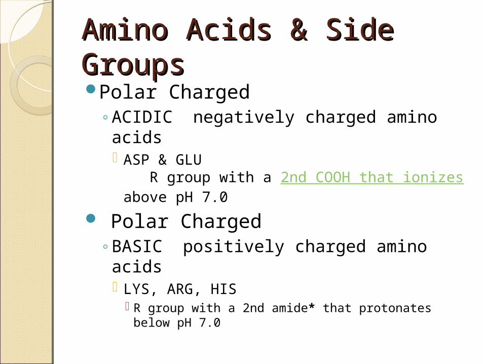
Amino Acids & Side Amino Acids & Side GroupsGroupsPolar Charged
◦ACIDIC negatively charged amino acids ASP & GLU
R group with a 2nd COOH that ionizes above pH 7.0
Polar Charged◦BASIC positively charged amino acids
LYS, ARG, HIS R group with a 2nd amide* that protonates below
pH 7.0
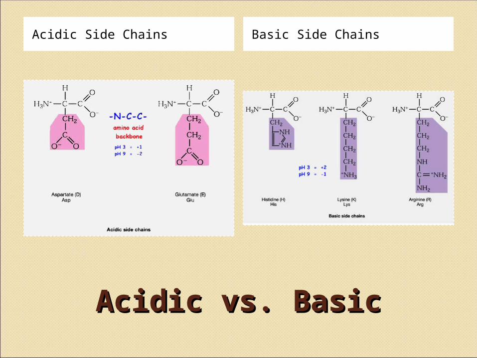
Acidic vs. BasicAcidic vs. Basic
Acidic Side Chains Basic Side Chains

Polar ChargedPolar Charged

Amino Acids & Side Amino Acids & Side GroupsGroupsPolar Uncharged
◦THR, TYR, ASN, GLN (cys) are soluble in water, i.e.,
hydrophilic (attract H-bonds) Contain hydroxyl or amino functional groups
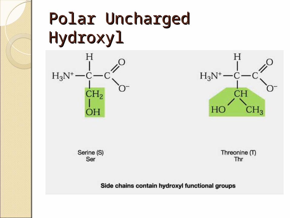
Polar UnchargedPolar UnchargedHydroxylHydroxyl
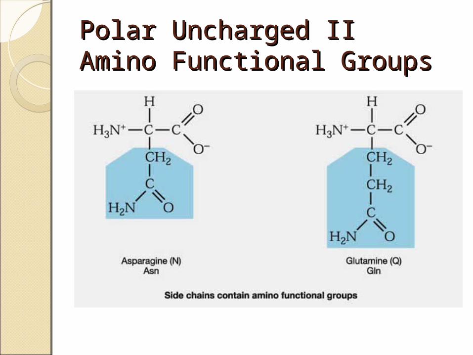
Polar Uncharged IIPolar Uncharged IIAmino Functional GroupsAmino Functional Groups

Polar Uncharged Amino Polar Uncharged Amino AcidsAcids
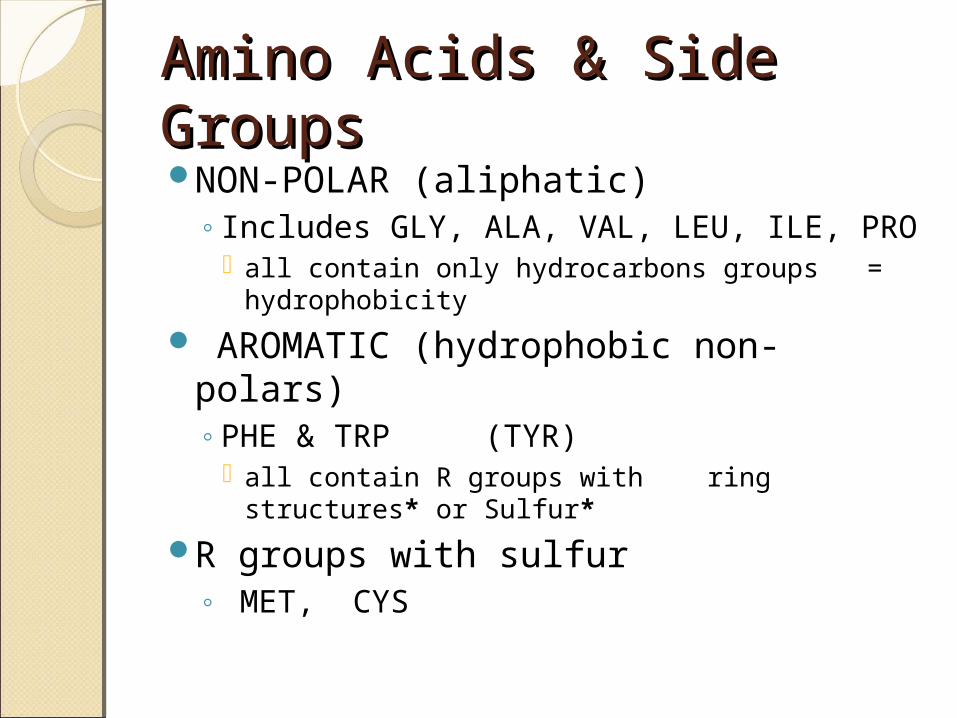
Amino Acids & Side Amino Acids & Side GroupsGroupsNON-POLAR (aliphatic)
◦Includes GLY, ALA, VAL, LEU, ILE, PRO all contain only hydrocarbons groups =
hydrophobicity
AROMATIC (hydrophobic non-polars) ◦PHE & TRP (TYR)
all contain R groups with ring structures* or Sulfur*
R groups with sulfur◦ MET, CYS
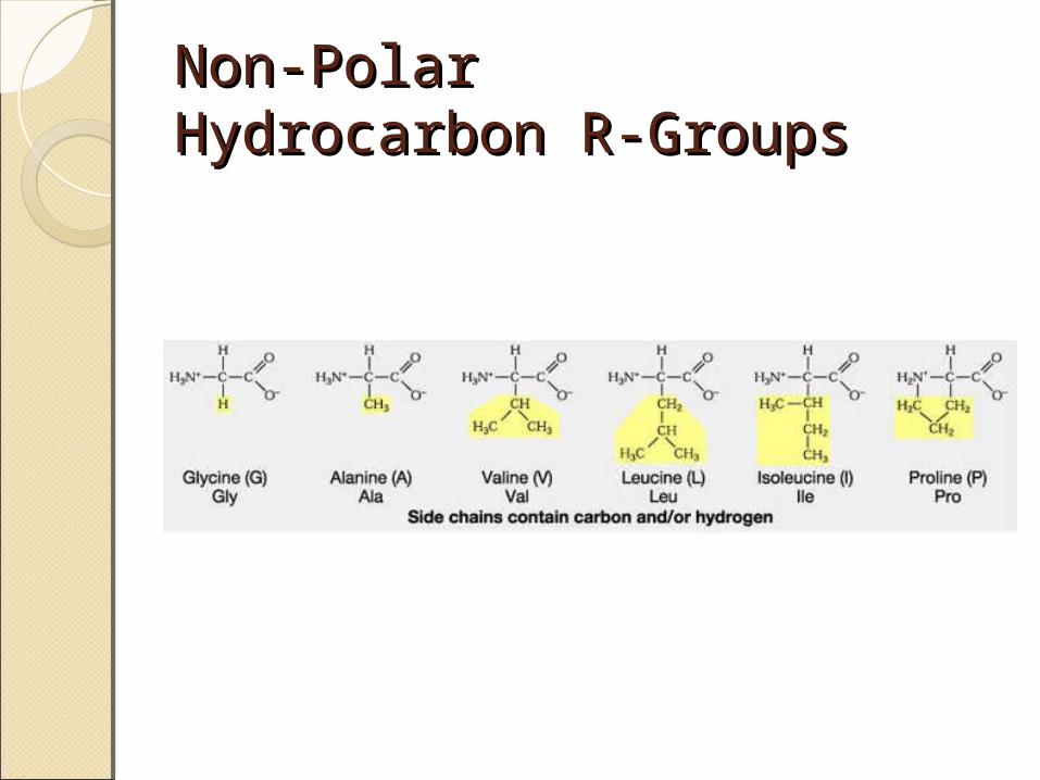
Non-PolarNon-PolarHydrocarbon R-GroupsHydrocarbon R-Groups

Non-PolarNon-PolarAromatic R-GroupsAromatic R-Groups

Non-PolarNon-PolarSulfur R GroupsSulfur R Groups

Secondary Protein Secondary Protein StructuresStructures
Alpha Helix Beta-pleated sheets
The most common polypeptide helix
Stabilized by extensive hydrogen bonding
Hydrogen bonds extend up from the oxygen from the carbonyl group to the NH group of a peptide linkage◦ This was shown in class via the
visuals
There are approximately 4 peptide bond links up stream between the atoms involved in the hydrogen bonds
Each turn of an alpha helix contains 3.6 amino acids.
• Unlike the alpha helix, composed of two or more peptide chains
• Polypeptide chains are joined by hydrogen bonds
• When the hydrogen bonds are formed between the polypeptide chains they are termed interchains.
• The polypeptide chains can run parallel to each other or anti-parallel– Recall the “ends” of a
polypeptide chain• C-terminus• N-terminus/Amino-terminus

Alpha HelixAlpha Helix

Beta-pleated sheetsBeta-pleated sheets

Beta-pleated Sheets and Beta-pleated Sheets and Alzheimer’s DiseaseAlzheimer’s Disease• The amyloid protein, a class of
fibrous proteins, is deposited in the brain.
• Individuals, that have Alzheimer’s Disease, have the amyloid protein composed of twisted Beta-pleated sheet fibrils whose three-dimentional structure is virtually identical to that of silk fibrils–Silk• Contain Beta-pleated sheet protein structures

Tertiary StructureTertiary StructureInteractions stabilizing Tertiary
Structures◦Four were mentioned in class
Disulfide Bonds Hydrophobic interactions Hydrogen bonds Ionic interactions
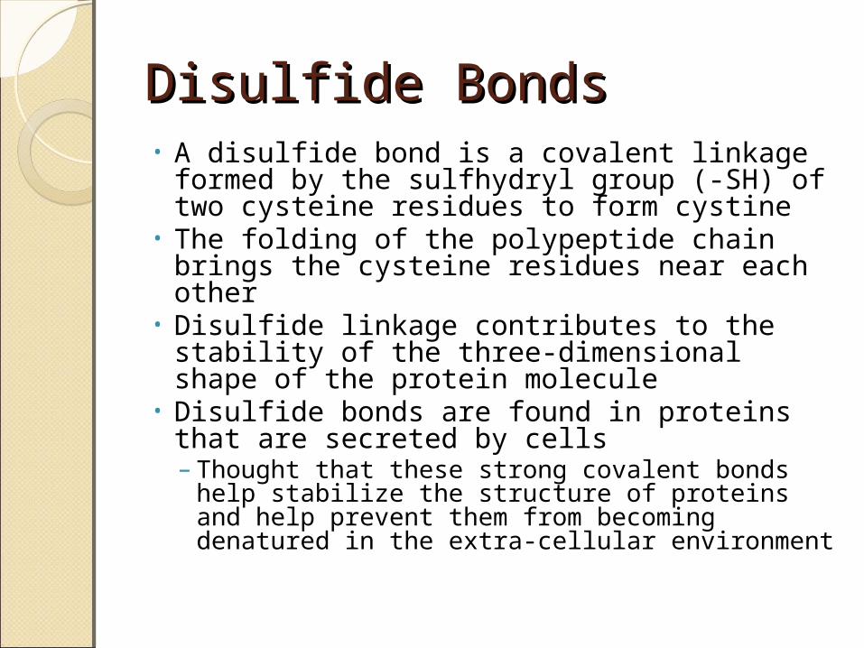
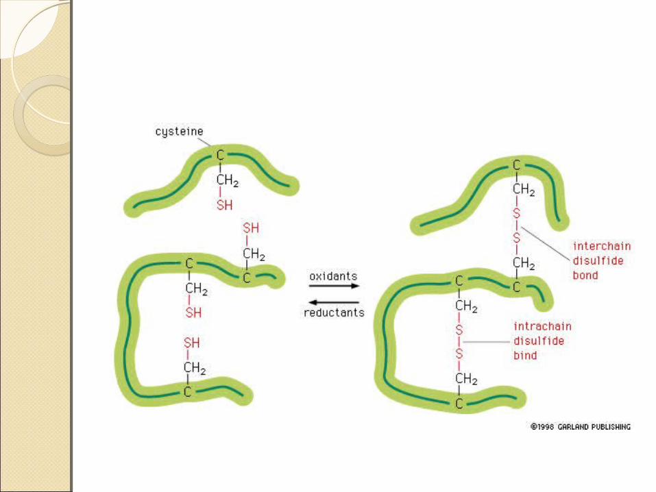
Disulfide Bonds Disulfide Bonds • A disulfide bond is a covalent linkage
formed by the sulfhydryl group (-SH) of two cysteine residues to form cystine
• The folding of the polypeptide chain brings the cysteine residues near each other
• Disulfide linkage contributes to the stability of the three-dimensional shape of the protein molecule
• Disulfide bonds are found in proteins that are secreted by cells– Thought that these strong covalent bonds
help stabilize the structure of proteins and help prevent them from becoming denatured in the extra-cellular environment
Hydrophobic InteractionsHydrophobic Interactions• Recall that amino acids with non-
polar side chains tend to be located in the interior of the polypeptide–Here, they associate with other
hydrophobic amino acids• Special Note–Proteins located in non-polar (lipid)
environments such as the phopholipid bilayer, tend to be in an opposite form• Hydrophobic amino acids are located on the
surface• Hydrophilic amino acids are located on the
interior

Ionic InteractionsIonic InteractionsNegatively charged groups
interact with positively charged groups◦Negatively charged groups
(-COO-) in the side chain of aspartate or glutamate
◦Positively charged groups (-NH3
+) in the side chain of lysine


Dipole MomentDipole MomentDipole Moment is
the measure of a molecule’s overall polarity
μ = Q * r◦ μ = Dipole
Moment◦ Q = charge◦ r = distance
between chargesMeasured in
debyes (\də-ˈbī\ )

Van der Waals ForcesVan der Waals ForcesA weak attractive
force between atoms or non-polar molecules caused by a temporary change in dipole moment ◦ Arising from a brief
shift of orbital electrons to one side of one atom or molecule, creating a similar shift in adjacent atoms or molecules.