amphioxus sarm involved in neural development … journal of immunology amphioxus sarm involved in...
TRANSCRIPT

of May 31, 2018.This information is current as
of TLR SignalingDevelopment May Function as a Suppressor Amphioxus SARM Involved in Neural
XuAnlongHuang, Huiling Liu, Xin Tao, Shengfeng Huang and
Shaochun Yuan, Kui Wu, Manyi Yang, Liqun Xu, Ling
http://www.jimmunol.org/content/184/12/6874doi: 10.4049/jimmunol.0903675May 2010;
2010; 184:6874-6881; Prepublished online 14J Immunol
Referenceshttp://www.jimmunol.org/content/184/12/6874.full#ref-list-1
, 12 of which you can access for free at: cites 31 articlesThis article
average*
4 weeks from acceptance to publicationFast Publication! •
Every submission reviewed by practicing scientistsNo Triage! •
from submission to initial decisionRapid Reviews! 30 days* •
Submit online. ?The JIWhy
Subscriptionhttp://jimmunol.org/subscription
is online at: The Journal of ImmunologyInformation about subscribing to
Permissionshttp://www.aai.org/About/Publications/JI/copyright.htmlSubmit copyright permission requests at:
Email Alertshttp://jimmunol.org/alertsReceive free email-alerts when new articles cite this article. Sign up at:
Print ISSN: 0022-1767 Online ISSN: 1550-6606. Immunologists, Inc. All rights reserved.Copyright © 2010 by The American Association of1451 Rockville Pike, Suite 650, Rockville, MD 20852The American Association of Immunologists, Inc.,
is published twice each month byThe Journal of Immunology
by guest on May 31, 2018
http://ww
w.jim
munol.org/
Dow
nloaded from
by guest on May 31, 2018
http://ww
w.jim
munol.org/
Dow
nloaded from

The Journal of Immunology
Amphioxus SARM Involved in Neural Development MayFunction as a Suppressor of TLR Signaling
Shaochun Yuan,1 Kui Wu,1 Manyi Yang, Liqun Xu, Ling Huang, Huiling Liu,
Xin Tao, Shengfeng Huang, and Anlong Xu
Among five Toll/IL-1R resistance adaptors, sterile a and Toll/IL-1R resistance motif containing protein (SARM) is the only one
conserved from Caenorhabditis elegans to human. However, its physiologic roles are hardly understood, and its involvement in
TLR signaling remains debatable. In this study, we first demonstrated a predominant expression of amphioxus SARM (Bran-
chiostoma belcheri tsingtauense SARM) in neural cells during embryogenesis and its predominant expression in the digestive
system from larva to adult, suggesting its primitive role in neural development and a potential physiologic role in immunity. We
further found that B. belcheri tsingtauense SARM was localized in mitochondria and could attenuate the TLR signaling via
interacting with amphioxus MyD88 and tumor necrosis receptor associated factor 6. Thus, amphioxus SARM appears unique in
that it may play dual functions in neural development and innate immunity by targeting amphioxus TLR signaling. The Journal
of Immunology, 2010, 184: 6874–6881.
The innate immune system relies on evolutionarily con-served TLRs to recognize diverse microbial molecularstructures (1). Most TLRs depend on a family of adaptor
proteins containing the Toll/IL-1R resistance (TIR) domain totransduce signals (2). In vertebrates, five TIR adaptors have beenidentified, including MyD88, MyD88 adaptor-like protein (MAL),TIR-domain containing adaptor inducing IFN-b (TRIF), tumornecrosis receptor associated factor 6 (TRAM), and sterile a andTIR motif containing protein (SARM) (2). MyD88, the first to bedescribed, plays a role in signal transduction by all TLRs exceptTLR3 (3, 4). When recruited by TLR4 and TLR2, MyD88reassembles with MAL for the early activation of NF-kB andMAPKs (5–7). TRIF is specifically used by TLR3 and TLR4 toactivate NF-kB and another transcription factor, IRF3. TRAMcoupled with TRIF is essential for TLR4 signals (6, 8, 9).
The fifth TIR adaptor, SARM, is the only one conserved fromCaenorhabditis elegans to mammals (10). It is the most recentTIR-containing adaptor protein to be characterized, and little isknown about its precise role in immune signaling. A SARM ortho-log contains evolutionarily conserved protein structure comprisingtwo sterile a motifs, an N-terminal heat armadillo repeat motif(ARM), and a C-terminal TIR domain (10). A study of the C.elegans SARM homolog (TIR-1) showed its importance to anefficient immune response and in the development of olfactoryneurons (11, 12). The knockdown of TIR-1 by RNA interferenceresulted in decreased C. elegans survival when challenged withfungal and bacterial infections (11). However, this effect was in-dependent of TOL-1, the only TLR homolog in C. elegans (11).Unlike other TIR-containing adaptors, overexpression of hu-
man SARM in vitro does not induce NF-kB activation. However,it can serve as a negative regulator of TRIF-dependent TLR sig-naling, but not of MyD88-dependent pathways (13). Overexpres-sion of human SARM leads to the reduction in TRIF-downstreamgene expression and enhances TRIF-dependent cytokine produc-tion (13). Recently, a SARM ortholog in the horseshoe crab withthe ability to downregulate human TRIF-dependent TLR signalingand response to infection was also characterized (14). However,macrophages from SARM knockout mice respond normally toTLR ligands, such as polyinosinic:polycytidylic acid, CpG, andLPS (15). Therefore, the authors concluded that, in contrast tothe results of Carty et al. (13), mouse SARM does not have a non-redundant role in regulating TLR signaling. Rather, mouse SARMis primarily expressed in neurons to regulate neuronal death duringoxygen and glucose deprivation (15), and its immune functionoccurs to restrict viral infection and neuronal injury by modulatingthe activation of resident CNS inflammatory cells (16).Because the first vertebrate TLR was identified in 1997, an in-
creasingly detailed picture is emerging about its intracellular sig-naling network. However, no more than 10 studies of SARM havebeen published, and such limited studies reported different obser-vations among species. Thus, investigating the physiologic roles ofSARM in amphioxus, an organism representing a key evolutionarystagefrominvertebrate tovertebrate,willhelp to revealhowits role inTLR signaling has been altered, and whether its crucial role in
State Key Laboratory of Biocontrol, National Engineering Research Center of SouthChina Sea Marine Biotechnology, Department of Biochemistry, College of LifeSciences, Sun Yat-sen (Zhongshan) University, Guangzhou, China
1S.Y. and K.W. contributed equally to this work.
Received for publication November 17, 2009. Accepted for publication April 13,2009.
This work was supported by Project 2007CB815800 of the National Basic ResearchProgram (973), Project 2008AA092603 of the State High-Tech Development Project(863), Project 2007DFA30840 of the International S&T Cooperation Program fromthe Ministry of Science and Technology of China, a key project (0107) from theMinistry of Education, the First Degree Supporting Project 20080440119 and theSecond Especial Supporting Project 200902341 of the China Postdoctoral ScienceFoundation, and Project 30901305 of the National Nature Science Foundation ofChina. A.X. is a recipient of the Outstanding Young Scientist award from the Na-tional Nature Science Foundation of China.
Address correspondence and reprint requests to Dr. Anlong Xu, Department of Bio-chemistry, College of Life Sciences, Sun Yat-Sen (Zhongshan) University, 135 Xin-GangXi Road, 510275, Guangzhou, China. E-mail address: [email protected]
Abbreviations used in this paper: ARM, armadillo repeat motif; bbtSARM, Bran-chiostoma belcheri tsingtauense sterile a and TIR motif containing protein;bbtMyD88, Branchiostoma belcheri tsingtauense MyD88; g, gill; HEK, human em-bryonic; i, intestine; MAL, MyD88 adaptor-like protein; n, notochord; o, ovary;PHB1, prohibitin 1; s, spermary; SARM, sterile a and TIR motif containing protein;TIR, Toll/IL-1R resistance; TRAF6, tumor necrosis receptor associated factor 6;TRIF, TIR-domain containing adaptor inducing IFN-b.
Copyright� 2010 by TheAmericanAssociation of Immunologists, Inc. 0022-1767/10/$16.00
www.jimmunol.org/cgi/doi/10.4049/jimmunol.0903675
by guest on May 31, 2018
http://ww
w.jim
munol.org/
Dow
nloaded from

neuronal development is conserved progressing from C. elegans tohumans via intermediate species, finally will help to understand thefunctional transition of TLRs from development to immunity.
Materials and MethodsAnimals, embryos, cells, and reagents
Adult Chinese amphioxus (Branchiostoma belcheri tsingtauense) wasobtained from Qingdao, China. During the breeding season, blastulas, gas-trulas, neurulas, and larvaewere collected. Human embryonic kidney (HEK)293T cells and Hela cells were maintained in DMEM supplemented with10% FCS. LPS from Escherichia coli (O111:B4), IL-1a and TNF-a humanrecombinants were all purchased from Sigma-Aldrich (St. Louis, MO).
Cloning of B. belcheri tsingtauense sterile a andTIR motif-containing protein cDNAs
Our previous annotation of the immune related genes in the B. floridaegenome identified an SARM ortholog. Based on this sequence, a partialsequence of B. belcheri tsingtauense SARM (bbtSARM) was cloned fromChinese amphioxus intestinal cDNA by specific primer pair derived fromB. floridae SARM. Subsequently, 59RACE and 39RACE were conductedaccording to the manufacturer’s protocol using a GeneRACE Kit (Invi-trogen, Carlsbad, CA) for full-length sequence cloning.
Wholemount and section in situ hybridization
A fragment of bbtSARM was amplified and cloned into T-easy vector(Promega, Madison, WI). DIG-labeled sense (synthesized by T7 RNA poly-merase) and anti-sense (synthesized by SP6 RNA polymerase) probes weregenerated based on the T-easy construct according to the manufacturer’sprotocol using a DIG RNA Labeling Kit (Roche, Mannheim, Germany).Whole-mount in situ hybridizations of embryos of different developmentalstages were performed according to the protocol by Holland (17). Sectionin situ hybridization used the same probes and followed the procedureindicated by Yuan et al. (18).
Acute immune challenges of adult amphioxus and real-timePCR
Either 105 CFU of Vibrio vulnificus or 15 ml per animal of LPS (1 mg/ml)in PBS was injected into the amphioxus coelom. The challenged animalswere cultured in separate tanks and collected at 2, 4, 8, 12, and 24 hpostinjection. Intestines from five individuals were combined in a singlesample for RNA extraction. Intestines from five PBS-injected animals(15 ml per animal) were also collected at the same time as controls.
Total RNA from infected samples was prepared, treated with DNAase,and reverse transcribed (Invitrogen). The PCRs were run in triplicate usingthese conditions: 2 min at 95˚C followed by 40 cycles of 30s at 95˚C, 15s at60˚C, and 1 min at 72˚C. Data were quantified using the 22DDCt methodbased on Ct values of bbtSARM and b-actin. For expression followingchallenge, folds were normalized to the expression in PBS injected ani-mals. Values were considered to be significant at p , 0.05.
Expression plasmids
For the expression of bbtSARM in 293T cells, PCR fragments encodingfor aas 1–734 of bbtSARM fused with 39Flag tag were inserted intopcDNA3.0. For the coimmunoprecipitation test, the full-length B. belcheritsingtauenseMyD88 (bbtMyD88) andB. belcheri tsingtauense tumor necrosisreceptor associated factor (TRAF6) from Chinese amphioxus fused with flag-tag, and the full length bbtSARM fused with hemaglutinin, were inserted intopcDNA3.0. The vector containing full-length humanMyD88 fusedwith Flag-tag was provided by Dr. H. Tang. For testing the subcellular location ofbbtSARM in Hela cells, PCR fragments encoding for aa 1–734, 1–400,401–734, and 550–734 of bbtSARMwere inserted into pEGFP-N1 expressionvector (Clontech, Mountain View, CA) and designated as SG-F, SG-1, SG-2, and SG-3.
Immunofluorescence imaging
Hela cells were seeded onto coverslips (10 310 mm) in a 24-well plate for16 h and transfected with 400 ng indicated expression plasmids. At 24 hposttransfection, cells were fixed for 15 min in a 4% formaldehyde solu-tion, washed twice in PBS, and permeabilized by the treatment for 10 minwith PBST (0.1% Triton X-100 in PBS). After blocking for 1 h, cells wereincubated with 5 mg/ml anti-Flag mAb for 45 min, washed twice in PBST,incubated with a second Ab, triple-washed in PBS, labeled with 0.2mg/ml DAPI in PBS for 5 min, washed for 3–10 min in PBS, and mounted
in MOWIOL R4-88 Reagent (Calbiochem) and photographed by ZEISSAxioVision 4 microscopy (Carl Zeiss MicroImaging, Thornwood, NY).
Transfection, reporter assays, and measurement of cytokineconcentrations
For the basic test, HEK293T cells were plated in 48-well plates (23 104 cellsper well) and transfected the next day with 400 ng per well DNA. The mixedDNA contained the indicated amount of expression vectors, 10 ng perwell of phRL-TK reporter plasmid (Promega) to allow normalization ofdata for transfection efficiency, and 100 ng per well of the NF-kBresponse promoter luciferase reporter and the addition of pcDNA3.0. Forcytokine stimulation, 100 ng/ml IL-1a or 100 ng/ml TNF-a were added tothe cell medium at 24 h posttransfection as described above, and cellswere incubated for an additional 12 h. All samples were measured bya luciferase reporter assay system (Promega) according to the manufac-turer’s instructions. For measurement of cytokine concentrations, cellswere transected with indicated plasmids, and incubated for 24 h; super-natants were collected, and IL-8 concentration was measured by ELISA(R&D Systems, Minneapolis, MN).
Coimmunoprecipitation
HEK293T cells in six-well dishes (53 106 cells per well) were transfectedwith 3 mg DNA plasmids (1.5 mg for each expression vector). At 48 hposttransfection, cells were lysed and incubated with primary Abs (4 mganti-FLAG [M2]; Sigma-Aldrich) at 4˚C overnight and then incubated withprotein G sepharose for an additional 4 h at 4˚C. Analysis was conductedusing SDS-PAGE followed byWestern blot, using the ECL protocol (Amer-sham, GE Healthcare, Buckinghamshire, U.K.), with anti-hemaglutinin(1:1000), and anti-Flag (1:1000) mAb.
ResultsCloning and sequence analyses of bbtSARM
A full-length cDNA of 2802 bp was isolated from a B. belcheritsingtauense intestine cDNA library and designated as bbtSARM.With a 600 bp 39-untranslated region, bbtSARMencoded a polypep-tide of 734 aas with a highly conserved protein structure comprisingan N-terminal ARM repeat, a C-terminal TIR domain, and two cen-tral sterile amotifs. The overall sequence identity of bbtSARMwithother SARMhomologs ranged from 33 to 54%, whereas the identityin theTIRdomain ranged from45 to 65%.Comparedwith horseshoecrab and human SARM homologs (CrSARM and hsSARM),bbtSARMwasmore similar to hsSARM,asCrSARMis significantlylonger than its vertebrate counterpart (Fig. 1A). Because the N-terminus of CrSARM protein does not contain any recognizablemotif, and the N terminal deletion of CrSARM (CrSARMDN)remains functional, the additional N-terminal tail may be redundantand be deleted during evolution (14). A phylogenetic tree showedthat the evolutionary position of bbtSARM, which shares a commonancestor with vertebrate SARM, is in accordance with the transitionstatus of amphioxus in the evolution of chordates (Fig. 1B). Thus, thecharacterization of bbtSARM is of particular importance in sheddinglight on the evolution and change in the function of SARM frominvertebrates to vertebrates and in resolving functional confusionwith other reported SARM homologs.
bbtSARM has dual functions in neuronal development and hostdefense
Because null-mutant Drosophila SARM is lethal and C. elegansSARM has a crucial function in the development of olfactoryneurons, we used whole-mount in situ hybridization to investigatethe potential function of bbtSARM during amphioxus embryogen-esis. As shown in Fig. 2, bbtSARM was abundant from zygote toblastula. As embryos developed through the late gastrula, the ex-pression of bbtSARM gradually concentrated in the anterior mes-oblast, which develops into the dorsal nerve cord during neuru-lation. Subsequently, transcripts of bbtSARM were abundant inthe mesoblast somite, nerve tube, and notochord from neurulathrough 24 h larva. In addition, bbtSARM was expressed in the
The Journal of Immunology 6875
by guest on May 31, 2018
http://ww
w.jim
munol.org/
Dow
nloaded from

archenteron (the anterior gut diverticulum) during these stages. At48 h, bbtSARM was detected only in the developing midgut di-verticulum, intestine, and atriopore (Fig. 2).
In adult female amphioxus, expression of bbtSARM was stronglydetected in the epithelial cells of the gut and weakly in the connectivetissue and cell membrane of mature oocytes (Fig. 3A, 3C), but not
FIGURE 1. Sequence analysis and comparison of
bbtSARM. A, Domain topology of bbtSARM com-
pared with CrSARM (horseshoe crab) and hsSARM
(human). B, Neighbor-joining tree of bbtSARM
with 23 other SARM sequences was constructed
with the MEGA version 3.1 (www.megasoftware.
net/) using the full-length sequence. The numbers
at the nodes indicate bootstrap values.
FIGURE 2. BbtSARM related to neural develop-
ment. A, Four-cell stage. BbtSARM was a maternal ex-
pression gene. B, BbtSARM was abundant in the
blastula. C and D, Blastopore view and side view of late
gastrula showed the expression of BbtSARM in the
arterial mesoblast, which develops into the dorsal nerve
cord at neurulation. E–G, Side view of late neurula, 16-h
larva, and 24-h larva, showing that bbtSARM is abun-
dant in mesoblast somites, nerve tube, notochord, and
archenteron. H, Two-day larva showing bbtSARM only
in developing midgut diverticulum, intestine, and atrio-
pore. Scale bar, 100 mm for whole embryos. Original
magnification 3200.
6876 FUNCTIONAL CHARACTERIZATION OF AMPHIOXUS SARM
by guest on May 31, 2018
http://ww
w.jim
munol.org/
Dow
nloaded from

abundant in gill epidermal cells and spermatic cells (Fig. 3B). Thispredominant expression in the amphioxus digestive system is con-sistent with that observed in the larva. Because the digestive systemis considered to be the primary line of defense in amphioxus, tofurther confirm the immune significance of bbtSARM, real-time RT-PCR analysis was performed to monitor the expression of bbtSARMtranscripts in adults challenged with the Gram-negative bacteriaV. vulnificus and its cell wall component LPS. In response to acuteimmune challenge, bbtSARM was not altered dramatically, but wasweakly upregulated by Gram-negative bacteria V. vulnificus and LPS(Fig. 3E, 3F). Considering the nature of SARM as an adaptor, thisexpression profile might imply the immune relevance of bbtSARMin amphioxus. Thus, bbtSARM plays an important role in neuronaldevelopment, as demonstrated in other species, and might participatein amphioxus immunity.
Amphioxus SARM located in mitochondria
The location of mouse SARM in mitochondria is important for thisconserved molecule to recruit JNK3 to regulate neuronal deathupon stress (15). To further investigate the functional implicationsof the subcellular location of bbtSARM, we transfected Helacells with a bbtSARM-GFP. Fluorescent imaging microscopy ofbbtSARM-GFP fusion protein showed green fluorescence in thecytoplasm at the location similar to that in the mitochondria ap-paratus (Fig. 4B). To investigate whether these punctuated struc-tures of bbtSARM were exactly colocalized with mitochondria,a mitochondria-targeting vector was constructed by fusing a pro-hibitin 1 (PHB1) sequence into the pEGFP-N1 vector, becausePHB1 is an evolutionally conserved mitochondrial protein local-ized in the inner membrane and has been used as a mitochondriamarker in many studies. Next, the PHB1-GFP vector was coex-pressed with a bbtSARM-Flag vector that was further costainedwith rhodamine in Hela cells (19). As shown in Fig. 4C, thebbtSARM-Flag fusion protein colocalized strongly with mito-chondria in structures along which mitochondria move. Similarto mouse SARM, the distribution and shape of mitochondria inHela cells varied depending on their expression of bbtSARM (Fig.4C). In cells expressing a low to moderate level of bbtSARM,mitochondria were distributed widely throughout the cytoplasm
(Fig. 4C). In cells with high levels of bbtSARM expression, mi-tochondria were round and clustered around the nucleus (Fig. 4C).Because we could not predict a mitochondria-targeting sequence
in bbtSARM, we used several truncated GFP constructs to verifywhich domain was crucial for its mitochondrial association (Fig.4A). The truncated segment with only an N terminal ARM repeatshowed similar localization, whereas a truncated segment lackingthe ARM repeat was instead distributed evenly throughout thecytoplasm (Fig. 4B). In contrast, a construct consisting of onlythe TIR domain was distributed evenly throughout the cytoplasmand the nuclei (Fig. 4B). Thus, ARM repeat controls the associa-tion of bbtSARM with mitochondria.
Amphioxus SARM can attenuate the NF-kB activationmediated by amphioxus MyD88
Given that SARM is a cytosolic TIR domain-containing protein,we sought to determine whether its function is similar to that ofother TIR domain-containing proteins. We first compared the abilityof bbtSARM and bbtMyD88 to drive transcription factor activationand gene induction. Constant to previous reports, we found that, al-though overexpression of bbtMyD88 led to activation of NF-kB in293T cells via a fashion similar human MyD88 (20), overexpressionof bbtSARM had no such effect. However, when coexpressed withbbtMyD88, bbtSARM can attenuate the NF-kB activation mediatedby bbtMyD88 in a dose-dependent manner (Fig. 5A).To further confirm the observed inhibition on amphioxus MyD88-
mediated NF-kB activation, we examined the effect of bbtSARMon human MyD88-dependent gene induction. Results showed thatthe production of IL-8 (solely mediated via MyD88), was affectedby bbtSARM expression in a dose-dependent manner (Fig. 5B). Inaddition, we showed that IL-1a–induced NF-kB activation, whichis mediated exclusively by MyD88, was affected by bbtSARMexpression (Fig. 5C). Because the activation of NF-kB is alsoa hallmark of TNF signaling, to determine whether the inhibitionby bbtSARM is restricted to TLR signaling, we investigated theeffect of bbtSARM expression on NF-kB activation induced byTNF-a. Results showed that overexpression of bbtSARM did notaffect the TNF-a–induced NF-kB activation (Fig. 5C), suggestingthat bbtSARM may be a specific suppressor for the TLR signaling.
FIGURE 3. The tissue distribution and expression pattern after bacterial challenge of bbtSARM. A, Section in situ hybridization analyses of bbtSARM
anti-sense probe show predominant expression in the intestine, connective tissue, and cell membrane of mature oocytes. B, BbtSARM was not abundant on
the epidermal cells of the gill, which is also thought to be the defense organ of the amphioxus. C, Macroscopic view of the hybridization signals of
bbtSARM in intestine and mature oocytes. D, Section in situ hybridization analysis of bbtSARM using sense probe as negative control. Scale bar, 500 mm
(A, B, D) or 200 mm (C). Original magnification 380 (A, B, D) or 3200 (C). E and F, Quantitative real-time RT-PCR analysis of the expression of
bbtSARM after LPS or Gram-negative bacterial challenge. Results were presented as fold-induction of mRNA expression after PBS injection using the 22
DDCtmethod from two parallel experiments done in triplicate. Endogenous control for quantification was cytoplasmic b-actin. Values were considered to be
significant at p , 0.05. g, gill; i, intestine; n, notochord; o, ovary; s, spermary.
The Journal of Immunology 6877
by guest on May 31, 2018
http://ww
w.jim
munol.org/
Dow
nloaded from

Amphioxus SARM may target MyD88 and TRAF6
To obtain further information on the mechanism of bbtSARMfunction in TLR signaling, we transfected the several truncatedmutants with amphioxus MyD88 for luciferase activity analyses. Asshown in Fig. 5D, all truncated mutants are functional. Given thepresence of the TIR homotypic interaction domain in those TIRdomain-containing adaptors, we speculated that SARM inhibition ofMyD88 signaling might be accomplished through its direct interac-tion with amphioxus MyD88. To test that possibility, we conductedcoimmunoprecipitation trials. Results showed bbtSARM to interactwith bbtMyD88 (Fig. 5F). We further observed that overexpressionof bbtSARM resulted in the inhibition of NF-kB activation mediated
by amphioxus TRAF6 (Fig. 5E), which shares high sequence
conservation and functions in a manner similar to human TRAF6
(18). Thus, sequence analysis was performed to discover whether
bbtSARM contained a PxExxAr/Ac TRAF6-binding motif, and then
two putative TRAF6-binding motifs were identified—one at aa po-
sition 299–304 and a second at 661–667 (Fig. 5G). Such motifs have
not been identified in other SARM homologs, but have been de-
scribed in IL-1R–associated kinases, MAL, and TRIF (21, 22). To
test whether bbtSARM can interact with bbtTRAF6, coimmunopre-
cipitation experiments were performed and demonstrated that bbt-
SARM interacted with bbtTRAF6 (Fig. 5F). We also observed that
bbtSARM can colocalize with bbtTRAF6 in Hela cells (Fig. 5H).
FIGURE 4. Mitochondria localization of bbtSARM.
A, Name and structure of fusion proteins used in this
study. B, Intracellular localization of bbtSARM and its
truncated mutants fused with EGFP in Hela cells. C,
bbtSARM was well colocalized with mitochondria,
and the shape of mitochondria in Hela cells varied
depending on their expression of bbtSARM. Nucleus
was stained by DAPI. Original magnification3400 (B),
3630 (C).
6878 FUNCTIONAL CHARACTERIZATION OF AMPHIOXUS SARM
by guest on May 31, 2018
http://ww
w.jim
munol.org/
Dow
nloaded from
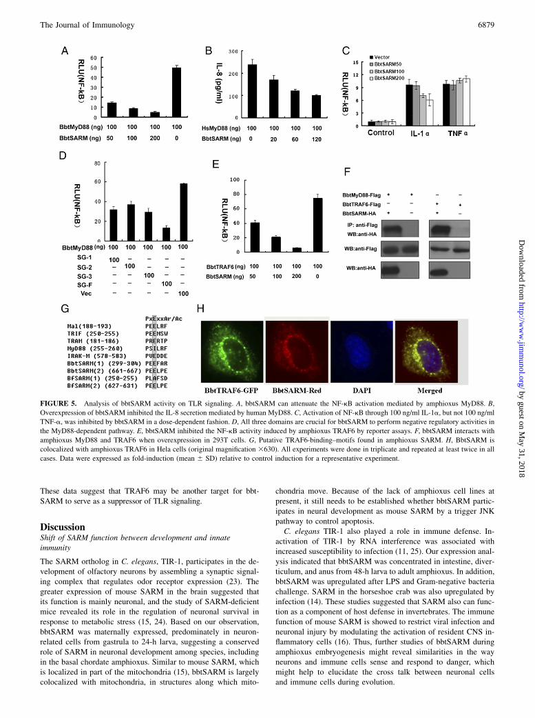
These data suggest that TRAF6 may be another target for bbt-SARM to serve as a suppressor of TLR signaling.
DiscussionShift of SARM function between development and innateimmunity
The SARM ortholog in C. elegans, TIR-1, participates in the de-velopment of olfactory neurons by assembling a synaptic signal-ing complex that regulates odor receptor expression (23). Thegreater expression of mouse SARM in the brain suggested thatits function is mainly neuronal, and the study of SARM-deficientmice revealed its role in the regulation of neuronal survival inresponse to metabolic stress (15, 24). Based on our observation,bbtSARM was maternally expressed, predominately in neuron-related cells from gastrula to 24-h larva, suggesting a conservedrole of SARM in neuronal development among species, includingin the basal chordate amphioxus. Similar to mouse SARM, whichis localized in part of the mitochondria (15), bbtSARM is largelycolocalized with mitochondria, in structures along which mito-
chondria move. Because of the lack of amphioxus cell lines atpresent, it still needs to be established whether bbtSARM partic-ipates in neural development as mouse SARM by a trigger JNKpathway to control apoptosis.C. elegans TIR-1 also played a role in immune defense. In-
activation of TIR-1 by RNA interference was associated withincreased susceptibility to infection (11, 25). Our expression anal-ysis indicated that bbtSARM was concentrated in intestine, diver-ticulum, and anus from 48-h larva to adult amphioxus. In addition,bbtSARM was upregulated after LPS and Gram-negative bacteriachallenge. SARM in the horseshoe crab was also upregulated byinfection (14). These studies suggested that SARM also can func-tion as a component of host defense in invertebrates. The immunefunction of mouse SARM is showed to restrict viral infection andneuronal injury by modulating the activation of resident CNS in-flammatory cells (16). Thus, further studies of bbtSARM duringamphioxus embryogenesis might reveal similarities in the wayneurons and immune cells sense and respond to danger, whichmight help to elucidate the cross talk between neuronal cellsand immune cells during evolution.
FIGURE 5. Analysis of bbtSARM activity on TLR signaling. A, bbtSARM can attenuate the NF-kB activation mediated by amphioxus MyD88. B,
Overexpression of bbtSARM inhibited the IL-8 secretion mediated by human MyD88. C, Activation of NF-kB through 100 ng/ml IL-1a, but not 100 ng/ml
TNF-a, was inhibited by bbtSARM in a dose-dependent fashion. D, All three domains are crucial for bbtSARM to perform negative regulatory activities in
the MyD88-dependent pathway. E, bbtSARM inhibited the NF-kB activity induced by amphioxus TRAF6 by reporter assays. F, bbtSARM interacts with
amphioxus MyD88 and TRAF6 when overexpression in 293T cells. G, Putative TRAF6-binding–motifs found in amphioxus SARM. H, BbtSARM is
colocalized with amphioxus TRAF6 in Hela cells (original magnification 3630). All experiments were done in triplicate and repeated at least twice in all
cases. Data were expressed as fold-induction (mean 6 SD) relative to control induction for a representative experiment.
The Journal of Immunology 6879
by guest on May 31, 2018
http://ww
w.jim
munol.org/
Dow
nloaded from
Potential function of SARM in innate immunity by targetingTLR signaling
Among five TIR adaptors, SARM is the only one conserved fromC. elegans to mammals. The prominent contribution of all otherknown TIR adaptors to innate immunity is well documented andhas fueled the expectation that SARM would play a similar role.However, although C. elegans TIR-1 has a positive function inimmunity, it does not appear to mediate signaling from C. elegansTOL, the sole TLR in C. elegans, but acts as a component of a p38MAPK signaling cassette (11, 12, 26). In contrast, human SARMis a specific negative regulator of TRIF signaling through its tar-geting of TRIF for innate immune responses (13). Subsequent tothe study by Carty et al. (13) a study of mouse SARM showed thatit does not have a nonredundant role in regulating macrophageresponses to polyinosinic:polycytidylic acid and LPS, which mayrule out a role in TLR signaling (15). Thus, the involvement ofSARM in TLR signaling is still debatable, and the molecular basisfor this functional difference remains unclear.Because the amphioxus genome does not contain an ortholog to
mammalian TRIF, IRF3, IRF7, and IFN-b, the MyD88-dependentpathway seems to be the key route of TLR signaling in the amphi-oxus (27). In addition, amphioxus MyD88 and TRAF6 share highsequence similarities and function in a fashion similar to their humancounterparts in HEK293T cells, indicating the molecular conserva-tion of the MyD88-dependent pathway between amphioxus andhumans (18, 20). Therefore, our observation that bbtSARM couldattenuate the NF-kB activation mediated by amphioxus MyD88and TRAF6 in 293T cells may present some natural roles ofthis conserved molecule in amphioxus cells, adding evidence thatSARM may be a negative regulator of TLR signaling at the basalchordate stage. Considering its roles in neural development, furtherstudy to reveal how bbtSARM regulates neural development bytargeting MyD88 dependent pathway would shed light on thealternation of TLR function between development and immunitywhen invertebrates developed into vertebrates.
Molecular basis for bbtSARM to participate in TLR signaling
Although the ARM repeat for mouse and amphioxus SARM isimportant for their localization with mitochondria (15), this spe-cific localization seems not to be indispensable for bbtSARM inTLR signaling, as the truncated mutant without ARM repeat isstill functional. The C. elegans TIR-1, when truncated to only thetwo sterile a motifs and the TIR domain, showed a stronger gain-of-function developmental phenotype than the full length protein(11, 25). The N terminus-deleted SARM in humans and horseshoecrab were also more efficient in the inhibition of TRIF-dependentpathways (13, 14). These data suggest that the negative roles ofSARM in TLR signaling may be determined by its sterile a motifsand TIR domain, which provide interface to interact with differentmolecules. Similar to other TIR adaptors, the TIR domain ofbbtSARM mediated the direct TIR–TIR interactions. For example,amphioxus SARM could interact with MyD88, whereas humanSARM interacts with TRIF. It may be that the binding of SARMto MyD88 prevents the formation of the MyD88 complex (28).Unlike the TIR domain, proteins with sterile a motifs exist in allsubcellular locations and could form multimeric complexes witha wide variety of proteins (29), providing another interface forbbtSARM to interact with some unidentified molecules involvedin TLR signaling. In addition, we identified two TRAF6-bindingmotifs in bbtSARM protein and confirmed their direct interactionwhen overexpression. Thus, another possibility may be that theinteraction of SARM with TRAF6 physically prevents engage-ment of TRAF6 with its upstream activators or downstream
effectors (30, 31). Therefore, we are not surprised to observe thatall three conserved protein domains found in bbtSARM are nec-essary for its inhibition activity in TLR signaling. Owing to thelack of amphioxus cell lines for functional analyses, we have toborrow a mammalian cell line system to study the functions ofamphioxus molecules, which may present limitations for thesecross-species approaches. Nevertheless, this first report of amphi-oxus SARM establishes its conserved roles in neural developmentand provides evidence for its involvement in innate immunity,which should help to provide a picture on the functional evolutionof SARM from C. elegans to humans.
AcknowledgmentsWe thank Dr. Hong Tang for providing human TRIF and human MyD88
plasmids and for constructive comments on this study.
DisclosuresThe authors have no financial conflicts of interest.
References1. Takeda, K., and S. Akira. 2007. Toll-like receptors. Curr. Protoc. Immunol. 14:
14.12.2. Kenny, E. F., and L. A. O’Neill. 2008. Signalling adaptors used by Toll-like
receptors: an update. Cytokine 43: 342–349.3. Jenkins, K.A., and A. Mansell. 2009. TIR-containing adaptors in Toll-like re-
ceptor signalling. Cytokine 29: 237–244.4. Medzhitov, R., P. Preston-Hurlburt, E. Kopp, A. Stadlen, C. Chen, S. Ghosh, and
C. A. Janeway, Jr. 1998. MyD88 is an adaptor protein in the hToll/IL-1 receptorfamily signaling pathways. Mol. Cell 2: 253–258.
5. Fitzgerald, K. A., E. M. Palsson-McDermott, A. G. Bowie, C. A. Jefferies,A. S. Mansell, G. Brady, E. Brint, A. Dunne, P. Gray, M. T. Harte, et al.2001. Mal (MyD88-adapter-like) is required for Toll-like receptor-4 signal trans-duction. Nature 413: 78–83.
6. Sheedy, F. J., and L. A. O’Neill. 2007. The Troll in Toll: Mal and Tram asbridges for TLR2 and TLR4 signaling. J. Leukoc. Biol. 82: 196–203.
7. Yamamoto, M., S. Sato, H. Hemmi, H. Sanjo, S. Uematsu, T. Kaisho,K. Hoshino, O. Takeuchi, M. Kobayashi, T. Fujita, et al. 2002. Essential role forTIRAP in activation of the signalling cascade shared by TLR2 and TLR4. Nature420: 324–329.
8. Han, K. J., X. Su, L. G. Xu, L. H. Bin, J. Zhang, and H. B. Shu. 2004. Mech-anisms of the TRIF-induced interferon-stimulated response element and NF-kappaB activation and apoptosis pathways. J. Biol. Chem. 279: 15652–15661.
9. Kagan, J. C., T. Su, T. Horng, A. Chow, S. Akira, and R. Medzhitov. 2008.TRAM couples endocytosis of Toll-like receptor 4 to the induction of interferon-beta. Nat. Immunol. 9: 361–368.
10. Mink, M., B. Fogelgren, K. Olszewski, P. Maroy, and K. Csiszar. 2001. A novelhuman gene (SARM) at chromosome 17q11 encodes a protein with a SAM motifand structural similarity to Armadillo/beta-catenin that is conserved in mouse,Drosophila, and Caenorhabditis elegans. Genomics 74: 234–244.
11. Couillault, C., N. Pujol, J. Reboul, L. Sabatier, J. F. Guichou, Y. Kohara, andJ. J. Ewbank. 2004. TLR-independent control of innate immunity in Caenorhab-ditis elegans by the TIR domain adaptor protein TIR-1, an ortholog of humanSARM. Nat. Immunol. 5: 488–494.
12. Tenor, J. L., and A. Aballay. 2008. A conserved Toll-like receptor is required forCaenorhabditis elegans innate immunity. EMBO Rep. 9: 103–109.
13. Carty, M., R. Goodbody, M. Schroder, J. Stack, P. N. Moynagh, and A. G. Bowie.2006. The human adaptor SARM negatively regulates adaptor protein TRIF-dependent Toll-like receptor signaling. Nat. Immunol. 7: 1074–1081.
14. Belinda, L. W., W. X. Wei, B. T. Hanh, L. X. Lei, H. Bow, and D. J. Ling. 2008.SARM: a novel Toll-like receptor adaptor, is functionally conserved from ar-thropod to human. Mol. Immunol. 45: 1732–1742.
15. Kim, Y., P. Zhou, L. Qian, J. Z. Chuang, J. Lee, C. Li, C. Iadecola, C. Nathan,and A. Ding. 2007. MyD88-5 links mitochondria, microtubules, and JNK3 inneurons and regulates neuronal survival. J. Exp. Med. 204: 2063–2074.
16. Szretter, K. J., M. A. Samuel, S. Gilfillan, A. Fuchs, M. Colonna, andM. S. Diamond. 2009. The immune adaptor molecule SARM modulates tumornecrosis factor alpha production and microglia activation in the brainstem andrestricts West Nile Virus pathogenesis. J. Virol. 83: 9329–9338.
17. Holland, P. W. 1999. Wholemount in situ hybridization to amphioxus embryos.Methods Mol. Biol. 97: 641–644.
18. Yuan, S., T. Liu, S. Huang, T. Wu, L. Huang, H. Liu, X. Tao, M. Yang, K. Wu,Y. Yu, et al. 2009. Genomic and functional uniqueness of the TNF receptor-associated factor gene family in amphioxus, the basal chordate. J. Immunol. 183:4560–4568.
19. Kasashima, K., M. Sumitani, M. Satoh, and H. Endo. 2008. Human prohibitin 1maintains the organization and stability of the mitochondrial nucleoids. Exp. CellRes. 314: 988–996.
20. Yuan, S., S. Huang, W. Zhang, T. Wu, M. Dong, Y. Yu, T. Liu, K. Wu, H. Liu,M. Yang, et al. 2009. An amphioxus TLR with dynamic embryonic expression
6880 FUNCTIONAL CHARACTERIZATION OF AMPHIOXUS SARM
by guest on May 31, 2018
http://ww
w.jim
munol.org/
Dow
nloaded from

pattern responses to pathogens and activates NF-kappaB pathway via MyD88.
Mol. Immunol. 46: 2348–2356.21. Keating, S. E., G. M. Maloney, E. M. Moran, and A. G. Bowie. 2007.
IRAK-2 participates in multiple toll-like receptor signaling pathways to NFkap-
paB via activation of TRAF6 ubiquitination. J. Biol. Chem. 282: 33435–33443.22. Mansell, A., E. Brint, J. A. Gould, L. A. O’Neill, and P. J. Hertzog. 2004. Mal
interacts with tumor necrosis factor receptor-associated factor (TRAF)-6 to me-
diate NF-kappaB activation by toll-like receptor (TLR)-2 and TLR4. J. Biol.
Chem. 279: 37227–37230.23. Chuang, C. F., and C. I. Bargmann. 2005. AToll-interleukin 1 repeat protein at the
synapse specifies asymmetric odorant receptor expression via ASK1 MAPKKK
signaling. Genes Dev. 19: 270–281.24. Dalod, M. 2007. Studies of SARM1 uncover similarities between immune and
neuronal responses to danger. Sci. STKE 2007: pe73.25. Fuchs, B. B., and E. Mylonakis. 2006. Using non-mammalian hosts to study
fungal virulence and host defense. Curr. Opin. Microbiol. 9: 346–351.26. Kurz, C. L., M. Shapira, K. Chen, D. L. Baillie, and M. W. Tan. 2007. Caeno-
rhabditis elegans pgp-5 is involved in resistance to bacterial infection and heavy
metal and its regulation requires TIR-1 and a p38 map kinase cascade. Biochem.Biophys. Res. Commun. 363: 438–443.
27. Huang, S., S. Yuan, L. Guo, Y. Yu, J. Li, T. Wu, T. Liu, M. Yang, K. Wu, H. Liu, et al.2008. Genomic analysis of the immune gene repertoire of amphioxus reveals ex-traordinary innate complexity and diversity. Genome Res. 18: 1112–1126.
28. Ohnishi, H., H. Tochio, Z. Kato, K. E. Orii, A. Li, T. Kimura, H. Hiroaki,N. Kondo, and M. Shirakawa. 2009. Structural basis for the multiple interactionsof the MyD88 TIR domain in TLR4 signaling. Proc. Natl. Acad. Sci. USA 106:10260–10265.
29. Leone, M., J. Cellitti, and M. Pellecchia. 2008. NMR studies of a heterotypicSam-Sam domain association: the interaction between the lipid phosphataseShip2 and the EphA2 receptor. Biochemistry 47: 12721–12728.
30. Lamothe, B., A. D. Campos, W. K. Webster, A. Gopinathan, L. Hur, andB. G. Darnay. 2008. The RING domain and first zinc finger of TRAF6 coordinatesignaling by interleukin-1, lipopolysaccharide, and RANKL. J. Biol. Chem. 283:24871–24880.
31. Verstrepen, L., T. Bekaert, T. L. Chau, J. Tavernier, A. Chariot, and R. Beyaert.2008. TLR-4, IL-1R and TNF-R signaling to NF-kappaB: variations on a com-mon theme. Cell. Mol. Life Sci. 65: 2964–2978.
The Journal of Immunology 6881
by guest on May 31, 2018
http://ww
w.jim
munol.org/
Dow
nloaded from