amphiphilic ionic liquid induced fusion of phospholipid
TRANSCRIPT

doi.org/10.26434/chemrxiv.12752408.v1
Amphiphilic Ionic Liquid Induced Fusion of Phospholipid LiposomesSandeep Kumar, Navleen Kaur, Venus Singh Mithu
Submitted date: 03/08/2020 • Posted date: 04/08/2020Licence: CC BY-NC-ND 4.0Citation information: Kumar, Sandeep; Kaur, Navleen; Mithu, Venus Singh (2020): Amphiphilic Ionic LiquidInduced Fusion of Phospholipid Liposomes. ChemRxiv. Preprint.https://doi.org/10.26434/chemrxiv.12752408.v1
Membrane fusion is a key biological phenomenon with potential applications in biotechnology. In this work, weprovide biophysical and structural evidence that liposomes composed of POPC/POPG phospholipids undergofusion in the presence of ionic liquids containing 1-alkyl-3-methyl-imidazolium cations. The fusionphenomenon is confirmed using dynamic light scattering based size measurements, and Fluorescence baseddye leakage and lipid mixing assays. 1H-1H NOESY measurements using solid-state NMR spectroscopy wereperformed to obtain insights into fusion mechanism. It is found that ionic liquid induced splaying ofphospholipid chains is crucial for overcoming the hydration barrier between the merging bilayers. Also,transiently lived fusion-holes are formed at the initial stages of bilayer mixing resulting in a leaky fusionphenomenon.
Although considered as “green” alternatives to conventional solvents, ionic liquids can exhibit cytotoxicity byaltering the structural integrity of cellular membrane. Our study provides mechanistic details of the evolution ofphospholipid membrane structure resulting in membrane fusion when subjected to increasing ionic liquidconcentrations. We believe that findings of this study will further our understanding of ionic liquids inducedcytotoxicity and non-protein assisted membrane fusion.
File list (2)
download fileview on ChemRxivIonic liquid_membrane fusion.pdf (1.35 MiB)
download fileview on ChemRxivIonic liquid_membrane fusion_Supp Info.pdf (1.49 MiB)

Amphiphilic Ionic Liquid Induced Fusion of
Phospholipid Liposomes
Sandeep Kumar#, Navleen Kaur
#, Venus Singh Mithu
#*
#Department of Chemistry, Guru Nanak Dev University, Amritsar-143005, India
AUTHOR INFORMATION
Corresponding Author:
KEYWORDS. Ionic liquid; Membrane fusion; Fusion-holes; Lipid Splay; solid-state NMR.

ABSTRACT. We have investigated the impact of increasing concentrations of imidazolium-
based ionic liquids ([CnMIM]+[Br]
-) on structural integrity of large unilamellar vesicles (LUVs)
made of pure phosphatidylcholine (PC) and phosphatidylglycerol (PG) lipids. Calcein based dye
leakage assays were used to monitor permeability of LUVs in presence of ionic liquids. As ionic
liquid concentration approaches the critical micelle value, vesicle fusion occurs resulting in
unexpected quenching which is accompanied by a rapid dye leakage due to formation of
transiently lived fusion-holes. Vesicle fusion is confirmed using dynamic light scattering based
size measurements and Fluorescence based lipid mixing assays. 1H-
1H NOESY measurements
using solid-state NMR spectroscopy were performed to obtain insights into fusion mechanism.
While POPC LUVs are more prone to membrane fusion, the overall extent of fusion is higher in
POPG. Ionic liquid induced splaying of phospholipid chains is crucial for overcoming the
hydration barrier between the merging bilayers.

INTRODUCTION
Merging of inner and outer leaflets of two bilayers, also known as membrane fusion, is a key
biological phenomenon with potential applications in biotechnology.1 It is known to play a
crucial role in several biological events such as fertilization, viral infections, cellular trafficking,
endocytosis, exocytosis, and muscle development.2-3
In vivo, proteins and peptides act as
fusogenic agents and assist the process of membrane fusion.4-6
In vitro, metals ions, lipids, small
organic ligands, polymers, drugs, and surfactants have been shown to act as fusogenic agents.7
Mechanistically, protein-free fusion begins with the merging of outer leaflets (OL) of lipid
bilayer leading to formation of the stalk. The stalk further evolves into a hemifusion-diaphragm
where lipids from the inner leaflets (IL) of the merging bilayers start to mix, and eventually form
a fusion pore allowing contents of two liposomes to mix.1, 7
Fusogens can be classified into two
categories; lipid-soluble and water-soluble. Lipid-soluble fusogens act by perturbing the
structure of hydrocarbon chain thereby increasing the membrane contact area due to enhanced
surface exposure of lipid chains.8-9
Water-soluble fusogens, like metal ions, alter the membrane
surface potential or hydration of the polar head-groups allowing the fusing membranes to
achieve desired proximity.10-11
In this context, amphiphilic ionic liquid molecules with their long
lyophobic chains and charged head-groups possess properties of both type of fusogens and
hence, are interesting candidates to be explored as fusogenic agents.
Ionic liquids are the molten salts with exciting physiochemical properties.12
Due to a size
mismatch in composing ions, their melting points are considerable lower (100C) than regular
ionic salts.12
Unique characteristics of ionic liquids include high ionic conductivity, non-
volatility and a wide range of viscosity.12
Moreover, their properties can be easily tuned by
changing the combination of composing ions.12
These properties make ionic liquids acceptable
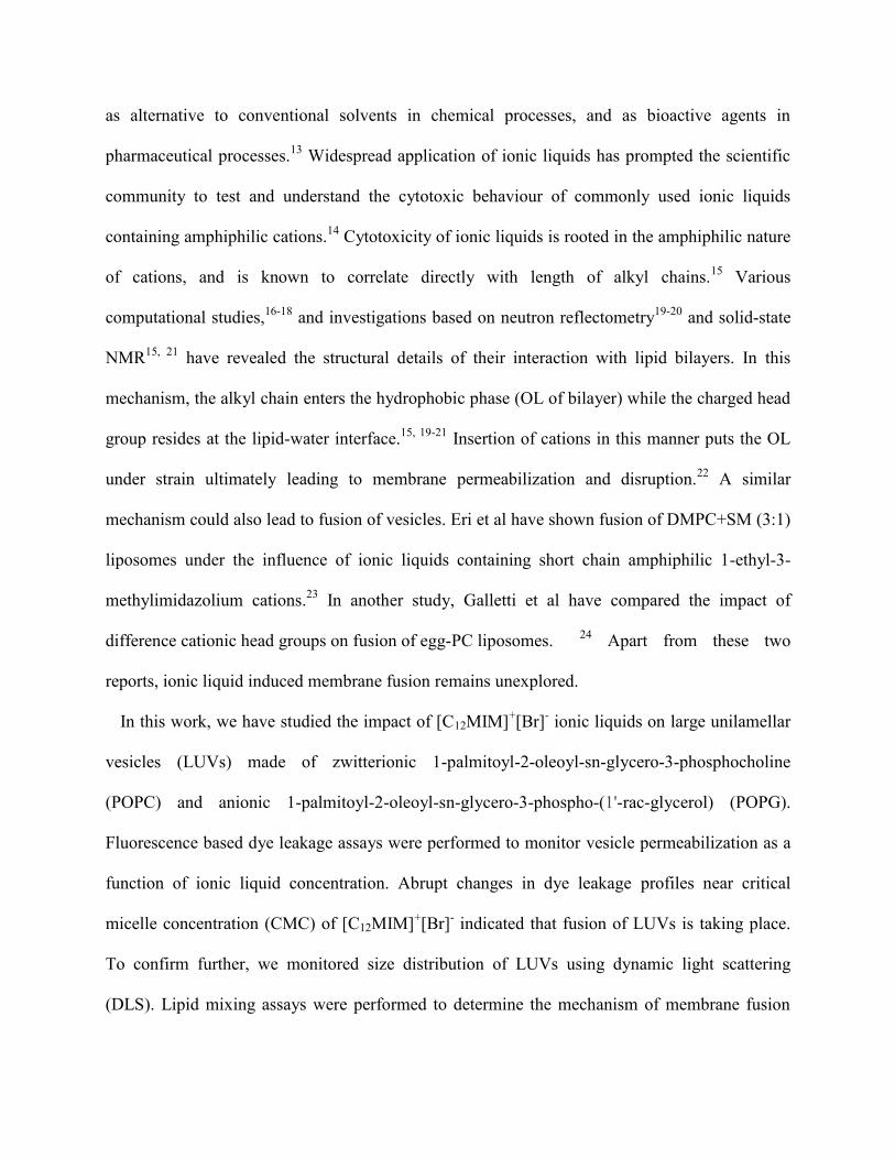
as alternative to conventional solvents in chemical processes, and as bioactive agents in
pharmaceutical processes.13
Widespread application of ionic liquids has prompted the scientific
community to test and understand the cytotoxic behaviour of commonly used ionic liquids
containing amphiphilic cations.14
Cytotoxicity of ionic liquids is rooted in the amphiphilic nature
of cations, and is known to correlate directly with length of alkyl chains.15
Various
computational studies,16-18
and investigations based on neutron reflectometry19-20
and solid-state
NMR15, 21
have revealed the structural details of their interaction with lipid bilayers. In this
mechanism, the alkyl chain enters the hydrophobic phase (OL of bilayer) while the charged head
group resides at the lipid-water interface.15, 19-21
Insertion of cations in this manner puts the OL
under strain ultimately leading to membrane permeabilization and disruption.22
A similar
mechanism could also lead to fusion of vesicles. Eri et al have shown fusion of DMPC+SM (3:1)
liposomes under the influence of ionic liquids containing short chain amphiphilic 1-ethyl-3-
methylimidazolium cations.23
In another study, Galletti et al have compared the impact of
difference cationic head groups on fusion of egg-PC liposomes. 24
Apart from these two
reports, ionic liquid induced membrane fusion remains unexplored.
In this work, we have studied the impact of [C12MIM]+[Br]
- ionic liquids on large unilamellar
vesicles (LUVs) made of zwitterionic 1-palmitoyl-2-oleoyl-sn-glycero-3-phosphocholine
(POPC) and anionic 1-palmitoyl-2-oleoyl-sn-glycero-3-phospho-(1'-rac-glycerol) (POPG).
Fluorescence based dye leakage assays were performed to monitor vesicle permeabilization as a
function of ionic liquid concentration. Abrupt changes in dye leakage profiles near critical
micelle concentration (CMC) of [C12MIM]+[Br]
- indicated that fusion of LUVs is taking place.
To confirm further, we monitored size distribution of LUVs using dynamic light scattering
(DLS). Lipid mixing assays were performed to determine the mechanism of membrane fusion

using LUVs containing 1,2-dipalmitoyl-sn-glycero-3-phosphoethanolamine-N-(7-nitro-2-1,3-
benzoxadiazol-4-yl) (NBD-PE) and 1,2-dioleoyl-sn-glycero-3-phosphoethanolamine-N-
(lissamine rhodamine B sulfonyl) (Rho-PE) as FRET probes. To gain molecular insights, ionic
liquid induced lipid splay was estimated using solid-state NMR spectroscopy. A membrane
fusion model has been developed based on these observed. Outcomes of this model were tested
using similar ionic liquids with shorter alkyl chains ([C8MIM]+[Br]
-).
EXPERIMENTAL METHODS
Materials. Chloroform stock solutions of 1-palmitoyl-2-oleoyl-sn-glycero-3-phosphocholine
(POPC), 1-palmitoyl-2-oleoyl-sn-glycero-3-phospho-(1'-rac-glycerol) (sodium salt) (POPG), L-
α-Phosphatidylethanolamine-N-(lissamine rhodamine B sulfonyl) (Ammonium Salt) (Rh-PE),
1,2-dioleoyl-sn-glycero-3-phosphoethanolamine-N-(7-nitro-2-1,3-benzoxadiazol-4-yl)
(ammonium salt) (NBD-PE) were purchased from Avanti Polar Lipids, Inc. (Alabaster, AL) and
used without further purification. Sephadex G-50 was purchased from Sigma Aldrich, India.
Calcein, Triton X-100, and anhydrous sodium phosphate monobasic (AR grade) were purchased
from Sisco Research Laboratories Pvt. Ltd. (Maharashtra, India). 4-amino-3-hydroxy-1-
naphthalene sulfonic acid (Purity 99.0%), 1-Bromooctane, 1-bromododecane (Purity 98.0%), and
sodium dithionite (DTN) were purchased from Alfa Aesar, India. Ammonium heptamolybdate
tetrahydrate, sodium sulphite anhydrous were purchased from Merck, India. Sodium
metabisulfite was purchased from Fisher Scientific, India. 1-Methyl imidazole was purchased
from Spectrochem, India. Tris hydrochloride 99% (Molecular biology grade), Sodium Chloride
(AR grade), Sulphuric acid 98% (AR grade) were purchased from Loba Chemie, Mumbai, India.
Diethylether (AR grade), and Sodium hydroxide pellets (AR grade) were purchased from SD

fine-chem limited, (Mumbai, India). Perchloric acid 70% (AR grade) was purchased from
Qualikems Fine Chem Pvt. Ltd., India. Ionic liquids [CnMIM]+Br
- (n = 8, 12) were synthesized
as reported previously.15
Methods. Dye Leakage Assay. Dye leakage assays were performed on LUVs containing self-
quenched calcein dye (70 mM) which were prepared as described previously.15
Stock solutions
of [C8MIM]+[Br]
- (1 M), and [C12MIM]
+[Br]
- (100 mM) were prepared in 7.7 mM Tris HCl
buffer containing 100 mM of NaCl (pH 7.4). Appropriate amount of dye filled LUVs were added
to the buffer containing [CnMIM]+[Br]
- (n=8, 12) achieving a final lipid concentration of 0.275 ±
0.015 mM in each case. The solutions were gently mixed and transferred to quartz cuvette to
perform fluorescence measurements (dead time = 30 s) using PerkinElmer LS-55 Luminescence
spectrometer. Calcein emission was measured at 520 nm with the excitation wavelength set at
485 nm using an excitation and emission slit width of 10 nm, each. The percentage of dye-
leakage was calculated by normalizing the observed fluorescence intensity at a given time point
( ) with respect to that obtained without addition of ionic liquid ( ), and that obtained after
addition of 1% Triton-X ( ) using following equation
Separate set of controls and were recorded for each LUV preparation. The
normalized data was smoothened by three-point averaging and plotted against time.
Dynamic light scattering (DLS) measurements. Stock solutions of 1 M (C8MIM+Br
-), and 100
mM (C12MIM+Br
-) were prepared in 7.7 mM Tris HCl buffer containing 100 mM of NaCl (pH
7.4). Size distribution measurements for POPC and POPG LUVs were performed in the absence
and presence of ionic liquids at 25 °C using Malvern instrument (Zeta sizer, Nano Series, nano-
ZS, Malvern, U.K.). Samples were thermally equilibrated for 10 min before each measurement.

The concentration of lipids in LUVs (POPC or POPG) was fixed at 0.275 mM in each
measurement.
Lipid splay measurements using solid-state NMR. Details of sample preparation and 1H-
1H
magic-angle-spinning (MAS) solid-state NMR measurements are as described previously.15
Briefly, multilamellar vesicles (MLVs) of POPC and POPG containing 0.768 moles of ionic
liquid per mole of lipid were packed in 4 mm high-resolution MAS rotors with spherical Kel-F
inserts for NMR measurements. A set of five two dimensional 1H-
1H MAS NOESY spectra
25-26
were recorded on each sample using mixing times of 0.1, 100, 200, 300, and 500 ms. NOESY
cross-peaks between all lipid segments are observed. As the spin diffusion effect can safely be
ruled out for the mixing times used in our study, this is due to the high molecular disorder and
mobility of lipid membranes in the liquid crystalline phase.27-28
All 1H MAS NMR measurements
were carried out on a Bruker Avance III 600 MHz NMR spectrometer using a 4 mm HR-MAS
probe at a MAS frequency of 6 kHz. The volume of the respective diagonal and cross peaks of -
(CH2)n- protons in lipid chains and the protons of the lipid head groups were integrated using the
Bruker Topspin 3.5 software package. NOE buildup curves were fitted to the spin pair model
yielding cross-relaxation rates (σij)29
and used as a measure of lipid splay.
Lipid mixing assay
Total lipid mixing. Probe dilution assay based on mixing of LUVs containing fluorescence
resonance energy transfer (FRET) pairs was used to determine total lipid mixing during
membrane fusion.30
LUVs containing FRET pair probes NBD-PE (donor) and Rho-PE (acceptor)
at a concentration of 1.5 mol % each were used. Two sets of LUVs, probe free and probe

containing, were prepared in 7.7 mM Tris HCl buffer containing 100 mM of NaCl (pH 7.4).
Probe containing LUVs were mixed with probe free LUVs at a ratio 1:4 (final lipid concentration
was 0.275 mM) and loaded in a 1 cm path length quartz cuvette. NBD emission was measured at
530 nm with the excitation wavelength set at 460 nm using an excitation and emission slit width
of 15 nm, each. A cut off filter at 515 nm was used between the sample and emission
monochromator to avoid scattering interference. Fluorescence dequenching of NBD-PE due to
dilution of FRET probes into probe free LUVs was monitored with time as a function of ionic
liquid concentration, and used to calculate percentage of total mixing as
where Xt is the fluorescence intensity at time t, X0,t is the fluorescence intensity in the absence
of ionic liquid. Xmax,t is the maximum fluorescence intensity (MFI) obtained after addition of 1%
(v/v) Triton X-100. A correction factor of 1.5 was applied to observed fluorescence in the last
case as Triton X-100 is known to affect NBD-PE fluorescence.31
Lipid mixing in inner (IL) Ionic liquid induced lipid-mixing in the inner and outer monolayers
(leaflets) was determined by a modification of the phospholipid-mixing measurement as reported
elsewhere.32
Next, contribution of IL mixing towards total lipid mixing was determined using
selective silencing of OL. Fluorescence intensity of NBD-PE located in the OL of bilayers was
completely quenched by treating LUVs containing 1.5 mol% of both NBD-PE and Rho-PE with
100 mM sodium dithionite for approximately 1-2 h on ice in the dark. Sodium dithionite was
then removed by the size exclusion chromatography through Sephadex G 50 eluted with a buffer
containing 7.7 mM Tris HCl buffer containing 100 mM of NaCl (pH 7.4). Percentage IL mixing
was calculated as

where is the fluorescence intensity at time t in the presence of ionic liquid, is the
fluorescence intensity in the absence of ionic liquid, and is MFI as obtained earlier.
Hemifusion %. Amount of FRET probes in each leaflet is first determined by following the
quench kinetics of NBD-PE molecules present in the OL. Fluorescence intensity of LUVs
containing 1.5 mol% of only NBD-PE was monitored as a function of time after adding 20 mM
sodium dithionite (Figure S4). From the fast component of quench kinetics, the percentage ( )
of NBD molecules in the IL was found to be 45% (in POPC) and 60% (in POPG). %
Hemifusion was then calculated by using the difference between the expected IL mixing in the
event of full fusion ( ) and the observed IL mixing as

RESULTS AND DISCUSSIONS
Imidazolium based ionic liquids are known for their ability to permeabilize the lipid bilayers.15,
21, 33 This is mainly studied by observing the fraction of trapped dye leaking out of LUVs as a
function of time. These molecules partition themselves into the lipid membrane altering its
structural integrity and stability, which results in the leakage of trapped dye.15
Figure 1 shows the
time dependent leakage of calcein dye trapped inside POPC or POPG LUVs (diameter 120 nm)
upon addition of increasing [C12MIM]+Br
- concentrations.

Figure 1. Time course of calcein leakage from (a) POPC, and (b) POPG LUVs upon adding
[C12MIM]+Br
- in indicated concentrations. Total lipid concentration in LUVs is 0.275 mM in
both cases.
An initial increase in [C12MIM]+Br
- concentration results in an enhancement of dye leakage
kinetics in both cases with maximum dye leakage reaching 100%. As observed previously,
POPC LUVs were found to be more leakage prone as compared to POPG.15, 21
However, a
sudden change in dye leakage profile is observed when [C12MIM]+Br
- concentration is increased
to 3 mM in POPC, and 4 mM in case of POPG. In both LUVs, maximum dye leakage is reduced
to only 48%, accompanied by a sharp enhancement in dye leakage kinetics. These changes could
be due to disruption of LUVs as shown by Jing and co-workers where [C12MIM]+Cl
- leads to
disruption of POPC SUVs when present in concentration exceeding its CMC value.34
[C12MIM]+Br
- has a CMC value of 3.3 mM in working buffer (Figure S1), which is fairly close

to the concentration at which change in leakage profile is observed. Moreover, a complete
disruption of LUVs should result in 100% leakage of dye as observed after the addition of 1%
(v/v) Triton X-100. Even higher [C12MIM]+Br
- concentrations yield a similar looking leakage
profile with slight enhancement in leakage extent. The fact that dye leakage saturates at value
less than 100% indicates trapping of dye molecules in a stable environment, possibly in a fused
vesicle.
Normally, vesicle fusion proceeding through “stalk-hemifusion diaphragm-fusion pore”
pathway results in complete mixing of vesicle contents.35
However, under some conditions
fusion proceeds through a “fusion-through-rupture” pathway which is accompanied by leakage
of inner contents.1, 35-36
This leakage is caused by the formation of fusion-holes close to the stalk
where the membrane is relatively thinner.37
Leakage rate through these holes depend on the
lifetime of holes and diffusion rate of dye molecules, and goes to zero once fusion is complete
and the holes are sealed. LUVs studied in this work are composed of cylinder shaped PC and PG
lipid molecules which make bilayers with negligible curvature.1 Insertion of inverted cone
shaped [C12MIM]+
molecules would disturb the packing of such bilayers. This would enhance
the positive curvature in the outer leaflet thereby putting the bilayer under strain. To reduce this
additional strain, the bilayer undergoes structural reorganization which results in membrane
permeability.15, 21
However, this mechanism does not seem to be sufficient to rescue the strained
bilayers when subjected to high [C12MIM]+
concentrations. As an additional counter measure,
the LUVs could fuse together to form bigger vesicles. These fused vesicles would possess a
larger surface area with reduced curvature and hence, lower strain. Dye leakage profiles of POPC
and POPG obtained after adding 3 and 4 mM of [C12MIM]+Br
- indicate that a similar mechanism
underway. In the time it takes for LUVs to fuse, nearly half of the trapped calcein dye (52% in

POPC and 52% in POPG) leaks out through the fusion-holes. As [C12MIM]+ concentration is
increased further, relatively higher amount of dye leaks out in the fusion time period and hence,
relatively higher saturation values are observed. These amphiphilic cations have been shown to
induce fusion in egg-PC24
and DOPC vesicles23
previously. However, these studies did not
comment on the leaky nature of membrane fusion or formation of fusion-holes.
Figure 2. Hydrodynamic radius (Dh) of (a) POPC, and (b) POPG LUVs as function of
[C12MIM]+Br
- concentrations.
To further verify [C12MIM]+Br
- induced LUV fusion, we monitored change in size of LUVs
using dynamic light scattering measurements (Figure 2). Average diameter of both LUVs is
~120 nm, which remains unchanged in presence of up to 2 and 3 mM of [C12MIM]+Br
- in POPC
and POPG LUVs, respectively. As expected, addition of [C12MIM]+Br
- in concentrations higher
than these results in appearance of a new size distribution peaks centered at ~1110 nm and ~955
nm in POPC and POPG LUVs, respectively. Similar effect was seen in case of mixed DOPC/SM
LUVs (100 nm) whose size increased up to 1.7 m in presence of 1-ethyl-3-methylimidazolium

cations due to fusion.23
Therefore, peaks centered at 1110 nm in POPC and 955 nm in POPG
LUVs most likely correspond to large sized fused vesicles. In addition to these peaks, smaller
size distributions centered at 10 nm were also observed in both DLS profiles. These peaks most
likely correspond to mixed [C12MIM]+-POPC micelles which are reported to form under these
conditions.34
These mixed micelles are possibly formed due to interaction of [C12MIM]+
micelles with LUVs causing a population exchange between the lipidic and micellar phases. DLS
measurements confirm that POPC and POPG LUVs undergo fusion in presence of high
[C12MIM]+Br
- concentrations. Since changes in DLS and dye leakage profiles take place at same
[C12MIM]+Br
- concentrations lends further support to the hypothesis that this fusion process is a
leaky one due to formation of fusion-holes.
Figure 3. Structures of (a) POPC and (b) POPG molecules with protons in the glycerol and
head-group region highlighted in blue and red colors, respectively. 1H-
1H NOESY average
cross-relaxation rates between these protons and -(CH2)n- protons in lipid chains of (c) POPC

and (d) POPG observed in the absence and presence of [C12MIM]+Br
-. Also shown are the
average cross-relaxation rates glycerol and head-group protons taken together.
For two LUVs to fuse, their bilayers must attain a minimal spatial proximity during a collision
event by overcoming the electrostatic and hydration barriers between them.38-39
A comparison
between POPC and POPG tells us that LUVs of former require lower [C12MIM]+Br
-
concentrations to fuse. This is counter-intuitive from an electrostatic point of view since
[C12MIM]+ bound POPC LUVs possess a net positive surface charge and hence should be less
prone to fusion as compared to near-neutral [C12MIM]+ bound POPG LUVs (Figure S3). It
seems that [C12MIM]+ induced membrane fusion is initiated by the hydrophobic interaction
between the merging bilayers. In absence of [C12MIM]+Br
-, POPG bilayers are softer in
comparison to POPC due to higher disorder in lipid chains.15
Average chain order parameter
(<S>) in pure POPC and POPG membranes is 0.163 and 0.150, respectively. However, insertion
of [C12MIM]+ molecules renders POPC bilayers more disordered (<S>=0.127) as compared to
POPG (<S>=0.154),15
a factor which could assist membrane fusion through surface exposure of
hydrophobic tails thus overcoming the hydration barrier.40
Some membrane fusion models have
suggested that formation of an initial contact between merging bilayers known as prestalk-
membrane.37, 41-42
Formation of this fusion intermediate is assisted by transient splaying of lipid
molecules which exposes the hydrophobic lipid chains to membrane surface.40, 43-45
Fusogens
like membrane-fusogenic soluble peptides46
and transmembrane domains47
are known to act by
promoting the lipid splay phenomenon in bilayers. Keeping this in view, we quantified the
impact of [C12MIM]+ insertion on lipid splay by determining the
1H-
1H NOESY cross-relaxation
rates between -(CH2)n- protons in the lipid chain and various protons in the glycerol and head-
group region (Figure S5 and S6). As suspected, incorporation of [C12MIM]+ leads to a very

significant enhancement in cross-relaxation rates of lipids from 0.0299 s-1
to 0.172 s-1
in POPC
bilayers (Figure 3). POPG lipids on the other hand incur a small reduction (30%) upon
incorporating [C12MIM]+, but yields a value 0.262 s
-1 which is still higher than POPC. At the
same time, a comparison of average chain order parameter (<S>=0.127) tells us that [C12MIM]+
incorporation makes the POPC lipids chains much more disordered as compared to POPG. This
discrepancy could be due to the fact that these distance dependent cross-relaxation rates indicate
the probability of contacts between protons and are observed due to conformational disorder of
the lipid bilayer which includes lipid splay and lipid fluxes along the bilayer.27, 29, 48
In a nutshell,
incorporation of [C12MIM]+ results in a very large enhancement in lipid splay in POPC which
would assist its bilayers in making a hydrophobic contact. When taken together with the fact that
[C12MIM]+ binds more strongly to POPC than POPG,
15 it explains why membrane fusion occurs
at relatively lower [C12MIM]+ concentrations in case of POPC.

Figure 4. The percentage total lipid mixing (black), OL mixing (red), IL mixing (blue) and
hemifused LUVs (green) observed 10 min after addition of [C12MIM]+Br
- in (a) POPC, and (b)
POPG LUVs.
Membrane fusion involves exchange of lipid populations between the bilayers of merging
membranes often termed as lipid mixing.49
This property makes it different from simple
aggregation of vesicles which also results in large size distributions. Accordingly, we have
determined the extent of lipid mixing using the “probe dilution” assay which is particularly
insensitive to simple aggregation of LUVs.50
In this assay, LUVs containing fluorescently
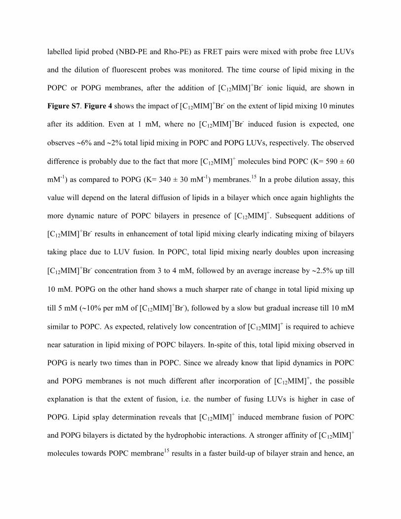
labelled lipid probed (NBD-PE and Rho-PE) as FRET pairs were mixed with probe free LUVs
and the dilution of fluorescent probes was monitored. The time course of lipid mixing in the
POPC or POPG membranes, after the addition of [C12MIM]+Br
- ionic liquid, are shown in
Figure S7. Figure 4 shows the impact of [C12MIM]+Br
- on the extent of lipid mixing 10 minutes
after its addition. Even at 1 mM, where no [C12MIM]+Br
- induced fusion is expected, one
observes 6% and 2% total lipid mixing in POPC and POPG LUVs, respectively. The observed
difference is probably due to the fact that more [C12MIM]+ molecules bind POPC (K= 590 ± 60
mM-1
) as compared to POPG (K= 340 ± 30 mM-1
) membranes.15
In a probe dilution assay, this
value will depend on the lateral diffusion of lipids in a bilayer which once again highlights the
more dynamic nature of POPC bilayers in presence of [C12MIM]+. Subsequent additions of
[C12MIM]+Br
- results in enhancement of total lipid mixing clearly indicating mixing of bilayers
taking place due to LUV fusion. In POPC, total lipid mixing nearly doubles upon increasing
[C12MIM]+Br
- concentration from 3 to 4 mM, followed by an average increase by 2.5% up till
10 mM. POPG on the other hand shows a much sharper rate of change in total lipid mixing up
till 5 mM (10% per mM of [C12MIM]+Br
-), followed by a slow but gradual increase till 10 mM
similar to POPC. As expected, relatively low concentration of [C12MIM]+ is required to achieve
near saturation in lipid mixing of POPC bilayers. In-spite of this, total lipid mixing observed in
POPG is nearly two times than in POPC. Since we already know that lipid dynamics in POPC
and POPG membranes is not much different after incorporation of [C12MIM]+, the possible
explanation is that the extent of fusion, i.e. the number of fusing LUVs is higher in case of
POPG. Lipid splay determination reveals that [C12MIM]+ induced membrane fusion of POPC
and POPG bilayers is dictated by the hydrophobic interactions. A stronger affinity of [C12MIM]+
molecules towards POPC membrane15
results in a faster build-up of bilayer strain and hence, an

early fusion. Though delayed, once the hydrophobic contact is established in POPG, its near
neutral LUV carrying a net surface charge of 3.93 ± 0.49 mV in presence of 4 mM [C12MIM]+
undergo rampant fusion (Figure S3). On the other hand, POPC LUVs carrying a net positive
surface charge (15.5 ± 3.3 mV at 3 mM [C12MIM]+) prevents their close approach. Thus, while
hydrophobic factors determine the initiation of [C12MIM]+ induced fusion, electrostatic factors
seem to control its overall extent.
A simple modification of lipid mixing assay allows one to determine the extent of lipid mixing
in each individual leaflet, i.e. the outer leaflet (OL) and the inner leaflet (IL) of the bilayer, thus
helping us gain further insight in to [C12MIM]+Br
- induced fusion pathway. The percentage of IL
lipid mixing (in blue, Figure 4) was calculated using selective silencing of the NBD labelled
lipids in OL of LUVs in presence of sodium dithionite as explained in material and methods
section. The OL lipid mixing (in red, Figure 4) was then calculated as the difference between
total lipid and IL mixing. % IL and % OL mixing, 10 minutes after addition of [C12MIM]+Br
- are
shown in Figure 4, and their time dependence are shown in Figure S7. Before the beginning of
the fusion, lipid mixing is dominated by OL mixing in both cases. This is probably because
insertion of [C12MIM]+
cations in the OL of POPC and POPG bilayers results in an enhanced
lipid dynamics.34
After fusion, contribution of IL mixing in POPC shows a steady increase with
concentration indicating the increasing dominance of the fully-fused states (% Hemifusion, Fig.
4). In contrast, OL mixing continues to remain a dominant factor in POPG even at higher
concentrations indicating a near-equal contribution of both hemi- and fully-fused states. It is
possible that addition of higher [C12MIM]+Br
- concentration (>10 mM) in POPG could result in a
fully-fused state. A better quantification of hemi-fused vs fully-fused state is provided by a
“content mixing assay” which is based on mixing of LUV trapped with TbCl3 (Terbium chloride)

and DPA (Dipicolinic acid).51
However, the leaky nature LUVs due to formation of fusion-holes
in the early stage of fusion prevented the application of this assay in our case.
Figure 5. Hydrodynamic radius (Dh) (a) POPC, and (b) POPG LUVs as observed in presence of
various [C8MIM]+Br
- concentrations.
Our studies have helped us in building a model for membrane fusion induced by ionic liquids
containing imidazolium based amphiphilic cations. According to this model, when present in
high concentrations (near CMC), [C12MIM]+ acts as a fusogenic agent for LUVs composed of
neutral (POPC; ionic liquid:lipid > 7:1) and negatively charged (POPG; ionic liquid:lipid > 11:1)
lipids. While the onset of fusion is critically dependent on [C12MIM]+ induced lipid splay, its
total extent depends on the net surface charge carried by the LUVs. At [C12MIM]+Br
-
concentrations studied here, fusion process is dominated by a hemi-fused state and involves
formation of leaky fusion-holes in the early stages of fusion. Before concluding our observations,
we tested this model using another imidazolium based ionic liquid containing amphiphilic
cations with shorter hydrophobic chain ([C8MIM]+Br
-). In comparison to [C12MIM]
+,
partitioning of [C8MIM]+
into POPC and POPG bilayers is relatively weak leading to a relatively
lower permeabilization at comparable concentrations.15
At the same time, insertion of these small

sized molecules results in large lipid chain disorder and bilayer softness as compared to its long
chain counterparts particularly in the POPC membranes.15
Figure 5 shows the impact of
increasing [C8MIM]+ concentrations on size of LUVs. As expected, membrane fusion requires
much higher [C8MIM]+ concentrations as compared to [C12MIM]
+ as size distributions
corresponding to fused vesicles (825 nm) are observed at 150 mM in both POPC and POPG
LUVs. Also observed are the smaller size distributions (in the range of 1-8 nm) corresponding to
mixed micelles at these concentrations. Lipid mixing assays also confirmed the process of
fusion, and like [C12MIM]+, extent of fusion was found to be larger in case of POPG (Figure 6a
and b). The time course of lipid mixing, OL, and IL mixing in the POPC or POPG membranes,
after the addition of [C8MIM]+Br
- ionic liquid, are shown in Figure S8. In contrast to
[C12MIM]+, % hemi-fusion is significantly smaller at the onset of fusion. This difference is
probably due to a highly disordered nature of POPC (<S> = 0.116) and POPG (<S> = 0.115)
lipid chains in presence of [C8MIM]+ resulting in high membrane fluidity and lipid mixing.
15
Also, at high IL concentrations (>100 mM) in POPG, IL mixing exceeds OL mixing values
resulting in negative % Hemifusion values. This is possibly due to formation of mixed micelles
in presence of abundant [C8MIM]+ thus providing an additional way to change FRET pair
distances apart from the lipid mixing. Figures 6c and 6d show the concentration dependent dye
leakage profiles of POPC and POPG LUVs, respectively. As expected, a change in leakage
kinetics is observed around [C8MIM]+
concentrations required to induce LUV fusion, confirming
its leaky nature due to formation of fusion-holes while the membranes merge together. Lastly,
partitioning of [C8MIM]+ molecules enhanced splaying of POPC lipids by a significant amount (
0.0299 s-1
to 0.2295 s-1
), while increasing it only a little in case of POPG (0.374 s-1
to 0.399 s-1
)

(Figure S9), thus supporting our hypothesis that LUV fusion is driven by hydrophobic
interactions.
Figure 6. The percentage total lipid mixing (black), OL mixing (red), IL mixing (blue) and
hemifused LUVs (green) observed 10 min after addition of [C8MIM]+Br
- in (a) POPC, and (b)
POPG LUVs. Time course of dye leakage from (c) POPC, and (d) POPG LUVs upon addition of
[C8MIM]+Br
- in concentration as indicated.

CONCLUSIONS
We have conducted a systematic investigation into mechanism of [CnMIM]+ induced fusion
of POPC and POPG LUVs. The kinetics, extent, and pathway of [CnMIM]+
induced membrane
fusion is critically dependent of lipid dynamics, which is regulated by the interaction of lipid and
[CnMIM]+ molecules. Insertion of [CnMIM]
+ molecules alters packing of lipid bilayers, thereby
putting them under strain. As a counter measure, lipid chains become more disordered resulting
in enhanced exposure of hydrophobic lipid chains to membrane surface through lipid splay. This
enables the fusing membrane to overcome the inter-bilayer hydration barrier and make a
hydrophobic contact, possibly forming a prestalk-membrane. Accordingly, relatively disordered
chains in presence of [CnMIM]+ cations result in an early onset of fusion in POPC LUVs. Once
the hydration barrier is crossed, the extent of fusion is predominantly dictated by electrostatic
factors, which favour fusion of near-neutral [CnMIM]+ inserted POPG LUVs. The fusion process
in both membranes seems to proceed through the stalk-hemifusion diaphragm-fusion pore
pathway. Early stages of fusion are accompanied by leakage of inner contents from vesicles due
to formation of fusion-holes at the stalk intermediate stage. These holes seal off as the membrane
evolves into the hemi-fused diaphragm. In a nutshell, our study provides a structure-property
model to explain the mechanism of membrane fusion induced by amphiphilic cations in
imidazole based ionic-liquids. The outcomes of this model can be used to regulate membrane
fusion by varying structural parameters of ionic-liquid components.

ASSOCIATED CONTENT
Supporting Information contains CMC determination of C12MIM+Br
- and C8MIM
+Br
-, -
potential measurements, dithionite bleaching, 1H-
1H MAS NOESY spectra, and time dependent
lipid mixing profiles of C12MIM+Br
- and C8MIM
+Br
-, and
1H-
1H MAS NOESY Cross-relaxation
rates of POPC and POPG in the presence and absence of C8MIM+Br
-.
AUTHOR INFORMATION
Notes
The authors declare no competing financial interests.
Author Contributions
S.K. synthesized ionic liquids and LUVs, performed dye- leakage and lipid mixing assays, and
analyzed ssNMR data. N.K. performed DLS and ζ-potential studies. V.S.M. conceptualized the
project, performed ssNMR studies, wrote, reviewed, and edited the manuscript with
contributions from all the co-authors.
Orcid Id
Venus S. Mithu: 0000-0003-2869-9776
Sandeep Kumar: 0000-0002-3867-5586
Navleen Kaur: 0000-0002-4916-5266

ACKNOWLEDGMENT
The authors like to acknowledge research funding from Department of Biotechnology, Govt.
of India (BT/PR22289/BRB/10/1566/2016). V.S.M. would like to acknowledge German
Academic Exchange Service (DAAD) for a research stay grant (ID: 57314018) to perform
ssNMR studied in Prof. Daniel Huster’s lab at University of Leipzig, Germany. S.K. is thankful
to the Council of Scientific and Industrial Research (CSIR), India, for Senior Research
Fellowship (SRF) no. 09/254(0267)/2017- EMR-I. N.K. is thankful to the University Grant
Commission (UGC), India, for Senior Research Fellowship (SRF) no. 105743.
REFERENCES
1. Chernomordik, L. V.; Kozlov, M. M. Mechanics of membrane fusion. Nat. Struct. Mol.
Biol. 2008, 15, 675.
2. Eckert, D. M.; Kim, P. S. Mechanisms of viral membrane fusion and its inhibition. Annu.
Rev. Biochem. 2001, 70, 777-810.
3. Duelli, D. and Lazebnik, Y. Cell-to-cell fusion as a link between viruses and cancer. Nat.
Rev. Cancer 2007, 7, 968-976.
4. Lentz, B. R.; Malinin, V.; Haque, M. E.; Evans, K. J. Protein machines and lipid assemblies:
current views of cell membrane fusion. Curr. Opin. Struct. Biol. 2000, 10, 607-615.
5. Haque, M. E.; Koppaka, V.; Axelsen, P. H.; Lentz, B. R. Properties and structures of the
influenza and HIV fusion peptides on lipid membranes: implications for a role in fusion.
Biophys. J. 2005, 89, 3183-94.

6. Kozlov, M. M.; Chernomordik, L. V., A mechanism of protein-mediated fusion: coupling
between refolding of the influenza hemagglutinin and lipid rearrangements. Biophys. J.
1998, 75, 1384-96.
7. Mondal Roy, S., Sarkar, M. Membrane fusion induced by small molecules and ions. J.
Lipids 2011, 2011.
8. Goodall, A. H.; Galloway, M. J.; Fisher, D.; Lucy, J. A. The interactions of dispersions of
lipid-soluble fusogens with hen erythrocytes. Portland Press Limited 1979, 937-939.
9. Mason, W.; Miller, N. G. A. Fusion of charged and uncharged liposomes by n-alkyl
bromides. Biochem. Biophys. Res. Commun. 1979, 91, 878-885.
10. Maggio, B.; Lucy, J. A. Interactions of water‐ soluble fusogens with phospholipids in
monolayers. FEBS Lett. 1978, 94, 301-304.
11. Cevc, G. Membrane electrostatics. Biochim. Biophys. Acta, Biomembr. 1990, 1031, 311-382.
12. Welton, T. Room-temperature ionic liquids. Solvents for synthesis and catalysis. Chem. Rev.
1999, 99, 2071-2084.
13. Egorova, K. S.; Gordeev, E. G.; Ananikov, V. P. Biological activity of ionic liquids and their
application in pharmaceutics and medicine. Chem. Rev. 2017, 117, 7132-7189.
14. Pham, T. P. T.; Cho, C. W.; Yun, Y. S. Environmental fate and toxicity of ionic liquids: a
review. Water Res. 2010, 44, 352-372.

15. Kumar, S.; Scheidt, H. A.; Kaur, N.; Kang, T. S.; Gahlay, G. K.; Huster, D.; Mithu, V. S.
Effect of the Alkyl Chain Length of Amphiphilic Ionic Liquids on the Structure and
Dynamics of Model Lipid Membranes. Langmuir 2019, 35, 12215-12223.
16. Bingham, R. J.; Ballone, P. Computational study of room-temperature ionic liquids
interacting with a POPC phospholipid bilayer. J. Phys. Chem. B 2012, 116, 11205-11216.
17. Yoo, B.; Shah, J. K.; Zhu, Y.; Maginn, E. J. Amphiphilic interactions of ionic liquids
with lipid biomembranes: a molecular simulation study. Soft Matter 2014, 10, 8641-8651.
18. Benedetto, A.; Ballone, P. Engineering, Room temperature ionic liquids meet biomolecules:
a microscopic view of structure and dynamics. ACS Sustainable Chem. Eng. 2015, 4, 392-
412.
19. Benedetto, A.; Heinrich, F.; Gonzalez, M. A.; Fragneto, G.; Watkins, E.; Ballone, P.
Structure and stability of phospholipid bilayers hydrated by a room-temperature ionic
liquid/water solution: a neutron reflectometry study. J. Phys. Chem. B 2014, 118, 12192-
12206.
20. Sharma, V.K.; Mukhopadhyay, R. Deciphering interactions of ionic liquids with
biomembrane. Biophys. Rev. 2018, 10, 721-734.
21. Kumar, S.; Scheidt, H. A.; Kaur, N.; Kaur, A.; Kang, T. S.; Huster, D.; Mithu, V. S.
Amphiphilic Ionic Liquid-Induced Membrane Permeabilization: Binding Is Not Enough. J.
Phys. Chem. B 2018, 122, 6763-6770.

22. Yoo, B.; Zhu, Y.; Maginn, E. J. Molecular mechanism of ionic-liquid-induced membrane
disruption: morphological changes to bilayers, multilayers, and vesicles. Langmuir 2016, 32,
5403-5411.
23. Hayakawa, E. H.; Mochizuki, E.; Tsuda, T.; Akiyoshi, K.; Matsuoka, H.; Kuwabata, S. The
effect of hydrophilic ionic liquids 1-ethyl-3-methylimidazolium lactate and choline lactate
on lipid vesicle fusion. Plos one 2013, 8, e85467.
24. Galletti, P.; Malferrari, D.; Samorì, C.; Sartor, G.; Tagliavini, E. Effects of ionic liquids on
membrane fusion and lipid aggregation of egg-PC liposomes. Colloids Surf., B 2015, 125,
142-150.
25. Jeener, J.; Meier, B.; Bachmann, P.; Ernst, R. R., Investigation of exchange processes by
two‐ dimensional NMR spectroscopy. J. Chem. Phys. 1979, 71, 4546-4553.
26. Kumar, A.; Wagner, G.; Ernst, R.R.; Wuethrich, K. Buildup rates of the nuclear Overhauser
effect measured by two-dimensional proton magnetic resonance spectroscopy: implications
for studies of protein conformation. J. Am. Chem. Soc., 1981, 103, 3654-3658.25.
27. Huster, D.; Arnold, K.; Gawrisch, K. Investigation of lipid organization in biological
membranes by two-dimensional nuclear Overhauser enhancement spectroscopy. J. Phys.
chem. B 1999, 103, 243-251.
28. Huster, D.; and Gawrisch, K. NOESY NMR crosspeaks between lipid headgroups and
hydrocarbon chains: spin diffusion or molecular disorder? J. Am. Chem. Soc. 1999, 121,
1992-1993.
29. Scheidt, H. A.; Huster, D. The interaction of small molecules with phospholipid membranes
studied by 1
H NOESY NMR under magic-angle spinning. Acta Pharmacol. Sin. 2008, 29,
35.
30. Struck, D. K.; Hoekstra, D.; Pagano, R. E. Use of resonance energy transfer to monitor
membrane fusion. Biochemistry 1981, 20, 4093-4099.
31. Torchilin, V.; Weissig, V. Liposomes: a practical approach. Oxford University Press. 2003,
264.
32. Meers, P.; Ali, S.; Erukulla, R.; Janoff, A. S. Novel inner monolayer fusion assays reveal
differential monolayer mixing associated with cation-dependent membrane fusion. Biochim.
Biophys. Acta, Biomembr. 2000, 1467, 227-243.
33. Evans, K. O. Physicochemical, S. A.; Aspects, E., Room-temperature ionic liquid cations act
as short-chain surfactants and disintegrate a phospholipid bilayer. Colloids Surf., A 2006,
274, 11-17.
34. Jing, B.; Lan, N.; Qiu, J.; Zhu, Y. Interaction of ionic liquids with a lipid bilayer: a
biophysical study of ionic liquid cytotoxicity. J. Phys. Chem. B 2016, 120, 2781-2789.
35. Frolov, V.; Dunina-Barkovskaya, A. Y.; Samsonov, A.; Zimmerberg, J. Membrane
permeability changes at early stages of influenza hemagglutinin-mediated fusion. Biophys. J.
2003, 85, 1725-1733.
36. Bu, B.; Crowe, M.; Diao, J.; Ji, B.; Li, D. Cholesterol suppresses membrane leakage by
decreasing water penetrability. Soft matter 2018, 14, 5277-5282.

37. Kozlovsky, Y.; Chernomordik, L. V.; Kozlov, M. M. Lipid intermediates in membrane
fusion: formation, structure, and decay of hemifusion diaphragm. Biophys. J. 2002, 83,
2634-2651.
38. Burgess, S. W.; McIntosh, T. J.; Lentz, B. R. Modulation of poly (ethylene glycol)-induced
fusion by membrane hydration: importance of inter bilayer separation. Biochemistry 1992,
31, 2653-2661.
39. Ohki, S. A mechanism of divalent ion-induced phosphatidylserine membrane fusion.
Biochim. Biophys. Acta, Biomembr. 1982, 689, 1-11.
40. Smirnova, Y. G.; Marrink, S. J.; Lipowsky, R.; Knecht, V. Solvent-exposed tails as prestalk
transition states for membrane fusion at low hydration. J. Am. Chem. Soc. 2010, 132, 6710-
6718.
41. Kozlovsky, Y.; Efrat, A.; Siegel, D. A.; Kozlov, M. M. Stalk phase formation: effects of
dehydration and saddle splay modulus. Biophys. J. 2004, 87, 2508-2521.
42. Kozlovsky, Y.; Kozlov, M. M. Stalk model of membrane fusion: solution of energy crisis.
Biophys. J. 2002, 82, 882-895.
43. Kinnunen, P. K.; Holopainen, J. M. Mechanisms of initiation of membrane fusion: role of
lipids. Biosci. Rep. 2000, 20, 465-482.
44. Mirjanian, D.; Dickey, A. N.; Hoh, J. H.; Woolf, T. B.; Stevens, M. J. Splaying of aliphatic
tails plays a central role in barrier crossing during liposome fusion. J. Phy. Chem. B 2010,
114, 11061-11068.

45. Kawamoto, S.; Shinoda, W. Free energy analysis along the stalk mechanism of membrane
fusion. Soft matter 2014, 10, 3048-3054.
46. Larsson, P.; Kasson, P. M. Lipid tail protrusion in simulations predicts fusogenic activity of
influenza fusion peptide mutants and conformational models. Plos one Comput. Biol. 2013,
9.
47. Scheidt, H. A.; Kolocaj, K.; Veje Kristensen, J.; Huster, D.; Langosch, D. Transmembrane
helix induces membrane fusion through lipid binding and splay. J. Phys. Chem. Lett. 2018,
9, 3181-3186.
48. Mihailescu, M.; Vaswani, R. G.; Jardón-Valadez, E.; Castro-Román, F.; Freites, J. A.;
Worcester, D. L.; Chamberlin, A. R.; Tobias, D. J.; White, S. H. Acyl-chain methyl
distributions of liquid-ordered and-disordered membranes. Biophys. J. 2011, 100, 1455-1462
49. Cevc, G.; Richardsen, H. Lipid vesicles and membrane fusion. Adv. Drug Deliv. Rev 1999,
38, 207-232.
50. Duzgunes, N.; Allen, T. M.; Fedor, J.; Papahadjopoulos, D. Lipid mixing during membrane
aggregation and fusion: why fusion assays disagree. Biochemistry 1987, 26, 8435-8442.
51. Wilschut, J.; Duzgunes, N.; Fraley, R.; Papahadjopoulos, D. Studies on the mechanism of
membrane fusion: kinetics of calcium ion induced fusion of phosphatidylserine vesicles
followed by a new assay for mixing of aqueous vesicle contents. Biochemistry 1980, 19,
6011-6021.

download fileview on ChemRxivIonic liquid_membrane fusion.pdf (1.35 MiB)

SUPPORTING INFORMATION
Amphiphilic Ionic Liquid Induced Fusion of
Phospholipid Liposomes
Sandeep Kumar#, Navleen Kaur
#, Venus Singh Mithu
#*
#Department of Chemistry, Guru Nanak Dev University, Amritsar-143005, India
AUTHOR INFORMATION
Corresponding Author:

Determination of critical micelles concentration of ionic liquids
CMC values of [CnMIM]+Br
- (n = 8, 12) were determined using steady state fluorescence
studies using PerkinElmer LS-55 Luminescence spectrometer. All measurements were
performed at 298.15 K using pyrene1 dye (2M) as an external fluorescent probe. The
emission spectrum was recorded in the range 350-450 nm keeping the excitation wavelength
fixed at 335 nm, and slit width at 3.0 nm in each case. The ratio of intensity of first (I1) to
third (I3) vibration bands of pyrene was plotted as a function of ionic liquid concentration to
obtain CMC values of 3.3 mM ([C12MIM]+Br
-, Figure S1) and 48 mM ([C8MIM]
+Br
-,
Figure S2) in 7.7 mM Tris HCl buffer containing 100 mM NaCl.

Figure S1. Ratio of intensity of first (I1) to third (I3) vibration bands of pyrene (I1/I3) plotted
as a function of [C12MIM]+Br
- concentrations. The value where I1/I3 value is reduced to half
of its starting value is taken as CMC.

Figure S2. Ratio of intensity of first (I1) to third (I3) vibration bands of pyrene (I1/I3) plotted
as a function of [C8MIM]+Br
- concentrations.

Figure S3. The change in zeta potential of POPC and POPG LUVs as function of
[C12MIM]+Br
- concentration.
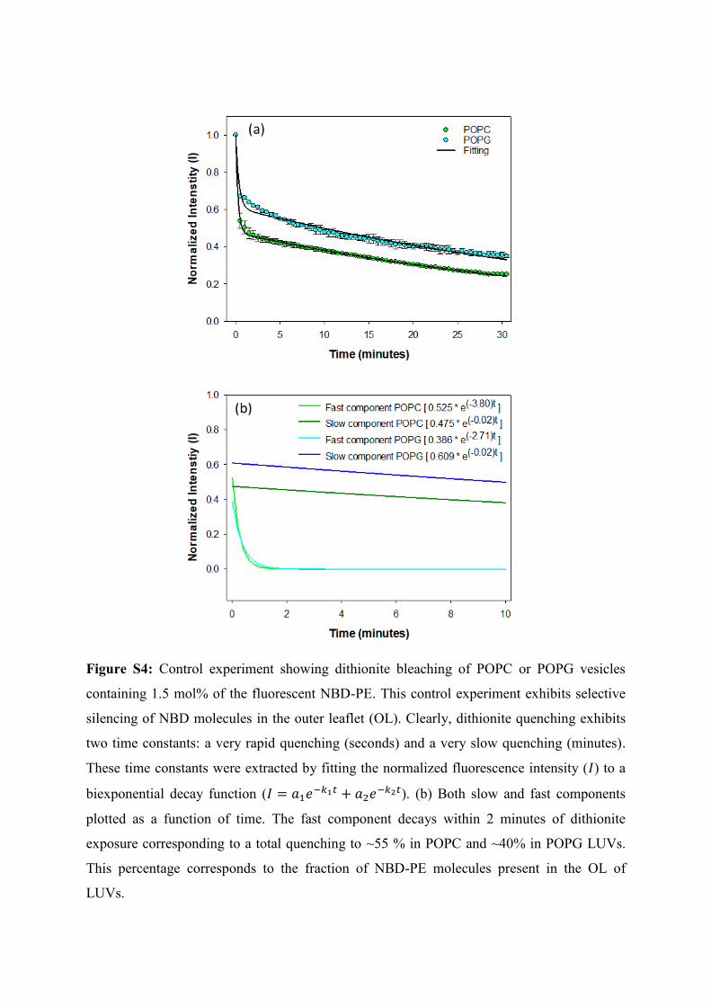
Figure S4: Control experiment showing dithionite bleaching of POPC or POPG vesicles
containing 1.5 mol% of the fluorescent NBD-PE. This control experiment exhibits selective
silencing of NBD molecules in the outer leaflet (OL). Clearly, dithionite quenching exhibits
two time constants: a very rapid quenching (seconds) and a very slow quenching (minutes).
These time constants were extracted by fitting the normalized fluorescence intensity ( ) to a
biexponential decay function (
). (b) Both slow and fast components
plotted as a function of time. The fast component decays within 2 minutes of dithionite
exposure corresponding to a total quenching to ~55 % in POPC and ~40% in POPG LUVs.
This percentage corresponds to the fraction of NBD-PE molecules present in the OL of
LUVs.

Figure
S5. (a) Molecular structures of POPC including the nomenclature. (b) 1H MAS NMR spectra
of POPC bilayer along with peak assignment. 1H MAS NMR spectra of POPC in presence of
(c) C8MIM+Br
- and (d) C12MIM
+Br
-. (e)
1H-
1H NOESY NMR spectrum of POPC at a
mixing time of 300 ms. The cross peaks between the -(CH2)n- protons of lipid chain and the
protons of lipid head group which were used in the analysis are indicated by black squares.

Figure S6. (a) Molecular structures of POPG including the nomenclature. (b) 1H MAS NMR
spectra of POPG bilayer along with peak assignment. 1H MAS NMR spectra of POPG in
presence of (c) C8MIM+Br
- and (d) C12MIM
+Br
-. (e)
1H-
1H NOESY NMR spectrum of POPG
at a mixing time of 300 ms. The cross peaks between the -(CH2)n- protons of lipid chain and
the protons of lipid head group which were used in the analysis are indicated by black
squares.

Figure S7: Time course of percentage of (a, b) total lipid mixing, and (c, d) OL mixing, and
(e, f) IL mixing in (a, c and e) POPC, and (b, d, and f) POPG LUVs upon adding C12MIM+Br
-
in concentrations as indicated in legend. The experimental error for each measurement is not
more than 3%.
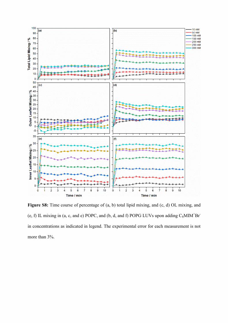
Figure S8: Time course of percentage of (a, b) total lipid mixing, and (c, d) OL mixing, and
(e, f) IL mixing in (a, c, and e) POPC, and (b, d, and f) POPG LUVs upon adding C8MIM+Br
-
in concentrations as indicated in legend. The experimental error for each measurement is not
more than 3%.

Figure S9: Structures of (a) POPC and (b) POPG molecules with protons in the glycerol and
head-group region highlighted in blue and red colours, respectively. 1H-
1H NOESY average
cross-relaxation rates between these protons and -(CH2)n- protons in lipid chains of (c) POPC
and (d) POPG observed in the absence and presence of [C8MIM]+Br
-. Also shown are the
average cross-relaxation rates glycerol and head-group protons taken together.

REFERENCE
1. Ananthapadmanabhan, K.; Goddard, E.; Turro, N.; Kuo, P. L., Fluorescence probes for
critical micelle concentration. Langmuir 1985, 1, 352-355.

download fileview on ChemRxivIonic liquid_membrane fusion_Supp Info.pdf (1.49 MiB)