an additional c-terminal loop in endonuclease iv, an apurinic/apyrimidinic endonuclease, controls...
Post on 06-Jul-2016
216 views
TRANSCRIPT
research papers
Acta Cryst. (2011). D67, 149–155 doi:10.1107/S0907444910052479 149
Acta Crystallographica Section D
BiologicalCrystallography
ISSN 0907-4449
An additional C-terminal loop in endonuclease IV,an apurinic/apyrimidinic endonuclease, controlsbinding affinity to DNA
Ryuichi Asano,a Hirohito
Ishikawa,a Shuhei Nakane,a
Noriko Nakagawa,a,b Seiki
Kuramitsua,b and Ryoji Masuia,b*
aDepartment of Biological Sciences,
Graduate School of Science, Osaka University,
1-1 Machikaneyama-cho, Toyonaka,
Osaka 560-0043, Japan, and bPhoton Science
Research Division, RIKEN SPring-8 Center,
Harima Institute, 1-1-1 Kouto, Sayo-cho,
Sayo-gun, Hyogo 679-5148, Japan
Correspondence e-mail:
# 2011 International Union of Crystallography
Printed in Singapore – all rights reserved
Endonuclease IV (EndoIV) is an endonuclease that acts at
apurinic/apyrimidinic (AP) sites and is classified as either
long-type or short-type. The crystal structures of representa-
tive types of EndoIV from Geobacillus kaustophilus and
Thermus thermophilus HB8 were determined using X-ray
crystallography. G. kaustophilus EndoIV (the long type) had a
higher affinity for double-stranded DNA containing an AP-
site analogue than T. thermophilus EndoIV (the short type).
Structural analysis of the two different EndoIVs suggested
that a C-terminal DNA-recognition loop that is only present in
the long type contributes to its high affinity for AP sites. A
mutation analysis showed that Lys267 in the C-terminal DNA-
recognition loop plays an important role in DNA binding.
Received 1 April 2010
Accepted 14 December 2010
PDB References: Geobacillus
kaustophilus endonuclease
IV, 3aal; Thermus thermo-
philus endonuclease IV,
3aam.
1. Introduction
The apurinic/apyrimidinic (AP) site is one of the most
common DNA lesions in vivo and is produced by spontaneous
hydrolysis, chemical toxins, radiation or DNA glycosylases
that remove abnormal DNA bases (Friedberg et al., 2006;
Seeberg et al., 1995; Demple & Sung, 2005). The AP site
prevents transcription and translation and thus must be
repaired by DNA-repair pathways, including base-excision
repair. In base-excision repair, an AP endonuclease recog-
nizes the AP site and cleaves the DNA backbone at the 50-side
of the AP site to generate a 50-deoxyribose phosphate group
and a free 30-hydroxyl end for DNA polymerase repair
synthesis (Levin et al., 1988; Barzilay & Hickson, 1995). AP
endonucleases are classified on the basis of their structure as
members of either the exonuclease III family or the endo-
nuclease IV (EndoIV) family. In yeast, deletion of the EndoIV
homologue increases the rate of transversion (AT to GC)
mutations and results in hypersensitivity to alkylating agents
and chemical oxidants (Kunz et al., 1994). EndoIV is induced
by superoxide for defence against oxygen radicals and nitric
oxide (Friedberg et al., 2006). All of the available information
highlights the physiological importance of EndoIV in DNA
repair. The EndoIV family has been widely identified in
archaea, bacteria and some eukaryotes but not in most
mammals. This makes EndoIV an attractive target for anti-
biotics (Kunz et al., 1994).
In bacteria, base-excision repair has been studied exten-
sively in the Gram-negative species Escherichia coli. The
tertiary structure of EndoIV has only been determined for the
E. coli homologue (Hosfield et al., 1999). In contrast, little
effort has been made to study EndoIV from Gram-positive
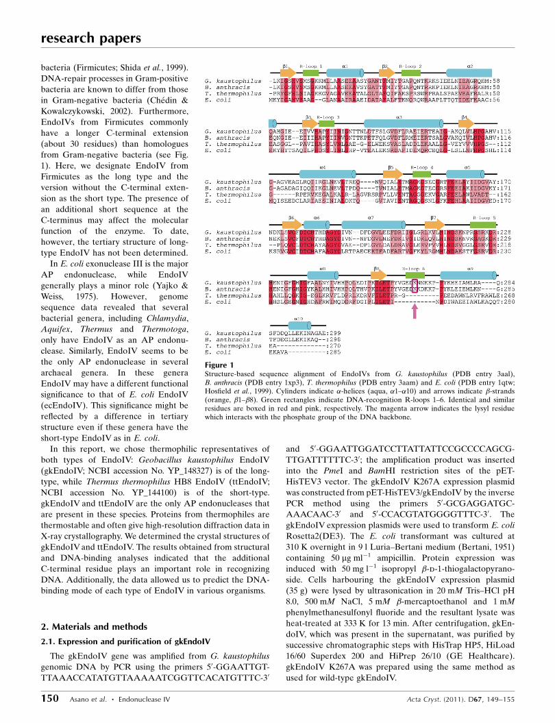
bacteria (Firmicutes; Shida et al., 1999).
DNA-repair processes in Gram-positive
bacteria are known to differ from those
in Gram-negative bacteria (Chedin &
Kowalczykowski, 2002). Furthermore,
EndoIVs from Firmicutes commonly
have a longer C-terminal extension
(about 30 residues) than homologues
from Gram-negative bacteria (see Fig.
1). Here, we designate EndoIV from
Firmicutes as the long type and the
version without the C-terminal exten-
sion as the short type. The presence of
an additional short sequence at the
C-terminus may affect the molecular
function of the enzyme. To date,
however, the tertiary structure of long-
type EndoIV has not been determined.
In E. coli exonuclease III is the major
AP endonuclease, while EndoIV
generally plays a minor role (Yajko &
Weiss, 1975). However, genome
sequence data revealed that several
bacterial genera, including Chlamydia,
Aquifex, Thermus and Thermotoga,
only have EndoIV as an AP endonu-
clease. Similarly, EndoIV seems to be
the only AP endonuclease in several
archaeal genera. In these genera
EndoIV may have a different functional
significance to that of E. coli EndoIV
(ecEndoIV). This significance might be
reflected by a difference in tertiary
structure even if these genera have the
short-type EndoIV as in E. coli.
In this report, we chose thermophilic representatives of
both types of EndoIV: Geobacillus kaustophilus EndoIV
(gkEndoIV; NCBI accession No. YP_148327) is of the long-
type, while Thermus thermophilus HB8 EndoIV (ttEndoIV;
NCBI accession No. YP_144100) is of the short-type.
gkEndoIV and ttEndoIV are the only AP endonucleases that
are present in these species. Proteins from thermophiles are
thermostable and often give high-resolution diffraction data in
X-ray crystallography. We determined the crystal structures of
gkEndoIV and ttEndoIV. The results obtained from structural
and DNA-binding analyses indicated that the additional
C-terminal residue plays an important role in recognizing
DNA. Additionally, the data allowed us to predict the DNA-
binding mode of each type of EndoIV in various organisms.
2. Materials and methods
2.1. Expression and purification of gkEndoIV
The gkEndoIV gene was amplified from G. kaustophilus
genomic DNA by PCR using the primers 50-GGAATTGT-
TTAAACCATATGTTAAAAATCGGTTCACATGTTTC-30
and 50-GGAATTGGATCCTTATTATTCCGCCCCAGCG-
TTGATTTTTTC-30; the amplification product was inserted
into the PmeI and BamHI restriction sites of the pET-
HisTEV3 vector. The gkEndoIV K267A expression plasmid
was constructed from pET-HisTEV3/gkEndoIV by the inverse
PCR method using the primers 50-GCGAGGATGC-
AAACAAC-30 and 50-CCACGTATGGGGTTTC-30. The
gkEndoIV expression plasmids were used to transform E. coli
Rosetta2(DE3). The E. coli transformant was cultured at
310 K overnight in 9 l Luria–Bertani medium (Bertani, 1951)
containing 50 mg ml�1 ampicillin. Protein expression was
induced with 50 mg l�1 isopropyl �-d-1-thiogalactopyrano-
side. Cells harbouring the gkEndoIV expression plasmid
(35 g) were lysed by ultrasonication in 20 mM Tris–HCl pH
8.0, 500 mM NaCl, 5 mM �-mercaptoethanol and 1 mM
phenylmethanesulfonyl fluoride and the resultant lysate was
heat-treated at 333 K for 13 min. After centrifugation, gkEn-
doIV, which was present in the supernatant, was purified by
successive chromatographic steps with HisTrap HP5, HiLoad
16/60 Superdex 200 and HiPrep 26/10 (GE Healthcare).
gkEndoIV K267A was prepared using the same method as
used for wild-type gkEndoIV.
research papers
150 Asano et al. � Endonuclease IV Acta Cryst. (2011). D67, 149–155
Figure 1Structure-based sequence alignment of EndoIVs from G. kaustophilus (PDB entry 3aal),B. anthracis (PDB entry 1xp3), T. thermophilus (PDB entry 3aam) and E. coli (PDB entry 1qtw;Hosfield et al., 1999). Cylinders indicate �-helices (aqua, �1–�10) and arrows indicate �-strands(orange, �1–�8). Green rectangles indicate DNA-recognition R-loops 1–6. Identical and similarresidues are boxed in red and pink, respectively. The magenta arrow indicates the lysyl residuewhich interacts with the phosphate group of the DNA backbone.
2.2. Expression and purification of ttEndoIV
The ttEndoIV gene was amplified from T. thermophilus
genomic DNA by PCR using the primers 50-ATATCATAT-
GCCACGCTACGGGTTCCACCTTTCC-30 and 50-ATATG-
GATCCTTATTAGGCCTCCTCGAGCCAGGCCCT-30; the
amplification product was inserted into the pT7Blue vector
using the EcoRV restriction site. The gene encoding ttEndoIV
was subcloned into the NdeI and BamHI sites of pET-15b
(Novagen) from pT7Blue/ttEndoIV and was then subcloned
into the NdeI and BamHI sites of pET-11a (Novagen) from
pET-15b/ttEndoIV. The ttEndoIV expression plasmid was
used to transform E. coli BL21(DE3). The E. coli transfor-
mant was cultured at 310 K overnight in 12 l Luria–Bertani
medium (Bertani, 1951) containing 50 mg ml�1 ampicillin.
Protein expression was induced with 50 mg l�1 isopropyl
�-d-1-thiogalactopyranoside. Cells harbouring the ttEndoIV
expression plasmid (15 g) were lysed by ultrasonication in
50 mM Tris–HCl pH 8.0 and 5 mM �-mercaptoethanol and the
resultant lysate was heat-treated at 338 K for 10 min. After
centrifugation, ttEndoIV, which was present in the super-
natant, was purified by successive chromatographic steps with
Toyopearl Phenyl, Toyopearl SuperQ (Tosoh), hydroxyapatite
(Nacalai) and Superdex75 10/300 GL (GE Healthcare).
2.3. DNA-binding assay
A 21 bp double-stranded DNA containing an AP-site
analogue (50-GGGTGTTGCFTTAGTTGTCAT-30 and 50-A-
TGACAACTAAGGCAACACCC-30, where F is tetrahydro-
furan, an AP-site analogue) was used for the binding assay.
The reaction mixture contained 20 mM Tris–HCl pH 8.0,
10 mM KCl, the 21 bp DNA and EndoIV. 0.5 mM EndoIV
solution (200 ml) containing 50 mM EDTA was titrated against
10 mM DNA and the fluorescence of the mixture was
measured at 340 nm with excitation at 295 nm at 298 K using
an F-4500 fluorescence spectrophotometer (Hitachi). The
binding curve was analyzed by a similar method as described
previously (Watanabe et al., 1994).
2.4. Crystallization, data collection and structuredetermination
gkEndoIV crystals were obtained using condition No. 6
(0.1 M cacodylate pH 6.5, 30% polyethylene glycol 600, 1 M
NaCl and 10% glycerol) of the Cryo II screen kit (Emerald
BioSystems) using the 96-well sitting-drop vapour-diffusion
method with drops consisting of 1 ml 20 mg ml�1 protein
solution and 1 ml reservoir solution equilibrated against 500 ml
well solution at 293 K. ttEndoIV crystals were obtained using
condition No. 43 (0.2 M ammonium phosphate monobasic and
20% polyethylene glycol 3350) of the PEG/Ion screen kit
(Hampton Research) with an additional 20 mM MnCl2 using
the hanging-drop method with drops consisting of 0.5 ml
protein solution at 19.7 mg ml�1 and 0.5 ml reservoir solution
equilibrated against 200 ml well solution at 293 K (Iino et al.,
2008). The dimensions of the harvested crystals were 0.05 �
0.05 � 0.2 mm. Data were collected under a liquid-nitrogen
stream on the RIKEN Structural Genomics Beamline II
(BL26B2; Ueno et al., 2006) at SPring-8 (Hyogo, Japan). The
data were processed using the HKL-2000 program suite
research papers
Acta Cryst. (2011). D67, 149–155 Asano et al. � Endonuclease IV 151
Table 1Data-collection and refinement statistics for gkEndoIV and ttEndoIV.
Values in parentheses are for the highest resolution shell.
Data set gkEndoIV ttEndoIV
Data collectionWavelength (A) 1.0000 1.0000Resolution (A) 50.00–1.60
(1.66–1.60)50.00–1.58
(1.64–1.58)Space group P6122 P21
Molecules per asymmetric unit 1 1Unit-cell parameters (A, �) a = b = 85.170,
c = 140.220,� = � = � = 120.00
a = 36.646,b = 85.273,c = 36.580,� = � = 90.00,� = 105.370
Measured reflections 599847 180015Unique reflections 40267 29574Completeness (%) 99.7 (97.6) 99.2 (93.9)Multiplicity 14.9 (6.6) 6.1 (4.0)hI/�(I)i 62.8 (6.4) 43.6 (8.4)Rmerge† (%) 4.9 (21.6) 6.8 (16.1)
RefinementResolution (A) 23.42–1.60 29.12–1.58Rwork‡/Rfree§ (%) 19.7/22.2 19.9/22.3No. of protein atoms 2316 2048No. of water atoms 110 223R.m.s.d. bond lengths (A) 0.013 0.004R.m.s.d. angles (�) 1.600 1.200Average B factor (A2) 17.6 13.8Ramachandran plot (%)
Most favoured 93.4 92.0Additional 6.2 8.0Disallowed 0.4 0.0
† Rmerge =P
hkl
Pi jIiðhklÞ � hIðhklÞij=
Phkl
Pi IiðhklÞ, where hI(hkl)i is the average of
individual measurements of Ii(hkl). ‡ Rwork =P
hkl
��jFobsj � jFcalcj
��=P
hkl jFobsj, where|Fobs| and |Fcalc| are the observed and calculated structure-factor amplitudes,respectively. § Rfree is calculated against a 10% random sampling of reflections thatwere removed before structure refinement.
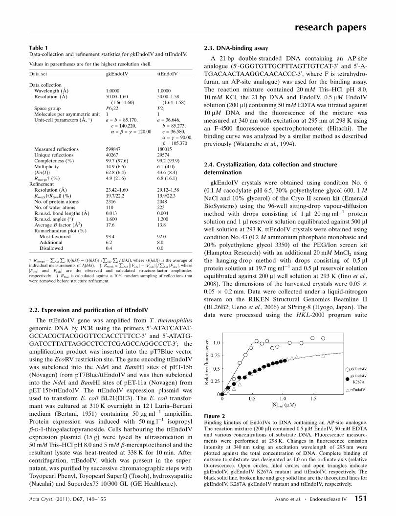
Figure 2Binding kinetics of EndoIVs to DNA containing an AP-site analogue.The reaction mixture (200 ml) contained 0.5 mM EndoIV, 50 mM EDTAand various concentrations of substrate DNA. Fluorescence measure-ments were performed at 298 K. Changes in fluorescence emissionintensity at 340 nm using an excitation wavelength of 295 nm wereplotted against the total concentration of DNA. Complete binding ofenzyme to substrate was designated as 1.0 on the ordinate axis (relativefluorescence). Open circles, filled circles and open triangles indicategkEndoIV, gkEndoIV K267A mutant and ttEndoIV, respectively. Theblack solid line, broken line and grey solid line are the theoretical lines forgkEndoIV, K267A gkEndoIV mutant and ttEndoIV, respectively.
(Otwinowski & Minor, 1997). The structures were solved using
Bacillus anthracis EndoIV (PDB code 1xp3; M. J. Fogg, V. M.
Levdikov, E. V. Blagova, J. A. Brannigan, A. J. Wilkinson &
K. S. Wilson, unpublished work) as the model for molecular
replacement using MOLREP (Vagin & Teplyakov, 2010). The
automatic tracing procedures in the program ARP/wARP
(Morris et al., 2003) were utilized to build the initial models.
Model refinements were carried out using the programs
XtalView (McRee, 1992) and CNS (Brunger et al., 1998).
According to the PROCHECK software (Laskowski et al.,
1993), 93.4 and 92.0% of the residues in the final models of
gkEndoIV and ttEndoIV, respectively, are located in the most
favoured region of the Ramachandran plot and 0.4 and 0% of
the residues, respectively, are located in disallowed regions.
Data-collection and data-processing statistics are presented
in Table 1. The metal ions were determined using X-ray
absorption fine structure (XAFS), measurement of inductively
coupled plasma (ICP), coordination number and an anom-
alous difference Fourier map. The coordinates are available
in the Protein Data Bank under accession codes 3aal and
3aam. The nucleotide sequence data reported are available in
the DDBJ/EMBL/GenBank databases under accession Nos.
AP006508 and AP008226. DNA-binding
models were constructed based on the
structure of ecEndoIV complexed with a
product DNA (PDB code 1qum; Hosfield et
al., 1999) using PyMOL (DeLano, 2002) and
CNS.
3. Results and discussion
3.1. Binding affinity of the AP site indouble-stranded DNA
We measured the binding affinity of
gkEndoIV and ttEndoIV for the substrate
analogue (a 21 bp double-stranded DNA
containing an AP-site analogue) using
intrinsic fluorescence of the protein from
tryptophan or tyrosine residues (Fig. 2).
When the ttEndoIV solution was titrated
against the DNA the fluorescence with an
emission maximum at 345 nm, probably
from a tryptophan, was quenched on DNA
binding. The fluorescence emission spec-
trum of gkEndoIV had an emission
maximum at 320 nm, but it also changed
when titrated against the DNA. The titra-
tion experiments gave the binding profiles
shown in Fig. 2. The affinity of gkEndoIV
for the substrate analogue was found to be
very high and its dissociation constant was
less than 0.01 mM. The break point of total
concentration in the presence of 0.5 mM
gkEndoIV was 0.5 mM (open circles in
Fig. 2). This indicates that one molecule of
gkEndoIV binds to one molecule of double-
stranded DNA. The affinity of ttEndoIV for
the substrate analogue was lower than that
of gkEndoIV (open triangles in Fig. 2).
Assuming a bimolecular reaction, its disso-
ciation constant was calculated to be
0.37 mM. These results indicate that
gkEndoIV has a higher affinity for the AP
site in double-stranded DNA than ttEn-
doIV. It should be noted that the DNA-
binding assay was performed at 298 K. The
absolute values of the dissociation constants
research papers
152 Asano et al. � Endonuclease IV Acta Cryst. (2011). D67, 149–155
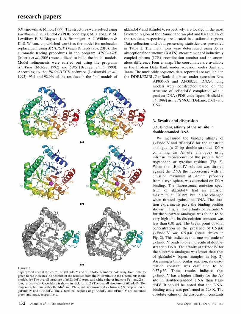
Figure 3Superposed crystal structures of gkEndoIV and ttEndoIV. Rainbow colouring from blue togreen to red indicates the position of the residues from the N-terminus to the C-terminus in themodels. (a) The overall structure of gkEndoIV. Aqua and white spheres indicate Fe2+ and Zn2+
ions, respectively. Cacodylate is shown in stick form. (b) The overall structure of ttEndoIV. Themagenta sphere indicates the Mn2+ ion. Phosphate is shown in stick form. (c) Superposition ofgkEndoIV and ttEndoIV. The C-terminal regions of gkEndoIV and ttEndoIV are colouredgreen and aqua, respectively.
may be changed at optimum temperatures for both thermo-
philes.
3.2. Structural determination
To investigate the differences between gkEndoIV and
ttEndoIV in their relative affinities for the substrate, we
determined their tertiary structures by X-ray crystallography.
The overall structures of gkEndoIV (Fig. 3a) and ttEndoIV
(Fig. 3b) were determined at resolutions of 1.60 and 1.58 A,
respectively. In both EndoIVs the first methionine in the
amino-acid sequence encoded by their genes was removed
in the determined structures. The crystallographic data are
summarized in Table 1. Each asymmetric unit contained one
molecule that adopted an eight-stranded �/�-barrel fold (TIM
barrel). The two structures were very similar, with a root-
mean-square deviation (r.m.s.d.) of only 1.65 A for the C�
atoms. gkEndoIV, with 299 amino-acid residues, is longer than
ttEndoIV by 29 residues (Fig. 1). The most significant differ-
ence between these structures was observed in the C-terminal
region, which contained an additional loop and an �-helix in
gkEndoIV (Fig. 3c). This structural difference may contribute
to the increased affinity of gkEndoIV for the substrate
(discussed later).
Another difference was observed with respect to the metal
ions bound to the proteins. In the determined structures
gkEndoIV contained two Fe2+ ions and one Zn2+ ion (Fig. 4a),
whereas ttEndoIV contained one Mn2+ ion (Fig. 4b). The
Mn2+ ion was probably derived from the crystallization
mother liquor. The Mn2+-ion site in ttEndoIV was verified
using an anomalous difference Fourier map for data collected
at the Zn peak wavelength (1.7000 A). The assignment of Fe2+
and Zn2+ ions in gkEndoIV was verified by XAFS and ICP
measurements. The numbers of these metal ions were also
determined by ICP measurements. A cacodylate ion was
bound to the active site of gkEndoIV and a phosphate ion was
bound to that of ttEndoIV. The cacodylate and phosphate ions
were components of the crystallization mother liquor. The
sites to which they bound were expected to be the site of the
DNA phosphate group.
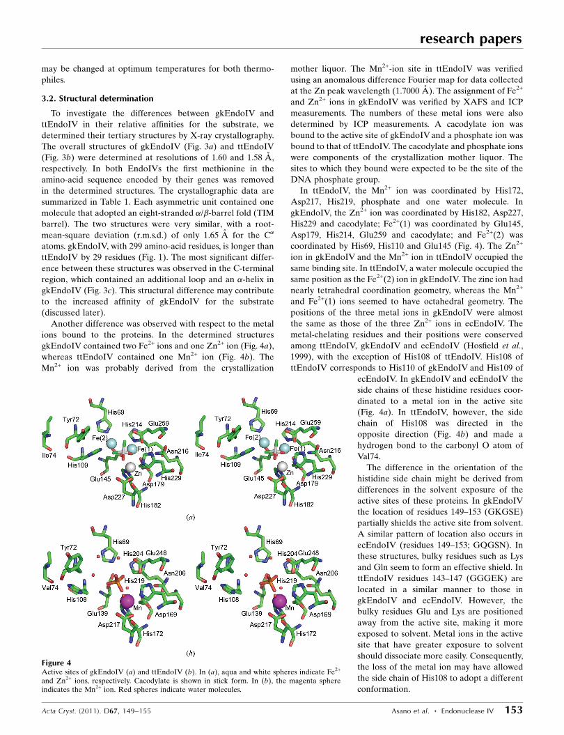
In ttEndoIV, the Mn2+ ion was coordinated by His172,
Asp217, His219, phosphate and one water molecule. In
gkEndoIV, the Zn2+ ion was coordinated by His182, Asp227,
His229 and cacodylate; Fe2+(1) was coordinated by Glu145,
Asp179, His214, Glu259 and cacodylate; and Fe2+(2) was
coordinated by His69, His110 and Glu145 (Fig. 4). The Zn2+
ion in gkEndoIV and the Mn2+ ion in ttEndoIV occupied the
same binding site. In ttEndoIV, a water molecule occupied the
same position as the Fe2+(2) ion in gkEndoIV. The zinc ion had
nearly tetrahedral coordination geometry, whereas the Mn2+
and Fe2+(1) ions seemed to have octahedral geometry. The
positions of the three metal ions in gkEndoIV were almost
the same as those of the three Zn2+ ions in ecEndoIV. The
metal-chelating residues and their positions were conserved
among ttEndoIV, gkEndoIV and ecEndoIV (Hosfield et al.,
1999), with the exception of His108 of ttEndoIV. His108 of
ttEndoIV corresponds to His110 of gkEndoIV and His109 of
ecEndoIV. In gkEndoIV and ecEndoIV the
side chains of these histidine residues coor-
dinated to a metal ion in the active site
(Fig. 4a). In ttEndoIV, however, the side
chain of His108 was directed in the
opposite direction (Fig. 4b) and made a
hydrogen bond to the carbonyl O atom of
Val74.
The difference in the orientation of the
histidine side chain might be derived from
differences in the solvent exposure of the
active sites of these proteins. In gkEndoIV
the location of residues 149–153 (GKGSE)
partially shields the active site from solvent.
A similar pattern of location also occurs in
ecEndoIV (residues 149–153; GQGSN). In
these structures, bulky residues such as Lys
and Gln seem to form an effective shield. In
ttEndoIV residues 143–147 (GGGEK) are
located in a similar manner to those in
gkEndoIV and ecEndoIV. However, the
bulky residues Glu and Lys are positioned
away from the active site, making it more
exposed to solvent. Metal ions in the active
site that have greater exposure to solvent
should dissociate more easily. Consequently,
the loss of the metal ion may have allowed
the side chain of His108 to adopt a different
conformation.
research papers
Acta Cryst. (2011). D67, 149–155 Asano et al. � Endonuclease IV 153
Figure 4Active sites of gkEndoIV (a) and ttEndoIV (b). In (a), aqua and white spheres indicate Fe2+
and Zn2+ ions, respectively. Cacodylate is shown in stick form. In (b), the magenta sphereindicates the Mn2+ ion. Red spheres indicate water molecules.
3.3. DNA-binding site
To obtain further insights into the possible reasons for the
differences in affinity for the substrate shown by the two types
of enzyme, we constructed DNA-binding models for ttEndoIV
and gkEndoIV based on the structure of ecEndoIV com-
plexed with product DNA (Hosfield et al., 1999; Fig. 5). The
ecEndoIV structure in the DNA complex could be super-
imposed on those of gkEndoIV and ttEndoIV with r.m.s.d.s
(for C� atoms) of 1.65 and 1.69 A, respectively. The five DNA-
recognition loops (R-loops) that have been defined previously
(Garcin et al., 2008) were identified in the model structures of
both gkEndoIV and ttEndoIV (Figs. 5a and 5b, respectively).
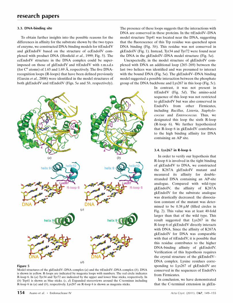
The presence of these loops suggests that the interactions with
DNA are conserved in these proteins. In the ttEndoIV–DNA
model structure Trp41 was located near the DNA, suggesting
that the fluorescence of this Trp residue was quenched upon
DNA binding (Fig. 5b). This residue was not conserved in
gkEndoIV (Fig. 1). Instead, Tyr34 and Tyr72 were found near
the DNA in the gkEndoIV–DNA model structure (Fig. 5a).
Unexpectedly, in the model structure of gkEndoIV com-
plexed with DNA an additional loop (265–269) between the
last two helices was identified and was presumed to interact
with the bound DNA (Fig. 5a). The gkEndoIV–DNA binding
model suggested a possible interaction between the phosphate
group of the DNA backbone and Lys267 in this loop (Fig. 5c).
In contrast, it was not present in
ttEndoIV (Fig. 5d). The amino-acid
sequence of this loop was not restricted
to gkEndoIV but was also conserved in
EndoIVs from other Firmicutes,
including Bacillus, Listeria, Staphylo-
coccus and Enterococcus. Thus, we
designated this loop the sixth R-loop
(R-loop 6). We further hypothesized
that R-loop 6 in gkEndoIV contributes
to the high binding affinity for DNA
containing an AP site.
3.4. Lys267 in R-loop 6
In order to verify our hypothesis that
R-loop 6 is involved in the tight binding
of gkEndoIV to DNA, we constructed
the K267A gkEndoIV mutant and
measured its affinity for double-
stranded DNA containing an AP-site
analogue. Compared with wild-type
gkEndoIV, the affinity of K267A
gkEndoIV for the substrate analogue
was drastically decreased: the dissocia-
tion constant of the mutant was deter-
mined to be 0.38 mM (filled circles in
Fig. 2). This value was at least 40-fold
larger than that of the wild type. This
result suggested that Lys267 in the
R-loop 6 of gkEndoIV directly interacts
with DNA. Since the affinity of K267A
gkEndoIV for DNA was comparable
with that of ttEndoIV, it is possible that
this residue contributes to the higher
DNA-binding affinity of gkEndoIV.
Verification of this hypothesis requires
the crystal structure of the gkEndoIV–
DNA complex. Lysine residues corre-
sponding to Lys267 of gkEndoIV are
conserved in the sequences of EndoIVs
from Firmicutes.
In conclusion, we have demonstrated
that the C-terminal extension in gkEn-
research papers
154 Asano et al. � Endonuclease IV Acta Cryst. (2011). D67, 149–155
Figure 5Model structures of the gkEndoIV–DNA complex (a) and the ttEndoIV–DNA complex (b). DNAis shown in yellow. R-loops are indicated by magenta loops with numbers. The red circle indicatesR-loop 6. In (a) Tyr34 and Tyr72 are indicated by the upper and lower blue sticks, respectively. In(b) Trp41 is shown as blue sticks. (c, d) Expanded stereoviews around the C-terminus includingR-loop 6 in (a) and (b), respectively. Lys267 on R-loop 6 is shown as magenta sticks.
doIV contributes to its tight binding to substrate DNA. This
conclusion can be extended to the long-type EndoIV. It
remains unclear why the long-type EndoIV binds more tightly
to DNA than the short-type. EndoIV is known to be asso-
ciated with other proteins in DNA-repair processes (Friedberg
et al., 2006). Firmicutes may require different cooperation
partners for these processes.
We thank Michiyo Takahara for her help in the purification
of gkEndoIV, Toshi Arima for her help in gkEndoIV crys-
tallization, Yuka Nonaka for her help in data collection from
gkEndoIV crystals and Ryoichi Arai for providing the pET-
HisTEV3 vector. This work was supported by Grants-in-Aid
for Scientific Research 17770089 (to NN) and 20570131 (to
RM) from the Ministry of Education, Science, Sports and
Culture of Japan.
References
Barzilay, G. & Hickson, I. D. (1995). Bioessays, 17, 713–719.Bertani, G. (1951). J. Bacteriol. 62, 293–300.Brunger, A. T., Adams, P. D., Clore, G. M., DeLano, W. L., Gros, P.,
Grosse-Kunstleve, R. W., Jiang, J.-S., Kuszewski, J., Nilges, M.,Pannu, N. S., Read, R. J., Rice, L. M., Simonson, T. & Warren, G. L.(1998). Acta Cryst. D54, 905–921.
Chedin, F. & Kowalczykowski, S. C. (2002). Mol. Microbiol. 43,823–834.
DeLano, W. L. (2002). PyMOL. http://www.pymol.org.Demple, B. & Sung, J.-S. (2005). DNA Repair, 4, 1442–1449.
Friedberg, E. C., Walker, G. C., Siede, W., Wood, R. D., Schultz, R. A.& Ellenberger, T. (2006). DNA Repair and Mutagenesis, 2nd ed.,pp. 9–69. Washington: ASM Press.
Garcin, E. D., Hosfield, D. J., Desai, S. A., Haas, B. J., Bjoras, M.,Cunningham, R. P. & Tainer, J. A. (2008). Nature Struct. Mol. Biol.15, 515–522.
Hosfield, D. J., Guan, Y., Haas, B. J., Cunningham, R. P. & Tainer, J. A.(1999). Cell, 98, 397–408.
Iino, H., Naitow, H., Nakamura, Y., Nakagawa, N., Agari, Y.,Kanagawa, M., Ebihara, A., Shinkai, A., Sugahara, M., Miyano, M.,Kamiya, N., Yokoyama, S., Hirotsu, K. & Kuramitsu, S. (2008). ActaCryst. F64, 487–491.
Kunz, B. A., Henson, E. S., Roche, H., Ramotar, D., Nunoshiba, D. &Demple, B. (1994). Proc. Natl Acad. Sci. USA, 91, 8165–8169.
Laskowski, R. A., MacArthur, M. W., Moss, D. S. & Thornton, J. M.(1993). J. Appl. Cryst. 26, 283–291.
Levin, J. D., Johnson, A. W. & Demple, B. (1988). J. Biol. Chem. 263,8066–8071.
McRee, D. E. (1992). J. Mol. Graph. 10, 44–46.Morris, R. J., Perrakis, A. & Lamzin, V. S. (2003). Methods Enzymol.
374, 229–244.Otwinowski, Z. & Minor, W. (1997). Methods Enzymol. 276, 307–
326.Seeberg, E., Eide, L. & Bjoras, M. (1995). Trends Biochem. Sci. 20,
391–397.Shida, T., Ogawa, T., Ogasawara, N. & Sekiguchi, J. (1999). Biosci.
Biotechnol. Biochem. 63, 1528–1534.Ueno, G., Kanda, H., Hirose, R., Ida, K., Kumasaka, T. & Yamamoto,
M. (2006). J. Struct. Funct. Genomics, 7, 15–22.Vagin, A. & Teplyakov, A. (2010). Acta Cryst. D66, 22–25.Watanabe, R., Masui, R., Mikawa, T., Takamatsu, S., Kato, R. &
Kuramitsu, S. (1994). J. Biochem. 116, 960–966.Yajko, D. M. & Weiss, B. (1975). Proc. Natl Acad. Sci. USA, 72,
688–692.
research papers
Acta Cryst. (2011). D67, 149–155 Asano et al. � Endonuclease IV 155