an anatomical study of arcuate foramen and its clinical
TRANSCRIPT
CASE REPORT Open Access
An anatomical study of arcuate foramenand its clinical implications: a case reportSalman Afsharpour1*, Kathryn T. Hoiriis2, R. Bruce Fox3 and Samuel Demons1
Abstract
Background: The objective of this paper is to describe the relationship of the vertebral artery (VA) to the Atlas (C1)in the sub-occipital region in the presence of arcuate foramen; and discuss the clinical implications related tomanual therapies and surgical implications related to screw placement. This study is an anatomical cadaveric casereport of symmetrical bilateral lateral and dorsal arcuate foramina on the C1 dorsal arch.
Case Presentation: Out of 40 cadavers that were available for use in teaching anatomy in the university setting,three presented with anomalies of the C1 dorsal arch. The sub-occipital regions were skillfully prosected to preserverelated structures, especially VAs, sub-occipital and greater occipital nerves. Visual observations, photographs,measurements, and radiographic examinations were performed between January 15, 2014 and August 25, 2014.One cadaver (Specimen A) presented with complete bilateral ossified arcuate foramina, and two presented withpartial ossification of the atlanto-occipital membrane. Specimen A presented the bilateral anomaly which is almostsymmetrical. The VAs were found passing through double foramina (lateral and dorsal) on each side.
Conclusions: Arcuate foramina have been shown to be commonly found anomalies with highly variable shapesand sizes, even in the same individual with a bilateral condition. This study found a rare type of the anomalyassociated with the C1 dorsal arch, which protected the VA against manual pressure. However, VA, in this case,would be more susceptible to dissection. The presence of the arcuate foramen would also complicate screwplacement during surgery. Clinical pre-screening for signs of vertebrobasilar insufficiency is important forchiropractic and manual therapies.
Keywords: Arcuate foramen, C1 dorsal arch, Vertebral artery
BackgroundThe Atlas, located at the cranio-cervical junction, is aring-shaped vertebra. Normally, the vertebral arteryglides easily with neck movements as it lies in thegroove at the supero-lateral aspect of the C1 dorsal arch.The relationship of the VA to the dorsal arch of C1 hasbeen well described. Cacciola, Ebraheim, and others havestudied the course of the artery and the parameters rele-vant during surgery in the region [1–8]. Cacciola, et al.injected colored silicone into the arteries and veins often cadaveric specimen; and the microsurgical anatomyof the VAs were evaluated along its course from the C3transverse process to its entrance into the vertebral for-amen at the occipito-atlantal (C0-C1) level with
particularly close inspection of the relationship to theC2 vertebra [1]. The authors concluded that the intimaterelationship makes the VA susceptible to injury duringthe surgical procedures in the region [1]. The multipleloops of the artery provide VA extra length which isprobably essential to avoid any stretch during neckmovements [1].Certain anomalies of the Atlas vertebra may have clin-
ical significance for surgery, chiropractic and other man-ual therapies [9–14]. Arcuate foramen have been shownto be commonly found anomalies with highly variableshapes and sizes [1–8, 15, 16]. A bony bridge is formedby ossification in the oblique part of the atlanto-occipitalmembrane above the passage of VA [15]. There are sev-eral names used for the bony bridge, among them: pos-terior ponticle, posticus ponticus, ponticulus posticus,kimmerle anomaly or arcuate foramen [15]. Of graveconcern is whether the posterolateral bony bridge could
* Correspondence: [email protected] Science Division, Department of Anatomy, Life University, College ofChiropractic, 1269 Barclay Circle, Marietta, GA 30060, USAFull list of author information is available at the end of the article
© 2016 Afsharpour et al. Open Access This article is distributed under the terms of the Creative Commons Attribution 4.0International License (http://creativecommons.org/licenses/by/4.0/), which permits unrestricted use, distribution, andreproduction in any medium, provided you give appropriate credit to the original author(s) and the source, provide a link tothe Creative Commons license, and indicate if changes were made. The Creative Commons Public Domain Dedication waiver(http://creativecommons.org/publicdomain/zero/1.0/) applies to the data made available in this article, unless otherwise stated.
Afsharpour et al. Chiropractic & Manual Therapies (2016) 24:4 DOI 10.1186/s12998-016-0082-2
involve VA compression and/or fixation (tethering).Cushing et al. reported that presence of arcuate foramencaused increased incidence of VA dissection because oftethering as it passed through the osseous bridges of ar-cuate foramina [9]. The number of patients treated withC1 lateral mass screws through the posterior arch hasdramatically increased recently, therefore it is importantto recognize the presence of arcuate foramen before per-forming the Goel procedure for placement of the screws[15–19].A meta-analysis, by Elliot and Tanweer, demonstrated
that arcuate foramen anomalies are not rare [15]. Theirreport described a systematic review of radiographic, ca-daveric, and surgical data. They reported overall preva-lence of arcuate foramina was 16.7 %; with 18.8 % incadaver studies, 17.2 % in computed tomography stud-ies, and 16.6 % in x-ray studies. Standard radiographscannot demonstrate bilateral versus unilateral arcuateforamina. Elliot and Tanweer reported complete foramenin 9.3 % of patients and a partial or incomplete foramenin 8.7 %. In 5.4 % of cases, complete foramina werepresent bilaterally. In 7.6 % of cases, it was unilateral.They found no difference in prevalence between malesand females [15].Measurements of arcuate foramina are of interest be-
cause of the highly variable nature of the anomalies andtheir clinical implications. The arcuate foramina maypossibly compress the VA. In a study by Krishnamurthyet al., 1044 complete undamaged dry human atlas verte-brae were examined [13]. These researchers found thetrait was present in 13.8 % of their samples. They mea-sured the mean length of the arcuate foramen at7.16 mm on the left side and 9.99 mm on the right sidein bilateral positive samples. It was 8.14 mm and9.26 mm respectively in unilateral positive samples. Theyreported mean vertical height of arcuate foramen was6.57 mm on the left side and 6.52 mm on the right sidein bilateral positive samples. It was 4.91 mm and5.38 mm respectively in unilateral positive samples. Be-sides genetic factors, the researchers discuss that mech-anical external factors, such as carrying heavy objects onthe head, could also play a role in the development ofbridges [13]. It has been suggested that healthcare pro-viders, including neurologists, neurosurgeons, and themedical community, in general, should have knowledgeabout the present variation and should try to look for itwhen dealing with the patients complaining of symp-toms of vertebrobasilar insufficiency like headache, ver-tigo, and shoulder and arm pain [9–13, 14, 20–22].This report describes an unusual finding during anat-
omy course instruction with cadavers provided by theLife University, College of Chiropractic. Anatomists gen-erally recognized that skillful prosection leads to bettervisualization, demonstration and description for student
learning [23, 24]. Anatomical descriptions of location,relation to neighboring structures, size and shape areoften supported by drawings, but not often by photo-graphic or radiographic images [23]. An increase in theuse of computers for teaching anatomy has been re-ported [24]. Our study provided photography and x-raysof this clinically important anomaly for use in teaching.This report will discuss the anomalies of arcuate for-amen found and clinical significance for surgery, as wellas chiropractic and other manual therapies.
Case presentationMethods and materialsProsections are used primarily in the teaching of anat-omy in disciplines as varied as human medicine, chiro-practic, veterinary medicine, and physical therapy. Therewere 40 cadavers (20 males and 20 females) available foruse in teaching anatomy in the university setting. Pro-sections of the suboccipital region were performed inroutine teaching practices to allow students to investi-gate the passage of VA between foramen transversariumof the Atlas and penetration of the dura mater towardthe cranial cavity. One male cadaver (Specimen A) pre-sented with the anomalies of complete bilateral arcuateforamen, although two other cadavers had partial for-amen (Specimens B and C). Visual observations werepreserved with digital photography and dimension mea-surements were recorded (Figs. 1, 2, 3 and 4). Radio-graphic images were obtained in the frontal, bilateraloblique and neutral lateral (right and left) positions ofthe prosected suboccipital region of Specimen A to com-pare the visual effects of this variation (Figs. 5 and 6).These procedures were done between January 15, 2014and August 25, 2014.
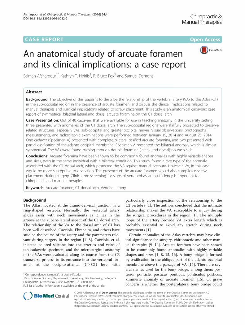
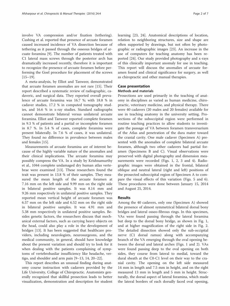
ResultsAmong the 40 cadavers, only one (Specimen A) showedthe presence of almost symmetrical bilateral dorsal bonybridges and lateral osseo-fibrous rings. In this specimen,VAs were found passing through the lateral foraminabut deep to the dorsal bony bridge, as shown in Fig. 1,and at higher magnification of the right side in Fig. 2.The detailed dissection showed only the sub-occipitalnerve (C1 dorsal ramus) along with accompanyingbranch of the VA emerging through the oval opening be-tween the dorsal and lateral arches (Figs. 1 and 2). VAswere found passing deep to the oval opening on bothsides, they course from lateral to medial, toward thedural sheath at the C0-C1 level on their way to the cra-nial cavity. The opening on the left side measured14 mm in length and 7.5 mm in height, and on the rightmeasured 13 mm in length and 5 mm in height. Struc-turally, the dorsal aspect of lateral foramina, which madethe lateral borders of each dorsally faced oval opening,
Afsharpour et al. Chiropractic & Manual Therapies (2016) 24:4 Page 2 of 7
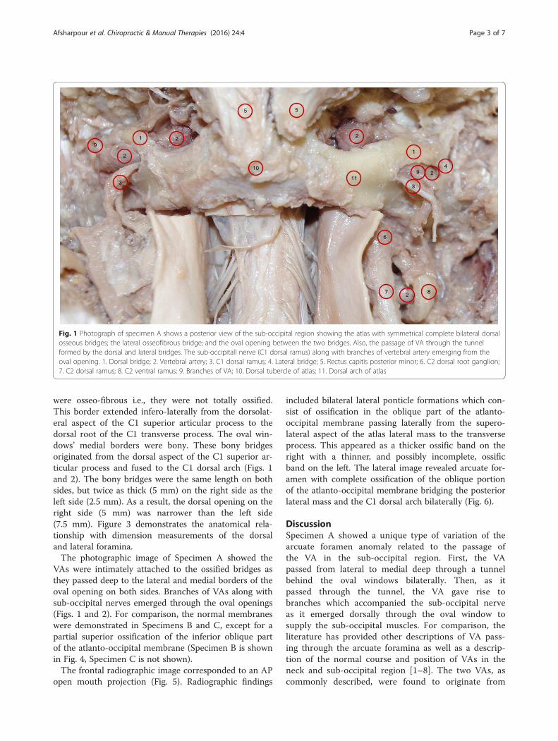
were osseo-fibrous i.e., they were not totally ossified.This border extended infero-laterally from the dorsolat-eral aspect of the C1 superior articular process to thedorsal root of the C1 transverse process. The oval win-dows’ medial borders were bony. These bony bridgesoriginated from the dorsal aspect of the C1 superior ar-ticular process and fused to the C1 dorsal arch (Figs. 1and 2). The bony bridges were the same length on bothsides, but twice as thick (5 mm) on the right side as theleft side (2.5 mm). As a result, the dorsal opening on theright side (5 mm) was narrower than the left side(7.5 mm). Figure 3 demonstrates the anatomical rela-tionship with dimension measurements of the dorsaland lateral foramina.The photographic image of Specimen A showed the
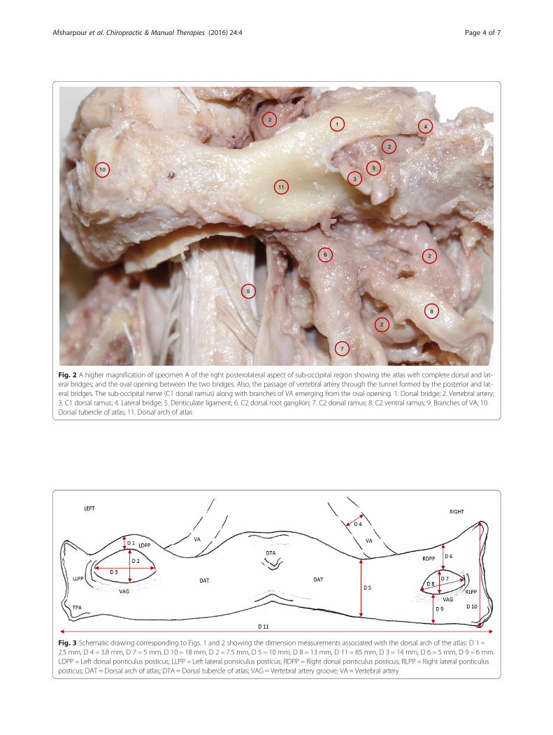
VAs were intimately attached to the ossified bridges asthey passed deep to the lateral and medial borders of theoval opening on both sides. Branches of VAs along withsub-occipital nerves emerged through the oval openings(Figs. 1 and 2). For comparison, the normal membraneswere demonstrated in Specimens B and C, except for apartial superior ossification of the inferior oblique partof the atlanto-occipital membrane (Specimen B is shownin Fig. 4, Specimen C is not shown).The frontal radiographic image corresponded to an AP
open mouth projection (Fig. 5). Radiographic findings
included bilateral lateral ponticle formations which con-sist of ossification in the oblique part of the atlanto-occipital membrane passing laterally from the supero-lateral aspect of the atlas lateral mass to the transverseprocess. This appeared as a thicker ossific band on theright with a thinner, and possibly incomplete, ossificband on the left. The lateral image revealed arcuate for-amen with complete ossification of the oblique portionof the atlanto-occipital membrane bridging the posteriorlateral mass and the C1 dorsal arch bilaterally (Fig. 6).
DiscussionSpecimen A showed a unique type of variation of thearcuate foramen anomaly related to the passage ofthe VA in the sub-occipital region. First, the VApassed from lateral to medial deep through a tunnelbehind the oval windows bilaterally. Then, as itpassed through the tunnel, the VA gave rise tobranches which accompanied the sub-occipital nerveas it emerged dorsally through the oval window tosupply the sub-occipital muscles. For comparison, theliterature has provided other descriptions of VA pass-ing through the arcuate foramina as well as a descrip-tion of the normal course and position of VAs in theneck and sub-occipital region [1–8]. The two VAs, ascommonly described, were found to originate from
1
1
8
3
55
4
3
2
2
2
27
11
2
6
910
9
Fig. 1 Photograph of specimen A shows a posterior view of the sub-occipital region showing the atlas with symmetrical complete bilateral dorsalosseous bridges; the lateral osseofibrous bridge; and the oval opening between the two bridges. Also, the passage of VA through the tunnelformed by the dorsal and lateral bridges. The sub-occipitall nerve (C1 dorsal ramus) along with branches of vertebral artery emerging from theoval opening. 1. Dorsal bridge; 2. Vertebral artery; 3. C1 dorsal ramus; 4. Lateral bridge; 5. Rectus capitis posterior minor; 6. C2 dorsal root ganglion;7. C2 dorsal ramus; 8. C2 ventral ramus; 9. Branches of VA; 10. Dorsal tubercle of atlas; 11. Dorsal arch of atlas
Afsharpour et al. Chiropractic & Manual Therapies (2016) 24:4 Page 3 of 7
9
11
8
7
6
5
4
3
2
1
2
2
2
10
Fig. 2 A higher magnification of specimen A of the right posterolateral aspect of sub-occipital region showing the atlas with complete dorsal and lat-eral bridges; and the oval opening between the two bridges. Also, the passage of vertebral artery through the tunnel formed by the posterior and lat-eral bridges. The sub-occipital nerve (C1 dorsal ramus) along with branches of VA emerging from the oval opening. 1. Dorsal bridge; 2. Vertebral artery;3. C1 dorsal ramus; 4. Lateral bridge; 5. Denticulate ligament; 6. C2 dorsal root ganglion; 7. C2 dorsal ramus; 8. C2 ventral ramus; 9. Branches of VA; 10.Dorsal tubercle of atlas; 11. Dorsal arch of atlas
Fig. 3 Schematic drawing corresponding to Figs. 1 and 2 showing the dimension measurements associated with the dorsal arch of the atlas: D 1 =2.5 mm, D 4 = 3.8 mm, D 7 = 5 mm, D 10 = 18 mm, D 2 = 7.5 mm, D 5 = 10 mm, D 8 = 13 mm, D 11 = 85 mm, D 3 = 14 mm, D 6 = 5 mm, D 9 = 6 mm.LDPP = Left dorsal ponticulus posticus; LLPP = Left lateral ponsiculus posticus; RDPP = Right dorsal ponticulus posticus; RLPP = Right lateral ponticulusposticus; DAT = Dorsal arch of atlas; DTA = Dorsal tubercle of atlas; VAG = Vertebral artery groove; VA = Vertebral artery
Afsharpour et al. Chiropractic & Manual Therapies (2016) 24:4 Page 4 of 7
the first part of the subclavian artery and then ascendthrough the transverse foramina of six cervical verte-brae (C6 to C1). In the sub-occipital region, theywere found to wind posterior to the atlanto-occipitaljoint, laying on the groove located on the superiorand lateral aspect of the dorsal arch of the atlas ontheir way to the cranium through the atlanto-occipitalmembrane then through the dorsolateral aspect of thespinal dural sheath at C0-C1 level and foramen mag-num. Specimen B and C demonstrated similar partial
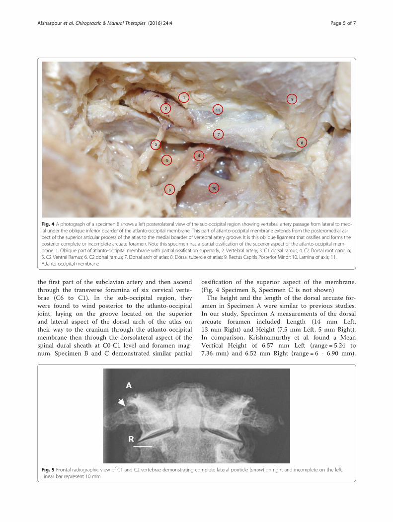
ossification of the superior aspect of the membrane.(Fig. 4 Specimen B, Specimen C is not shown)The height and the length of the dorsal arcuate for-
amen in Specimen A were similar to previous studies.In our study, Specimen A measurements of the dorsalarcuate foramen included Length (14 mm Left,13 mm Right) and Height (7.5 mm Left, 5 mm Right).In comparison, Krishnamurthy et al. found a MeanVertical Height of 6.57 mm Left (range = 5.24 to7.36 mm) and 6.52 mm Right (range = 6 - 6.90 mm).
1
11
10
9
8
7
6
54
3
2
Fig. 4 A photograph of a specimen B shows a left posterolateral view of the sub-occipital region showing vertebral artery passage from lateral to med-ial under the oblique inferior boarder of the atlanto-occipital membrane. This part of atlanto-occipital membrane extends from the posteromedial as-pect of the superior articular process of the atlas to the medial boarder of vertebral artery groove. It is this oblique ligament that ossifies and forms theposterior complete or incomplete arcuate foramen. Note this specimen has a partial ossification of the superior aspect of the atlanto-occipital mem-brane. 1. Oblique part of atlanto-occipital membrane with partial ossification superiorly; 2. Vertebral artery; 3. C1 dorsal ramus; 4. C2 Dorsal root ganglia;5. C2 Ventral Ramus; 6. C2 dorsal ramus; 7. Dorsal arch of atlas; 8. Dorsal tubercle of atlas; 9. Rectus Capitis Posterior Minor; 10. Lamina of axis; 11.Atlanto-occipital membrane
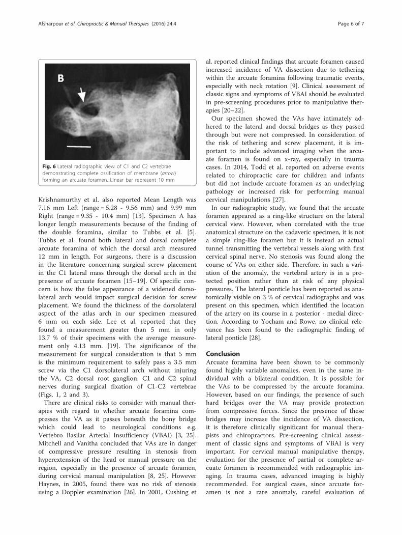
Fig. 5 Frontal radiographic view of C1 and C2 vertebrae demonstrating complete lateral ponticle (arrow) on right and incomplete on the left.Linear bar represent 10 mm
Afsharpour et al. Chiropractic & Manual Therapies (2016) 24:4 Page 5 of 7
Krishnamurthy et al. also reported Mean Length was7.16 mm Left (range = 5.28 - 9.56 mm) and 9.99 mmRight (range = 9.35 - 10.4 mm) [13]. Specimen A haslonger length measurements because of the finding ofthe double foramina, similar to Tubbs et al. [5].Tubbs et al. found both lateral and dorsal completearcuate foramina of which the dorsal arch measured12 mm in length. For surgeons, there is a discussionin the literature concerning surgical screw placementin the C1 lateral mass through the dorsal arch in thepresence of arcuate foramen [15–19]. Of specific con-cern is how the false appearance of a widened dorso-lateral arch would impact surgical decision for screwplacement. We found the thickness of the dorsolateralaspect of the atlas arch in our specimen measured6 mm on each side. Lee et al. reported that theyfound a measurement greater than 5 mm in only13.7 % of their specimens with the average measure-ment only 4.13 mm. [19]. The significance of themeasurement for surgical consideration is that 5 mmis the minimum requirement to safely pass a 3.5 mmscrew via the C1 dorsolateral arch without injuringthe VA, C2 dorsal root ganglion, C1 and C2 spinalnerves during surgical fixation of C1-C2 vertebrae(Figs. 1, 2 and 3).There are clinical risks to consider with manual ther-
apies with regard to whether arcuate foramina com-presses the VA as it passes beneath the bony bridgewhich could lead to neurological conditions e.g.Vertebro Basilar Arterial Insufficiency (VBAI) [3, 25].Mitchell and Vanitha concluded that VAs are in dangerof compressive pressure resulting in stenosis fromhyperextension of the head or manual pressure on theregion, especially in the presence of arcuate foramen,during cervical manual manipulation [8, 25]. HoweverHaynes, in 2005, found there was no risk of stenosisusing a Doppler examination [26]. In 2001, Cushing et
al. reported clinical findings that arcuate foramen causedincreased incidence of VA dissection due to tetheringwithin the arcuate foramina following traumatic events,especially with neck rotation [9]. Clinical assessment ofclassic signs and symptoms of VBAI should be evaluatedin pre-screening procedures prior to manipulative ther-apies [20–22].Our specimen showed the VAs have intimately ad-
hered to the lateral and dorsal bridges as they passedthrough but were not compressed. In consideration ofthe risk of tethering and screw placement, it is im-portant to include advanced imaging when the arcu-ate foramen is found on x-ray, especially in traumacases. In 2014, Todd et al. reported on adverse eventsrelated to chiropractic care for children and infantsbut did not include arcuate foramen as an underlyingpathology or increased risk for performing manualcervical manipulations [27].In our radiographic study, we found that the arcuate
foramen appeared as a ring-like structure on the lateralcervical view. However, when correlated with the trueanatomical structure on the cadaveric specimen, it is nota simple ring-like foramen but it is instead an actualtunnel transmitting the vertebral vessels along with firstcervical spinal nerve. No stenosis was found along thecourse of VAs on either side. Therefore, in such a vari-ation of the anomaly, the vertebral artery is in a pro-tected position rather than at risk of any physicalpressures. The lateral ponticle has been reported as ana-tomically visible on 3 % of cervical radiographs and waspresent on this specimen, which identified the locationof the artery on its course in a posterior - medial direc-tion. According to Yocham and Rowe, no clinical rele-vance has been found to the radiographic finding oflateral ponticle [28].
ConclusionArcuate foramina have been shown to be commonlyfound highly variable anomalies, even in the same in-dividual with a bilateral condition. It is possible forthe VAs to be compressed by the arcuate foramina.However, based on our findings, the presence of suchhard bridges over the VA may provide protectionfrom compressive forces. Since the presence of thesebridges may increase the incidence of VA dissection,it is therefore clinically significant for manual thera-pists and chiropractors. Pre-screening clinical assess-ment of classic signs and symptoms of VBAI is veryimportant. For cervical manual manipulative therapy,evaluation for the presence of partial or complete ar-cuate foramen is recommended with radiographic im-aging. In trauma cases, advanced imaging is highlyrecommended. For surgical cases, since arcuate for-amen is not a rare anomaly, careful evaluation of
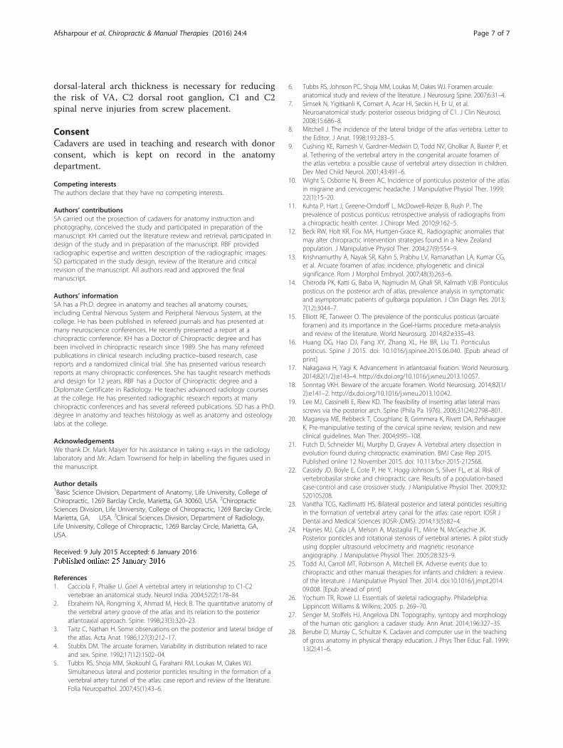
Fig. 6 Lateral radiographic view of C1 and C2 vertebraedemonstrating complete ossification of membrane (arrow)forming an arcuate foramen. Linear bar represent 10 mm
Afsharpour et al. Chiropractic & Manual Therapies (2016) 24:4 Page 6 of 7
dorsal-lateral arch thickness is necessary for reducingthe risk of VA, C2 dorsal root ganglion, C1 and C2spinal nerve injuries from screw placement.
ConsentCadavers are used in teaching and research with donorconsent, which is kept on record in the anatomydepartment.
Competing interestsThe authors declare that they have no competing interests.
Authors’ contributionsSA carried out the prosection of cadavers for anatomy instruction andphotography, conceived the study and participated in preparation of themanuscript. KH carried out the literature review and retrieval, participated indesign of the study and in preparation of the manuscript. RBF providedradiographic expertise and written description of the radiographic images.SD participated in the study design, review of the literature and criticalrevision of the manuscript. All authors read and approved the finalmanuscript.
Authors’ informationSA has a Ph.D. degree in anatomy and teaches all anatomy courses,including Central Nervous System and Peripheral Nervous System, at thecollege. He has been published in refereed journals and has presented atmany neuroscience conferences. He recently presented a report at achiropractic conference. KH has a Doctor of Chiropractic degree and hasbeen involved in chiropractic research since 1989. She has many refereedpublications in clinical research including practice–based research, casereports and a randomized clinical trial. She has presented various researchreports at many chiropractic conferences. She has taught research methodsand design for 12 years. RBF has a Doctor of Chiropractic degree and aDiplomate Certificate in Radiology. He teaches advanced radiology coursesat the college. He has presented radiographic research reports at manychiropractic conferences and has several refereed publications. SD has a PhD.degree in anatomy and teaches histology as well as anatomy and osteologylabs at the college.
AcknowledgementsWe thank Dr. Mark Maiyer for his assistance in taking x-rays in the radiologylaboratory and Mr. Adam Townsend for help in labelling the figures used inthe manuscript.
Author details1Basic Science Division, Department of Anatomy, Life University, College ofChiropractic, 1269 Barclay Circle, Marietta, GA 30060, USA. 2ChiropracticSciences Division, Life University, College of Chiropractic, 1269 Barclay Circle,Marietta, GA, USA. 3Clinical Sciences Division, Department of Radiology,Life University, College of Chiropractic, 1269 Barclay Circle, Marietta, GA,USA.
Received: 9 July 2015 Accepted: 6 January 2016
References1. Cacciola F, Phalke U. Goel A vertebral artery in relationship to C1-C2
vertebrae: an anatomical study. Neurol India. 2004;52(2):178–84.2. Ebraheim NA, Rongming X, Ahmad M, Heck B. The quantitative anatomy of
the vertebral artery groove of the atlas and its relation to the posterioratlantoaxial approach. Spine. 1998;23(3):320–23.
3. Taitz C, Nathan H. Some observations on the posterior and lateral bridge ofthe atlas. Acta Anat. 1986;127(3):212–17.
4. Stubbs DM. The arcuate foramen. Variability in distribution related to raceand sex. Spine. 1992;17(12):1502–04.
5. Tubbs RS, Shoja MM, Skokouhl G, Farahani RM, Loukas M, Oakes WJ.Simultaneous lateral and posterior ponticles resulting in the formation of avertebral artery tunnel of the atlas: case report and review of the literature.Folia Neuropathol. 2007;45(1):43–6.
6. Tubbs RS, Johnson PC, Shoja MM, Loukas M, Oakes WJ. Foramen arcuale:anatomical study and review of the literature. J Neurosurg Spine. 2007;6:31–4.
7. Simsek N, Yigitkanli K, Comert A, Acar HI, Seckin H, Er U, et al.Neuroanatomical study: posterior osseous bridging of C1. J Clin Neurosci.2008;15:686–8.
8. Mitchell J. The incidence of the lateral bridge of the atlas vertebra. Letter tothe Editor. J Anat. 1998;193:283–5.
9. Cushing KE, Ramesh V, Gardner-Medwin D, Todd NV, Gholkar A, Baxter P, etal. Tethering of the vertebral artery in the congenital arcuate foramen ofthe atlas vertebra: a possible cause of vertebral artery dissection in children.Dev Med Child Neurol. 2001;43:491–6.
10. Wight S, Osborne N, Breen AC. Incidence of ponticulus posterior of the atlasin migraine and cervicogenic headache. J Manipulative Physiol Ther. 1999;22(1):15–20.
11. Kuhta P, Hart J, Greene-Orndorff L, McDowell-Reizer B, Rush P. Theprevalence of posticus ponticus: retrospective analysis of radiographs froma chiropractic health center. J Chiropr Med. 2010;9:162–5.
12. Beck RW, Holt KR, Fox MA, Hurtgen-Grace KL. Radiographic anomalies thatmay alter chiropractic intervention strategies found in a New Zealandpopulation. J Manipulative Physiol Ther. 2004;27(9):554–9.
13. Krishnamurthy A, Nayak SR, Kahn S, Prabhu LV, Ramanathan LA, Kumar CG,et al. Arcuate foramen of atlas: incidence, phylogenetic and clinicalsignificance. Rom J Morphol Embryol. 2007;48(3):263–6.
14. Chitroda PK, Katti G, Baba IA, Najmudin M, Ghali SR, Kalmath VJB. Ponticulusposticus on the posterior arch of atlas, prevalence analysis in symptomaticand asymptomatic patients of gulbarga population. J Clin Diagn Res. 2013;7(12):3044–7.
15. Elliott RE, Tanweer O. The prevalence of the ponticulus posticus (arcuateforamen) and its importance in the Goel-Harms procedure: meta-analysisand review of the literature. World Neurosurg. 2014;82:e335–43.
16. Huang DG, Hao DJ, Fang XY, Zhang XL, He BR, Liu TJ. Ponticulusposticus. Spine J 2015. doi: 10.1016/j.spinee.2015.06.040. [Epub ahead ofprint]
17. Nakagawa H, Yagi K. Advancement in atlantoaxial fixation. World Neurosurg.2014;82(1/2):e143–4. http://dx.doi.org/10.1016/j.wneu.2013.10.057.
18. Sonntag VKH. Beware of the arcuate foramen. World Neurosurg. 2014;82(1/2):e141–2. http://dx.doi.org/10.1016/j.wneu.2013.10.042.
19. Lee MJ, Cassinelli E, Riew KD. The feasibility of inserting atlas lateral massscrews via the posterior arch. Spine (Phila Pa 1976). 2006;31(24):2798–801.
20. Magareya ME, Rebbeck T, Coughlanc B, Grimmera K, Rivett DA, RefshaugeeK. Pre-manipulative testing of the cervical spine review, revision and newclinical guidelines. Man Ther. 2004;9:95–108.
21. Futch D, Schneider MJ, Murphy D, Grayev A. Vertebral artery dissection inevolution found during chiropractic examination. BMJ Case Rep 2015.Published online 12 November 2015. doi: 10.113/bcr-2015-212568.
22. Cassidy JD, Boyle E, Cote P, He Y, Hogg-Johnson S, Silver FL, et al. Risk ofvertebrobasilar stroke and chiropractic care. Results of a population-basedcase-control and case crossover study. J Manipulative Physiol Ther. 2009;32:S2010S208.
23. Vanitha TCG, Kadlimatti HS. Bilateral posterior and lateral ponticles resultingin the formation of vertebral artery canal for the atlas: case report. IOSR JDental and Medical Sciences (IOSR-JDMS). 2014;13(5):82–4.
24. Haynes MJ, Cala LA, Melson A, Mastaglia FL, Milne N, McGeachie JK.Posterior ponticles and rotational stenosis of vertebral arteries. A pilot studyusing doppler ultrasound velocimetry and magnetic resonanceangiography. J Manipulative Physiol Ther. 2005;28:323–9.
25. Todd AJ, Carroll MT, Robinson A, Mitchell EK. Adverse events due tochiropractic and other manual therapies for infants and children: a reviewof the literature. J Manipulative Physiol Ther. 2014. doi:10.1016/j.jmpt.2014.09.008. [Epub ahead of print]
26. Yochum TR, Rowe LJ. Essentials of skeletal radiography. Philadelphia:Lippincott Williams & Wilkins; 2005. p. 269–70.
27. Senger M, Stoffels HJ, Angelova DN. Topography, syntopy and morphologyof the human otic ganglion: a cadaver study. Ann Anat. 2014;196:327–35.
28. Berube D, Murray C, Schultze K. Cadaver and computer use in the teachingof gross anatomy in physical therapy education. J Phys Ther Educ Fall. 1999;13(2):41–6.
Afsharpour et al. Chiropractic & Manual Therapies (2016) 24:4 Page 7 of 7