an approach to hypercalcemia - khoo teck puat hospital … 1... · 2018-02-02 · of anemia, back...
TRANSCRIPT

An Approach to Hypercalcemia
Endocrine GP Symposium
23rd July 2016
Dr. Daphne Lee

Issues Pertinent to Primary Care?
• What the clinical features of hypercalcemia?
• How should I investigate?
• When should I refer urgently?
• When should I refer to Endocrine?

Terminologies
• What is:
– Total Calcium?
– Ionized Calcium?
– Adjusted Calcium?

Distribution of Calcium in the Body

Measuring Calcium
• Total Calcium = – Ionized calcium + protein bound calcium + anion
bound calcium
• Corrected (adjusted) Calcium = – Hypoalbuminemia affects protein bound calcium
– Corrected calcium = what total calcium would be if patient had normal albumin level
– Measured total calcium (mmol/L) + 0.02 (40-serum albumin in g/L)
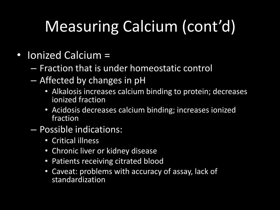
Measuring Calcium (cont’d)
• Ionized Calcium = – Fraction that is under homeostatic control – Affected by changes in pH
• Alkalosis increases calcium binding to protein; decreases ionized fraction
• Acidosis decreases calcium binding; increases ionized fraction
– Possible indications: • Critical illness • Chronic liver or kidney disease • Patients receiving citrated blood • Caveat: problems with accuracy of assay, lack of
standardization

Biological Importance of Calcium
• Crucial regulator of various cellular events:
– Muscle contraction
– Cell signaling
– Hormone secretion
– Neuromuscular transmission
– Co-factor for many steps involved in coagulation

Calcium Regulation
• Key players: – 3 Regulators:
• Parathyroid hormone (PTH)
• Vitamin D
• Calcitonin
– 4 Organs: • Parathyroid glands
• Bone
• Kidneys
• Gut

Parathyroid Hormone (PTH): Retains Calcium in the Circulation
• Actions of PTH: – Increases bone
resorption
– Increases calcium reabsorption at DCT
– Decreases phosphate reabsorption
– Increases activation of 1,25 OH Vit D • Increases calcium and
phosphate absorption from intestine
Increases calcium
Decreases phosphate

Vitamin D
• Actions of Vitamin D:
– Increases calcium and phosphate absorption in gut
– Increases bone resorption when calcium levels are low
– Increases calcium and phosphate reabsorption in the kidneys
Increases calcium
Increases phosphate

“Bones, Stones, Psychic Moans”
• Bones: – Bone pain – Pseudogout – Chondrocalcinosis – Osteoporosis
• Stones (Renal): – Polyuria – Nephrogenic DI – Nephrolithiasis – Renal impairment
• Psychic Moans (Psychiatric): – Anxiety – Depression – Cognitive dysfunction – Psychosis
• Cardiovascular: – Hypertension – Arrhythmias – Short QTc – Calcification of valves, coronary
arteries
• GIT: – Constipation – Anorexia – Nausea vomiting – pancreatitis
• Neurological – Hypotonia – Hyporeflexia – Myopathy – Paresis

Clinical Features of Hypercalcemia
• Symptoms can be non specific
• Related to severity and rate of change of serum calcium
• Symptoms of underlying diseases causing hypercalcemia may dominate the clinical picture

Asymptomatic, non specific symptoms
Coma, confusion, psychosis,
arrhythmias Polyuria, polydipsia, anorexia,
changes in sensorium
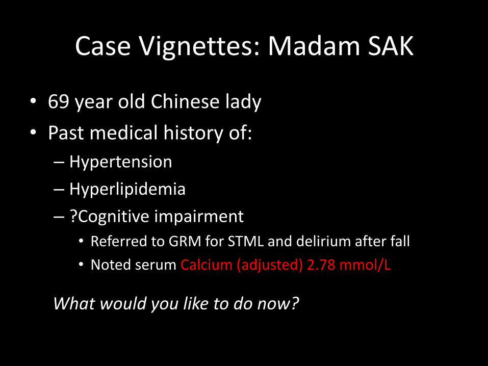
Case Vignettes: Madam SAK
• 69 year old Chinese lady
• Past medical history of:
– Hypertension
– Hyperlipidemia
– ?Cognitive impairment
• Referred to GRM for STML and delirium after fall
• Noted serum Calcium (adjusted) 2.78 mmol/L
What would you like to do now?

Madam SAK
1. Enquire regarding symptoms of hypercalcemia
2. Drug history especially thiazides
3. Ask for symptoms of underlying malignancy
4. Repeat calcium with PTH, phosphate
5. Review old results for previous calcium levels
6. Look for end-organ involvement (renal, osteoporosis)
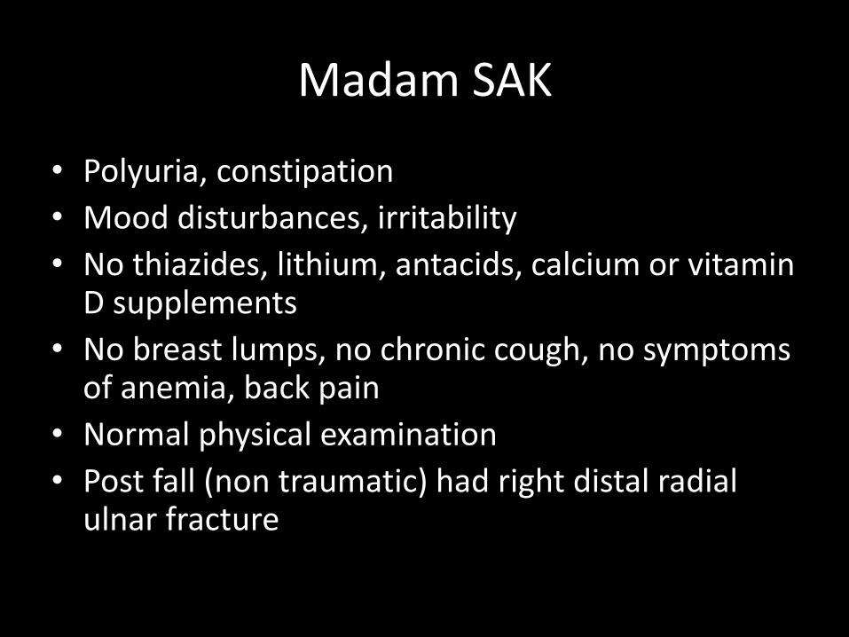
Madam SAK
• Polyuria, constipation
• Mood disturbances, irritability
• No thiazides, lithium, antacids, calcium or vitamin D supplements
• No breast lumps, no chronic cough, no symptoms of anemia, back pain
• Normal physical examination
• Post fall (non traumatic) had right distal radial ulnar fracture

Madam SAK
• Corrected Calcium 3.10 mmol/L (2.15-2.50) • Phosphate 0.70 mmol/L (0.85-1.45) • PTH 15.6 pmol/L (1.6-6.9) • Vitamin D 17.4 ug/L (30-100) • Creatinine 38 umol/L (45-84) • FBC normal • LFT normal
• Differentials?
Madam SAK
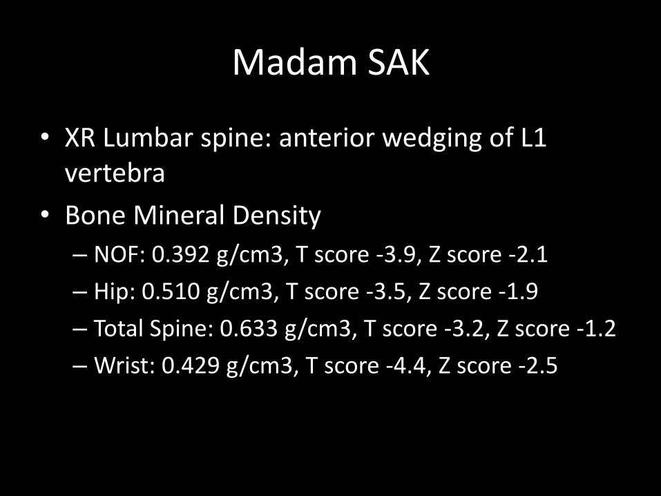
• XR Lumbar spine: anterior wedging of L1 vertebra
• Bone Mineral Density
– NOF: 0.392 g/cm3, T score -3.9, Z score -2.1
– Hip: 0.510 g/cm3, T score -3.5, Z score -1.9
– Total Spine: 0.633 g/cm3, T score -3.2, Z score -1.2
– Wrist: 0.429 g/cm3, T score -4.4, Z score -2.5

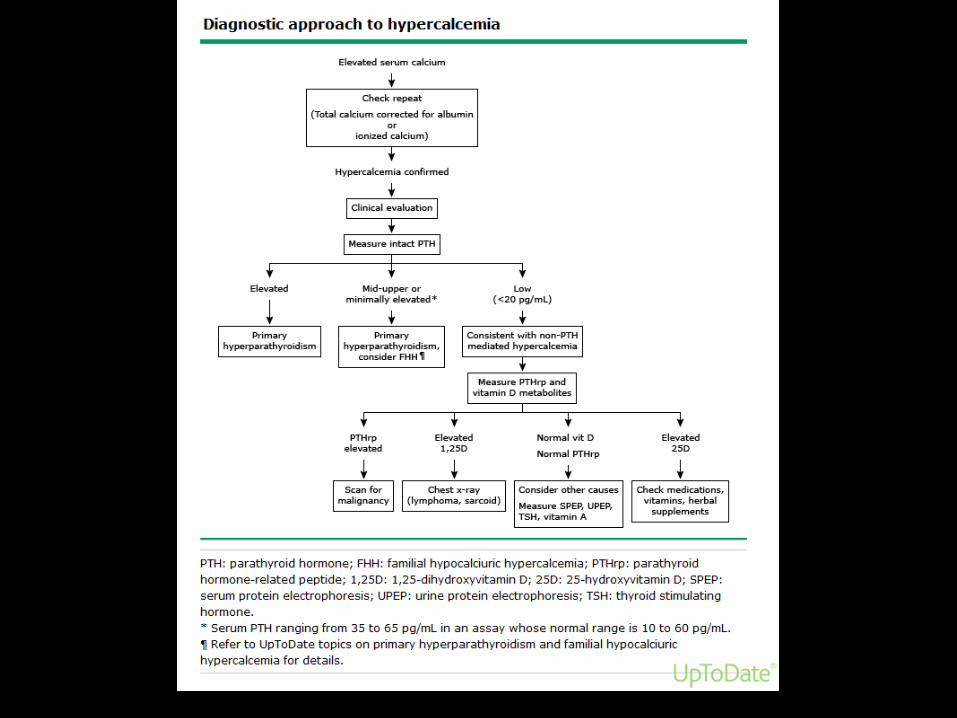
Inverse Relationship Between Calcium and Parathyroid Hormone (PTH)

PTH mediated
Non-PTH mediated
PTH level suppressed (<2 pmol/L)
PTH level normal or elevated

Primary Hyperparathyroidism
• Parathyroid glands were the last major organ to be recognized in humans – First described by Ivar
Sandstrom in 1879
• First parathyroidectomy performed for HPT in Vienna in 1925 by Dr. Felix Mandl
• Primary hyperparathyroidism was first diagnosed in the US in 1926 by Dr. Eugene DuBois

Primary Hyperparathyroidism
• Excessive secretion of parathyroid hormone (PTH) by parathyroid glands
• Classical disease is characterized by: – Symptomatic hypercalcemia
– Renal calculi
– Bone disease: Osteitis fibrosa cystica, brown tumors, osteoporosis
• May present with parathyroid crisis

Evolution of the Clinical Presentation of Primary Hyperparathyroidism
• Development of the multichannel autoanalyser in the 1970s allowed calcium levels to be routinely available
• Increasingly sophisticated PTH immunoassays – First PTH immunoassay was developed by Berson and
Yalow in the 1960s
• Increasing incidence of asymptomatic hyperparathyroidism – Robert Coffey reported increased incidence of
asymptomatic HPT from 5% (pre-1970) to 40% (post 1970)
– Mayo Clinic reported 64% of their cases of HPT during 1974-1980 were asymptomatic

Causes of Primary Hyperparathyroidism
• Occurs at any age; majority are above age of 50
• Single gland adenoma is the most common cause (75-85%)
• Multi-gland disease (10-15%) – Usually in the context of
familial diseases e.g. MEN 1, 2a
• Parathyroid carcinoma (~1%)

Surgical Management of Primary Hyperparathyroidism
• Gold standard of management is parathyroid surgery
• Unilateral parathyroidectomy vs. bilateral neck exploration
– Role of pre-op localisation studies and intra-op PTH monitoring
– Bilateral approach is still indicated for those with familial disease


Medical Management
• General Measures – Ensure good hydration – Avoid thiazide diuretics – Encourage ambulation – Avoid excessive calcium intake – Modest replacement of vitamin D 800-1000IU/day
• Pharmacologic agents – Bisphosphonates for symptomatic
hypercalcemia/osteoporosis – CaSR modulators e.g. cinacalcet to control calcium
levels
Familial Hypocalciuric Hypercalcemia (FHH)
• Autosomal-dominant condition • Caused by mutation in the calcium sensing
receptor gene • Patients have moderate hypercalcemia from an
early age with relatively low urinary calcium excretion
• PTH levels can be normal or mildly elevated • Differentiated from HPTH by calcium creatinine
clearance of <0.01 • Parathyroidectomy is not beneficial
Madam SAK
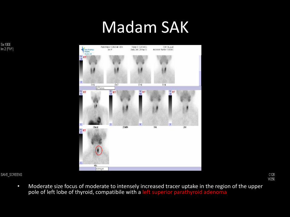
• Moderate size focus of moderate to intensely increased tracer uptake in the region of the upper pole of left lobe of thyroid, compatibile with a left superior parathyroid adenoma

Madam SAK
• Underwent left upper parathyroidectomy – Intraop: baseline PTH 23.9 pmol/L-> 15 minute post
excision 5.5 pmol/L – Histology:
• 2x1.5x0.9cm specimen weighing 1.57g • Hyperplastic parathyroid gland with attenuated capsule in
keeping with parathyroid adenoma. No evidence of malignancy.
• Post op follow up: – Calcium 2.27 mmol/L – Phospate 1.16 mmol/L – PTH 5.4 pmol/L
Case Vignette: Mr. TAC
• 73 year old Chinese man
• Past medical history: – Stage 4 diffuse large b cell lymphoma defaulted
follow up for 1 year
– DM
– Hypertension
– Hyperlipidemia
– CKD, baseline Cr 110
– IHD

Mr. TAC
• Admitted for one month history of vomiting, weakness, lethargy and functional decline
• On day of admission, noted to be confused and drowsy
• On clinical examination, GCS 10, dehydrated

Mr. TAC
• Creatinine 370 umol/L (59-104)
• Urea 26.3 mmol/L (2.8-7.6)
• Ca, adjusted 4.11 mmol/L (2.15-2.50)
• Phosphate 1.52 mmol/L (0.85-1.45)
• PTH 1.0 pmol/L (1.6-6.9)
• Vitamin D 17.8 ug/L (30-100)

Hypercalcemic Crisis
• MEDICAL EMERGENCY
• Ca usually >4.0 mmol/L
• Primary symptoms include oliguria/anuria and mental state changes (including somnolence and eventually coma)
• Majority of cases are due to primary hyperparathyroidism and malignancy

• Priority is to stabilize the patient
• Hydration, hydration, hydration
• Refer to nearest ED as soon as possible
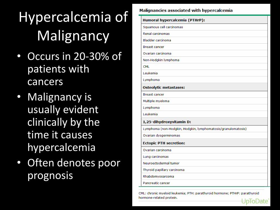
Hypercalcemia of Malignancy
• Occurs in 20-30% of patients with cancers
• Malignancy is usually evident clinically by the time it causes hypercalcemia
• Often denotes poor prognosis

Mechanisms Causing Hypercalcemia of Malignancy

Miscellaneous Causes of Hypercalcemia
• Drugs – Thiazides: lower urinary calcium excretion, can unmask hypercalcemia
in a patient with underlying hyperparathyroidism – Lithium: exact mechanism unknown
• increase in the set point at which calcium suppresses PTH release, leads to increased secretion of PTH and mild hypercalcemia
• Significant association with multigland disease • Cessation of therapy may not correct the hypercalcemia/hyperparathyroidism
• Milk alkali syndrome – Occurs in the setting of high intake of milk or calcium carbonate
coupled with absorbable alkali – More common when absorbable antacids were standard treatment for
peptic ulcer disease
• Prolonged immobilisation – Due to increased bone resoprtion

Endocrine Causes of Hypercalcemia
• Hyperthyroidism – Mild hypercalcemia occurs in 15-20% of thyroxic patients – Due to thyroid hormone mediated increase in bone
resorption
• Adrenal Insufficiency – Multifactorial including increased bone resoprtion, volume
contraction, hemoconcentration, increased c calcium reabsorption
• Phaeochromocytoma – Usually due to concurrent hyperparathyroidism in the
context of MEN 2 – Phaeochromocytomas can also secrete PTHrp

Suggested Approach
• Review history:
– Classical presentation very rare
– Subtle manifestations more common
• Look for associated conditions/complications
• Review medications
• Pursue symptoms of underlying malignancy
Suggested Approach
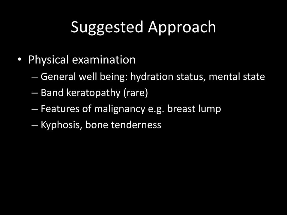
• Physical examination
– General well being: hydration status, mental state
– Band keratopathy (rare)
– Features of malignancy e.g. breast lump
– Kyphosis, bone tenderness
Suggested Approach
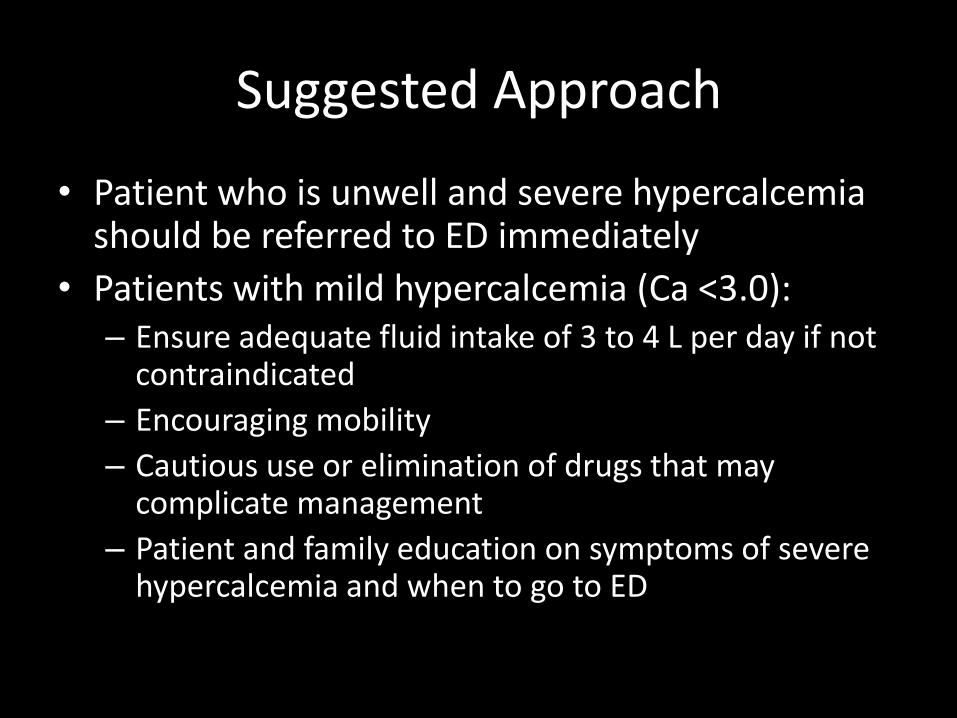
• Patient who is unwell and severe hypercalcemia should be referred to ED immediately
• Patients with mild hypercalcemia (Ca <3.0): – Ensure adequate fluid intake of 3 to 4 L per day if not
contraindicated
– Encouraging mobility
– Cautious use or elimination of drugs that may complicate management
– Patient and family education on symptoms of severe hypercalcemia and when to go to ED


• What the symptoms and signs of hypercalcemia?
• How should I investigate?
• When should I refer urgently?
• When should I refer to Endocrine?

THE END