an automated motion detection and reward system for animal
TRANSCRIPT

Received 07/25/2015 Review began 07/27/2015 Review ended 11/23/2015 Published 12/04/2015
© Copyright 2015Miller et al. This is an open access articledistributed under the terms of theCreative Commons Attribution LicenseCC-BY 3.0., which permits unrestricteduse, distribution, and reproduction in anymedium, provided the original author andsource are credited.
An Automated Motion Detection and RewardSystem for Animal TrainingBrad Miller , Audrey N. Lim , Arnold F. Heidbreder , Kevin J. Black
1. Psychiatry, Washington University School of Medicine 2. Electronics shop, Washington University School ofMedicine 3. Departments of Psychiatry, Neurology, Radiology, and Anatomy & Neurobiology, Washington UniversitySchool of Medicine
Corresponding author: Kevin J. Black, [email protected]
AbstractA variety of approaches has been used to minimize head movement during functional brain imaging studiesin awake laboratory animals. Many laboratories expend substantial effort and time training animals toremain essentially motionless during such studies. We could not locate an “off-the-shelf” automatedtraining system that suited our needs.
We developed a time- and labor-saving automated system to train animals to hold still for extended periodsof time. The system uses a personal computer and modest external hardware to provide stimulus cues,monitor movement using commercial video surveillance components, and dispense rewards. A customcomputer program automatically increases the motionless duration required for rewards based onperformance during the training session but allows changes during sessions. This system was used to traincynomolgus monkeys (Macaca fascicularis) for awake neuroimaging studies using positron emissiontomography (PET) and functional magnetic resonance imaging (fMRI).
The automated system saved the trainer substantial time, presented stimuli and rewards in a highlyconsistent manner, and automatically documented training sessions. We have limited data to prove thetraining system's success, drawn from the automated records during training sessions, but we believe othersmay find it useful. The system can be adapted to a range of behavioral training/recording activities forresearch or commercial applications, and the software is freely available for non-commercial use.
Categories: Psychology, Radiology, OtherKeywords: neuroimaging, neuroscience, positron emission tomography (pet), nonhuman primate, macacafascicularis, video recording, operant conditioning, reward, computers, magnetic resonance imaging
IntroductionTraining animals for science experiments can be a time-consuming and labor-intensive process. Automatedtraining systems reduce the labor commitment and have the potential to apply more consistent criteria andrewards than a human trainer who might be distracted, fatigued, or overwhelmed by demands of the trainingparadigm.
In many behavioral paradigms, an animal is required to complete a task in order to obtain a reward; forexample, a bird might be required to peck a specific key. We faced the slightly different problem of trainingthe animal to do nothing, or more accurately, to remain awake but motionless during the pharmacologicalchallenge of fMRI (functional magnetic resonance imaging) and PET (positron emission tomography) brainimaging studies.
One purpose for imaging awake animals was because anesthesia might interfere with the function of thesystem of interest. Our study examined the role of a dopaminergic D1 receptor agonist on brain function,
and anesthesia can alter regional cerebral blood flow responses to dopaminergic drugs in primates [1-2].
Two considerations motivated training the animal to remain motionless for our studies. First, even with ahead restraint, body movement can produce slight head movements that can cause motion artifacts in brainimages [3]. Second, all movements are initiated by brain activity, which produces changes in blood flow,metabolism, and blood oxygenation levels that lead to the signals detected by PET and fMRI [4-7]. A numberof additional strategies have been used to reduce such artifacts, including adaptations of image acquisitionor reconstruction, scanner hardware, or animal restraint systems [8].
This report describes an automated system using a charge coupled device (CCD) camera connected to acomputer to detect motion and dispense rewards after specified durations of motionlessness. Theexperimenter set up the equipment and started the program, after which all other elements of the trainingsessions were controlled by computer software that provided stimulus cues, detected motion objectively,increased reward intervals as appropriate, administered rewards, and logged all of these events.
1 1 2 3
Open Access TechnicalReport DOI: 10.7759/cureus.397
How to cite this articleMiller B, Lim A N, Heidbreder A F, et al. (December 04, 2015) An Automated Motion Detection and Reward System for Animal Training. Cureus7(12): e397. DOI 10.7759/cureus.397
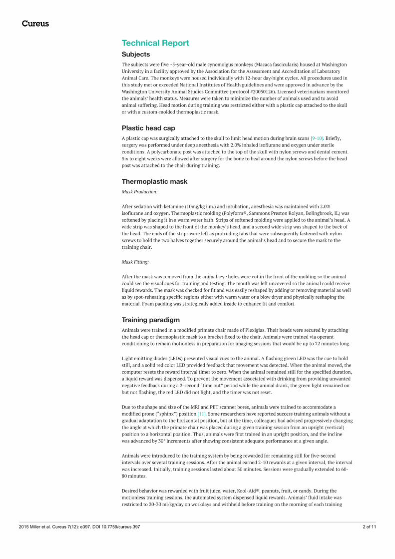
Technical ReportSubjectsThe subjects were five ~5-year-old male cynomolgus monkeys (Macaca fascicularis) housed at WashingtonUniversity in a facility approved by the Association for the Assessment and Accreditation of LaboratoryAnimal Care. The monkeys were housed individually with 12-hour day/night cycles. All procedures used inthis study met or exceeded National Institutes of Health guidelines and were approved in advance by theWashington University Animal Studies Committee (protocol #20050126). Licensed veterinarians monitoredthe animals’ health status. Measures were taken to minimize the number of animals used and to avoidanimal suffering. Head motion during training was restricted either with a plastic cap attached to the skullor with a custom-molded thermoplastic mask.
Plastic head capA plastic cap was surgically attached to the skull to limit head motion during brain scans [9-10]. Briefly,surgery was performed under deep anesthesia with 2.0% inhaled isoflurane and oxygen under sterileconditions. A polycarbonate post was attached to the top of the skull with nylon screws and dental cement.Six to eight weeks were allowed after surgery for the bone to heal around the nylon screws before the headpost was attached to the chair during training.
Thermoplastic maskMask Production:
After sedation with ketamine (10mg/kg i.m.) and intubation, anesthesia was maintained with 2.0%isoflurane and oxygen. Thermoplastic molding (Polyform®, Sammons Preston Rolyan, Bolingbrook, IL) wassoftened by placing it in a warm water bath. Strips of softened molding were applied to the animal’s head. Awide strip was shaped to the front of the monkey’s head, and a second wide strip was shaped to the back ofthe head. The ends of the strips were left as protruding tabs that were subsequently fastened with nylonscrews to hold the two halves together securely around the animal’s head and to secure the mask to thetraining chair.
Mask Fitting:
After the mask was removed from the animal, eye holes were cut in the front of the molding so the animalcould see the visual cues for training and testing. The mouth was left uncovered so the animal could receiveliquid rewards. The mask was checked for fit and was easily reshaped by adding or removing material as wellas by spot-reheating specific regions either with warm water or a blow dryer and physically reshaping thematerial. Foam padding was strategically added inside to enhance fit and comfort.
Training paradigmAnimals were trained in a modified primate chair made of Plexiglas. Their heads were secured by attachingthe head cap or thermoplastic mask to a bracket fixed to the chair. Animals were trained via operantconditioning to remain motionless in preparation for imaging sessions that would be up to 72 minutes long.
Light emitting diodes (LEDs) presented visual cues to the animal. A flashing green LED was the cue to holdstill, and a solid red color LED provided feedback that movement was detected. When the animal moved, thecomputer resets the reward interval timer to zero. When the animal remained still for the specified duration,a liquid reward was dispensed. To prevent the movement associated with drinking from providing unwantednegative feedback during a 2-second “time out” period while the animal drank, the green light remained onbut not flashing, the red LED did not light, and the timer was not reset.
Due to the shape and size of the MRI and PET scanner bores, animals were trained to accommodate amodified prone (“sphinx”) position [11]. Some researchers have reported success training animals without agradual adaptation to the horizontal position, but at the time, colleagues had advised progressively changingthe angle at which the primate chair was placed during a given training session from an upright (vertical)position to a horizontal position. Thus, animals were first trained in an upright position, and the inclinewas advanced by 30° increments after showing consistent adequate performance at a given angle.
Animals were introduced to the training system by being rewarded for remaining still for five-secondintervals over several training sessions. After the animal earned 2-10 rewards at a given interval, the intervalwas increased. Initially, training sessions lasted about 30 minutes. Sessions were gradually extended to 60-80 minutes.
Desired behavior was rewarded with fruit juice, water, Kool-Aid®, peanuts, fruit, or candy. During themotionless training sessions, the automated system dispensed liquid rewards. Animals’ fluid intake wasrestricted to 20-30 ml/kg/day on workdays and withheld before training on the morning of each training
2015 Miller et al. Cureus 7(12): e397. DOI 10.7759/cureus.397 2 of 11

session. The amount of fluid consumed during the training session was monitored and supplemental waterwas given after each session to ensure adequate hydration. On weekends, three-fold the weekday ration wasprovided. Body weight was recorded regularly and monitored by the veterinary staff to ensure adequatehydration.
Training system: hardwareThe computer was a Dell Optiplex GX280 with an Intel Pentium 4 CPU 3.20 GHz, 512 MB RAM, and an ATIRadeon X300 video card with 128 MB, running Microsoft Windows® XP Professional. We added a HuperLabH1004S video capture card (Huper Laboratories Co. Ltd., Taipei, Taiwan) and PC Witness Pro (DVR 2400)software (CCTV Wholesalers, New Orleans, LA). The card allowed smooth recording and display of video at640x480 resolution at 30 frames per second (fps). A Sony SSC-M183 Super HAD CCD black and white videocamera equipped with a Tamron 5-50mm 1:1.4 CCTV lens was connected to the capture card via a standardBNC cable.
[Note: We subsequently adapted this system to a different experiment that required recording from twocameras. Recording two video feeds required a faster video capture card; the Huper Laboratories Co. Ltd. 2404Q-PCI interface card (purchased from Anova Microsystems, Inc., Milpitas, CA) allowed smooth video capture withtwo cameras at 640x480 resolution at 30 fps each.]
Because the head was secured to the chair during training sessions, and to avoid detecting mouth/jawmovements associated with drinking the rewards, the video camera was directed toward the body, and ifneeded, extraneous portions of the camera's visual field were masked from motion detection using the PCWitness Pro DVR 2400 software.
A 60 cc syringe (sans plunger) was used as a reservoir for juice rewards and mounted ~1 m above the animal’shead so that liquid was delivered by gravity. It was connected to flexible silicone rubber tubing that ran tothe animal’s mouth through a solenoid-controlled pinch valve (Bio-Chem Valve Inc., part # 100P2NC12-05SQ, Boonton, New Jersey). The pinch valve closed the tubing by default and was activated (opened) tosupply rewards. The duration of the reward flow was adjustable through the software’s control panel and acontrol box adjacent to the pinch valve. A hand-held push-button cable was connected to the solenoid valvecontrol box to permit manual rewards. The physical arrangement of equipment in our training set-up isshown in Figure 1.
FIGURE 1: Illustration of the training system layoutSee text for details.
2015 Miller et al. Cureus 7(12): e397. DOI 10.7759/cureus.397 3 of 11

Training system: SoftwareThe automated training system involved a commercially available surveillance software program, PCWitness Pro DVR 2400 (then version 1.54), which detects motion in the video feed, integrated with customsoftware (Monkey Motion) written by one of the authors (AFH) using LabVIEW (National Instruments,Austin, TX, http://www.ni.com/trylabview/). The graphical source code file for Monkey Motion is freelyavailable for non-commercial use at Zenodo.org (doi: 10.5281/zenodo.21157). Running this programrequires the appropriate LabVIEW runtime software from National Instruments Corporation. MonkeyMotion monitored an output signal from the surveillance software when motion was detected to control allother functions related to the training paradigm: the visual cues for the animal (LED lights), interval timingand reward administration.
The stock DVR 2400 PC Witness Pro software (originally intended for security surveillance and recording)required a minimum event duration of 1 second to signal an event. At our request, the softwareprogrammers at Huper Laboratories under the direction of Geoff Wang modified the PC Witness Pro softwareto provide a nearly instantaneous electrical signal output to the custom LabVIEW software when motionwas detected.
Figure 2 shows the front panel of the Monkey Motion program, with annotations explained in the figurelegend. The active screen display shows when the subject is still, when a movement is detected, when areward is given, and whether the program is paused. Additional displays on the window show convenientreal-time information, such as current still time, best still time, and countdown time until the reward isgiven. Buttons on the front panel of the Monkey Motion program allow the user to start or pause training,give additional rewards, save data, or quit the program (Figure 2). The program records an ASCII text log fileof all events and settings.
FIGURE 2: Screen shot of the Monkey Motion software interface withseveral features annotated by numbers, letters, braces, and arrowsThe black box shows a live record of the animal’s behavior (A = Still; B = Moving), reward delivery (C), andthe program’s status (running or paused). In this example, a reward is delivered automatically 5 seconds afterthe most recent movement (#9). Controls on a purple background can be set at program startup or during asession.
Legend:
1. Animal ID.
2. Experimenter name.
3. Timeout. Time allowed for the subject to drink the reward, during which movement is not “detected.”
2015 Miller et al. Cureus 7(12): e397. DOI 10.7759/cureus.397 4 of 11
4. Status: No Motion, Motion, or Drinking.
5. Set N Periods: Number of consecutive earned rewards that triggers a change in the reward interval.
6. Period. Current number of consecutive rewarded periods.
7. Auto. Number of seconds the minimum reward interval will change by after the number of rewardedintervals set in #5.
8. Set Start. Initial reward interval (seconds).
9. Current. Current reward interval (seconds).
10. Set Max. Maximum reward interval period.
11. Current Still Time. A live count of the current time the subject has remained motionless (reset to zerowhen the subject moves).
12. Best Still Time. The longest period the subject has remained motionless in the current training session.
13. Bonus Period. Motionless interval required to receive an extra reward.
14. Bonus Countdown. Time remaining until the bonus reward.
15. Normal Count Down. Time remaining until the current motionless interval will be rewarded.
16. Elapsed Time. Duration of the training session.
17. Drinking Period. Time allowed for the subject to drink.
18. Reward Duration (milliseconds). Controls the size of the reward.
19. Bonus X. The size of the bonus reward as a multiple of the usual reward.
20. Read Every (mSec). Frequency of the sampling/recording intervals.
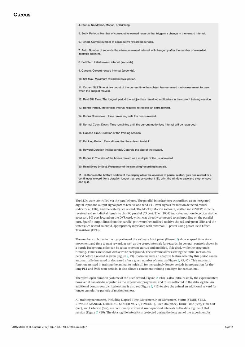
21. Buttons on the bottom portion of the display allow the operator to pause, restart, give one reward or acontinuous reward (for a duration longer than set by control #18), print the window, save and stop, or saveand quit.
The LEDs were controlled via the parallel port. The parallel interface port was utilized as an integrateddigital input and output signal port to receive and send TTL level signals for motion detected, visualindicators (LEDs), and the water/juice reward. The Monkey Motion software, written in LabVIEW, directlyreceived and sent digital signals to this PC parallel I/O port. The H1004S indicated motion detection via theaccessory I/O port located on the DVR card, which was directly connected to an input line on the parallelport. Specific output lines from the parallel port were then utilized to drive the red and green LEDs and thewater/juice reward solenoid, appropriately interfaced with external DC power using power Field EffectTransistors (FETs).
The numbers in boxes in the top portion of the software front panel (Figure 2) show elapsed time sincemovement and time to next reward, as well as the preset intervals for rewards. In general, controls shown ina purple background color can be set at program startup and modified, if desired, while the program isrunning. Timers are shown with a white background. The software allows setting the initial motionlessperiod before a reward is given (Figure 2, #9). It also includes an adaptive feature whereby this period can beautomatically increased or decreased after a given number of rewards (Figure 2, #5, #7). This automaticfunction assisted in training the animal to hold still for increasingly longer periods in preparation for thelong PET and fMRI scan periods. It also allows a consistent training paradigm for each animal.
The valve-open duration (volume of the juice reward, Figure 2, #18) is also initially set by the experimenter;however, it can also be adjusted as the experiment progresses, and this is reflected in the data log file. Anadditional bonus reward criterion time is also set (Figure 2, #13) to give the animal an additional reward forlonger cumulative periods of motionlessness.
All training parameters, including Elapsed Time, Movement/Non-Movement, Status (START, STILL,REWARD, MANUAL, DRINKING, SENSED MOVE, TIMEOUT), Juice On (mSec), Drink Time (Sec), Time Out(Sec), and Criterion (Sec), are continually written at user-specified intervals to the data log file of thatsession (Figure 2, #20). The data log file integrity is protected during the long run of the experiment by
2015 Miller et al. Cureus 7(12): e397. DOI 10.7759/cureus.397 5 of 11

periodically writing to a temporary data file on disk. At the end of the experiment, a summary of theperformance throughout the training session is prepended to the log file and includes percent still time, thenumber of rewards that were given, duration of training time, longest “motionless” period, and starting andending “motionless” interval criteria. Figure 3 shows an example of the output text file.
FIGURE 3: Example log fileAn excerpt from a text log file produced by the Monkey Motion program during one training session.
ResultsThe data available to demonstrate the training system's success are drawn from the automated recordscreated during training sessions. We show two sets of behavioral data.
Results: Early trainingThe first data set shows the animals’ performance during the transition from training sessions in a verticalposition to the horizontal position required for the brain scans. Previously, the monkeys had beenhabituated to working with humans, the primate chair, and the training room. The monkeys had also beentrained with the LED visual cues. These data are from the early stages of training, while the automatedcomputer system was being developed and before it was completely functional. During these sessions, thecomputer operated the LED visual cues and recorded movement and still intervals, but the human trainerwas the motion detector and recorded the animal’s movement by pressing the space bar on the computerkeyboard. The human trainer also gave the rewards manually. The “best motionless” period from eachtraining session represents the longest single period that the monkey held still during a training session(ignoring movement while drinking), and is one of several measurements used to track an animal’sperformance. The best motionless period from each training session during the transition from vertical tohorizontal training is plotted in Figure 4. The mean duration of motionless periods over each animal's lastsix training sessions was 22, 27, 265, 23, and 19 seconds, one animal performing much better than theothers.
2015 Miller et al. Cureus 7(12): e397. DOI 10.7759/cureus.397 6 of 11
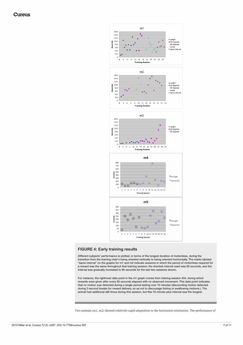
FIGURE 4: Early training resultsDifferent subjects' performance is plotted, in terms of the longest duration of motionless, during thetransition from the training chair's being oriented vertically to being oriented horizontally. The marks labeled“same interval” on the graphs for m1 and m2 indicate sessions in which the period of motionless required fora reward was the same throughout that training session; the shortest interval used was 60 seconds, and theinterval was gradually increased to 95 seconds for the last two sessions shown.
For instance, the rightmost data point in the m1 graph comes from training session #34, during whichrewards were given after every 95 seconds elapsed with no observed movement. This data point indicatesthat no motion was detected during a single period lasting over 10 minutes (discounting motion detectedduring 2-second breaks for reward delivery, so as not to discourage licking or swallowing motions.). Theanimal had additional still times during this session, but this 10-minute-plus interval was the longest.
Two animals (m1, m2) showed relatively rapid adaptation to the horizontal orientation. The performance of
2015 Miller et al. Cureus 7(12): e397. DOI 10.7759/cureus.397 7 of 11

m1 began to decline once the chair was placed horizontally, but improved again after about 10 sessions.Performance declined briefly for m2 when the experimenter changed the starting interval (see description inFigure 4) but rebounded quickly. The third animal’s progress was slow. He showed gradual improvementover time and eventually improved with the chair at an angle, but his performance lagged far behind that ofthe other two monkeys, and his training was not completed. Two additional animals (m4, m5) showedrelatively rapid adaptation to the horizontal orientation, but training data are available only for their initialtraining sessions, as by this time the grant funding the data collection was ending.
By the end of training, all animals were able to hold still the majority of the time. Over each animal's last sixtraining sessions, the percent still time was 70%, 85%, N/A, 74%, and 84%.
Results: Re-trainingThe second set of data shows the re-training of one monkey whose original surgically attached head caploosened and had to be removed after several months of initial training. Training was interrupted for 14weeks to allow the head cap site to heal. Subsequent training sessions and imaging sessions with thisanimal used a removable thermoplastic mask instead. The automated system was fully operational by thispoint and was used exclusively for subsequent retraining, automatically controlling all aspects of thetraining paradigm: visual cues, motion detection, interval recording, and reward administration.
Figure 5 shows three different measures of this animal’s performance from the Monkey Motion program datalogs: the number of movements detected, the percent still time, and the average cumulative still time. Theanimal’s performance on these measures while being retrained solely by the Monkey Motion system(connected dots in each graph in Figure 5) is compared with its performance before the head cap wasremoved (single dot at the left of each graph). The monkey’s performance on all measures at the end of 10weeks of retraining equaled or exceeded its performance before head cap removal.
2015 Miller et al. Cureus 7(12): e397. DOI 10.7759/cureus.397 8 of 11
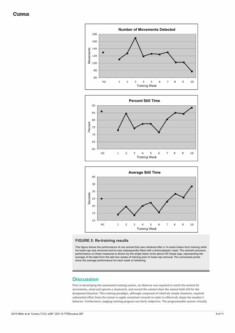
FIGURE 5: Re-training resultsThis figure shows the performance of one animal that was retrained after a 14-week hiatus from training whilehis head cap was removed and he was subsequently fitted with a thermoplastic mask. The animal’s previousperformance on these measures is shown by the single black circle above HC (head cap), representing theaverage of the data from the last two weeks of training prior to head cap removal. The connected pointsshow the average performance for each week of retraining.
DiscussionPrior to developing the automated training system, an observer was required to watch the animal formovements, mind and operate a stopwatch, and reward the animal when the animal held still for thedesignated duration. This training paradigm, although composed of relatively simple elements, requiredsubstantial effort from the trainer to apply consistent rewards in order to effectively shape the monkey’sbehavior. Furthermore, judging training progress was fairly subjective. The programmable system virtually
2015 Miller et al. Cureus 7(12): e397. DOI 10.7759/cureus.397 9 of 11

eliminated the need for human participation during the training procedures, except for the initial setup andto make sure that there was sufficient liquid in the reward dispenser.
The data presented show that the automated training paradigm effectively taught the monkeys to remainmotionless for increasingly longer periods of time. The data also reflect individual differences in learning,even under highly reproducible conditions (Figure 4). The Monkey Motion system effectively maintainedbehaviors initially taught one-on-one by a human trainer (Figure 4) and quickly retrained one animal after along absence from any training and with a new type of head restraint (Figure 5).
LimitationsThe data we present to document learning progress comes from the training sessions rather than fromsubsequent imaging sessions. Nevertheless, our imaging studies required the monkey to remain motionless
for 40-minute fMRI scans and 72-minute 18F-fluorodeoxyglucose PET scans. The longest recordedmotionless intervals were around 25 to 35 minutes. These durations are quite good for monkeys, consideringthat lying prone and motionless is not their typical behavior. Other researchers have reported that well-trained monkeys will sometimes fall asleep in their chair when not actively training [11], but that was notour experience at all.
Hardware costs for this system were about $2,300, and programming expertise was required to write theMonkey Motion program and tailor it to our specific needs. On the other hand, the automated system savedcountless hours of hands-on training, and the session logs text file documented trainingprogress continuously and objectively.
Our purpose in developing this system was primarily practical, so we did not directly compare the motiondetection accuracy of the automated motion detection system with the accuracy of the human motiondetector. We did, however, observe the animal during the sessions using the automated system and weresatisfied at the time that it was detecting essentially the same movements we would have detected. Finally,our experience with this system did not include training older animals or females, or training without anyphysical head restraint, so performance in those situations may differ.
Advantages of an automated systemThe reduced animal/human contact has advantages for humans, such as fewer hazardous interactions withpotentially dangerous animals. The automated training system appeared to provide advantages for theanimal as well, including a more consistent presentation of visual cues, rewards, and less distraction by theactivities of the human trainer. An additional benefit is the elimination of inadvertent experimenter biasbetween animals since the system ensures highly reproducible rewards.
An additional advantage was observed when using the system during some brain scanning sessions toprovide consistent rewards for holding still. Where the full setup could not easily be used (as in the MRsuite), there was flexibility to use the Monkey Motion software without the camera with a human “motiondetector” pressing a key to record observed movements. The record of animal movements and the timing ofrewards was helpful for post-experiment data processing.
This system has other potential uses, including training animals, recording behavior, monitoring patientactivity, or any study in which movement is an experimental variable [12-14].
ConclusionsWe developed a time- and labor-saving automated system for animal training using readily availablecomponents to control and monitor various features of a behavioral paradigm that included providingstimulus cues, monitoring movement, and dispensing rewards. Although the system's components are nowsomewhat dated, and the proof of its efficacy is limited, we feel that this system, or portions of it, may be ofuse in a variety of behavioral training or recording activities for clinical, research, or commercial purposes.
Additional InformationDisclosuresHuman subjects: All authors have confirmed that this study did not involve human participants or tissue.Animal subjects: Washington University Animal Studies Committee Issued protocol number 20050126.Conflicts of interest: In compliance with the ICMJE uniform disclosure form, all authors declare thefollowing: Payment/services info: This research was funded by the National Institutes of Health (R01NS044598 for the original data, and for manuscript preparation R21 MH081080, an ARRA supplement forthat award and K24 MH087913) and the McDonnell Center for Higher Brain Function (now the McDonnellCenter for Systems Neuroscience at Washington University). Financial relationships: All authors havedeclared that they have no financial relationships at present or within the previous three years with anyorganizations that might have an interest in the submitted work. Other relationships: All authors have
2015 Miller et al. Cureus 7(12): e397. DOI 10.7759/cureus.397 10 of 11

declared that there are no other relationships or activities that could appear to have influenced thesubmitted work.
AcknowledgementsThe authors thank Vince Lai for technical assistance and Geoff Wang of Huper Laboratories for hisexceptional software support. The affiliations listed for the first three authors are for the time period whenthis work was done.
References1. Hershey T, Black KJ, Carl JL, Perlmutter JS: Dopa-induced blood flow responses in nonhuman primates . Exp
Neurol. 2000, 166:342–49. 10.1006/exnr.2000.75222. Zhang Z, Andersen AH, Avison MJ, Gerhardt GA, Gash DM: Functional MRI of apomorphine activation of the
basal ganglia in awake rhesus monkeys. Brain Res. 2000, 852:290–96. 10.1016/S0006-8993(99)02243-X3. Ferris CF, Febo M, Luo F, Schmidt K, Brevard M, Harder JA, Kulkarni P, Messenger T, King JA: Functional
magnetic resonance imaging in conscious animals: A new tool in behavioural neuroscience research. JNeuroendocrinol. 2006, 18:307–18. 10.1111/j.1365-2826.2006.01424.x
4. Leslie RA, James MF: Pharmacological magnetic resonance imaging: a new application for functional MRI .Trends Pharmacol Sci. 2000, 21:314–18. 10.1016/S0165-6147(00)01507-8
5. Raichle ME: A brief history of human functional brain mapping . Brain Mapping: The Systems. Toga AW,Mazziotta JC (ed): Academic Press, San Diego; 2000. 33–75.
6. Raichle ME: Cognitive neuroscience: Bold insights. Nature. 2001, 412:128–30. 10.1038/350843007. Norris DG: Principles of magnetic resonance assessment of brain function . J Magn Reson Imaging. 2006,
23:794–807. 10.1002/jmri.205878. Varivar R, Vanduffel W: Functional MRI of awake behaving macaques using standard equipment . Advanced
Brain Neuroimaging Topics in Health and Disease - Methods and Applications. Papageorgiou TD,Christopoulos GI, Smirnakis SM (ed): InTech, http://www.intechopen.com/books/advanced-brain-neuroimaging-topics-in-health-and-disease-methods-and-applications/functional-mri-of-awake-behaving-macaques-using-standard-equipment; 2014. Chapter 6:10.5772/58281
9. Perlmutter JS, Lich LL, Margenau W, Buchholz S: PET measured evoked cerebral blood flow responses in anawake monkey. J. Cereb. Blood Flow Metab. 1991, 11:229–235. 10.1038/jcbfm.1991.54
10. Baker JT, Patel GH, Corbetta M, Snyder LH: Distribution of activity across the monkey cerebral corticalsurface, thalamus and midbrain during rapid, visually guided saccades. Cereb. Cortex. 2006, 16:447–459.10.1093/cercor/bhi124
11. Andersen AH, Zhang Z, Barber T, Rayens WS, Zhang J, Grondin R, Hardy P, Gerhardt GA, Gash DM:Functional MRI studies in awake rhesus monkeys: methodological and analytical strategies . J. Neurosci.Methods. 2002, 118:141–152. 10.1016/S0165-0270(02)00123-1
12. Anagnostaras SG, Josselyn SA, Frankland PW, Silva AJ: Computer-assisted behavioral assessment ofPavlovian fear conditioning in mice. Learn. Mem. 2000, 7:58–72. 10.1101/lm.7.1.58
13. Contarino A, Baca L, Kennelly A, Gold LH: Automated assessment of conditioning parameters for contextand cued fear in mice. Learn. Mem. 2002, 9:89–96. 10.1101/lm.43002
14. Greene DJ, Koller JM, Robichaux-Viehoever A, Bihun E, Schlaggar BL, Black KJ: Reward enhances ticsuppression in children within months of tic disorder onset. Dev Cogn Neurosci. 2015, 11:65–74.10.1016/j.dcn.2014.08.005
2015 Miller et al. Cureus 7(12): e397. DOI 10.7759/cureus.397 11 of 11