an automated system for high-throughput generation … · an automated system for high-throughput...
TRANSCRIPT
An automated system for high-throughput generation and optimization ofmicrodropletsZongjie Wang, Roya Samanipour, Mohamed Gamaleldin, Kabilan Sakthivel, and Keekyoung Kim
Citation: Biomicrofluidics 10, 054110 (2016); doi: 10.1063/1.4963666View online: http://dx.doi.org/10.1063/1.4963666View Table of Contents: http://aip.scitation.org/toc/bmf/10/5Published by the American Institute of Physics

An automated system for high-throughput generationand optimization of microdroplets
Zongjie Wang, Roya Samanipour, Mohamed Gamaleldin, Kabilan Sakthivel,and Keekyoung Kima)
School of Engineering, University of British Columbia, Kelowna,British Columbia V1V 1V7, Canada
(Received 27 July 2016; accepted 14 September 2016; published online 27 September 2016)
Microdroplets have been widely used in various biomedical applications. During
droplet generation, parameters are manually adjusted to achieve the desired size of
droplets. This process is tedious and time-consuming. In this paper, we present a
fully automated system for controlling the size of droplets to optimize droplet
generation parameters in a microfluidic flow-focusing device. The developed
system employed a novel image processing program to measure the diameter
of droplets from recorded video clips and correspondingly adjust the flow rates of
syringe pumps to obtain the required diameter of droplets. The system was tested
to generate phosphate-buffered saline and 8% polyethylene (glycol) diacrylate pre-
polymer droplets and regulate its diameters at various flow rates. Experimental
results demonstrated that the difference between droplet diameters from the image
processing and manual measurement is not statistically significant and the results
are consistent over five repetitions. Taking the advantages of the accurate image
processing method, the size of the droplets can be optimized in a precise and robust
manner via automatically adjusting flow rates by the feedback control. The system
was used to acquire quantitative data to examine the effects of viscosity and flow
rates. Droplet-based experiments can be greatly facilitated by the automatic droplet
generation and optimization system. Moreover, the system is able to provide quan-
titative data for the modelling and application of droplets with various conditions
in a high-throughput way. Published by AIP Publishing.[http://dx.doi.org/10.1063/1.4963666]
I. INTRODUCTION
Microdroplets are useful for various applications, including surface decontamination,1 food
processing,2 optical devices fabrication,3 beam manipulation,4 and drug delivery.5 In recent
years, microdroplets have been also extensively used in tissue engineering, which is the field of
study to restore or improve damaged tissues or organs.6 For example, hydrogel microdroplets
have been used as a building block for macroscale tissue generation7 and a high-throughput
platform for co-culturing cells.8 Among several microdroplet fabrication methods, the most
widely used method is the microfluidics-based method due to its high controllability of droplets,
high throughput, low fabrication cost, and relatively simple device fabrication process.9
There are three different methods to continuously generate microdroplets using the micro-
fluidic devices: T-junction, flow-focusing, and co-axial flow methods.10 In addition to the contin-
uous flow methods, drop-on-demand method, such as a liquid bridge, offers great control to the
size and shape of single or small volume of droplets,11,12 and has been used to create nanoliter
droplets for microarrays.13 However, this method is not suitable to generate bulk of droplets for
scalable applications, such as tissue regeneration.12 Among the continuous methods, the flow-
focusing method has been widely used due to the advantage of manipulating droplet size easily
a)Author to whom correspondence should be addressed. Electronic mail: [email protected]
1932-1058/2016/10(5)/054110/11/$30.00 Published by AIP Publishing.10, 054110-1
BIOMICROFLUIDICS 10, 054110 (2016)

and simple device fabrication process.14 Flow-focusing devices were used to generate hydrogel
microdroplets for seeding cells,15 cell-laden hydrogel microdroplets,16 and magnetically control-
lable droplets.17 Fig. 1 is the snapshot of the cross junction in a flow-focusing device during
droplet generation process. In the flow-focusing device, droplets are formed by the laminar flow
of the water-based dispersed phase when it is surrounded by the oil-based continuous phase at
the junction. These two phases are infused into two different inlets, respectively, and meet each
other at the junction. There are two main forces playing important roles to the interaction. The
viscous force of the continuous phase tends to stretch and pull the interface between two phases
downstream. At the same time, the interfacial force of the dispersed phase tries to retain the con-
nection of aqueous solution. According to Rayleigh instability theory,14 the interfacial area of
the aqueous solution is minimized to reduce the surface tension. Therefore, when the viscous
force of the oil overcomes the interfacial force of aqueous solution, the aqueous solution starts
to form droplets to minimize its surface tension. By changing the flow rates of the two different
phases, the flow-focusing device is able to control the size of droplets.
As summarized by Teh et al., fine control over the size, shape, and monodispersity of
droplets is of the utmost importance for droplet-based microfluidic systems.18 Therefore, it is
essential to characterize the size of droplets at various conditions according to the purpose of
application. The diameter of droplets is a key indicator of the size of droplets and has been
widely used to characterize droplet generation mechanism.8,10,14,15,19 The two most common
methods to measure the diameter of droplets are optical sensing and electrical sensing. The
electrical sensing is a low-cost, scalable method and uses the electrical property difference
between aqueous and oil phase to detect the diameter of droplets.20 However, the electrical
sensing is relatively sensitive to electrical properties of different types of liquids and not suit-
able for the biological applications which mainly use highly conductive media. On the contrary,
the optical sensing method using a microscope is able to provide consistent results in various
biological liquids and still used in the majority of the proof-of-concept devices.20
Although the mechanism of generating droplets was theoretically modeled by Anna
et al.,14 it is still hard to directly use the model to predict the diameter of droplets. The defec-
tive geometric structure of the flow-focusing devices causes the different result of droplet
diameter from the theoretical modeling, since the devices fabricated through standard soft-
lithography can be easily deformed.21,22 Also, the change of infused liquids with different viscosi-
ties can affect the characteristics of droplet generation significantly.23 This effect is hard to pre-
dict by theoretical modeling, especially in the case of biomaterials. Previous research has shown
the notable difference between the experimental results and 3D computational simulation results
FIG. 1. Flow-focusing droplet generation. Scale bar¼ 100 lm.
054110-2 Wang et al. Biomicrofluidics 10, 054110 (2016)

of the hydrogel droplet generation.19 In addition, for the biomedical applications such as cell
encapsulation, cells can also introduce the instability to the droplet generation process as reported
by Jung and Oh.16 Therefore, researchers usually estimate the diameter of droplets approximately
by using either computational simulation or theoretical analysis. Such rough estimation, however,
is not enough for many biological applications which benefit from an automated method to gener-
ate droplets with desired and uniform size. For example, Siepmann et al. reported that the release
behavior of drugs encapsulated inside the poly(lactic-co-glycolic acid) hydrogel droplet was sig-
nificantly affected by its diameter.24 Compared to droplets with the diameter of 56–72 lm, those
with the diameter of 72–125 lm released about 15% more absolute amounts of drugs at day 7. In
addition, stem cell behavior is sensitive to cellular microenvironments that are affected by the
size of droplets. It has been reported that the embryonic stem cells have higher cardiac differenti-
ation and less endothelial differentiation with the decrease of the size of hydrogel microenviron-
ments.25 Therefore, the diameter of hydrogel droplets has to be controlled precisely to regulate
drug delivery profile and stem cell differentiation.
In previous works, the diameter of droplets at various flow rates was controlled manu-
ally.15,19 This manual process is ineffective, time-consuming, and labor intensive, since the
process includes first manually adjusting the syringe pumps to control the flow rates, waiting
until the flows reach equilibrium, and checking whether the diameter of specific droplets meet
their requirement or not. Here, for the first time, we present an automated droplet generation
and optimization system which is based on the image processing and digital feedback control.
The system was built by connecting syringe pumps and a low-cost digital microscope camera,
as an optical sensing tool, to a host computer. The video clips captured by the microscope cam-
era were analyzed by MATLAB-based image processing program to determine the diameter of
droplets in a high-throughput way. Based on the results, the feedback control program automati-
cally adjusted the flow rates of syringe pumps. After the new flow rate was reached equilib-
rium, a new video clip was captured for another measurement until the diameters meet the
requirement. To check the feasibility and reliability of the system, we employed the system to
control the generation of phosphate-buffered saline (PBS) and polyethylene (glycol) diacrylate
(PEGDA) hydrogel droplets. The diameter of droplets determined by the image processing
matched well with the one by manual measurement. Also, the system demonstrated great reli-
ability through five repetitions of characterization process. Taken together, the system showed
its feasibility to characterize droplet generation process and provide quantitative data in an easy
and automated way for various applications.
II. MATERIALS AND METHODS
A. Material preparation
We prepared two different aqueous phases, including phosphate buffered saline (PBS, Life
Technologies, Carlsbad, CA, USA) and 8% w/v polyethylene (glycol) diacrylate (PEGDA,
molecular weight: 700 Da, Sigma-Aldrich, St. Louis, MO, USA) hydrogel prepolymer solution
with 0.5% w/v Irgacure 2959 (Ciba Chemicals, Basel, Switzerland) in PBS. The oil phase was
prepared using the white high purity mineral oil (VWR International, Radnor, PA, USA) with
20% v/v Span 80 surfactant (Sigma-Aldrich, St. Louis, MO, USA). As reported by Samanipour
et al., adding more surfactant can increase the viscosity of oil phase and thus help the droplet
generation process, especially in the case of relatively viscous hydrogel prepolymer solution.19
Therefore, 20% surfactant has been widely-used for generating hydrogel droplets.15,16 The vis-
cosity of PBS and 8% PEGDA solution was measured by a viscometer (Cannon Instrument,
State College, PA, USA).
B. Device fabrication
The flow-focusing device for generating droplets was fabricated through photolithography
and softlithography techniques, as previously described in Samanipour et al.19 Briefly, a nega-
tive photoresist (SU8-1025, Microchem, MA, USA) layer was coated on a silicon wafer at
054110-3 Wang et al. Biomicrofluidics 10, 054110 (2016)

500 rpm for 10 s, and then 2500 rpm for 30 s. The wafer was baked at 65 �C for 5 min, and
then at 95 �C for 10 min. Subsequently, the wafer was exposed to 365 nm ultra-violet (UV)
light (Intensity: 11 mW/cm2) through a photomask for 50 s. The exposed wafer was then hard-
baked for 10 min at 95 �C. The microchannel structure was formed by developing the photore-
sist. The wafer with microchannel as a mold for the softlithography process was placed in a
petri dish. Then, polydimethylsiloxane (PDMS, SYLGARD 184) and curing agent (both from
Dow Corning, Midland, MI, USA) were mixed at the ratio of 10:1. The PDMS mixture was
poured into the petri dish to cover the entire mold. After the bubble inside the mixture was
removed by a vacuum pump, the petri dish was cured at 60 �C for 8 h. In the end, the PDMS
microchannel was peeled off from the mold and bonded to a glass slide through an oxygen
plasma treatment (BD-20, Electro-Technic Products, Chicago, IL, USA).
C. System setup and experimental procedure
Two syringe pumps (Kent Scientific, Torrington, CT, USA) were used to infuse the oil and
aqueous phase flows, respectively. The pumps were connected to a host computer through a
USB 2.0 cable (Fig. 2(a)). Digital microscope (ToupTek, Hangzhou, China, resolution: 640 �480, frame rates: 30) was set under the microfluidic device to monitor the droplet generation at
the junction of the flow-focusing device (Fig. 2(b)). The digital microscope was also connected
to the host computer via a USB cable. The schematic illustrating the entire setup is shown in
Fig. 2(c). The control program written by MATLAB 2015b (Mathworks, Natick, MA, USA)
was used to operate the entire system. Based on the video clip obtained from the microscope,
the program measured the diameters of droplets through image processing and compared the
value to the user-defined diameter. If the diameter was not met with the required value, the pro-
gram changed the flow rate by sending commands to syringe pumps and started a new round.
The workflow of the control program is illustrated in Fig. 3. Typically, the system waited for
five minutes to let the droplet generation reach the stable condition after initializing the system
or changing the flow rate. Once the droplet generation became stable, the digital microscope
started to capture and store a 30-s (equivalently, 900 frames) video clip. The stored video clip
was then analyzed by the image processing algorithm to measure the average size of droplets.
FIG. 2. Experimental setup and system configuration. (a) and (b) Photographs of the automated droplet generation and opti-
mization system built around two syringe pumps and a digital microscope. (c) Schematic configuration of the working prin-
ciple of the system.
054110-4 Wang et al. Biomicrofluidics 10, 054110 (2016)

The averaged size with the standard deviation was returned by the algorithm to compare with
the user-defined size of droplets.
To test the accuracy of the system, we used the system for automatically regulating the
diameter of droplets. During the experiment, 8% PEGDA hydrogel prepolymer solution at the
flow rate of 0.5 lL/min was used as the aqueous phase. The initial flow rate of the oil phase
was 1.0 lL/min. The flow rate change was set to 60.5 lL/min per each cycle. The required
diameter of droplets was set to 100 6 10 lm. The waiting time to get the stable flow was 5 min
per each change. To test the reliability of the system, we conducted the experiments with
0.5 lL/min 8% PEGDA for five repetitions. We also tested the system for the high-throughput
generation and characterization of droplets with combinations of various aqueous phases (PBS
and 8% PEGDA) at the flow rates of 0.5 and 0.75 lL/min and oil phases started from 1.0 and
1.5 lL/min.
D. Image processing program
MATLAB toolkit was used to program the image processing algorithm for measuring the
diameter of droplets from the recorded video clips. Before the experiments, an image of the
flow-focusing junction without infusing any liquid inside was captured as a reference frame.
Once the MATLAB program initiates to record a video clip during the experiment, the built-in
image acquisition function was activated and recorded a video clip with 900 image frames per
30 s. Then, the video clip was read into a frame-based matrix and processed frame-by-frame.
To measure the diameter of droplets, we used the image from a specific frame to subtract
the reference frame. Then, the subtracted image was cropped to remove the area covering the
flow-focusing junction. The cropped image was converted to a binary (black and white) image
through a threshold function and the white color represented the boundary of droplets. Since
the boundary of some droplets was not completed properly, subsequent image processing was
required. First, the binary image was eroded to remove the residue of the channel edge resulted
FIG. 3. Flow chart of the program for controlling the diameter of droplets.
054110-5 Wang et al. Biomicrofluidics 10, 054110 (2016)
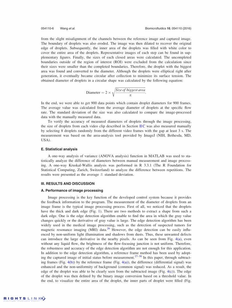
from the slight misalignment of the channels between the reference image and captured image.
The boundary of droplets was also eroded. The image was then dilated to recover the original
edge of droplets. Subsequently, the inner area of the droplets was filled with white color to
cover the entire area of the droplets. Representative images of each step can be found in sup-
plementary figures. Finally, the sizes of each closed areas were calculated. The uncompleted
boundaries outside of the region of interest (ROI) were excluded from the calculation since
their sizes were smaller than the completed boundaries. Therefore, the droplet with the biggest
area was found and converted to the diameter. Although the droplets were elliptical right after
generation, it eventually became circular after collection to minimize its surface tension. The
obtained diameter of droplets in a circular shape was calculated by the following equation:
Diameter ¼ 2�ffiffiffiffiffiffiffiffiffiffiffiffiffiffiffiffiffiffiffiffiffiffiffiffiffiffiffiffiffiffiffiffiffiffiffiffiffiffiSize of biggest area
p
r:
In the end, we were able to get 900 data points which contain droplet diameters for 900 frames.
The average value was calculated from the average diameter of droplets at the specific flow
rate. The standard deviation of the size was also calculated to compare the image-processed
data with the manually measured data.
To verify the accuracy of measured diameters of droplets through the image processing,
the size of droplets from each video clip described in Section II C was also measured manually
by selecting 8 droplets randomly from the different video frames with the gap at least 3 s. The
measurement was based on the area-analysis tool provided by ImageJ (NIH, Bethesda, MD,
USA).
E. Statistical analysis
A one-way analysis of variance (ANOVA analysis) function in MATLAB was used to sta-
tistically analyze the difference of diameters between manual measurement and image process-
ing. A one-way Kruskal-Wallis analysis was performed in R 3.3.1 (The R Foundation for
Statistical Computing, Zurich, Switzerland) to analyze the difference between repetitions. The
results were presented as the average 6 standard deviation.
III. RESULTS AND DISCUSSION
A. Performance of image processing
Image processing is the key function of the developed control system because it provides
the feedback information to the program. The measurement of the diameter of droplets from an
image frame is the typical image processing process. First of all, we noticed that the droplets
have the thick and dark edge (Fig. 1). There are two methods to extract a shape from such a
dark edge. One is the edge detection algorithm enable to find the area in which the gray value
changes quickly or the derivative of gray value is large. The edge detection algorithm has been
widely used in the medical image processing, such as the detection of suspected tumors for
magnetic resonance imaging (MRI) data.26 However, the edge detection can be easily influ-
enced by non-uniform light illumination and shadows from dusts. Thus, these unwanted defects
can introduce the large derivative in the nearby pixels. As can be seen from Fig. 4(a), even
without any liquid flow, the brightness of the flow-focusing junction is not uniform. Therefore,
the robustness and accuracy of the edge detection algorithm are not enough for this application.
In addition to the edge detection algorithm, a reference frame method has been used by adopt-
ing the captured image of initial status before measurement.27–30 In this paper, through subtract-
ing frames (Fig. 4(b)) by the reference frame (Fig. 4(a)), the difference (differential signal) was
enhanced and the non-uniformity of background (common signal) was reduced. As a result, the
edge of the droplet was able to be clearly seen from the subtracted image (Fig. 4(c)). The edge
of the droplet was then defined by the binary image conversion based on a threshold value. In
the end, to visualize the entire area of the droplet, the inner parts of droplet were filled (Fig.
054110-6 Wang et al. Biomicrofluidics 10, 054110 (2016)

4(d)). Through our testing, the reference image-based image processing described above gave
the best result for the measurement of the diameter of droplets.
In our applications, we further set the ROI with the size of 200 � 200 pixels which confine
the area of the microfluidic channel right after the flow-focusing junction. With the confined
ROI, we accelerated the processing speed as fewer pixels were involved in the image process-
ing. More importantly, the differential signals generated by the ‘tail’ of the droplets at the junc-
tion could be avoided. Therefore, the shape of droplet was the only differential signals in the
ROI. The representative images of the ROI are given in Figs. 4(e)–4(g). As can be seen from
Fig. 4(g), the filled area is the shape of one completed droplet.
In the case of small droplets typically less than 200 lm in diameter, there was more than
one droplet in a frame. The system was programmed to obtain the data from only the droplet
that has the biggest diameter. This strategy was reasonable since the sizes of droplets were rela-
tively uniform at the constant flow rate.23 On the other hand, in the case of big droplets typically
greater than 200 lm in diameter, there was no completed droplet in the frame from time to time
and thus the droplet area was not fully filled. Therefore, the size of the biggest area detected in
the frame was dropped down dramatically (Fig. 4(h)). Such data were removed automatically by
FIG. 4. Image processing. Microscopic images were chosen for (a) reference frame image, (b) captured frame image, (c)
subtracted image, and (d) processed image. (e)–(g) Captured, subtracted and processed images were taken from representa-
tive images of ROI. (h) The measured diameters of droplets in certain ranges of frames. All the images were taken from
8% PEGDA at the flow rate of 0.75 lL/min and mineral oil at the flow rate of 1.5 lL/min. Scale bar¼ 100 lm.
054110-7 Wang et al. Biomicrofluidics 10, 054110 (2016)

calculating the first order derivative of data, which was the difference between adjacent elements
in the matrix of droplet sizes. By excluding those bad frames, the detected diameters of droplets
were very stable. In the supplementary material, we included a video clip showing the original
and processed video. The detection result of the frame numbers of 150–300 was plotted in
Fig. 4(h).
B. Accuracy and repeatability
The accuracy and repeatability are very important parameters for the automated droplet con-
trol system. The image processing algorithm determines the accuracy of the system since the
feedback control signal comes from the result of image processing. Fig. 5 shows the representa-
tive images captured from the microscope and the image processing of the droplets generated
by 8% PEGDA at the flow rate of 0.75 lL/min and oil at the various flow rates. Based on the
figures, when the flow rate of the aqueous phase was fixed, the diameter of droplets was getting
smaller with the increment of the oil phase flow rate. By visually comparing the original image
with the processed image, we found that the developed algorithm could detect the areas of drop-
lets correctly. For the droplets generated right after the junction, it was hard to detect their edge
since the droplets were moved rapidly at the high flow rate of oil phase (i.e., 3.5 lL/min).
However, once it reached to the wide channel, the droplet moving speed was slowed down and
the area of droplets could be detected easily. To further analyze the accuracy of the system, we
compared the diameter of droplets from the manual measurement with that from the image proc-
essing (Fig. 6(a)). The difference between the two methods at the same flow rate was not statisti-
cally significant (p> 0.05), quantitatively proving the accuracy of the developed reference
image-based image processing algorithm.
Repeatability is another key factor of the system. To demonstrate the repeatability of the
automated droplet control system, the average diameter of droplets generated by each repeated
experiment needs to be consistent when the input parameters are same. We conducted experi-
ments of the droplet generation of 8% PEGDA at the flow rates of 0.5 lL/min for five repetitions.
Since the data from various flow rates in the same repetition did not follow a normal distribution,
Kruskal-Wallis test was used to analyze the statistical significance between repetitions. As pre-
sented in Fig. 6(b), the p-value of Kruskal-Wallis was greater than 0.95, indicating that the mean
values of diameters from different repetitions were similar and not statistically significant. This
result verified the high repeatability of the developed system.
As shown in Figs. 6(a) and 6(b), the standard deviation of diameters was less than 65 lm in
general. It indicates that the diameter of droplets was very stable at equilibrium state. The high
monodispersity is preferred for the most of droplet generation experiments.31 Monodispersed
droplets with a user-defined droplet size can be an ideal analytical platform for the high-
throughput combinatorial chemistry, proteomics, and genomics.32 In addition, monodispersed
droplets can be used in synthesizing advanced materials for biological assays.33
FIG. 5. Representative images of droplets at different flow rates of the oil phase. All data were taken from 8% PEGDA at
the flow rate of 0.75 lL/min. Scale bar¼ 200 lm.
054110-8 Wang et al. Biomicrofluidics 10, 054110 (2016)

C. Effect of various parameters
We used the developed system to characterize the effects of the flow rate and viscosity of
the aqueous phase. All the data were obtained from the developed system automatically, which
led to the significant reduction of the characterization process time. From the images given in
Fig. 5, Figs. 6(a) and 6(b), the diameter of droplets was significantly decreased at the high flow
rate of the oil phase. The flow rate of aqueous phase also played a major role in controlling the
diameter of droplets (Fig. 6(c)). The diameter of droplets was increased at the high flow rate of
aqueous phase and fixed flow rate of the oil phase. Taken together, to reduce the diameter of
droplets, there are two methods that are either decreasing the flow rate of the aqueous phase or
increasing the flow rate of the oil phase. In this paper, we focused on regulating the flow rate
of the oil phase to achieve the desired diameter of droplets. However, the system is capable of
regulating the flow rates of both aqueous and oil phases for the purpose of the experiments.
In addition to the flow rate, the type of infused liquid solutions also affects the droplet
generation process (Fig. 6(d)). Droplets from 8% PEGDA hydrogel prepolymer solution are sig-
nificantly bigger than those from PBS in the same condition. The differences between their
sizes were around 10 lm. The viscosity of 8% PEGDA solution at the room temperature was
around 1.17 mPa�S, which was approximately 1.4 times greater than PBS solution (0.90
mPa�S). The increment of viscosity required the higher interfacial force to break the droplets of
the aqueous phase, which further contributed to the increment of droplet size.10 Taken together,
the developed system was capable of controlling droplet size and generating accurate data
automatically to facilitate the droplet size optimization process in a quantitative and high-
throughput way under various conditions.
Overall, the developed system worked properly for the droplet characterization and optimi-
zation process. However, as the image processing algorithm detects the edge of droplets to
FIG. 6. Various experiments using the developed system. (a) Accuracy analysis was done by comparing the manual mea-
surement with the image processing using 8% PEGDA at the flow rate of 0.5 lL/min and oil phase with various flow rates
(n¼ 8 for manual counting and n> 50 for image processing). The statistical analysis indicated that the difference between
two measurements were not significant where p> 0.05. (b) Repeatability analysis was done by comparing 5 repetitions
using 8% PEGDA at the flow rate of 0.5 lL/min and oil with various flow rates. The statistical analysis indicated that differ-
ence among five repetitions were not significant where p> 0.95 (df¼ 4). (c) The effects of the flow rate of aqueous phase
from 8% PEGDA and oil phase with various flow rates. (d) The effects of different types of flows at the flow rate of the
aqueous phase of 0.5 lL/min and oil phase with various flow rates.
054110-9 Wang et al. Biomicrofluidics 10, 054110 (2016)

characterize the droplet size, the system will become less feasible if the edge of droplets is not
clearly captured by the microscope camera. Since the microscope camera system requires a cer-
tain exposure time and has a limited frame rate per second (e.g., 30 fps in the current system)
during capturing video clips, the image of droplets will become blurred if the droplet generation
speed is too fast (e.g., >30 Hz). Therefore, a camera with high frame rates (i.e., 120 fps or
greater) will be required to get high-quality image of fast-moving droplets for detection. In
addition, the total time of optimization depends on the initial flow rate given to the system.
Choosing a proper initial flow rate, which is close to the optimized flow rate, will shorten the
time of optimization. In general, at the first time, most of the characterization process can be
done within 10 loops (�50 min) under the current setup. The maximum amount of aqueous
phase waste will be 25 lL with the flow rate of 0.5 lL/min.
IV. CONCLUSIONS
In this paper, for the first time, we reported a fully automated control system for the high-
throughput characterization and optimization of flow-focusing droplet generation devices. The
system was used to regulate the diameter of droplets generated in bulk in a flow-focusing device
through the image processing of the video clips taken from a digital camera microscope. The
video clip was converted to a group of frames. Each frame was subtracted from the reference
frame image to enhance detecting the edge of droplets. Based on the edge, the system automati-
cally characterized the diameters of droplets and adjusted the flow rates of syringe pumps until
desired sizes were achieved. The system was highly accurate and repeatable as determined by
statistical analysis. The system will be able to be customized to meet any experimental require-
ments. The developed automate droplet generation and control system can significantly simplify
and accelerate the droplet characterization and optimization process of microfluidic devices. It is
also capable of providing high quality quantitative data for further modeling and analysis
purpose.
SUPPLEMENTARY MATERIAL
See supplementary materials for a figure showing representative images from each step of
the image processing and a video clip presenting representative frames of the original and proc-
essed video.
ACKNOWLEDGMENTS
This work was supported by the Natural Sciences and Engineering Research Council of Canada
(NSERC) Discovery Grant (No. RGPIN-2014-04010).
1T. Hamouda, M. M. Hayes, Z. Cao, R. Tonda, K. Johnson, D. C. Wright, J. Brisker, and J. R. Baker, J. Infect. Dis. 180,1939 (1999).
2C. Wibowo and K. M. Ng, AIChE J. 47, 2746 (2001).3G. De Filpo, J. Lanzo, F. P. Nicoletta, and G. Chidichimo, J. Appl. Phys. 84, 3581 (1998).4B. Born, E. L. Landry, and J. F. Holzman, IEEE Photonics J. 2, 873 (2010).5H. Shibata, Y. J. Heo, T. Okitsu, Y. Matsunaga, T. Kawanishi, and S. Takeuchi, Proc. Natl. Acad. Sci. U.S.A. 107, 17894(2010).
6R. Langer and J. P. Vacanti, Science 260, 920 (1993).7Y. T. Matsunaga, Y. Morimoto, and S. Takeuchi, Adv. Mater. 23, H90 (2011).8E. Tumarkin, L. Tzadu, E. Csaszar, M. Seo, H. Zhang, A. Lee, R. Peerani, K. Purpura, P. W. Zandstra, and E.Kumacheva, Integr. Biol. (Camb). 3, 653 (2011).
9B. G. Chung, K.-H. Lee, A. Khademhosseini, and S.-H. Lee, Lab Chip 12, 45 (2012).10G. F. Christopher and S. L. Anna, J. Phys. D: Appl. Phys. 40, R319 (2007).11B. S. Lee, H.-J. Cho, J.-G. Lee, N. Huh, J.-W. Choi, and I. S. Kang, J. Colloid Interface Sci. 302, 294 (2006).12D. Moon, D. J. Im, T. Uhm, and I. S. Kang, Int. J. Chem. Eng. Appl. 3, 1 (2012).13J. G. Lee, H. J. Cho, N. Huh, C. Ko, W. C. Lee, Y. H. Jang, B. S. Lee, I. S. Kang, and J. W. Choi, Biosens. Bioelectron.
21, 2240 (2006).14S. L. Anna, N. Bontoux, and H. A. Stone, Appl. Phys. Lett. 82, 364 (2003).15C. Cha, J. Oh, K. Kim, Y. Qiu, M. Joh, S. R. Shin, X. Wang, G. Camci-unal, K. Wan, R. Liao, and A. Khademhosseini,
Biomacromolecules 15, 283 (2014).16J. Jung and J. Oh, Biomicrofluidics 8, 36503 (2014).
054110-10 Wang et al. Biomicrofluidics 10, 054110 (2016)
17D. Oh, S. Lee, J. Kim, H. Choi, J. Seo, and K. I. Koo, in Proceedings of the 36th Annual International Conference onIEEE Engineering in Medicine and Biological Society (EMBC) (2014), pp. 1390–1393.
18S.-Y. Teh, R. Lin, L.-H. Hung, and A. P. Lee, Lab Chip 8, 198 (2008).19R. Samanipour, Z. Wang, A. Ahmadi, and K. Kim, J. Appl. Polym. Sci. 133, 43701 (2016).20C. Elbuken, T. Glawdel, D. Chan, and C. L. Ren, Sens. Actuators, A 171, 55 (2011).21Y. Xia and G. M. Whitesides, Annu. Rev. Mater. Sci. 28, 153 (1998).22T. W. Lee, O. Mitrofanov, and J. W. P. Hsu, Adv. Funct. Mater. 15, 1683 (2005).23T.-D. Dang, Y. H. Kim, H. G. Kim, and G. M. Kim, J. Ind. Eng. Chem. 18, 1308 (2012).24J. Siepmann, N. Faisant, J. Akiki, J. Richard, and J. P. Benoit, J. Controlled Release 96, 123 (2004).25J. M. Cha, H. Bae, N. Sadr, S. Manoucheri, F. Edalat, K. Kim, S. B. Kim, I. K. Kwon, Y.-S. Hwang, and A.
Khademhosseini, Macromol. Res. 23, 245 (2015).26Z. Wang, X. Xiao, H. Song, L. Wang, and Q. Li, IEEE Antennas Wirel. Propag. Lett. 13, 1757 (2014).27S. B. Kim, K. Koo, H. Bae, M. R. Dokmeci, G. A. Hamilton, A. Bahinski, S. M. Kim, D. E. Ingber, and A.
Khademhosseini, Lab Chip 12, 3976 (2012).28K. Koo, S. B. Kim, K. Kim, and J. Oh, Biotechnol. Lett. 36, 1089 (2014).29X. Dong, P. Song, and X. Liu, in Proceedings of the IEEE International Conference on Robotics and Automation (ICRA)
(2015), p. 996.30P. Song, X. Dong, and X. Liu, Biomicrofluidics 10, 011912 (2016).31V. van Steijn, P. M. Korczyk, L. Derzsi, A. R. Abate, D. A. Weitz, and P. Garstecki, Biomicrofluidics 7, 024108 (2013).32A. Huebner, S. Sharma, M. Srisa-Art, F. Hollfelder, J. B. Edel, and A. J. deMello, Lab Chip 8, 1244 (2008).33R. Seemann, M. Brinkmann, T. Pfohl, and S. Herminghaus, Rep. Prog. Phys. 75, 016601 (2012).
054110-11 Wang et al. Biomicrofluidics 10, 054110 (2016)