an electrochemical ensor based ons reduced …abechem.com/no. 2-2016/2016, 8(2), 219-233.pdfanal....
TRANSCRIPT
Anal. Bioanal. Electrochem., Vol. 8, No. 2, 2016, 219-233
Full Paper
An Electrochemical Sensor based on Reduced Graphene oxide-Silver Nanocomposite Modified Carbon Paste Electrode for Acetaminophen Determination
Ebrahim Tavakolian and Javad Tashkhourian*
Department of Chemistry, Faculty of Science, Shiraz University, Shiraz 71454, Iran
*Corresponding Author, Tel.: +98 7136137110; Fax: +98 7136460788
E-Mail: [email protected]
Received: 18 December 2015 / Accepted: 17 March 2016 / Published online: 31 March 2016
Abstract- An electrochemical sensor based on modification of carbon paste electrode by reduced graphene oxide-silver nanocomposite (RGO-Ag/CPE) was prepared for voltammetric determination of acetaminophen. The morphology and structure of the resulting products were characterized by transmission electron micrograph, X-ray diffraction and infrared spectroscopy. The electrochemical behaviours of acetaminophen on resulting modified electrode were investigated by cyclic voltammetry and differential pulse voltammetry. Differential pulse voltammetry shows that peak current increase linearly by increasing in acetaminophen concentration. Acetaminophen was determined in the range of 5.0–480.0, and the limit of detection was determined as 0.18 µM using differential pulse voltammetry under optimization conditions. This electrode obtains good and satisfactory results in the determination of acetaminophen in a commercial tablet.
Keywords- Reduced graphene oxide, Silver nanoparticles, Modified carbon paste electrode, Acetaminophen
1. INTRODUCTION
Graphene (Gr) is the name of a two-dimensional sheet of sp2-hybridized carbon [1] that is the thinnest known material in the universe [2]. Resulting single- and few-layer flakes were pinned to the substrate by only van der Waals forces and could be made free-standing by etching away the substrate [3-6]. Graphene has unique properties such as high specific
Analytical & Bioanalytical Electrochemistry
2016 by CEE
www.abechem.com

Anal. Bioanal. Electrochem., Vol. 8, No. 2, 2016, 219-233 220
surface area [7], good mechanical strength [8], excellent thermal and electrical conductivity [4,9,10], that allowed scientists to probe graphene’s properties [11]. Furthermore, it had reported that large quantities production of graphene needs much lower cost than that of carbon nanotubes [12].
Moreover, graphene or reduced graphene oxide (RGO) could act as an excellent substrate for supporting metal and/or semiconductor nanoparticles. Recently, reduced graphene oxide-or graphene–metal nanocomposite have been used as a proper electrode materials for electrochemical applications [13]. Metal nanoparticles such as Ag, Pd, Pt and Au have large surface area to volume ratios at the nanoscale that makes them excellent choice materials where large surface areas are required [14]. So far, a variety of successful approaches for preparing reduced graphene oxide–metal nanocomposite have been reported. For example, chemical reduction [15-17], epitaxial deposition [18], electrodeposition [19,20], and chemical vapor deposition [21] have been used. Among metal nanoparticles, silver nanoparticles (AgNPs) have been shown good conductivity and high electrochemical catalytic activity [22,23]. The availability of AgNPs will expand the possibilities for the preparation of Ag-doped nanomaterials and extend its application in biosensor [22,23]. It has been reported that the using of carbon-based materials and metal nanoparticles usually causes synergistic effects in electrocatalytic applications [24].
Acetaminophen (AP), also known as N-acetyl-p-aminophenol or paracetamol, is an effective analgesic and antipyretic drug with rather limited anti-inflammatory properties [25] and is used in the management of cancer or postoperative pain [26]. AP is rapidly and extensively metabolized by undergoing glucuronidation and sulfation to inactive metabolites which are eliminated in urine along with 5% of AP being eliminated unchanged. At the recommended dosage, there are no side effects but, overdoses will cause liver and kidney damage [27]. It is thought that a metabolite of acetaminophen is the actual hepatotoxic agent [28]. Several methods have been used for the determination of AP for quality control analysis in pharmaceutical formulations and for medical control in biological fluids including electrochemical [29-31], spectrophotometry [32], flow-injection [33] and chromatographic methods [34].
Since the RGO-Ag nanocomposites for the electrochemical application have been rarely reported in this work, a simple electrochemical sensor based on RGO–Ag nanocomposites was designed and reported for highly sensitive AP detection. Firstly, RGO-Ag nanocomposites were prepared by in situ deposition of AgNPs on to RGO sheet. Then, the electrooxidation of paracetamol on prepared carbon paste electrode modified with silver nanoparticle–reduced graphene oxide (RGO-Ag) was investigated. The prepared electrode showed good electrocatalytic activity for AP and supposed a sufficient limit of detection and linear range.

Anal. Bioanal. Electrochem., Vol. 8, No. 2, 2016, 219-233 221
2. EXPERIMENTAL
2.1. Reagent and solutions
Graphite powder (particle size<100 μm) used for constructing electrodes and synthesis was purchased from Fluka. Paraffin oil for constructing electrode was purchased from Merck. Hydrazine monohydrate for synthesis was purchased from Merck. Phosphate buffer solutions (PBS) were prepared by mixing the stock solutions of 0.1 M KH2PO4 (Merck) and 0.1 M K2HPO4 (Merck), and adjusting the pH with 0.1 M H3PO4 (Merck) or 0.1 M NaOH (Merck). All buffer solutions were freshly made prior to experiments. All solutions were prepared with double-distilled water. Ammonia solution (28 wt.%, Merck) was used for synthesis.
2.2. Apparatus
Electrochemical measurements were carried out with an electrochemical analyzer Autolab PGSTAT 302 N (Metrohm Autolab B.V., Utrecht, the Netherlands). A conventional three-electrode system was used throughout the experiments at room temperature. The working electrode was RGO-Ag/CPE and the auxiliary electrode was a platinum wire. Besides, a Ag/AgCl-KCl saturated was taken as the reference electrode. All potentials in this study refer to this reference electrode. Fourier transform infrared spectroscopy (FT-IR) measurements of graphene oxide (GO), reduced graphene oxide (RGO) and reduced graphene oxide–silver nanocomposite (RGO–Ag) were performed with a Spectrum RX 1 (Perkin Elmer, 940 Winter Street, Waltham, Massachusetts 02451, USA). Measurements of pH were made with a Denver Instrument Model 780 pH meter equipped with a Metrohm glass electrode. Transmission electron micrograph (TEM) was taken with Zeiss-EM10C-80 KV and XRD spectra were obtained by D8ADVANCE type (BRUKER-Germany).
2.3. Synthesis of reduced graphene oxide – silver nanocomposite
A modified Hummers method was used to synthesize the oxidized graphite powder [35]. In a typical procedure for reduction of graphene oxide to graphene [36], the centrifuged brown graphene oxide (100 ml) suspension was mixed with 150 µL hydrazine monohydrate and the pH of the suspension was adjusted to ~10 with ammonia solution (28 wt.% in water) in a 150 mL glass beaker to ionize the carboxylic acid groups on the graphene oxide sheets. After being vigorously stirred for a few minutes, the mixture was transferred into a 100 mL glass beaker flask to reduce the evaporation and then put in a water bath (98 °C) for 1 h, yielding the aqueous dispersion of graphene. The silver nitrate[36] aqueous solution (10.0 mL, 10-3 M) was added drop by drop to the RGO dispersion solution with vigorous stirring to obtain the RGO - Ag nanocomposite dispersion. The unreacted hydrazine in RGO dispersion solution was used to further reduce the silver ions to obtain the silver nanoparticles. The RGO
Anal. Bioanal. Electrochem., Vol. 8, No. 2, 2016, 219-233 222
and the RGO - Ag nanocomposite was prepared after centrifuging the dispersion solution and dried it in a vacuum oven at room temperature for 3 h.
2.4. Electrode Preparation
Unmodified carbon paste electrode was prepared by hand mixing of 70.0% of graphite powder and 30.0% of paraffin oil. A modified carbon paste electrode was prepared in a similar fashion, except that the graphite powder was mixed with a desired weight of RGO–Ag nanocomposite. Both unmodified and modified carbon paste electrodes were constructed by packing the paste into a tube (3 mm inner diameter). The electrode surface was polished by rubbing it against a piece of paper while a slight manual pressure was applied to the piston whenever regeneration of the electrode was required, a thin layer of the surface was removed with a spatula and replaced by fresh paste. A stainless steel rod was inserted in the tube for electrical connection and also as a piston for removing a thin layer of carbon paste for the electrode surface renewing process.
2.5. General procedure
Stock solution of AP was freshly prepared prior to use. Electrochemical experiments were carried out in the phosphate buffered solutions (PBS pH=4.0) of AP. For differential pulse voltammetric (DPV) experiments, the modified carbon paste electrode was immersed in 10.0 mL of buffer solution containing a known amount of analyte. Finally, the differential pulse voltammograms were recorded from 0.3 to 0.8 V (DPV were performed with pulse potential of 50.0 mV, pulse duration of 50.0 ms and pulse period of 0.2 s). All measurements were carried out at room temperature (25±1 °C).
3. RESULTS AND DISCUSSION
3.1. Characterization of reduced graphene oxide-silver nanocomposite
The XRD patterns of GO, RGO and RGO-Ag nanocomposite are shown in Figure 1. Compared to the typical diffraction peak at 2θ=12.6° corresponding to the (0 0 1) lattice plane of GO (Figure 1. a), those of RGO and RGO–Ag (Figure 1. b) have smaller intensities that shows reduction of GO, and these patterns also show major diffraction peaks at 2θ=38.3° and the other at 2θ=44.4° along with the broad and diffused diffraction peak at 2θ=24.0°. According to the database these peaks are indexed to (1 1 1) and (2 0 0) reflections of silver nanocrystals with face centered cubic symmetry, respectively, whereas the diffused diffraction peaks at 2θ=24° is attributed to the (0 0 2) reflection of graphene nanosheets. Figure 2 shows the FT-IR spectra of GO, RGO and RGO-Ag nanocomposite. The typical peaks of GO at 1730 cm-1 (C O= carboxylic), 3410 cm-1 (stretching vibration of C–OH) and

Anal. Bioanal. Electrochem., Vol. 8, No. 2, 2016, 219-233 223
1400 cm-1 (deformation vibration of C–OH) are observed. The peak at 1060 cm-1 confirms the presence of epoxide groups (C–O–C) and the peak at 1630 cm-1 is ascribed to the skeletal vibration of graphitic skeleton. After the reduction, the peaks corresponding to the oxygen-containing groups gradually decrease in intensity with increasing the synthesis temperature and even the peak of carboxylic acid vibration (C O= at 1730 cm-1) disappears. Also, the size and structure of the RGO-Ag were evaluated using transmission electron microscopy (TEM). According to the TEM (Figure 3), it was observed that the Ag has nanodimension ranging about 15 nm.
Fig. 1. XRD spectra of (a) GO and (b) RGO, and RGO-Ag
0 20 40 60 80
Inte
nsity
2θ
GO
(0 0 1)
0 20 40 60 80
Inte
nsity
2θ
RGO-Ag
RGO
(0 0 2)
(0 0 2)
(1 1 1)(2 0 0)
(a)
(b)

Anal. Bioanal. Electrochem., Vol. 8, No. 2, 2016, 219-233 224
Fig. 2. FTIR spectra of samples GO, RGO, and RGO-Ag
Fig. 3. TEM image of RGO-Ag nanocomposite
3.2. Electrochemical behaviour of acetaminophen
The behavior of the electrode in the presence or and absence of acetaminophen was studied. Based on Figure 4, the electrode in the absence of the analyte does not indicate an electrochemical response. Despite expectations [37], oxidation peak of the silver is not observed. The absence of Ag characteristic peak in voltammograms could be referred to the very low extent and small sizes of Ag NPs in composite [38]. Probably, this behaviour is due to low decorated silver nanoparticles on RGO that is clear in TEM image, too (Figure 3 in
100015002000250030003500400045005000
Tran
smit
ance
(%)
Wavenumber(cm-1)
RGO-Ag
RGO
GO
1730 1400 10603410

Anal. Bioanal. Electrochem., Vol. 8, No. 2, 2016, 219-233 225
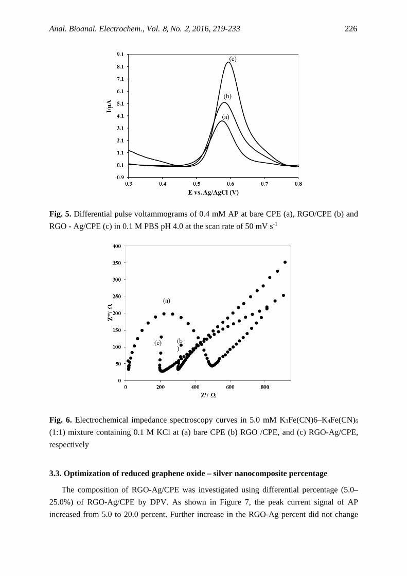
section 3.1). Electrooxidation behavior of AP at bare CPE (a), RGO/CPE (b) and RGO-Ag/CPE (c) in 0.1 M PBS pH 4.0 was investigated at the scan rate of 50mV s-1 using differential pulse voltammetry (DPV). As shown in Figure 5, at the bare CPE, acetaminophen shows an oxidation peak at Ep=0.58 V. When the modified CPE with RGO was used, there was a positive shift in peak potential to Ep=0.60 V with a little change in peak current. These results demonstrate (Figure 5) that the electrochemical reactivity of AP is improved on the RGO - Ag/CPE. As shown in Figure 4.1.5, the enhancement of oxidative peak current of AP could be attributed to the synergetic effect between the RGO and Ag NPs. However, the characteristic peaks of Ag were obvious in XRD spectra and these particles were clearly distinguishable in TEM image. Figure 6 depicts the electrochemical impedance spectroscopy (EIS) of bare CPE (a), RGO/CPE (b) and RGO-Ag (c) in PBS containing 5 mM Fe(CN)6 3-/4- and 0.1 M KCl, including a constant phase element (Cdl), charge transfer resistance (Rct), Warburg impedance (Zw) and the uncompensated solution resistance (Rs). As shown in this figure RGO-Ag/CPE had the lowest Rct value that revealing its faster electron transfer ability.
Fig. 4. Differential pulse voltammograms obtained in the absence (a) and presence (b) of 480 µM of acetaminophen in 0.1 M PBS pH 4.0 at the scan rate of 50 mV s-1
Anal. Bioanal. Electrochem., Vol. 8, No. 2, 2016, 219-233 226
Fig. 5. Differential pulse voltammograms of 0.4 mM AP at bare CPE (a), RGO/CPE (b) and RGO - Ag/CPE (c) in 0.1 M PBS pH 4.0 at the scan rate of 50 mV s-1
Fig. 6. Electrochemical impedance spectroscopy curves in 5.0 mM K3Fe(CN)6–K4Fe(CN)6 (1:1) mixture containing 0.1 M KCl at (a) bare CPE (b) RGO /CPE, and (c) RGO-Ag/CPE, respectively
3.3. Optimization of reduced graphene oxide – silver nanocomposite percentage
The composition of RGO-Ag/CPE was investigated using differential percentage (5.0–25.0%) of RGO-Ag/CPE by DPV. As shown in Figure 7, the peak current signal of AP increased from 5.0 to 20.0 percent. Further increase in the RGO-Ag percent did not change

Anal. Bioanal. Electrochem., Vol. 8, No. 2, 2016, 219-233 227
the current response of AP oxidation. The percent of RGO - Ag in the CPE was kept constant at 20.0 percent throughout the rest of this study.
Fig. 7. The peak current vs RGO - Ag percentage in composite electrode. 0.4 mM AP in 0.1 M PBS pH 4.0 at the scan rate of 50 mV s-1
3.4. Effects of pH on response of electrode
Nematollahi et al. [39] demonstrated the electrochemical oxidation of acetaminophen in various pHs using cyclic voltammetry and controlled-potential coulometry. AP is electrochemically oxidized via a pH-dependent two-electron, two-proton process to N-acetyl-p-quinone-imine (abbreviated NAPQI). The rate constants were estimated by comparing the experimental cyclic voltammetric responses with the digital simulated results. The results indicated that electrochemically generated NAPQI participates in different types of reactions depending on solution’s pH. It has been reported[40] that the stability of NAPQI was substantially affected by the sample pH with a highest stability in the pH range of 4.0–9.0 and a half-life equal to 47 min at pH 7.4. The effect of solution pH on the electrochemical redox reaction of AP on the RGO-Ag/CPE was investigated in the range of pH 2.0 - 9.0. Figure 8 shows the differential pulse voltammograms of acetaminophen in various pHs (2.0 to 9.0). In this work, phosphate buffer solution (0.1 mol L -1, pH=4.0) was used as supporting electrolyte for further experiments. Inset is the plot of peak potential vs. pH value, the slope of this plot is near to Nurnstian slope, so the number of electron and proton is same which are in agreement with the literature and shows two-electron, two-proton process [40].

Anal. Bioanal. Electrochem., Vol. 8, No. 2, 2016, 219-233 228
Fig. 8. Differential pulse voltammograms of 0.4 mM acetaminophen at RGO–Ag/CPE, in buffer solution with various pHs. Scan rate: 50 mV s−1. Inset is the plot of peak potential vs. pH value 3.5. Effect of scan rate
Cyclic voltammetry of AP was carried out at different scan rates ranging from 10.0 to 250.0 mV s-1 (Figure 9). The results indicated that there is a linear relationship between the anodic peak currents and the square root of scan rate (ν1/2) in the range of 10.0 to 250.0 mV s-1. These results indicate the existence of a diffusion-controlled mechanism for AP in the mentioned potential scan rates.
Fig. 9. Cyclic voltammorgrams of 0.4 mM AP on RGO-Ag/CPE at different scan rates from 10 to 250 mV/s. Inset is the plot of peak current vs. square root of scan rate

Anal. Bioanal. Electrochem., Vol. 8, No. 2, 2016, 219-233 229
3.6. Voltammetric determination of acetaminophen
Figure 10 depicts the DPV curves of different concentrations of acetaminophen at RGO-Ag/CPE modified electrode. The calibration curve for AP shows linear range from 5.0 µM to 4.8×10² µM with linear regression equation of Ip (µA)=0.0173CAP (µM)+0.282 (R²=0.9902). The limit of detection was 0.183 µM (LOD=3σblank /m, in which σblank is standard deviation of blank for n=5 and ‘m’ is the slope of the calibration curve).
Fig. 10. Relationship of current responses to acetaminophen concentration on RGO-Ag/CPE for different acetaminophen concentrations in 0.1 M phosphate buffer (pH=4.0)
Table 1. Comparison of the electroanalytical data for determination of AP
Electrode Linear range (μM)
Detection limit (μM)
Ref.
Polyphenol oxidase/glassy carbon paste electrode Up to 70.0 7.8 [41] MWCNT/GCE–PANI 100.00–1.00 0.25 [42]
graphite electrode-vaseline-Polyphenol oxidase 5800-120 88 [43] Electropolymerized-molecularly imprinted PPy/pencil graphite electrode
500.00–5.00 0.79 [44]
SPE/PEDOT 400–4 1.39 [25] Nafion coated glassy carbon tubular electrode Up to 500 17 [45]
CS/GCE–GR 1.0-100.0 0.3 [46]
CFE 100-0.02 0.034 [47] /polymer coated GCE2TiO-Nano 120.0–12.0 2.0 [48]
RGO-Ag/CPE 2×104.8–5.0 0.18 This work
PANI: polyaniline, MWCNT: multi-walled carbon nanotube, GCE: glassy carbon electrode, SPE: Screen-Printed Electrode, PEDOT: Poly (3,4-ethylenedioxythiophene), GR–CS: Graphene–chitosan, CFE: micro-crystalline natural graphite–polystyrene composite film electrode
Anal. Bioanal. Electrochem., Vol. 8, No. 2, 2016, 219-233 230
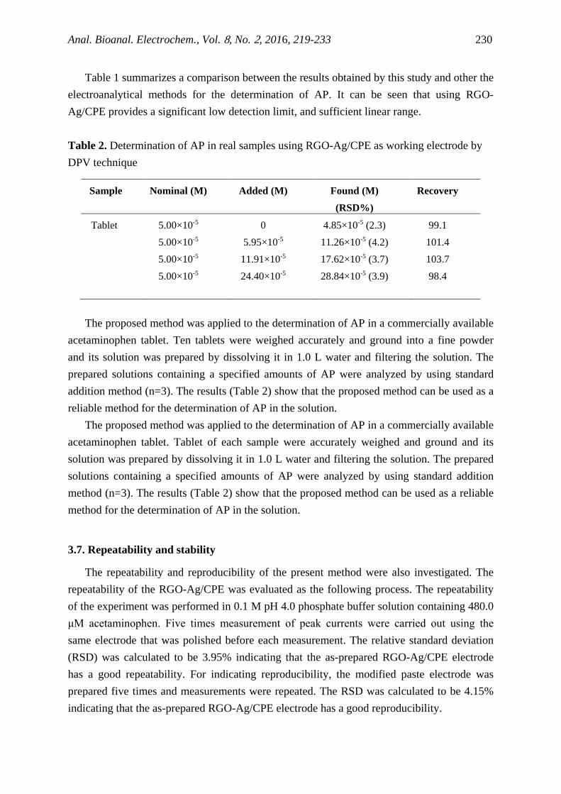
Table 1 summarizes a comparison between the results obtained by this study and other the electroanalytical methods for the determination of AP. It can be seen that using RGO-Ag/CPE provides a significant low detection limit, and sufficient linear range. Table 2. Determination of AP in real samples using RGO-Ag/CPE as working electrode by DPV technique
Sample Nominal (M) Added (M) Found (M) (RSD%)
Recovery
Tablet 5-×105.00 0 (2.3) 5-×104.85 99.1 5-×105.00 5-×105.95 (4.2) 5-×1011.26 101.4 5-×105.00 5-×1011.91 (3.7) 5-×1017.62 103.7 5-×105.00 5-×1024.40 (3.9) 5-×1028.84 98.4
The proposed method was applied to the determination of AP in a commercially available acetaminophen tablet. Ten tablets were weighed accurately and ground into a fine powder and its solution was prepared by dissolving it in 1.0 L water and filtering the solution. The prepared solutions containing a specified amounts of AP were analyzed by using standard addition method (n=3). The results (Table 2) show that the proposed method can be used as a reliable method for the determination of AP in the solution.
The proposed method was applied to the determination of AP in a commercially available acetaminophen tablet. Tablet of each sample were accurately weighed and ground and its solution was prepared by dissolving it in 1.0 L water and filtering the solution. The prepared solutions containing a specified amounts of AP were analyzed by using standard addition method (n=3). The results (Table 2) show that the proposed method can be used as a reliable method for the determination of AP in the solution.
3.7. Repeatability and stability
The repeatability and reproducibility of the present method were also investigated. The repeatability of the RGO-Ag/CPE was evaluated as the following process. The repeatability of the experiment was performed in 0.1 M pH 4.0 phosphate buffer solution containing 480.0 μM acetaminophen. Five times measurement of peak currents were carried out using the same electrode that was polished before each measurement. The relative standard deviation (RSD) was calculated to be 3.95% indicating that the as-prepared RGO-Ag/CPE electrode has a good repeatability. For indicating reproducibility, the modified paste electrode was prepared five times and measurements were repeated. The RSD was calculated to be 4.15% indicating that the as-prepared RGO-Ag/CPE electrode has a good reproducibility.
Anal. Bioanal. Electrochem., Vol. 8, No. 2, 2016, 219-233 231
3.8. Interference studies and real samples analysis
The electrochemical behaviors of the coexisting electroactive species, which often cause serious interference with the determination of AP, such as p-aminophenol, ascorbic acid, glucose, urea, dopamine, were investigated by DPV. The modified electrode could virtually eliminate the interference of p-aminophenol, ascorbic acid, glucose, dopamine and uric acid at 500-fold concentration of AP (at 90.0 μM), and it has been satisfactorily used for the voltammetric determination of AP.
4. CONCLUSION
We have demonstrated the application of the RGO-Ag/CPE for determination of acetaminophen. However, many researchers have been performed to synthesis this kind of composite, but there are very low works suggested for the electrochemically performance of this composite. This study suggested a new kind of silver supported material that not shown electrochemical disturbance of silver activity. The composite film modified electrode was successfully employed for the voltammetric determination of acetaminophen with low detection limit, good linear range and high selectivity. Acknowledgements We gratefully acknowledge the support of this work by Shiraz University Research Council.
REFERENCES
[1] X. Feng, Y. Zhang, J. Zhou, Y. Li, S. Chen, L. Zhang, Y. Ma, L. Wang, and X. Yan, Nanoscale 7 (2015) 2427.
[2] A. K. Geim, Science 324 (2009) 1530. [3] J. C. Meyer, A. Geim, M. Katsnelson, K. Novoselov, T. Booth, and S. Roth, Nature 446
(2007) 60. [4] K. I. Bolotin, K. Sikes, Z. Jiang, M. Klima, G. Fudenberg, J. Hone, P. Kim, and H.
Stormer, Solid State Commun. 146 (2008) 351. [5] K. Bolotin, K. Sikes, J. Hone, H. Stormer, and P. Kim, Phys. Rev. Lett. 101 (2008)
096802. [6] K. Novoselov, D. Jiang, F. Schedin, T. Booth, V. Khotkevich, S. Morozov, and A.
Geim, Proc. Natl. Acad. Sci. U.S.A. 102 (2005) 10451. [7] M. D. Stoller, S. Park, Y. Zhu, J. An, and R. S. Ruoff, Nano Lett. 8 (2008) 3498. [8] C. Lee, X. Wei, J. W. Kysar, and J. Hone, Science 321 (2008) 385. [9] A. A. Balandin, S. Ghosh, W. Bao, I. Calizo, D. Teweldebrhan, F. Miao, and C. N. Lau,
Nano Lett. 8 (2008) 902.

Anal. Bioanal. Electrochem., Vol. 8, No. 2, 2016, 219-233 232
[10] X. Feng, Y. Zhang, J. Song, N. Chen, J. Zhou, Z. Huang, Y. Ma, L. Zhang, and L. Wang, Electroanalysis 27 (2015) 353.
[11] M. J. Allen, V. C. Tung, and R. B. Kaner, Chem. Rev. 110 (2009) 132. [12] D. Li, R. B. Kaner, Science 320 (2008) 1170. [13] K. S. Kim, I. J. Kim, and S. J. Park, Synth. Met. 160 (2010) 2355. [14] A. Bello, M. Fabiane, D. Dodoo-Arhin, K. I. Ozoemena, and N. Manyala, J. Phys.
Chem. Solids 75 (2014) 109. [15] S. Liu, J. Tian, L. Wang, and X. Sun, Carbon 49 (2011) 3158. [16] Y. K. Yang, C. E. He, W. J. He, L. J. Yu, R. G. Peng, X. L. Xie, X. B. Wang, and Y. W.
Mai, J. Nanopart. Res. 13 (2011) 5571. [17] J. Tian, S. Liu, Y. Zhang, H. Li, L. Wang, Y. Luo, A. M. Asiri, A. O. Al-Youbi, and X.
Sun, Inorg. Chem. 51 (2012) 4742. [18] G. F. Ndlovu, W. D. Roos, Z. M. Wang, J. K. Asante, M. G. Mashapa, C. J. Jafta, B. W.
Mwakikunga, and K. T. Hillie, Nanoscale Res. lett. 7 (2012) 1-8. [19] X. Sun, M. Xie, G. Wang, H. Sun, A. S. Cavanagh, J. J. Travis, S. M. George, and J.
Lian, J. Electrochem. Soc. 159 (2012) A364. [20] X. Dong, Y. Cao, J. Wang, M. B. Chan-Park, L. Wang, W. Huang, and P. Chen, RSC
Adv. 2 (2012) 4364. [21] Y. H. Kahng, S. Lee, W. Park, G. Jo, M. Choe, J. H. Lee, H. Yu, T. Lee, and K. Lee,
Nanotechnology 23 (2012) 075702. [22] K. J. Huang, L. Wang, H. B. Wang, T. Gan, Y. Y. Wu, J. Li, and Y. M. Liu, Talanta
114 (2013) 43. [23] H. Chen, D. Tang, B. Zhang, B. Liu, Y. Cui, and G. Chen, Talanta 91 (2012) 95. [24] Q. Zhang, Q. Ren, Y. Miao, J. Yuan, K. Wang, F. Li, D. Han, and L. Niu, Talanta 89
(2012) 391. [25] W.Y. Su, and S. H. Cheng, Electroanalysis 22 (2010) 707. [26] F. F. Daly, J. S. Fountain, L. Murray, A. Graudins, and N. A. Buckley, Med. J.
Australia 188 (2008) 296. [27] M. Mazloum-Ardakani, M. A. Sheikh-Mohseni, M. Abdollahi-Alibeik, and A. Benvidi,
Sens. Actuators B 171 (2012) 380. [28] H. Filik, G. Çetintaş, A. A. Avan, S. N. Koç, and İ. Boz, Int. J. Electrochem. Sci. 8
(2013) 5724. [29] M. Mazloum-Ardakani, H. Beitollahi, M. A. S. Mohseni, A. Benvidi, H. Naeimi, M.
Nejati-Barzoki, and N. Taghavinia, Colloids Surf. B 76 (2010) 82. [30] Z.A. Alothman, N. Bukhari, S.M. Wabaidur, and S. Haider, Sens. Actuators B 146
(2010) 314. [31] F. Ghorbani-Bidkorbeh, S. Shahrokhian, A. Mohammadi, and R. Dinarvand,
Electrochim. Acta 55 (2010) 2752.

Anal. Bioanal. Electrochem., Vol. 8, No. 2, 2016, 219-233 233
[32] E. Souri, S. A. M. Nasab, M. Amanlou, and M. B. Tehrani, J. Anal. Chem. 70 (2015) 333-8.
[33] N. Wangfuengkanagul, and O. Chailapakul, J. Pharm. Biomed. Anal. 28 (2002) 841. [34] J. H. An, H. J. Lee, and B. H. Jung, Biomed. Chromatog. 26 (2012) 1596. [35] W. S. Hummers Jr, and R. E. Offeman, J. Am. Chem. Soc. 80 (1958) 1339. [36] Z. Xu, H. Gao, and H. Guoxin, Carbon 49 (2011) 4731. [37] V. Pifferi, V. Marona, M. Longhi, and L. Falciola, Electrochim. Acta 109 (2013) 447. [38] Electrochemical Methods. Fundamentals and Applications, ed. A. J. Bard, and L. R.
Faulkner, Wiley, 2nd edn (2001). [39] D. Nematollahi, H. Shayani-Jam, M. Alimoradi, S. Niroomand, Electrochim. Acta 54
(2009) 7407. [40] J. J. Van Benschoten, J. Y. Lewis, W. R. Heineman, D. A. Roston, and P. T. Kissinger,
J. Chem. Educ. 60 (1983) 772. [41] M. C. Rodrı́guez, and G. A. Rivas, Anal. Chim. Acta 459 (2002) 43. [42] M. Li, and L. Jing, Electrochim. Acta 52 (2007) 3250. [43] O. Fatibello-Filho, K. O. Lupetti, and I. C. Vieira, Talanta 55 (2001) 685. [44] L. Özcan, and Y. Şahin, Sens. Actuators. B 127 (2007) 362. [45] M. L. S. Silva, M. B. Q. Garcia, J. L. Lima, and E. Barrado, Talanta 72 (2007) 282. [46] M. Zheng, F. Gao, Q. Wang, X. Cai, S. Jiang, L. Huang, and F. Gao, Mater. Sci. Eng.
33 (2013) 1514. [47] A. R. Khaskheli, J. Fischer, J. Barek, V. Vyskočil, M. I. Bhanger, Electrochim. Acta
101 (2013) 238. [48] S. A. Kumar, C. F. Tang, and S. M. Chen, Talanta 76 (2008) 997.
Copyright © 2016 by CEE (Center of Excellence in Electrochemistry)
ANALYTICAL & BIOANALYTICAL ELECTROCHEMISTRY (http://www.abechem.com)
Reproduction is permitted for noncommercial purposes.