an experimental study of muscular injury repair in a mouse...
TRANSCRIPT

Abat et al. BMC Sports Science, Medicine, and Rehabilitation (2015) 7:7 DOI 10.1186/s13102-015-0002-0
RESEARCH ARTICLE Open Access
An experimental study of muscular injury repairin a mouse model of notexin-induced lesion withEPI® techniqueFerran Abat1, Soraya-L Valles2, Pablo-Eduardo Gelber3,4, Fernando Polidori5, Adrian Jorda2, Sergio García-Herreros2,Joan-Carles Monllau3,6,7 and Jose-Manuel Sanchez-Ibáñez5*
Abstract
Background: The mechanisms of muscle injury repair after EPI® technique, a treatment based on electricalstimulation, have not been described. This study determines whether EPI® therapy could improve muscle damage.
Methods: Twenty-four rats were divided into a control group, Notexin group (7 and 14 days) and a Notexin + EPI group.To induce muscle injury, Notexin was injected in the quadriceps of the left extremity of rats. Pro-inflammatory interleukin1-beta (IL-1beta) and tumoral necrosis factor-alpha (TNF-alpha) were determined by ELISA. The expression of receptorperoxisome gamma proliferator activator (PPAR-gamma), vascular endothelial growth factor (VEGF) and vascularendothelial growth factor receptor-1 (VEGF-R1) were determined by western-blot.
Results: The plasma levels of TNF-alpha and IL-1beta in Notexin-injured rats showed a significant increase comparedwith the control group. EPI® produced a return of TNF-alpha and IL-1beta values to control levels. PPAR-gammaexpression diminished injured quadriceps muscle in rats. EPI® increased PPAR-gamma, VEGF and VEGF-R1 expressions.EPI® decreased plasma levels of pro-inflammatory TNF-alpha and IL-1beta and increased anti-inflammatory PPAR-gammaand proangiogenic factors as well as VEGF and VEGF-R1 expressions.
Conclusion: The EPI® technique may affect inflammatory mediators in damaged muscle tissue and influences the newvascularization of the injured area. These results suggest that EPI® might represent a useful new therapy for the treatmentof muscle injuries. Although our study in rats may represent a valid approach to evaluate EPI® treatment, studies designedto determine how the EPI® treatment may affect recovery of injury in humans are needed.
Keywords: EPI, Technique, Notexin-induced, Muscle, Injury
BackgroundSoft tissue injuries are recurrent in sports and have anincidence rate of some 30% [1]. An overly conservativetherapeutic approach conflicts with patients’ economicsinterests and the ability to practice their chosen sport.Some authors have proposed qualitative and histopatho-logical classifications of muscle injuries directly relatedto the appearance of the lesion and its evolution [2].The inflammatory process is one of the most import-
ant parts of the immune system’s response to injury. It isdue to the fact that the biochemical mechanism and the
* Correspondence: [email protected] of Sports Rehabilitation, Cerede Sports Medicine, Barcelona,SpainFull list of author information is available at the end of the article
© 2015 Abat et al.; licensee BioMed Central. TCommons Attribution License (http://creativecreproduction in any medium, provided the orDedication waiver (http://creativecommons.orunless otherwise stated.
signal cascade are consistent and durable, independentof the underlying cause of the wound [3]. Non-musclecells such as leukocytes, phagocytes, macrophages, cyto-kines or growth factors play an important role in theinflammatory process in terms of recovery and regener-ation following injury to the muscle as well as in the sec-ondary damage that occurs during the inflammatoryprocess. Certain substances, such as interleukin 1-β(IL-1β), released from the muscle injury act as intercellu-lar messengers, start the process of inflammation and re-pair [4]. Moreover, tumor necrosis factor-alpha (TNF-α)is an important mediator of the inflammatory responseafter injury [5] whereas activation of PPAR, an anti-inflammatory protein, suppresses pro-inflammatory pro-cesses [6,7]. As a result of muscle injury, localized
his is an Open Access article distributed under the terms of the Creativeommons.org/licenses/by/4.0), which permits unrestricted use, distribution, andiginal work is properly credited. The Creative Commons Public Domaing/publicdomain/zero/1.0/) applies to the data made available in this article,

Abat et al. BMC Sports Science, Medicine, and Rehabilitation (2015) 7:7 Page 2 of 7
vasodilatation induced by two mechanisms comes aboutthrough the release of histamines from the cells presentwithin the damaged area and by activating the route ofthe vascular endothelial growth factor and nitric oxide(VEGF-NO) [8]. VEGF is the most important capillarygrowth factor in skeletal muscle [7] and is essential tobasal capillarization in the tissue and increased capillarygrowth in response to different mechanical stimuli [9].Electrical stimulations are likely to serve as an integra-
tor to organize cells into structured tissues in woundhealing, development and tissues regeneration. Becausecells possess signaling systems that make for electricstimulation, the exogenous application of therapeutic cur-rents for wound healing is considered to have effects aswell. The difficulties lie in the technical details such as typesof electrodes, stimulation parameters, stimulation position,and the variability of intrinsic resistance [10].The EPI® technique is an ultrasound guided physiothera-
peutic and medical technique that consists in causing, bymeans of a galvanic current transmitted through an acu-puncture needle, localized lysis in the damaged and/ordegenerated tissue [11-13]. The application of a galvaniccurrent brings about a chemical reaction, which causes thedissociation of molecules of sodium chloride and water.This process results in the formation of molecules of so-dium hydroxide, which cause the destruction of the dam-aged tissue and activate the inflammatory repair response.The application of EPI® can stimulate the inflammatory re-sponse and promote wound healing in degenerated patellartendon in rats [11] and has proven effective in the treat-ment of chronic patellar tendinopathy [12,13].Currently there is no published basic research relative
to the effect on muscle tissue injury upon applying thistreatment. Accordingly, the objective of this study wasto determine whether the application of EPI® therapycould have a beneficial effect on damaged muscle. Anexperimental design was carried out with the EPI® treat-ment after 7 days of Notexin-induced injury. Notexinhas been described as inducing necrosis of skeletalmuscle fibers in experimental inflammation models.Notexin, a presynaptic phospholipase A2 neurotoxinisolated from snake venom, produced inflammatoryevents associated with enzymatic activity and the re-lease of arachidonic acid metabolites or mechanismrelated to phospholipid hydrolysis [14].The experimental hypothesis is that the application of
intratissue percutaneous electrolysis therapy after Notex-ina induced muscle damage causes muscular effects thatmay be conducive to the recovery of injured muscletissue.
MethodsTwenty-four Sprague-Dawley rats weighing 250-300gwere divided into four groups. To induce muscle injury,
200 μl of Notexin was injected intramuscularly at a con-centration of 10 μg/ml in the quadriceps of the left ex-tremity, causing total degeneration of the muscle. Ascontrol, a group of rats (n = 6) were injected with 200 μlof saline solution. At seven days, rats were sacrificed andsamples were obtained to determine the effects ofNotexin-induced muscle injury. To study the effects ofEPI® treatment on tissue injury, a specific approved EPI®device (EPI Advanced Medicine, Barcelona, Spain) wasused. The following protocol was performed: on dayseven of Notexin-induced muscle injury, one group ofrats (n = 6) were treated with EPI®. This treatment con-sists in the application of a continuous current of 4pulses at an intensity of 3 mA for 5 seconds conveyed tothe muscle. As an electrode, an acupuncture needle witha diameter of 0.32 mm was used. To study how the in-jury evolves without receiving EPI® treatment, anothergroup of rats (n = 6) was maintained dover 14 days afterNotexin-induced injury. Previous to each treatment, ratswere anesthetized intraperitoneally with sodium pento-barbital (90 mg/kg). The evolution of the muscle tissueinjury was assessed by means of ultrasound images. Thesame evaluations were carried out after seven days ofEPI® treatment. After the protocol, rats were sacrificedand muscle tissue was removed from the treatment areaand samples were analyzed by using Western blot. Add-itionally blood samples were collected to detect TNF-α andIL-1β cytokines plasma levels with ELISA. The EthicalCommittee of the University of Medicine of Valencia, Spain(A1301314899794) approved the study. All animalprocedures were carried out in accordance with theEuropean legislation on the use and care of laboratoryanimals (CEE 86/609).Ultrasonography was performed before and after
EPI® treatment to follow up on the muscle tissue in-jury induced by Notexin (Figure 1). This examinationwas performed according to the protocol previouslydescribed [15].Plasma levels of cytokines IL-1β and tumor necrosis
factor-α (TNF-α) were determined with ELISA kits(Thermo Scientific Laboratories, Rockford, USA) follow-ing manufactorer’s recommendations.Muscle tissues were homogenized in a lysis buffer of
(in mM) 50 Tris-HCl, 125 NaCl, 1 EDTA, 1 EGTA and1% Nonidet (NP-40 containing 5% Complete Mini-tabcocktail proteinase inhibitor (Roche Biochemicals). Itwas then centrifuged at 10000 rpm for 15 min at 4°C.The protein concentration was determined using amodified Lowry method. Protein was resolved in 12%SDS-PAGE and electrophoretically transferred onto aPVDF-membrane using a Mini Trans-Blot cell (BioRadlaboratories, California). Membranes were put in blocksin 5% skim milk for 1 hour at room temperature and thenincubated with the corresponding antibodies following the
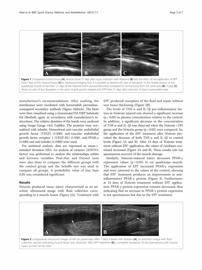
Figure 1 Comparison control tissue (A), muscle tissue 21 days after injury induction with Notexina (B) and the effect of the application of EPI®from7 days of the induced lesion (C) in ultrasound imaging (US). It is possible to observe the area of disruption in the middle portion of thequadriceps muscle of rats from 21 days of the induced lesion (surrounded area), compared to normal tissue from the same area (B). Image (C)shows an area of less disruption in the same muscle portion treated with EPI® from 21 days after induction of injury (surrounded area).
Abat et al. BMC Sports Science, Medicine, and Rehabilitation (2015) 7:7 Page 3 of 7
manufacturer’s recommendations. After washing, themembranes were incubated with horseradish peroxidase-conjugated secondary antibody (Sigma Aldrich). The blotswere then visualized using a InmunostarTM HRP SubstrateKit (BioRad), again, in accordance with manufacturer’s in-structions. The relative densities of the bands were analyzedusing Image Gauge v4.0, Fujifilm. The proteins were nor-malized with tubulin. Monoclonal anti-vascular endothelialgrowth factor (VEGF) (1:500), anti-vascular endothelialgrowth factor receptor 1 (VEGF-R1) (1:500), anti-PPAR-γ(1:500) and anti-tubulin (1:1000) were used.For statistical analysis, data are expressed as mean ±
standard deviation (SD). An analysis of variance (ANOVAfactor) was performed to analyze the relationships withinand between variables. Post-Hoc and Dunnet testswere also done to compare the different groups withthe control group and the Scheffe test was used tocompare all groups. A probability value of less than0.05 was considered significant.
ResultsNotexin produced tissue injury characterized as an an-echoic ultrasound image with fluid collection corre-sponding to a muscle lesion (Figure 2A). Treatment with
Figure 2 Longitudinal ultrasound images of left rat quadriceps. After 7collection (arrow) indicating muscle lesion was observed. After EPI® trearepair (arrow) can be seen.
EPI® produced resorption of the fluid and repair withoutscar tissue thickening (Figure 2B).The levels of TNF-α and IL-1β pro-inflammatory fac-
tors in Notexin injured rats showed a significant increase(p < 0.05) in plasma concentration relative to the control.In addition, a significant decrease in the concentrationof TNF-α and IL-1β was observed when the Notexin + EPIgroup and the Notexin group (p < 0.05) were compared. So,the application of the EPI® treatment after Notexin pro-voked the decrease of both TNF-α and IL-1β to controllevels (Figure 3A and B). After 14 days of Notexin treat-ment without EPI® application, the values of cytokines con-tinued increased (Figure 3A and B). These results rule outspontaneous recovery of the muscle damage.Similarly, Notexin-induced injury decreases PPAR-γ
expression values (p < 0.05) in rat quadriceps muscle.The application of EPI® increased PPAR-γ expressionand were returned to the values of the control, showingthat EPI® treatment produces an improvement in anti-inflammatory PPAR-γ protein (Figure 4). Furthermore,at 14 days of Notexin treatment without EPI® applica-tion, PPAR-γ protein expression remains decreased, thusindicating that an increase in PPAR-γ protein expressionis not spontaneous but due to the EPI® treatment.
days treated with Notexin (A), an anechoic image with fluidtment (B) a complete resorption of the haematoma with muscle

Figure 3 Plasma levels of IL-1β (A) and TNF-α (B) in control (C), Notexin (N7d, N14 d) and Notexin + EPI (N + E) groups. Values were measured byELISA assay as indicated in methods. Data are mean ± SD of six independent experiments. *p < 0.05 vs control group; # p < 0.05 vs bothNotexin groups.
Abat et al. BMC Sports Science, Medicine, and Rehabilitation (2015) 7:7 Page 4 of 7
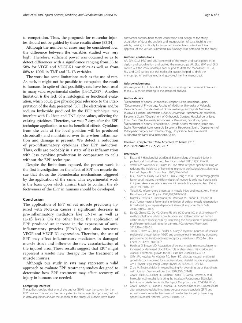
Notexin (7 and 14 days) treatment produced an in-crease in both VEGF and VEGF-R1 protein expressioncompared with the control (p < 0.05). Furthermore, EPI®treatment significantly potentiated the increase in VEGFand VEGF-R1 protein expression induced by Notexin(Figure 5).No adverse events were presented during the study.
DiscussionThe main findings of this study is that EPI® applied afterNotexin-induced muscle injury in rats decreases theproduction of the inflammatory mediators TNF-α and IL-1β, increases the protein expression of anti-inflammatoryfactor PPAR-γ and the angiogenic involved proteins VEGFand VEGF-R1.An increase in the TNF-α plasma levels was described
in the first days of tissular injury [16,17] and remainedelevated due to its action on cellular necrosis [18]. TNF-α disrupts the differentiation process and can promotecell catabolism thereby accelerating protein degradation[5]. Furthermore, TNF-α inhibits myogenesis throughredox-dependent and independent pathways [19]. One
Figure 4 PPAR-γ protein expression (relative densitometric units) in controwere determined in left rat quadriceps muscles by Western blot. A represeof protein. Data are mean ± SD of six independent experiments. *p < 0.05 v
potential mechanism by which TNF-α might directlystimulate catabolism is by inhibiting myoblast differenti-ation, an action that might limit the regenerative re-sponse of satellite cells to muscle injury [5]. A secondmechanism, apoptosis, appears less important. The thirdmechanism consists in a direct catabolic effect onmuscle tissue. In a muscular cell culture, TNF-α directlydecreases total muscle protein and the loss of muscle-specific proteins, including adult fast-type myosin heavychain [5,19].Our data shows an increase in the plasma level of
TNF-α due to Notexin-induced injury. EPI® treatmentnormalized the levels of TNF-α to reach control groupvalues. By contrast, in the group of rats without EPI®treatment, the TNF-α levels remained elevated with re-spect to the control group at 14 days after application.TNF-α action is also sensitive to other ligand/receptor
interactions (e.g. interleukin-1 and interleukin-6). Notexincaused a significant increase of IL-1β compared to the con-trol group. The maintenance of IL-1β over time has beenassociated with its condition as a pro-inflammatory cyto-kine more than for its action on tissue necrosis [16].
l (C), Notexin (N7d, N14 d) and Notexin + EPI (N + E) groups. Valuesntative inmunoblot is shown and tubulin was used as control amounts control group; # p < 0.05 vs both Notexin groups.
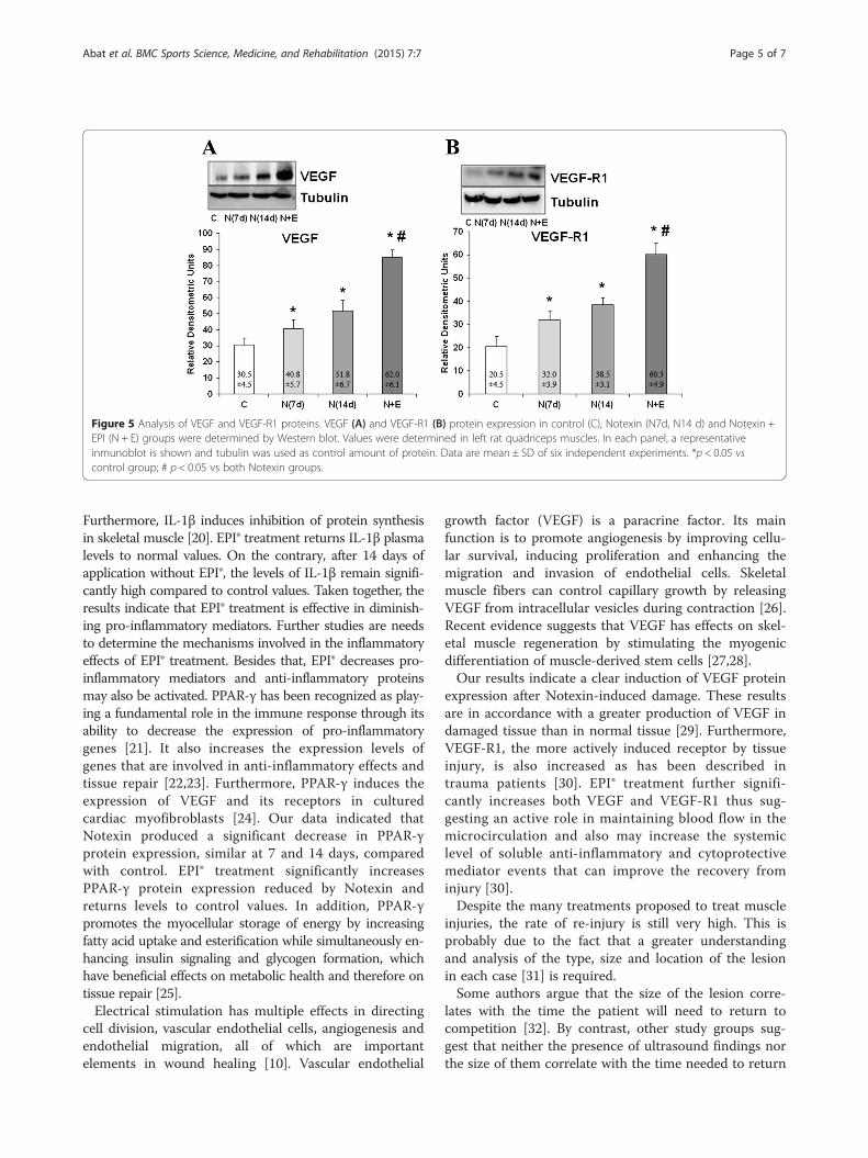
Figure 5 Analysis of VEGF and VEGF-R1 proteins. VEGF (A) and VEGF-R1 (B) protein expression in control (C), Notexin (N7d, N14 d) and Notexin +EPI (N + E) groups were determined by Western blot. Values were determined in left rat quadriceps muscles. In each panel, a representativeinmunoblot is shown and tubulin was used as control amount of protein. Data are mean ± SD of six independent experiments. *p < 0.05 vscontrol group; # p < 0.05 vs both Notexin groups.
Abat et al. BMC Sports Science, Medicine, and Rehabilitation (2015) 7:7 Page 5 of 7
Furthermore, IL-1β induces inhibition of protein synthesisin skeletal muscle [20]. EPI® treatment returns IL-1β plasmalevels to normal values. On the contrary, after 14 days ofapplication without EPI®, the levels of IL-1β remain signifi-cantly high compared to control values. Taken together, theresults indicate that EPI® treatment is effective in diminish-ing pro-inflammatory mediators. Further studies are needsto determine the mechanisms involved in the inflammatoryeffects of EPI® treatment. Besides that, EPI® decreases pro-inflammatory mediators and anti-inflammatory proteinsmay also be activated. PPAR-γ has been recognized as play-ing a fundamental role in the immune response through itsability to decrease the expression of pro-inflammatorygenes [21]. It also increases the expression levels ofgenes that are involved in anti-inflammatory effects andtissue repair [22,23]. Furthermore, PPAR-γ induces theexpression of VEGF and its receptors in culturedcardiac myofibroblasts [24]. Our data indicated thatNotexin produced a significant decrease in PPAR-γprotein expression, similar at 7 and 14 days, comparedwith control. EPI® treatment significantly increasesPPAR-γ protein expression reduced by Notexin andreturns levels to control values. In addition, PPAR-γpromotes the myocellular storage of energy by increasingfatty acid uptake and esterification while simultaneously en-hancing insulin signaling and glycogen formation, whichhave beneficial effects on metabolic health and therefore ontissue repair [25].Electrical stimulation has multiple effects in directing
cell division, vascular endothelial cells, angiogenesis andendothelial migration, all of which are importantelements in wound healing [10]. Vascular endothelial
growth factor (VEGF) is a paracrine factor. Its mainfunction is to promote angiogenesis by improving cellu-lar survival, inducing proliferation and enhancing themigration and invasion of endothelial cells. Skeletalmuscle fibers can control capillary growth by releasingVEGF from intracellular vesicles during contraction [26].Recent evidence suggests that VEGF has effects on skel-etal muscle regeneration by stimulating the myogenicdifferentiation of muscle-derived stem cells [27,28].Our results indicate a clear induction of VEGF protein
expression after Notexin-induced damage. These resultsare in accordance with a greater production of VEGF indamaged tissue than in normal tissue [29]. Furthermore,VEGF-R1, the more actively induced receptor by tissueinjury, is also increased as has been described intrauma patients [30]. EPI® treatment further signifi-cantly increases both VEGF and VEGF-R1 thus sug-gesting an active role in maintaining blood flow in themicrocirculation and also may increase the systemiclevel of soluble anti-inflammatory and cytoprotectivemediator events that can improve the recovery frominjury [30].Despite the many treatments proposed to treat muscle
injuries, the rate of re-injury is still very high. This isprobably due to the fact that a greater understandingand analysis of the type, size and location of the lesionin each case [31] is required.Some authors argue that the size of the lesion corre-
lates with the time the patient will need to return tocompetition [32]. By contrast, other study groups sug-gest that neither the presence of ultrasound findings northe size of them correlate with the time needed to return
Abat et al. BMC Sports Science, Medicine, and Rehabilitation (2015) 7:7 Page 6 of 7
to competition. Thus, the prognosis for muscular injur-ies should not be guided by these results alone [33,34].Although the number of cases may be considered low,
the difference between the variables studied was veryhigh. Therefore, sufficient power was obtained so as todetect differences with a significance ranging from 55 to58% for VEGF and VEGF-R1 variables as well as from88% to 100% in TNF and IL-1B variables.The work has some limitations such as the use of rats.
As such, it might not be possible to extrapolate the resultto humans. In spite of that possibility, rats have been usedin many valid experimental studies [14-17,20,27]. Anotherlimitation is the lack of a histological or functional evalu-ation, which could give physiological relevance to the inter-pretation of the data presented [35]. The electrolysis and/orsodium hydroxide produced by the EPI® technique mayinterfere with IL-1beta and TNF-alpha values, affecting theexisting cytokines. Therefore, we wait 7 days after the EPI®technique application to see its beneficial effects. Cytokinesfrom the cells at the local position will be producedchronically and maintained over time when inflamma-tion and damage is present. We detect a reductionof pro-inflammatory cytokines after EPI® induction.Thus, cells are probably in a state of less inflammationwith less cytokine production in comparison to cellswithout the EPI® technique.Despite the limitations exposed, the present work is
the first investigation on the effect of EPI® on muscle tis-sue that shows the biomolecular mechanisms triggeredby the application of the same. This experimental workis the basis upon which clinical trials to confirm the ef-fectiveness of the EPI® in humans should be developed.
ConclusionThe application of EPI® on rat muscle previously in-jured with Notexin causes a significant decrease inpro-inflammatory mediators like TNF-α as well asIL-1β levels. On the other hand, the application ofEPI® produced an increase in the expression of anti-inflammatory proteins (PPAR-γ) and also increasesVEGF and VEGF-R1 expression. Therefore, the use ofEPI® may affect inflammatory mediators in damagedmuscle tissue and influence the new vascularization ofthe injured area. These results suggest that EPI® mightrepresent a useful new therapy for the treatment ofmuscle injuries.Although our study in rats may represent a valid
approach to evaluate EPI® treatment, studies designed todetermine how EPI® treatment may affect recovery ofinjury in humans are needed.
Competing interestsThe authors declare that one of the author (SIJM) have the patent for theEPI® devices. This author has participated in the intervention process, but notin data acquisition and/or the analysis of this study. All authors have made
substantial contributions to the conception and design of the study,acquisition of data, the analysis and interpretation of data, drafting thearticle, revising it critically for important intellectual content and finalapproval of the version submitted. No fundings was obtained for this study.
Authors’ contributionsAF, SLV, SIJM, PEG and MJC conceived of the study, and participated in itsdesign and coordination and drafted the manuscript. AF, SLV, SIJM and GHScarried out the immunoassays and helped to draft the manuscript. PF, JA,SLV and GHS carried out the molecular studies helped to draft themanuscript. All authors read and approved the final manuscript.
AcknowledgementsWe are grateful to E. Goode for his help in editing the manuscript. We alsothank G. Gich for assisting in the statistical analysis.
Author details1Department of Sports Orthopedics, ReSport Clinic, Barcelona, Spain.2Department of Physiology, Faculty of Medicine, University of Valencia,Valencia, Spain. 3Catalan Institut of Traumatology and Sports Medicine(ICATME), Hospital Universitari Dexeus, Universitat Autónoma de Barcelona,Barcelona, Spain. 4Department of Orthopedic Surgery, Hospital de la SantaCreu i Sant Pau, University Autonoma of Barcelona, Barcelona, Spain.5Department of Sports Rehabilitation, Cerede Sports Medicine, Barcelona,Spain. 6Universitat Autónoma de Barcelona, Barcelona, Spain. 7Department ofOrthopedic Surgery and Traumatology, Hospital del Mar, UniversitatAutónoma de Barcelona, Barcelona, Spain.
Received: 2 September 2014 Accepted: 26 March 2015
References1. Ekstrand J, Hägglund M, Waldén M. Epidemiology of muscle injuries in
professional football (soccer). Am J Sports Med. 2011;39(6):1226–32.2. Verrall GM, Slavotinek JP, Barnes PG. The effect of sports specific training on
reducing the incidence of hamstring injuries in professional Australian rulesfootball players. Br J Sports Med. 2005;39(6):363–8.
3. Li Y, Foster W, Deasy BM, Chan Y, Prisk V, Tang Y, et al. Transforming growthfactor-beta1 induces the differentiation of myogenic cells into fibrotic cellsin injured skeletal muscle: a key event in muscle fibrogenesis. Am J Pathol.2004;164(3):1007–19.
4. Tidball JG. Inflammatory processes in muscle injury and repair. Am J PhysiolRegul Integr Comp Physiol. 2005;288(2):R345–53.
5. Moresi V, Pristerà A, Scicchitano BM, Molinaro M, Teodori L, Sassoon D,et al. Tumor necrosis factor-alpha inhibition of skeletal muscle regenerationis mediated by a caspase-dependent stem cell response. Stem Cells.2008;26(4):997–1008.
6. Liu CS, Chang CC, Du YC, Chang FR, Wu YC, Chang WC, et al. 2-hydroxy-4′-methoxychalcone inhibits proliferation and inflammation of humanaortic smooth muscle cells by increasing the expression of peroxisomeproliferator-activated receptor gamma. J Cardiovasc Pharmacol.2012;59(4):339–51.
7. Thom R, Rowe GC, Jang C, Safdar A, Arany Z. Hypoxic induction of vascularendothelial growth factor (VEGF) and angiogenesis in muscle by truncatedperoxisome proliferator-activated receptor γ coactivator (PGC)-1α. J BiolChem. 2014;289(13):8810–7.
8. Hudlicka O, Brown MD. Adaptation of skeletal muscle microvasculature toincreased or decreased blood flow: role of shear stress, nitric oxide andvascular endothelial growth factor. J Vasc Res. 2009;46(5):504–12.
9. Olfert IM, Howlett RA, Wagner PD, Breen EC. Myocyte vascular endotelialgrowth factor is required for exercise-induced skeletal muscle angiogenesis.Am J Physiol Regul Integr Comp Physiol. 2010;299(4):R1059–67.
10. Zhao M. Electrical fields in wound healing-An overriding signal that directscell migration. Semin Cell Dev Biol. 2009;20(6):674–82.
11. Abat F, Valles SL, Gelber PE, Polidori F, Stitik TP, García-Herreros S, et al.Molecular repair mechanisms using the Intratissue Percutaneous Electrolysistechnique in patellar tendonitis. Rev Esp Cir Ortop Traumatol. 2014;58(4):201–5.
12. Abat F, Gelber PE, Polidori F, Monllau JC, Sanchez-Ibañez JM. Clinical resultsafter ultrasound-guided intratissue percutaneous electrolysis (EPI®) andeccentric exercise in the treatment of patellar tendinopathy. Knee SurgSports Traumatol Arthrosc. 2014;23(4):1046–52.
Abat et al. BMC Sports Science, Medicine, and Rehabilitation (2015) 7:7 Page 7 of 7
13. Abat F, Diesel WJ, Gelber PE, Polidori F, Monllau JC, Sanchez-Ibañez JM.Effectiveness of the Intratissue Percutaneous Electrolysis (EPI®) techniqueand isoinertial eccentric exercise in the treatment of patellar tendinopathyat two years follow-up. Muscles Ligaments Tendons J. 2014;4(2):188–93.
14. Head SI, Houweling PJ, Chan S, Chen G, Hardeman EC. Properties ofregenerated mouse extensor digitorum longus muscle following notexininjury. Exp Physiol. 2014;99(4):664–74.
15. Joensen J, Gjerdet NR, Hummelsund S, Iversen V, Lopes-Martins RA, BjordalJM. An experimental study of low-level laser therapy in rat Achilles tendoninjury. Lasers Med Sci. 2012;27(1):103–11.
16. Meador BM, Krzyszton CP, Johnson RW, Huey KA. Effects of IL-10 and ageon IL-6, IL-1beta, and TNF-alpha responses in mouse skeletal and cardiacmuscle to an acute inflammatory insult. J Appl Physiol. 2008;104(4):991–7.
17. Crassous B, Richard-Bulteau H, Deldicque L, Serrurier B, Pasdeloup M,Francaux M, et al. Lack of effects of creatine on the regeneration of soleusmuscle after injury in rats. Med Sci Sports Exerc. 2009;41(9):1761–9.
18. Bhatnagar S, Panguluri SK, Gupta SK, Dahiya S, Lundy RF, Kumar A. Tumornecrosis factor-α regulates distinct molecular pathways and gene networksin cultured skeletal muscle cells. PLoS One. 2010;12;5(10):e13262.
19. Langen RC, Schols AM, Kelders MC, Van Der Velden JL, Wouters EF,Janssen-Heininger YM. Tumor necrosis factor-alpha inhibits myogenesisthrough redox-dependent and -independent pathways. Am J Physiol CellPhysiol. 2002;283(3):C714–21.
20. Borghi SM, Zarpelon AC, Pinho-Ribeiro FA, Cardoso RD, Cunha TM,Alves-Filho JC, et al. Targeting interleukin-1β reduces intense acuteswimming-induced muscle mechanical hyperalgesia in mice. J PharmPharmacol. 2014;66(7):1009–20.
21. Bertin B, Dubuquoy L, Colombel JF, Desreumaux P. PPAR-gamma inulcerative colitis: a novel target for intervention. Curr Drug Targets.2013;14(12):1501–7.
22. von Knethen A, Neb H, Morbitzer V, Schmidt MV, Kuhn AM, Kuchler L, et al.PPARγ stabilizes HO-1 mRNA in monocytes/macrophages which affectsIFN-β expression. Free Radic Biol Med. 2011;51(2):396–405.
23. Lea S, Plumb J, Metcalfe H, Spicer D, Woodman P, Fox JC, et al. The effectof peroxisome proliferator-activated receptor-γ ligands on in vitro andin vivo models of COPD. Eur Respir J. 2014;43(2):409–20.
24. Chintalgattu V, Harris GS, Akula SM, Katwa LC. PPAR-gamma agonists inducethe expression of VEGF and its receptors in cultured cardiac myofibroblasts.Cardiovasc Res. 2007;74(1):140–50.
25. Hu S, Yao J, Howe AA, Menke BM, Sivitz WI, Spector AA, et al. Peroxisomeproliferator-activated receptor γ decouples fatty acid uptake from lipid inhibitionof insulin signaling in skeletal muscle. Mol Endocrinol. 2012;26(6):977–88.
26. Hoier B, Prats C, Qvortrup K, Pilegaard H, Bangsbo J, Hellsten Y. Subcellularlocalization and mechanism of secretion of vascular endothelial growthfactor in human skeletal muscle. FASEB J. 2013;27(9):3496–504.
27. Bouchentouf M, Benabdallah BF, Bigey P, Yau TM, Scherman D, Tremblay JP.Vascular endothelial growth factor reduced hypoxia-induced death ofhuman myoblasts and improved their engraftment in mouse muscles.Gene Ther. 2008;15(6):404–14.
28. Beckman SA, Chen WC, Tang Y, Proto JD, Mlakar L, Wang B, et al. Beneficialeffect of mechanical stimulation on the regenerative potential of muscle-derivedstem cells is lost by inhibiting vascular endothelial growth factor. ArteriosclerThromb Vasc Biol. 2013;33(8):2004–12.
29. Rignault-Clerc S, Bielmann C, Delodder F, Raffoul W, Waeber B, Liaudet L,et al. Functional late outgrowth endotelial progenitors isolated fromperipheral blood of burned patients. Burns. 2013;39(4):694–704.
30. Ostrowski SR, Sørensen AM, Windeløv NA, Perner A, Welling KL, WanscherM, et al. High levels of soluble VEGF receptor 1 early after trauma areassociated with shock, sympathoadrenal activation, glycocalyx degradationand inflammation in severely injured patients: a prospective study. Scand JTrauma Resusc Emerg Med. 2012;10:20–7.
31. Askling CM, Tengvar M, Tarassova O, Thorstensson A. Acute hamstringinjuries in Swedish elite sprinters and jumpers: a prospective randomisedcontrolled clinical trial comparing two rehabilitation protocols. Br J SportsMed. 2014;48(7):532–9.
32. Connell DA, Schneider-Kolsky ME, Hoving JL, Malara F, Buchbinder R,Koulouris G, et al. Longitudinal study comparing sonographic and MRIassessments of acute and healing hamstring injuries. AJR Am J Roentgenol.2004;183(4):975–84.
33. Petersen J, Thorborg K, Nielsen MB, Skjødt T, Bolvig L, Bang N, et al. Thediagnostic and prognostic value of ultrasonography in soccer players withacute hamstring injuries. Am J Sports Med. 2014;42(2):399–404.
34. Prior M, Guerin M, Grimmer K. An evidence-based approach tohamstring strain injury: a systematic review of the literature. SportsHealth. 2009;1(2):154–64.
35. Delos D, Leineweber MJ, Chaudhury S, Alzoobaee S, Gao Y, Rodeo SA. Theeffect of platelet-rich plasma on muscle contusion healing in a rat model.Am J Sports Med. 2014;42(9):2067–74.
Submit your next manuscript to BioMed Centraland take full advantage of:
• Convenient online submission
• Thorough peer review
• No space constraints or color figure charges
• Immediate publication on acceptance
• Inclusion in PubMed, CAS, Scopus and Google Scholar
• Research which is freely available for redistribution
Submit your manuscript at www.biomedcentral.com/submit