an α-helical minimal binding domain within the h3 domain of syntaxin is required for snap-25...
TRANSCRIPT
An R-Helical Minimal Binding Domain within the H3 Domain of Syntaxin IsRequired for SNAP-25 Binding†
Pingyu Zhong,‡ Yu A. Chen,§ Deborah Tam,‡ David Chung,‡ Richard H. Scheller,§ and George P. Miljanich*,‡
Neurex Corporation, 3760 HaVen AVenue, Menlo Park, California 94025, and Department of Molecular and CellularPhysiology, Howard Hughes Medical Institute, Stanford UniVersity, Stanford, California 94305
ReceiVed October 9, 1996; ReVised Manuscript ReceiVed January 30, 1997X
ABSTRACT: The interaction between the proteins syntaxin 1A and SNAP-25 is a key step in synapticvesicle docking and fusion. To define the SNAP-25 binding domain on syntaxin, we have preparedpeptides that span the syntaxin H3 domain (residues 191-266), the region previously shown to be importantfor binding to SNAP-25, and then determined the affinities of these peptides for binding to SNAP-25. Aminimal binding domain was identified within a region of 32 amino acids (residues 189-220). Its affinityfor SNAP-25 is substantially enhanced by C-terminal extension (residues 221-266). Circular dichroismrevealed the presence of substantialR-helicity in the H3 domain and in the 32-mer minimal bindingdomain, but not in H3 peptides that do not bind to SNAP-25. At temperatures that denature theR-helixof the minimal binding domain peptide, SNAP-25 binding is lost. Selected mutations in evolutionarilyconserved residues of the amphiphilicR-helix within the minimal binding domain (e.g., residues 205 and209) greatly reduce the affinity for SNAP-25 but have no major effect on secondary structure, suggestingthat these residues may interact directly with SNAP-25. The H3 domain peptide and the minimal bindingdomain peptide inhibit norepinephrine release from PC12 cells. These results suggest that specific aminoacid residues in the H3 domain, positioned by the underlyingR-helical structure, are important for itsbinding to SNAP-25 and support the notion that this interaction is important for presynaptic vesicularexocytosis.
Neurons communicate with each other by secreting neu-rotransmitters from membranous vesicles in presynapticnerve terminals. The molecular mechanisms by whichsynaptic vesicles are directed to release sites and undergoexocytosis upon calcium entry have been extensively studiedover the past few years, leading to the identification of manyproteins that play critical roles in this process. The emergingtheme from these studies is that vesicular docking and fusionare mediated through a series of dynamic protein-proteininteractions (So¨llner et al., 1993a,b; Scheller, 1995) thatprovide specificity for vesicle targeting and the machineryfor docking and membrane fusion (Bennett & Scheller, 1994;Jahn & Su¨dhof, 1994). As proposed by a current model forvesicle docking and fusion, a 7S complex composed of twosynaptic vesicle proteins (vSNAREs),1 VAMP (also knownas synaptobrevin) and synaptotagmin, and two plasmamembrane proteins (tSNAREs), SNAP-25 and syntaxin, isassembled. The binding between vSNAREs and tSNAREsbrings the vesicle within close proximity of the release sitesat the plasma membrane and thus serves as the molecular
docking mechanism. Subsequent binding of the cytosolicproteins, RSNAP and NSF, concurrent with a loss ofsynaptotagmin, results in the formation of a 20S complexcomposed of syntaxin, SNAP-25, VAMP,RSNAP, and NSF(Sollner et al., 1993a,b). Hydrolysis of ATP by NSFactivates the complex, leading to membrane fusion throughunknown intermediates (Morgan et al., 1994). Thus, theinteraction between SNAP-25 and syntaxin appears to be ofcritical importance in the vesicle docking and fusion process.This conclusion is supported byin Vitro binding studies.SNAP-25 binds to syntaxin with moderate affinityin Vitro(Pevsner et al., 1994). Similarly, binding of VAMPII tosyntaxin is substantially stabilized by the presence of SNAP-25, leading to a nearly 10-fold increase in affinity (Pevsneret al., 1994). Interestingly, this potentiation is syntaxinisoform-specific; VAMPII binding to syntaxin 1A, the majorneuronal isoform of syntaxin, can be enhanced in dramaticfashion compared to that of other syntaxin isoforms (Pevsneret al., 1994), consistent with the expression of SNAP-25predominantly in neurons (Bark & Wilson, 1993; Bark etal., 1995). It is, therefore, reasonable to postulate that thesyntaxin-SNAP-25 interaction is crucial for promoting thedocking of VAMP-containing vesicles at plasmalemmalrelease sites with high affinity and high specificity.
The SNAP-25 binding region on syntaxin is located withinthe H3 domain (amino acids 191-266), which is next tothe C-terminal membrane anchor (Figure 1). The H3 domainmediates not only SNAP-25 binding but also the binding ofsyntaxin to VAMP,RSNAP, synaptotagmin, and, in part,n-sec1. Within the H3 domain, the N-terminal portion(amino acids∼191-221) is likely to contain the SNAP-25binding site (Chapman et al., 1994; Kee et al., 1995).
† Y.A.C. was supported by a predoctoral fellowship from the HowardHughes Medical Institute.* Correspondence to Dr. George P. Miljanich, Neurex Corp., 3760
Haven Ave., Menlo Park, CA 94025. Telephone: 415-833-1218.Fax: 415-853-1538.
‡ Neurex Corp.§ Stanford University.X Abstract published inAdVance ACS Abstracts,March 15, 1997.1 Abbreviations: CD, circular dichroism; GST, glutathioneS-
transferase; HPLC, high-performance liquid chromatography; PAGE,polyacrylamide gel electrophoresis; NE, norepinephrine; NSF,N-ethylmaleimide-sensitive fusion factor; SNAP, soluble NSF attachmentprotein; SNARE, SNAP receptor; vSNARE, vesicle SNARE; tSNARE,target SNARE; SNAP-25, synaptosomal-associated protein of 25 kDa;VAMP, vesicle-associated membrane protein.
4317Biochemistry1997,36, 4317-4326
S0006-2960(96)02540-8 CCC: $14.00 © 1997 American Chemical Society

Sequence analysis of the H3 domain predicts a strongpropensity forR-helical formation. Further analysis revealsa repeating pattern of hydrophobic amino acids within thehelical heptad repeats characteristic of amphiphilicR-helices(Inoue et al., 1992; Spring et al., 1993). That is, within eachset of seven amino acids, (abcdefg)n, positionsa andd areregularly occupied by hydrophobic residues. These aminoacids commonly form an interface for protein-proteininteractions through coiled coils. Consistent with theirfunctional importance, positionsa andd constitute the mostevolutionarily conserved sites in the H3 domain. Althoughdirect evidence for the existence of amphiphilicR-helicityin the H3 domain and its relationship to SNAP-25 bindinghas not been previously reported, one expects that alteringthe specific sequence in the H3 domain responsible forSNAP-25 binding or disrupting theR-helical structurepresumed to be required for this binding would interfere withsyntaxin-SNAP-25 interaction. Kee et al. (1995) demon-strated that a syntaxin 1A mutant containing point mutationsat amino acid residues 198, 202, 205, and 209, which are ata and d positions in the predicted heptad repeats, hassignificantly reduced SNAP-25 binding activity. However,protein-protein interactions in this and other studies wereassayed by SDS-PAGE and Western blot, which provideonly a qualitative measure of affinity. Furthermore, it is notknown whether the loss of SNAP-25 binding activity of thesesyntaxin mutants is in fact due to the alteration of the primarysequence alone or to disruption of the putative underlyinghelical structure.In the present study, we focus on further defining the
molecular features of syntaxin 1A binding to SNAP-25. Afamily of peptides spanning different sequences of the H3domain of syntaxin 1A was prepared through chemicalsynthesis or recombinant expression and partial proteasedigestion. The affinities of these peptides for SNAP-25 weredetermined in a quantitativein Vitro binding assay. On thebasis of this analysis, a putative minimal SNAP-25 bindingdomain spanning amino acids 189-220 was identified. Itwas also found that a supporting domain located in the regionC-terminal to the minimal binding domain significantlyenhances the affinity of syntaxin for SNAP-25 but, by itself,does not bind to SNAP-25. To investigate the secondarystructural requirements for the binding, we studied thesolution structure of the SNAP-25 binding domain ofsyntaxin with circular dichroism (CD) spectroscopy. Thesemeasurements revealed that substantial amphiphilicR-helicalstructure is present in the H3 domain and in the minimalbinding domain and strongly suggested that the helicalstructure is necessary for syntaxin-SNAP-25 binding. Inaddition, selected point mutations in evolutionarily conservedresidues of the putative amphiphilicR-helix within the
minimal binding domain greatly reduce the affinity forSNAP-25 without affecting the secondary structure. Finally,the affinities of the H3 domain peptides for SNAP-25 arecorrelated with their potencies for inhibiting norepinephrinesecretion from PC12 cells. These results support the notionthat the interaction between syntaxin and SNAP-25 isrequired for presynaptic vesicle exocytosis.
EXPERIMENTAL PROCEDURE
Recombinant Protein Preparation.GST (glutathioneS-transferase) fusion protein constructs of syntaxin 1A11(residues 4-266), SP191-266 (residues 191-266), SP220-266 (residues 220-266), and syntaxin 1A11 point mutants(m1-m6, m12, m13, m34, and m35) were subcloned in theexpression vector pGEX-KG. Syntaxin 1A11 is derivedfrom syntaxin 1A and lacks the first three N-terminal aminoacids and the C-terminal membrane anchor. Previous studieshave shown that syntaxin 1A11 contains sufficient sequencefor binding to a number of exocytotic proteins, includingSNAP-25 (Pevsner et al., 1994; Chapman et al., 1994;Calakos et al., 1994; Hayashi et al., 1994; Kee et al., 1995;Kee & Scheller, 1996). Positions of the point mutations areas follows: m1 (R198A and I202A, i.e., Arg198 and Ile202were mutated to Ala), m2 (L205A and I209A), m3 (L212Aand F216A), m4 (V223A and Q226A), m5 (I230A andI233A), m6 (A240V and V244A), m12 (R198A, I202A,L205A, and I209A), m13 (R198A, I202A, L212A, andF216A), m34 (L212A, F216A, V223A, and Q226A), andm35 (V223A, Q226A, I230A, and I233A). Purification wasperformed by binding the fusion proteins to a glutathione-agarose column followed by thrombin cleavage as previouslydescribed (Pevsner et al., 1994; Kee et al., 1995). Forcompetition binding assays, protein concentrations weredetermined by SDS-PAGE and scanning densitometry. Forcircular dichroism (CD) spectroscopy, and PC12 cell secre-tion assays, proteins from the glutathione-agarose columnand thrombin cleavage were further purified by reverse phaseHPLC. Protein concentrations were determined by aminoacid analysis.Preparation of Peptide Fragments of the H3 Domain.
Peptide sequences and preparative sources are listed in Table1. Synthetic peptides SP189-220, SP204-220, SP186-211, SP209-240, and SP238-266 were synthesized on thebasis of the sequence of the H3 domain of syntaxin 1A. P1,whose sequence is unrelated to the H3 domain, was used asa control peptide. Solid phase synthesis of the peptides wasperformed on an Applied Biosystems 430A peptide synthe-sizer, starting fromp-methylbenzhydrylamine resin. Peptideswere cleaved from the resin with hydrogen fluoride andpurified by reverse phase HPLC. Peptide concentrationswere determined by amino acid analysis. For peptidefragments from partial protease digestion, a panel of peptidesof different sizes was prepared by partial digestion (chy-motrypsin, 100:1 protein:enzyme by weight, 37°C for 20min) of recombinant SP191-266, or of synthetic peptidesdescribed above, followed by fractionation by reverse phaseHPLC. The purity of each peptide preparation was con-firmed by analytical HPLC. The concentration of eachpurified peptide was determined by amino acid analysis.In Vitro Transcription and in Vitro Translation.A 0.6
kbBamHI fragment of rat SNAP-25 cDNA was ligated intopBluescript KSII- to generate RNA using T7 polymerase.
FIGURE 1: Functional domains of syntaxin 1A. A schematicrepresentation of various functional domains of syntaxin 1A isillustrated. The three regions, H1-H3, are predicted to be amphi-philic R-helices. Within the H3 domain, a minimal binding domain(MBD) for SNAP-25 and a supporting domain (SD) are locatedN-terminally and C-terminally, respectively. Leucine 205 andisoleucine 209 are likely to be key residues for the binding ofSNAP-25. The last 20 amino acid residues of syntaxin serve as amembrane anchor (MA).
4318 Biochemistry, Vol. 36, No. 14, 1997 Zhong et al.
The RNA template was used to performin Vitro translationin the presence of [35S]methionine. The specific activity ofthe 35S-labeled SNAP-25 was about 14 000 Ci/mmol.Scintillation Proximity Binding Assay.A simple microtiter
plate binding assay employing scintillation proximity assaymethodology was developed to study synaptic vesicleprotein-protein interactions. Syntaxin GST fusion proteindiluted in 50 mM Tris at pH 8.0 was first coated onto wellsof a Scintistrip, a microtiter plate in which scintillant iscovalently incorporated, by incubation overnight at 4°C. Theremaining nonspecific binding sites were blocked by incuba-tion in 0.01% BSA for 1 h at 4°C. [35S]Methionine-labeledSNAP-25 generated throughin Vitro translation was dilutedin binding buffer [20 mM Hepes (pH 7.4), 120 mMpotassium acetate, 2 mM EDTA, and 0.05% Tween 20], andaliquots of the diluted labeled SNAP-25 were added into theprecoated and preblocked wells to reach a final concentrationof approximately 0.03 nM. The binding incubation wasperformed in the presence of a soluble test competitor atroom temperature for 1.5 h, except where indicated other-wise. Finally, the microtiter plate was washed in cold buffer(0.05% Tween 20 replaced by 5% glycerol in the bindingbuffer) to remove the soluble ligand and free [35S]methionine.After air-drying, bound [35S]SNAP-25 was determined using
a 96-well liquid scintillation counter (Wallac Microbeta). Forall experiments, the total bound counts per minute in theabsence of competing ligand was approximately 5% of thetotal specific counts per minute loaded. Data were expressedas a percentage of maximal binding determined in theabsence of competitor.CD Spectroscopy.CD spectra were obtained at 0°C using
an Aviv 60 DS spectropolarimeter. Samples were dissolvedto a concentration of approximately 0.03-0.06 mM in 1 mMpotassium phosphate at pH 7.0 and 100 mM potassiumfluoride for spectroscopic studies or 1 M NaCl and 1 mMsodium borate, sodium phosphate, and sodium citrate at pH7.0 for thermal denaturation studies. Mean residue ellipticityis defined as the ellipticity normalized by the molarconcentration of amino acid residues of a tested peptide. Totalellipticity is defined as the ellipticity normalized by the molarconcentration of a tested peptide. Thermal denaturation wasstudied at 222 nm with step increases (2-5 °C) in temper-ature. Melting point (Tm) is defined as the temperature atwhich 50% of theR-helical structure is denatured; i.e., meanresidue ellipticity at 222 nm decreases to 50% of the maximalvalue obtained at 0°C.[ 3H]Norepinephrine (NE) Secretion Assay in PC12 Cells.
Procedures to prepare permeabilized PC12 cells by “cell
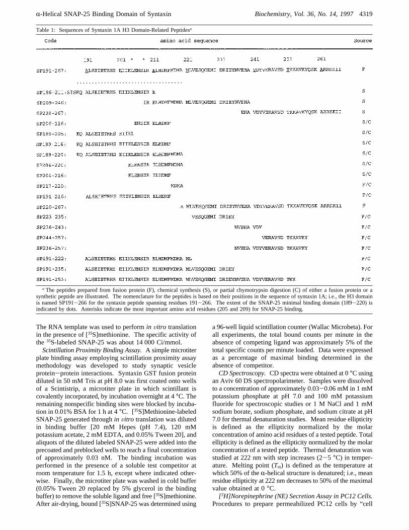
Table 1: Sequences of Syntaxin 1A H3 Domain-Related Peptidesa
a The peptides prepared from fusion protein (F), chemical synthesis (S), or partial chymotrypsin digestion (C) of either a fusion protein or asynthetic peptide are illustrated. The nomenclature for the peptides is based on their positions in the sequence of syntaxin 1A; i.e., the H3 domainis named SP191-266 for the syntaxin peptide spanning residues 191-266. The extent of the SNAP-25 minimal binding domain (189-220) isindicated by dots. Asterisks indicate the most important amino acid residues (205 and 209) for SNAP-25 binding.
R-Helical SNAP-25 Binding Domain of Syntaxin Biochemistry, Vol. 36, No. 14, 19974319
cracking” are described in detail elsewhere (Hay & Martin,1992). In brief, PC12 cells labeled with [3H]NE werepermeabilized by a single passage through a stainless steelball homogenizer. One to two million cracked cell ghostswere then added to KGlu buffer [20 mM Hepes (pH 7.2),120 mM potassium glutamate, 20 mM potassium acetate, 2mM EGTA, and 0.1% BSA] containing 2 mM ATP, rat braincytosol (∼1 mg/mL final protein concentration), 1µM freecalcium, and a test compound as indicated, for 1 h ofpreincubation on ice. NE release was triggered by raisingthe temperature to 22 or 35°C. Secretion was terminatedafter 15 min by chilling on ice followed by centrifugation at2000g for 40 min. The released [3H]NE in the supernatantwas quantified by scintillation counting. Pellets weresolubilized in 1% Triton X-100 and similarly counted.[3H]NE release is expressed as a percentage of the totalcellular [3H]NE: [100([3H] in supernatant)]/([3H] in super-natant+ [3H] in pellet).Materials. [35S]Methionine and [3H]NE were purchased
from Amersham. Kits forin Vitro translation andin Vitrotranscription were purchased from Promega (San Diego, CA).Glutathione-agarose beads, glutathione (reduced form),human plasma thrombin, and chymotrypsin were purchasedfrom Sigma Chemicals (St. Louis, Mo). Scintistrip microtiterplates were purchased from Wallac Inc. (Turko, Finland).
RESULTS
Minimal SNAP-25 Binding Domain of Syntaxin 1A.Thesyntaxin-SNAP-25 binding assay relies on the ability ofradioactive label bound to the well wall of a microtiter plateimpregnated with scintillant to produce photons; unboundradiolabeled ligand will not. In the present study,35S-radiolabeled SNAP-25 is brought into proximity of thescintillant by specific binding to the GST-syntaxin 1A11fusion protein that is preimmobilized on the well wall.The specificity of binding of SNAP-25 to syntaxin in this
assay was evaluated in two ways. In the first, the bindingassay was performed in the presence of increasing concentra-tions of soluble SNAP-25. The IC50 for competition by thesoluble SNAP-25 was found to be 0.45( 0.03µM (mean( SE). This value is in good agreement with the previouslyreportedKd value for SNAP-25 binding to syntaxin 1A (0.4µM; Pevsner et al., 1994). In the second, the binding assaywas performed in the presence of increasing concentrationsof either soluble syntaxin 1A11 or SP191-266 (the intactH3 domain, Figure 1 and Table 1). These proteins bind to[35S]SNAP-25 to form soluble complexes which are re-moved during the wash following incubation. Thus, thepresence of soluble syntaxin peptide reduces the amount of[35S]SNAP-25 binding to the syntaxin 1A11 fusion proteinimmobilized on the well wall in a concentration-dependentmanner. The IC50 for the competition by syntaxin or syntaxinpeptide can be used as a relative measure of its affinity forSNAP-25 (Table 2). The level of nonspecific binding,determined by the residual counts per minute in the presenceof saturating concentrations of SNAP-25, syntaxin 1A11, orSP191-266, accounted for approximately 15% of the totalbinding observed in the absence of competitor.Semiquantitative studies previously showed that a C-
terminally truncated syntaxin 1A that contains amino acids4-221 binds to SNAP-25, whereas another truncated syn-taxin 1A that is only 31 residues shorter on the C-terminal
side does not (Kee et al., 1995). Moreover, the H3 domainof syntaxin 1A (amino acids 191-266) was shown to haveSNAP-25 binding activity (Kee et al., 1995). On the basisof these results, it was postulated that a putative bindingdomain for SNAP-25 resides approximately within the regionfrom amino acids 191 to 221 of syntaxin. However, morequantitative studies of the syntaxin-SNAP-25 interaction arerequired to define this binding domain at a higher resolution.Furthermore, it is not clear whether this region by itself issufficient for SNAP-25 binding, whether a smaller fragmentwithin this region is sufficient, or what contribution theadditional C-terminal region of the H3 domain makes tosyntaxin-SNAP-25 interactions. Thus, a number of syn-thetic peptides and chymotryptic fragments of the H3 domainwere generated and tested for their affinities for [35S]SNAP-25 in the competition binding assay (Figure 2 and Table 2).Peptides SP189-220 and SP191-222 are both 32-mers andoverlap the putative binding domain [amino acids 191-221,from Pevsner et al. (1994) and Kee et al. (1995)]. SP186-211 and SP209-240 partially overlap the putative bindingdomain and contain sequences either N-terminal or C-terminal, respectively, to the region containing the putativeSNAP-25 binding domain. SP191-216, SP204-220, andits chymotryptic fragments were produced in order to more
Table 2: SNAP-25 Binding Affinities of Peptides of the H3Domain and Syntaxin 1A Mutantsa
H3 peptides IC50 (µM)syntaxin 1Amutants IC50 (µ)
SP191-266 0.043( 0.004 1A11 0.062( 0.008SP186-211 >100 m1 0.35( 0.05SP209-240 >100 m2 1.7( 0.28SP238-266 >100 m3 0.43( 0.04SP189-205 >100 m4 0.09( 0.002SP189-216 >100 m5 0.22( 0.04SP189-220 2.7( 0.5 m6 0.04( 0.005SP204-220 >100 m12 2.2( 0.33SP191-216 >100 m13 3.5( 0.47SP191-222 4.6( 0.9 m34 1.2( 0.47SP191-235 0.18( 0.025 m35 0.24( 0.03SP191-253 0.05( 0.006SP220-266 >100a Binding affinities were determined as described in Experimental
Procedures. Maximal binding in the absence of competitor (control)was approximately 3000-4000 cpm, whereas the background bindingwas about 15% of the control. IC50’s were calculated using computer-ized curve fitting. The data are expressed as the mean( sd (n g 3).
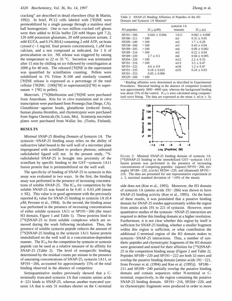
FIGURE 2: Minimal SNAP-25 binding domain of syntaxin 1A.[35S]SNAP-25 binding to the immobilized GST-syntaxin 1A11fusion protein was performed in the presence of increasingconcentrations of competing peptides: (square) SP191-266, (tri-angle) SP189-220, (circle) SP204-220, and (diamond) SP191-216. The data are presented for one representative experiment (ng 3, maximal standard deviation of<10% of the mean).
4320 Biochemistry, Vol. 36, No. 14, 1997 Zhong et al.
precisely pinpoint critical epitopes within the putative bindingdomain. Furthermore, SP238-266 and its related peptidesand SP220-266 were generated to encompass variousepitopes in the C-terminal region and the entire C-terminalregion, respectively, outside the putative binding domain.Finally, an orderly increase in the size of the putative bindingdomain was generated by increasing the length of the peptidein the C-terminal direction (SP189-220, SP191-235, SP191-253, and SP191-266, the entire H3 domain).As shown in Figure 2 and Table 2, a minimal binding
domain for SNAP-25, a sequence that substantially overlapsthe region of amino acids 191-221, is sufficient for SNAP-25 binding. SP189-220, the 32-mer peptide that spansamino acid 189-220, inhibits [35S]SNAP-25 binding toimmobilized syntaxin 1A11 with an IC50 of 2.7( 0.5 µM.Another peptide within this region (SP191-222; see Table2) appears to have a somewhat decreased affinity. SP204-220, a 17-mer peptide that has a sequence identical to theC-terminal portion of the minimal binding domain, has avery low level of activity, suggesting a requirement for theN terminus of this domain. On the other hand, peptidescontaining the N-terminal sequence alone, e.g., SP191-216,did not displace [35S]SNAP-25 binding. IC50’s for allpeptides assayed are given in Table 2. Note that SP189-216 lacks only the last four amino acids (MDMA) of SP189-220 but is inactive. Taken together, these results show thatonly peptides that contain virtually the entire core of the 32-mer region are active. This 32-mer from amino acids 189-220 is therefore the minimal binding domain for theinteraction of syntaxin and SNAP-25.While the minimal binding domain (residues 189-220)
is an effective competitor of [35S]SNAP-25 binding, it isabout 50-fold less potent than SP191-266 (IC50 ) 0.043(0.004µM), which represents the entire H3 domain (Figure2 and Table 2). This observation is consistent with previousstudies demonstrating that truncation of the C-terminal regionresults in a substantial reduction of SNAP-25 binding(Chapman et al., 1994). In peptide SP191-235 (Figure 2),15 additional residues extend C-terminally from the 32-merminimal binding domain. This extension enhances theaffinity for SNAP-25 by more than 10-fold (Table 1).Extension C-terminally by 33 residues (SP191-253) in-creases the affinity to that of SP191-266 and syntaxin 1A11(Table 2). In order to define whether the C-terminal regioncontains additional binding sites for SNAP-25, peptides thatrepresent the sequence of this region were tested for[35S]SNAP-25 competitive binding. Interestingly, all pep-tides spanning various C-terminal epitopes of the H3 domain(SP209-240, etc.) and the peptide spanning the entireC-terminal region (SP220-266) but lacking amino acids189-220 fail to bind to SNAP-25. Therefore, it appearsthat the C-terminal portion of the H3 domain is not sufficientfor SNAP-25 binding.Previous studies have also demonstrated that the H3
domain is required for binding of syntaxin 1A to otherSNARE proteins such as VAMPII (Kee et al., 1995).Interestingly, a heterotrimeric complex can be formed amongsyntaxin 1A, SNAP-25, and VAMPII (Pevsner et al., 1994).Using the binding assay described above, we have observedthat SP191-266, and to a lesser extent SP189-220, en-hanced [35S]SNAP-25 binding to the immobilized VAMPII-GST fusion protein in a concentration-dependent manner(data not shown). This suggests that the SNAP-25 minimal
binding domain is also involved in the binding of syntaxinto VAMPII.SNAP-25 Binding and theR-Helical Content of the H3
Domain. Sequence analysis predicts that the H3 domain ofsyntaxin 1A contains substantialR-helical content (Inoue etal., 1992; Spring et al., 1993). In the present study,experiments were directed toward answering the followingthree questions regarding the relationship of theR-helicalstructure of the H3 domain to its SNAP-25 binding activity.(1) Does the predictedR-helical structure in the H3 domainin fact exist? (2) If there areR-helices in the H3 domain,how does the 32-mer domain contribute to thisR-helicity?(3) IsR-helical structure required for SNAP-25 binding? Toanswer these questions, circular dichroism (CD) spectroscopywas performed to analyze the secondary structures ofSP191-266 and related peptides.(1) Is the H3 domainR-helical as predicted? As predicted,
the H3 domain (SP191-266) yields a CD spectrum typicalof anR-helical conformation. That is, a characteristic doublepeak with maxima at 208 and 222 nm is observed (Figure3). In parallel, SP189-220, the SNAP-25 minimal bindingdomain, and SP191-235, a peptide containing the minimalbinding domain and a C-terminal extension, also show thepresence ofR-helicity. Similar CD spectra were observedfor SP191-222 and SP191-253 (data not shown). The CDintensity at 222 nm suggests that SP189-220 is ∼70%helical and SP191-235 and SP191-266 are∼80% helical.In sharp contrast, littleR-helix was found in SP204-220and SP191-216, inactive peptides lacking only short N-terminal or C-terminal sequences of the minimal bindingdomain, respectively.(2) How does the 32-mer minimal binding domain
contribute to the formation of theR-helix of the H3 domain?Surprisingly, compared to the high helical content of SP191-266, minimalR-helix was found in the peptide that spansthe entire C terminus of H3 but lacks the 32-mer minimalbinding domain (SP220-266, Figure 3). Other smallerC-terminal peptides (e.g. SP238-266) also show insignificantR-helical content (data not shown). Therefore, the C-terminalregion of the H3 domain cannot adopt anR-helical confor-mation independent of the minimal binding domain, eventhough its amino acid sequence is enriched in helix-formingamino acids and contains the seven heptad repeats typicalof amphiphilicR-helices.
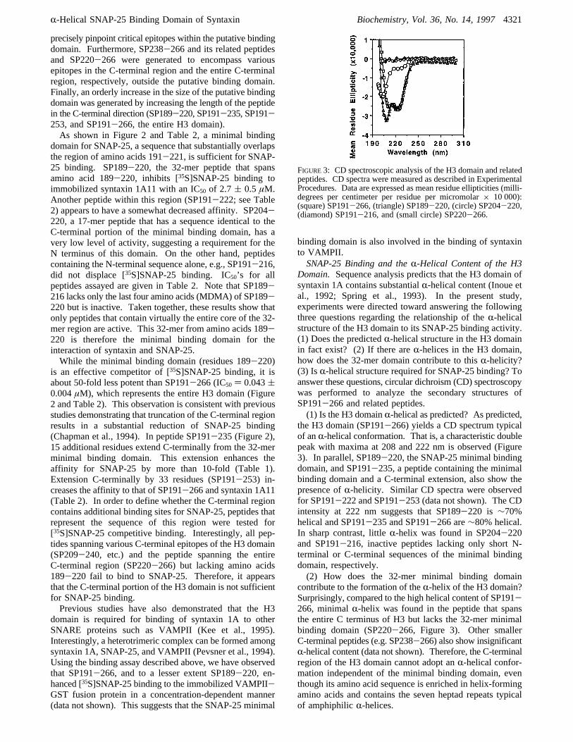
FIGURE3: CD spectroscopic analysis of the H3 domain and relatedpeptides. CD spectra were measured as described in ExperimentalProcedures. Data are expressed as mean residue ellipticities (milli-degrees per centimeter per residue per micromolar× 10 000):(square) SP191-266, (triangle) SP189-220, (circle) SP204-220,(diamond) SP191-216, and (small circle) SP220-266.
R-Helical SNAP-25 Binding Domain of Syntaxin Biochemistry, Vol. 36, No. 14, 19974321
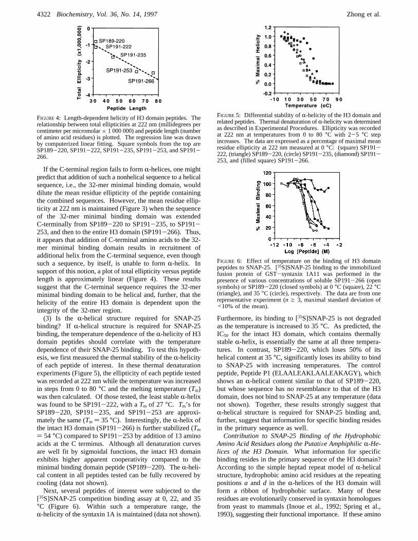
If the C-terminal region fails to formR-helices, one mightpredict that addition of such a nonhelical sequence to a helicalsequence, i.e., the 32-mer minimal binding domain, woulddilute the mean residue ellipticity of the peptide containingthe combined sequences. However, the mean residue ellip-ticity at 222 nm is maintained (Figure 3) when the sequenceof the 32-mer minimal binding domain was extendedC-terminally from SP189-220 to SP191-235, to SP191-253, and then to the entire H3 domain (SP191-266). Thus,it appears that addition of C-terminal amino acids to the 32-mer minimal binding domain results in recruitment ofadditional helix from the C-terminal sequence, even thoughsuch a sequence, by itself, is unable to formR-helix. Insupport of this notion, a plot of total ellipticity versus peptidelength is approximately linear (Figure 4). These resultssuggest that the C-terminal sequence requires the 32-merminimal binding domain to be helical and, further, that thehelicity of the entire H3 domain is dependent upon theintegrity of the 32-mer region.(3) Is the R-helical structure required for SNAP-25
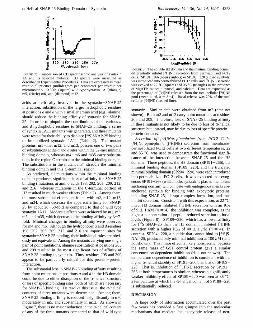
binding? If R-helical structure is required for SNAP-25binding, the temperature dependence of theR-helicity of H3domain peptides should correlate with the temperaturedependence of their SNAP-25 binding. To test this hypoth-esis, we first measured the thermal stability of theR-helicityof each peptide of interest. In these thermal denaturationexperiments (Figure 5), the ellipticity of each peptide testedwas recorded at 222 nm while the temperature was increasedin steps from 0 to 80°C and the melting temperature (Tm)was then calculated. Of those tested, the least stableR-helixwas found to be SP191-222, with aTm of 27 °C. Tm’s forSP189-220, SP191-235, and SP191-253 are approxi-mately the same (Tm ) 35 °C). Interestingly, theR-helix ofthe intact H3 domain (SP191-266) is further stabilized (Tm) 54 °C) compared to SP191-253 by addition of 13 aminoacids at the C terminus. Although all denaturation curvesare well fit by sigmoidal functions, the intact H3 domainexhibits higher apparent cooperativity compared to theminimal binding domain peptide (SP189-220). TheR-heli-cal content in all peptides tested can be fully recovered bycooling (data not shown).Next, several peptides of interest were subjected to the
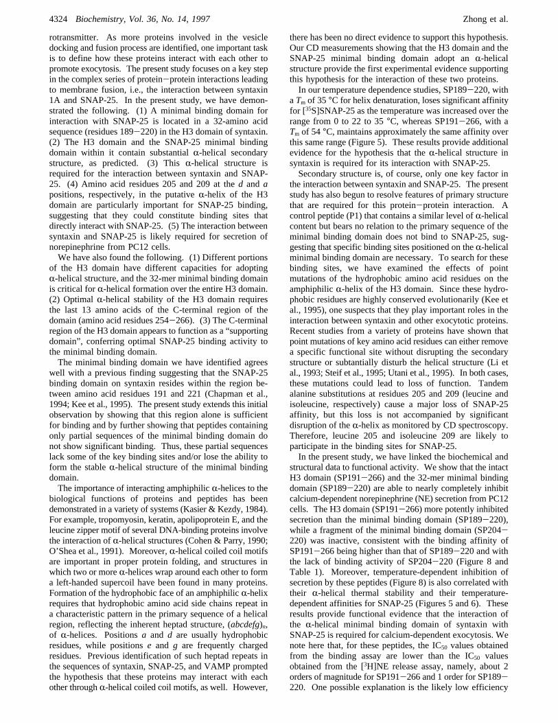
[35S]SNAP-25 competition binding assay at 0, 22, and 35°C (Figure 6). Within such a temperature range, theR-helicity of the syntaxin 1A is maintained (data not shown).
Furthermore, its binding to [35S]SNAP-25 is not degradedas the temperature is increased to 35°C. As predicted, theIC50 for the intact H3 domain, which contains thermallystableR-helix, is essentially the same at all three tempera-tures. In contrast, SP189-220, which loses 50% of itshelical content at 35°C, significantly loses its ability to bindto SNAP-25 with increasing temperatures. The controlpeptide, Peptide P1 (ELAALEAKLAALEAKAGY), whichshows anR-helical content similar to that of SP189-220,but whose sequence has no resemblance to that of the H3domain, does not bind to SNAP-25 at any temperature (datanot shown). Together, these results strongly suggest thatR-helical structure is required for SNAP-25 binding and,further, suggest that information for specific binding residesin the primary sequence as well.Contribution to SNAP-25 Binding of the Hydrophobic
Amino Acid Residues along the PutatiVe AmphiphilicR-He-lices of the H3 Domain.What information for specificbinding resides in the primary sequence of the H3 domain?According to the simple heptad repeat model ofR-helicalstructure, hydrophobic amino acid residues at the repeatingpositionsa and d in the R-helices of the H3 domain willform a ribbon of hydrophobic surface. Many of theseresidues are evolutionarily conserved in syntaxin homologuesfrom yeast to mammals (Inoue et al., 1992; Spring et al.,1993), suggesting their functional importance. If these amino
FIGURE 4: Length-dependent helicity of H3 domain peptides. Therelationship between total ellipticities at 222 nm (millidegrees percentimeter per micromolar× 1 000 000) and peptide length (numberof amino acid residues) is plotted. The regression line was drawnby computerized linear fitting. Square symbols from the top areSP189-220, SP191-222, SP191-235, SP191-253, and SP191-266.
FIGURE5: Differential stability ofR-helicity of the H3 domain andrelated peptides. Thermal denaturation ofR-helicity was determinedas described in Experimental Procedures. Ellipticity was recordedat 222 nm at temperatures from 0 to 80°C with 2-5 °C stepincreases. The data are expressed as a percentage of maximal meanresidue ellipticity at 222 nm measured at 0°C: (square) SP191-222, (triangle) SP189-220, (circle) SP191-235, (diamond) SP191-253, and (filled square) SP191-266.
FIGURE 6: Effect of temperature on the binding of H3 domainpeptides to SNAP-25. [35S]SNAP-25 binding to the immobilizedfusion protein of GST-syntaxin 1A11 was performed in thepresence of various concentrations of soluble SP191-266 (opensymbols) or SP189-220 (closed symbols) at 0°C (square), 22°C(triangle), and 35°C (circle), respectively. The data are from onerepresentative experiment (n g 3, maximal standard deviation of<10% of the mean).
4322 Biochemistry, Vol. 36, No. 14, 1997 Zhong et al.
acids are critically involved in the syntaxin-SNAP-25interaction, substitution of the larger hydrophobic residuesat positionsa anddwith a smaller amino acid (e.g., alanine)should reduce the binding affinity of syntaxin for SNAP-25. In order to pinpoint the contributions of the variousaandd hydrophobic residues to SNAP-25 binding, a seriesof syntaxin 1A11 mutants was generated, and these mutantswere tested for their ability to displace [35S]SNAP-25 bindingto immobilized syntaxin 1A11 (Table 2). The mutantproteins, m1-m3, m12, and m13, possess one or two pairsof substitutions at thea andd sites within the 32-mer minimalbinding domain, whereas m4-m6 and m35 carry substitu-tions in the region C-terminal to the minimal binding domain.The substitutions in the mutant m34 straddle the minimalbinding domain and this C-terminal region.As predicted, all mutations within the minimal binding
domain produced significant loss of affinity for SNAP-25binding (mutations at amino acids 198, 202, 205, 209, 212,and 216), whereas mutations in the C-terminal portion ofH3 resulted in much weaker effects (Table 2). Among them,the most substantial effects are found with m2, m12, m13,and m34, which decrease the apparent affinity for SNAP-25 by about 20-50-fold as compared to that of wild typesyntaxin 1A11. Moderate effects were achieved by m1, m3,m5, and m35, which decreased the binding affinity by 3-7-fold. Minimal changes in binding affinity were observedfor m4 and m6. Although the hydrophobica andd residues198, 202, 205, 209, 212, and 216 are important sites forsyntaxin-SNAP-25 binding, their individual roles are obvi-ously not equivalent. Among the mutants carrying one singlepair of point mutations, alanine substitution at positions 205and 209 resulted in the most significant loss of affinity ofSNAP-25 binding to syntaxin. Thus, residues 205 and 209appear to be particularly critical for this protein-proteininteraction.The substantial loss in SNAP-25 binding affinity resulting
from point mutations at positionsa andd in the H3 domaincould be due to either disruption of theR-helical structureor loss of specific binding sites, both of which are necessaryfor SNAP-25 binding. To resolve this issue, theR-helicalcontents of three mutants were determined. Among them,SNAP-25 binding affinity is reduced insignificantly in m6,moderately in m5, and substantially in m12. As shown inFigure 7, there is no major reduction in theR-helical contentof any of the three mutants compared to that of wild type
syntaxin. Similar data were obtained from m2 (data notshown). Both m2 and m12 carry point mutations at residues205 and 209. Therefore, loss of SNAP-25 binding affinityin these mutants is not likely to be due to loss ofR-helicalstructure but, instead, may be due to loss of specific protein-protein contacts.Secretion of [3H]Norepinephrine from PC12 Cells.
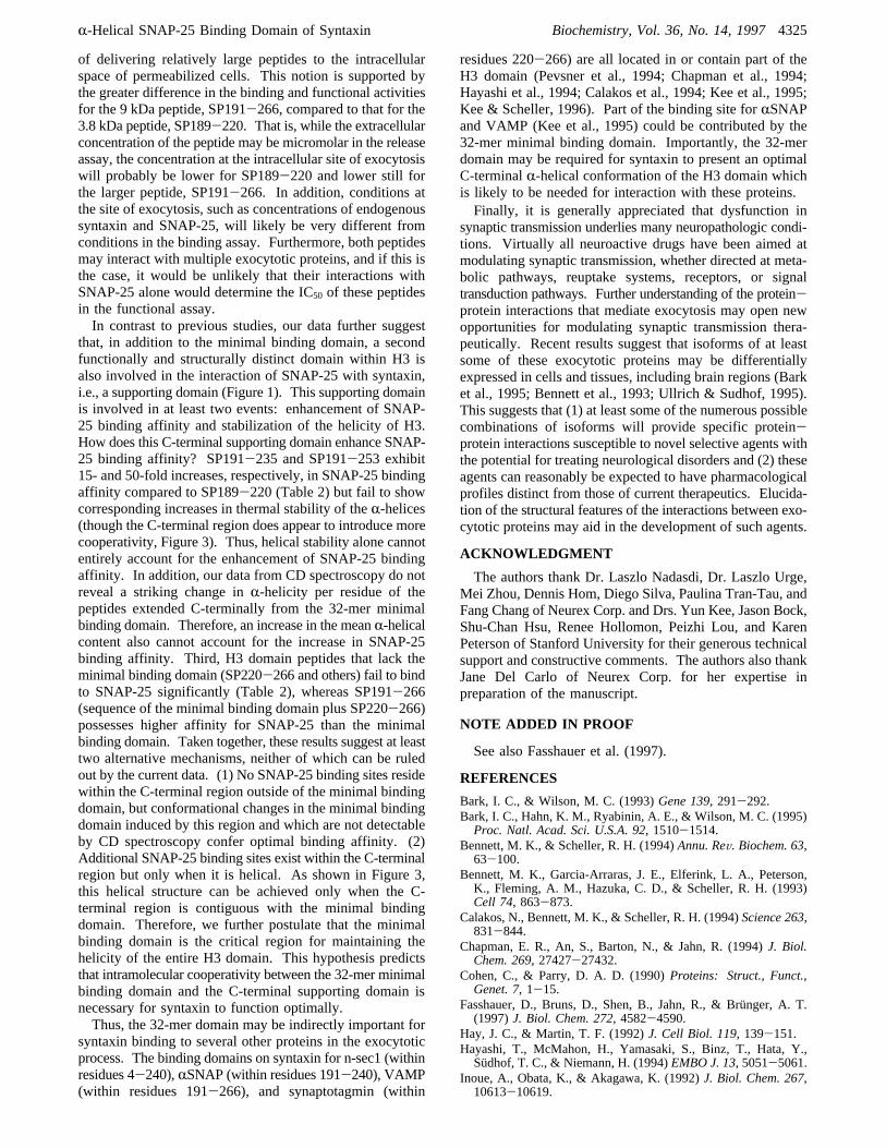
[3H]Norepinephrine ([3H]NE) secretion from membrane-permeabilized PC12 cells at two different temperatures, 22and 35°C, was used to demonstrate the functional signifi-cance of the interaction between SNAP-25 and the H3domain. Three peptides, the H3 domain (SP191-266), theminimal binding domain (SP189-220), and the truncatedminimal binding domain (SP204-220), were each introducedinto permeabilized PC12 cells. It was expected that exog-enous SP191-266 (which lacks syntaxin’s plasma membraneanchoring domain) will compete with endogenous membrane-anchored syntaxin for binding with exocytotic proteins,including SNAP-25, disrupt complex formation, and thusinhibit secretion. Consistent with this expectation, at 22°C,intact H3 domain inhibited [3H]NE secretion with an IC50of 9 ( 1 µM (n ) 4); the inhibition was complete, as thehighest concentration of peptide reduced secretion to basallevels (Figure 8). SP189-220, which has a lower affinityfor [35S]SNAP-25 than the H3 domain, inhibited [3H]NEsecretion with a higher IC50 of 40 ( 1 µM (n ) 4). Incontrast, SP204-220, a peptide that cannot bind to [35S]S-NAP-25, produced only minimal inhibition at 100µM (datanot shown). This minor effect is likely nonspecific, becausethe same mass of GST control protein gave a similarconcentration-dependent inhibition (data not shown). Thetemperature dependence of inhibition is consistent with thehigherR-helical stability of SP191-266 than that of SP189-220. That is, inhibition of [3H]NE secretion by SP191-266 at both temperatures is similar, whereas a significantlyweaker inhibitory effect of SP189-220 was seen at 35°C,a temperature at which theR-helical content of SP189-220is substantially reduced.
DISCUSSION
A large body of information accumulated over the pastfew years has provided a first glimpse into the molecularmechanisms that mediate the exocytotic release of neu-
FIGURE 7: Comparison of CD spectroscopic analysis of syntaxin1A and its selected mutants. CD spectra were measured asdescribed in Experimental Procedures. Data are expressed as meanresidue ellipticities (millidegrees per centimeter per residue permicromolar× 10 000: (square) wild type syntaxin 1A, (triangle)m5, (circle) m6, and (diamond) m12.
FIGURE8: The soluble H3 domain and the minimal binding domaindifferentially inhibit [3H]NE secretion from permeabilized PC12cells. SP191-266 (open symbols) or SP189-220 (closed symbols)was introduced into permeabilized PC12 cells, and [3H]NE secretionwas evoked at 22°C (square) and 35°C (triangle) in the presenceof MgATP, rat brain cytosol, and calcium. Data are expressed asthe percentage of [3H]NE released from the total cellular [3H]NEpool (mean( sd,n ) 3-4). Basal release was 20% of the totalcellular [3H]NE (dashed line).
R-Helical SNAP-25 Binding Domain of Syntaxin Biochemistry, Vol. 36, No. 14, 19974323
rotransmitter. As more proteins involved in the vesicledocking and fusion process are identified, one important taskis to define how these proteins interact with each other topromote exocytosis. The present study focuses on a key stepin the complex series of protein-protein interactions leadingto membrane fusion, i.e., the interaction between syntaxin1A and SNAP-25. In the present study, we have demon-strated the following. (1) A minimal binding domain forinteraction with SNAP-25 is located in a 32-amino acidsequence (residues 189-220) in the H3 domain of syntaxin.(2) The H3 domain and the SNAP-25 minimal bindingdomain within it contain substantialR-helical secondarystructure, as predicted. (3) ThisR-helical structure isrequired for the interaction between syntaxin and SNAP-25. (4) Amino acid residues 205 and 209 at thed andapositions, respectively, in the putativeR-helix of the H3domain are particularly important for SNAP-25 binding,suggesting that they could constitute binding sites thatdirectly interact with SNAP-25. (5) The interaction betweensyntaxin and SNAP-25 is likely required for secretion ofnorepinephrine from PC12 cells.We have also found the following. (1) Different portions
of the H3 domain have different capacities for adoptingR-helical structure, and the 32-mer minimal binding domainis critical forR-helical formation over the entire H3 domain.(2) OptimalR-helical stability of the H3 domain requiresthe last 13 amino acids of the C-terminal region of thedomain (amino acid residues 254-266). (3) The C-terminalregion of the H3 domain appears to function as a “supportingdomain”, conferring optimal SNAP-25 binding activity tothe minimal binding domain.The minimal binding domain we have identified agrees
well with a previous finding suggesting that the SNAP-25binding domain on syntaxin resides within the region be-tween amino acid residues 191 and 221 (Chapman et al.,1994; Kee et al., 1995). The present study extends this initialobservation by showing that this region alone is sufficientfor binding and by further showing that peptides containingonly partial sequences of the minimal binding domain donot show significant binding. Thus, these partial sequenceslack some of the key binding sites and/or lose the ability toform the stableR-helical structure of the minimal bindingdomain.The importance of interacting amphiphilicR-helices to the
biological functions of proteins and peptides has beendemonstrated in a variety of systems (Kasier & Kezdy, 1984).For example, tropomyosin, keratin, apolipoprotein E, and theleucine zipper motif of several DNA-binding proteins involvethe interaction ofR-helical structures (Cohen & Parry, 1990;O’Shea et al., 1991). Moreover,R-helical coiled coil motifsare important in proper protein folding, and structures inwhich two or moreR-helices wrap around each other to forma left-handed supercoil have been found in many proteins.Formation of the hydrophobic face of an amphiphilicR-helixrequires that hydrophobic amino acid side chains repeat ina characteristic pattern in the primary sequence of a helicalregion, reflecting the inherent heptad structure, (abcdefg)n,of R-helices. Positionsa and d are usually hydrophobicresidues, while positionse and g are frequently chargedresidues. Previous identification of such heptad repeats inthe sequences of syntaxin, SNAP-25, and VAMP promptedthe hypothesis that these proteins may interact with eachother throughR-helical coiled coil motifs, as well. However,
there has been no direct evidence to support this hypothesis.Our CD measurements showing that the H3 domain and theSNAP-25 minimal binding domain adopt anR-helicalstructure provide the first experimental evidence supportingthis hypothesis for the interaction of these two proteins.In our temperature dependence studies, SP189-220, with
aTm of 35 °C for helix denaturation, loses significant affinityfor [35S]SNAP-25 as the temperature was increased over therange from 0 to 22 to 35°C, whereas SP191-266, with aTm of 54 °C, maintains approximately the same affinity overthis same range (Figure 5). These results provide additionalevidence for the hypothesis that theR-helical structure insyntaxin is required for its interaction with SNAP-25.Secondary structure is, of course, only one key factor in
the interaction between syntaxin and SNAP-25. The presentstudy has also begun to resolve features of primary structurethat are required for this protein-protein interaction. Acontrol peptide (P1) that contains a similar level ofR-helicalcontent but bears no relation to the primary sequence of theminimal binding domain does not bind to SNAP-25, sug-gesting that specific binding sites positioned on theR-helicalminimal binding domain are necessary. To search for thesebinding sites, we have examined the effects of pointmutations of the hydrophobic amino acid residues on theamphiphilicR-helix of the H3 domain. Since these hydro-phobic residues are highly conserved evolutionarily (Kee etal., 1995), one suspects that they play important roles in theinteraction between syntaxin and other exocytotic proteins.Recent studies from a variety of proteins have shown thatpoint mutations of key amino acid residues can either removea specific functional site without disrupting the secondarystructure or subtantially disturb the helical structure (Li etal., 1993; Steif et al., 1995; Utani et al., 1995). In both cases,these mutations could lead to loss of function. Tandemalanine substitutions at residues 205 and 209 (leucine andisoleucine, respectively) cause a major loss of SNAP-25affinity, but this loss is not accompanied by significantdisruption of theR-helix as monitored by CD spectroscopy.Therefore, leucine 205 and isoleucine 209 are likely toparticipate in the binding sites for SNAP-25.In the present study, we have linked the biochemical and
structural data to functional activity. We show that the intactH3 domain (SP191-266) and the 32-mer minimal bindingdomain (SP189-220) are able to nearly completely inhibitcalcium-dependent norepinephrine (NE) secretion from PC12cells. The H3 domain (SP191-266) more potently inhibitedsecretion than the minimal binding domain (SP189-220),while a fragment of the minimal binding domain (SP204-220) was inactive, consistent with the binding affinity ofSP191-266 being higher than that of SP189-220 and withthe lack of binding activity of SP204-220 (Figure 8 andTable 1). Moreover, temperature-dependent inhibition ofsecretion by these peptides (Figure 8) is also correlated withtheir R-helical thermal stability and their temperature-dependent affinities for SNAP-25 (Figures 5 and 6). Theseresults provide functional evidence that the interaction ofthe R-helical minimal binding domain of syntaxin withSNAP-25 is required for calcium-dependent exocytosis. Wenote here that, for these peptides, the IC50 values obtainedfrom the binding assay are lower than the IC50 valuesobtained from the [3H]NE release assay, namely, about 2orders of magnitude for SP191-266 and 1 order for SP189-220. One possible explanation is the likely low efficiency
4324 Biochemistry, Vol. 36, No. 14, 1997 Zhong et al.
of delivering relatively large peptides to the intracellularspace of permeabilized cells. This notion is supported bythe greater difference in the binding and functional activitiesfor the 9 kDa peptide, SP191-266, compared to that for the3.8 kDa peptide, SP189-220. That is, while the extracellularconcentration of the peptide may be micromolar in the releaseassay, the concentration at the intracellular site of exocytosiswill probably be lower for SP189-220 and lower still forthe larger peptide, SP191-266. In addition, conditions atthe site of exocytosis, such as concentrations of endogenoussyntaxin and SNAP-25, will likely be very different fromconditions in the binding assay. Furthermore, both peptidesmay interact with multiple exocytotic proteins, and if this isthe case, it would be unlikely that their interactions withSNAP-25 alone would determine the IC50 of these peptidesin the functional assay.In contrast to previous studies, our data further suggest
that, in addition to the minimal binding domain, a secondfunctionally and structurally distinct domain within H3 isalso involved in the interaction of SNAP-25 with syntaxin,i.e., a supporting domain (Figure 1). This supporting domainis involved in at least two events: enhancement of SNAP-25 binding affinity and stabilization of the helicity of H3.How does this C-terminal supporting domain enhance SNAP-25 binding affinity? SP191-235 and SP191-253 exhibit15- and 50-fold increases, respectively, in SNAP-25 bindingaffinity compared to SP189-220 (Table 2) but fail to showcorresponding increases in thermal stability of theR-helices(though the C-terminal region does appear to introduce morecooperativity, Figure 3). Thus, helical stability alone cannotentirely account for the enhancement of SNAP-25 bindingaffinity. In addition, our data from CD spectroscopy do notreveal a striking change inR-helicity per residue of thepeptides extended C-terminally from the 32-mer minimalbinding domain. Therefore, an increase in the meanR-helicalcontent also cannot account for the increase in SNAP-25binding affinity. Third, H3 domain peptides that lack theminimal binding domain (SP220-266 and others) fail to bindto SNAP-25 significantly (Table 2), whereas SP191-266(sequence of the minimal binding domain plus SP220-266)possesses higher affinity for SNAP-25 than the minimalbinding domain. Taken together, these results suggest at leasttwo alternative mechanisms, neither of which can be ruledout by the current data. (1) No SNAP-25 binding sites residewithin the C-terminal region outside of the minimal bindingdomain, but conformational changes in the minimal bindingdomain induced by this region and which are not detectableby CD spectroscopy confer optimal binding affinity. (2)Additional SNAP-25 binding sites exist within the C-terminalregion but only when it is helical. As shown in Figure 3,this helical structure can be achieved only when the C-terminal region is contiguous with the minimal bindingdomain. Therefore, we further postulate that the minimalbinding domain is the critical region for maintaining thehelicity of the entire H3 domain. This hypothesis predictsthat intramolecular cooperativity between the 32-mer minimalbinding domain and the C-terminal supporting domain isnecessary for syntaxin to function optimally.Thus, the 32-mer domain may be indirectly important for
syntaxin binding to several other proteins in the exocytoticprocess. The binding domains on syntaxin for n-sec1 (withinresidues 4-240),RSNAP (within residues 191-240), VAMP(within residues 191-266), and synaptotagmin (within
residues 220-266) are all located in or contain part of theH3 domain (Pevsner et al., 1994; Chapman et al., 1994;Hayashi et al., 1994; Calakos et al., 1994; Kee et al., 1995;Kee & Scheller, 1996). Part of the binding site forRSNAPand VAMP (Kee et al., 1995) could be contributed by the32-mer minimal binding domain. Importantly, the 32-merdomain may be required for syntaxin to present an optimalC-terminalR-helical conformation of the H3 domain whichis likely to be needed for interaction with these proteins.Finally, it is generally appreciated that dysfunction in
synaptic transmission underlies many neuropathologic condi-tions. Virtually all neuroactive drugs have been aimed atmodulating synaptic transmission, whether directed at meta-bolic pathways, reuptake systems, receptors, or signaltransduction pathways. Further understanding of the protein-protein interactions that mediate exocytosis may open newopportunities for modulating synaptic transmission thera-peutically. Recent results suggest that isoforms of at leastsome of these exocytotic proteins may be differentiallyexpressed in cells and tissues, including brain regions (Barket al., 1995; Bennett et al., 1993; Ullrich & Sudhof, 1995).This suggests that (1) at least some of the numerous possiblecombinations of isoforms will provide specific protein-protein interactions susceptible to novel selective agents withthe potential for treating neurological disorders and (2) theseagents can reasonably be expected to have pharmacologicalprofiles distinct from those of current therapeutics. Elucida-tion of the structural features of the interactions between exo-cytotic proteins may aid in the development of such agents.
ACKNOWLEDGMENT
The authors thank Dr. Laszlo Nadasdi, Dr. Laszlo Urge,Mei Zhou, Dennis Hom, Diego Silva, Paulina Tran-Tau, andFang Chang of Neurex Corp. and Drs. Yun Kee, Jason Bock,Shu-Chan Hsu, Renee Hollomon, Peizhi Lou, and KarenPeterson of Stanford University for their generous technicalsupport and constructive comments. The authors also thankJane Del Carlo of Neurex Corp. for her expertise inpreparation of the manuscript.
NOTE ADDED IN PROOF
See also Fasshauer et al. (1997).
REFERENCES
Bark, I. C., & Wilson, M. C. (1993)Gene 139, 291-292.Bark, I. C., Hahn, K. M., Ryabinin, A. E., & Wilson, M. C. (1995)Proc. Natl. Acad. Sci. U.S.A. 92, 1510-1514.
Bennett, M. K., & Scheller, R. H. (1994)Annu. ReV. Biochem. 63,63-100.
Bennett, M. K., Garcia-Arraras, J. E., Elferink, L. A., Peterson,K., Fleming, A. M., Hazuka, C. D., & Scheller, R. H. (1993)Cell 74, 863-873.
Calakos, N., Bennett, M. K., & Scheller, R. H. (1994)Science 263,831-844.
Chapman, E. R., An, S., Barton, N., & Jahn, R. (1994)J. Biol.Chem. 269, 27427-27432.
Cohen, C., & Parry, D. A. D. (1990)Proteins: Struct., Funct.,Genet. 7, 1-15.
Fasshauer, D., Bruns, D., Shen, B., Jahn, R., & Bru¨nger, A. T.(1997)J. Biol. Chem. 272, 4582-4590.
Hay, J. C., & Martin, T. F. (1992)J. Cell Biol. 119, 139-151.Hayashi, T., McMahon, H., Yamasaki, S., Binz, T., Hata, Y.,Sudhof, T. C., & Niemann, H. (1994)EMBO J. 13, 5051-5061.
Inoue, A., Obata, K., & Akagawa, K. (1992)J. Biol. Chem. 267,10613-10619.
R-Helical SNAP-25 Binding Domain of Syntaxin Biochemistry, Vol. 36, No. 14, 19974325
Jahn, R., & Su¨dhof, T. C. (1994)Annu. ReV. Neurosci. 17, 219-246.
Kasier, E. T., & Kezdy, F. J. (1984)Science 259, 249-255.Kee, Y., & Scheller, R. H. (1996)J. Neurosci. 16, 1975-1981.Kee, Y., Lin, R. C., Hsu, S., & Scheller, R. H. (1995)Neuron 14,991-998.
Li, X., Rock, F., Chong, P., Cockle, S., Keating, A., Ziltener, H.,& Klein, M. (1993) J. Biol. Chem. 268(30), 22377-22384.
Morgan, A., Dimaline, R., & Burgoyne, R. D. (1994)J. Biol. Chem.269, 29347-29350.
O’Shea, E. K., Klemm, J. D., Kim, P. S., & Alber, T. (1991)Science254, 539.
Pevsner, J., Hsu, S.-C., Braun, J. E. A., Calakos, N., Ting, A. E.,Bennett, M. K., & Scheller, R. H. (1994)Neuron 13, 353-361.
Scheller, R. H. (1995)Neuron 14, 1-5.
Sollner, T., Whiteheart, S. W., Brunner, M., Erdjument-Bromage,H., Geromanos, S., Tempst, P., & Rothman, J. E. (1993a)Nature362, 318-324.
Sollner, T., Bennet, M. K., Whiteheart, S. W., Scheller, R. H., &Rothman, J. E. (1993b)Cell 75, 409-418.
Spring, J. K., Kato, M., & Bernfield, M. (1993)Trends Biochem.Sci. 18, 124-125.
Steif, C., Hinz, H.-J., & Cesarieni, G. (1995)Proteins 23, 83-96.
Ullrich, B., & Sudhof, T. C. (1995)Neuropharmacology 34, 1371-1377.
Utani, A., Nomizu, M., Sugiyama, S., Miyamoto, S., Roller, P., &Yamada, Y. (1995)J. Biol. Chem. 270(7), 3292-3298.
BI9625408
4326 Biochemistry, Vol. 36, No. 14, 1997 Zhong et al.