an iatrogenically unmasked life threatening disease ... · brugada syndrome is a life threatening...
TRANSCRIPT
426 Copyright © 2013 The Korean Society of Cardiology
Korean Circulation Journal
Introduction
Brugada syndrome, first described in 1992, is a life threatening disease and is usually overlooked during emergency service admis-sions.1) It is characterized by typical electrocardiography (ECG) re-sembling right bundle branch block (RBBB), static or dynamic ST-seg-ment elevation in leads V 1-3. There is familial tendency in some ca-ses.2) A majority of patients have a structurally normal heart and are likely to remain asymptomatic, however they may present to emer-gency departments with syncope and various arrhythmias (atrial fi-brillation, ventricular tachycardia, ventricular fibrillation-cardiac ar-rest and etc.).1)2) Therefore it is crucially important for emergency medicine physicians not to omit this potential diagnosis. Herein we report a case with Brugada syndrome which was iatrogenically un-
Case Report
http://dx.doi.org/10.4070/kcj.2013.43.6.426Print ISSN 1738-5520 • On-line ISSN 1738-5555
An Iatrogenically Unmasked Life Threatening Disease: Brugada SyndromeSuleyman Ercan, MD1, Muhammed Oylumlu, MD2, Gokhan Altunbas, MD3, and Vedat Davutoglu, MD1
1Department of Cardiology, Gaziantep University School of Medicine, Gaziantep, 2Department of Cardiology, S̨ehitkamil State Hospital, Gaziantep,3Department of Cardiology, Kilis State Hospital, Kilis, Turkey
Brugada syndrome is a life threatening disease that is usually overlooked during emergency service admissions. It is characterized by typical electrocardiography resembling right bundle branch block, static or dynamic ST-segment elevation in leads V 1-3. There is familial tendency in some cases. A majority of patients have a structurally normal heart and are likely to remain asymptomatic, however they may present to emergency departments with syncope and various serious arrhythmias. Therefore it is crucially important for emergency medicine physicians not to omit this potential diagnosis. Herein we report a case with Brugada syndrome which was iatrogenically unmasked after propafenone administration for atrial fibrillation. (Korean Circ J 2013;43:426-428)
KEY WORDS: Brugada syndrome; Propafenone; Atrial fibrillation; Acute myocardial infarction.
Received: June 19, 2012Revision Received: July 11, 2012Accepted: August 10, 2012Correspondence: Suleyman Ercan, MD, Department of Cardiology, Ga-ziantep University School of Medicine, 27310, Gaziantep, Turkey Tel: 90 342 3606060/76286, Fax: 90 342 3603928E-mail: [email protected]
• The authors have no financial conflicts of interest.
This is an Open Access article distributed under the terms of the Creative Commons Attribution Non-Commercial License (http://creativecommons.org/licenses/by-nc/3.0) which permits unrestricted non-commercial use, distribution, and reproduction in any medium, provided the original work is properly cited.
masked after propafenone administration for atrial fibrillation.
Case
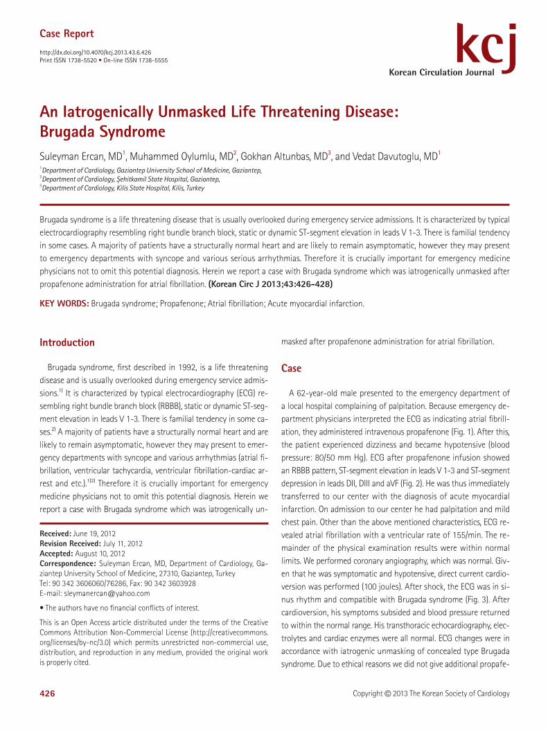
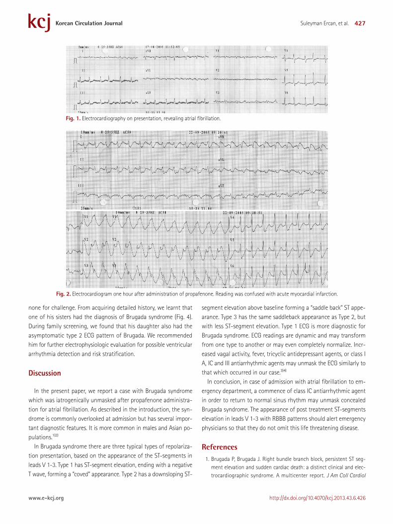
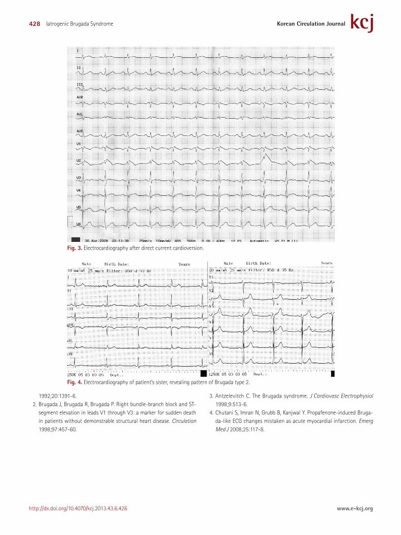
A 62-year-old male presented to the emergency department of a local hospital complaining of palpitation. Because emergency de-partment physicians interpreted the ECG as indicating atrial fibrill-ation, they administered intravenous propafenone (Fig. 1). After this, the patient experienced dizziness and became hypotensive (blood pressure: 80/50 mm Hg). ECG after propafenone infusion showed an RBBB pattern, ST-segment elevation in leads V 1-3 and ST-segment depression in leads DII, DIII and aVF (Fig. 2). He was thus immediately transferred to our center with the diagnosis of acute myocardial infarction. On admission to our center he had palpitation and mild chest pain. Other than the above mentioned characteristics, ECG re-vealed atrial fibrillation with a ventricular rate of 155/min. The re-mainder of the physical examination results were within normal limits. We performed coronary angiography, which was normal. Giv-en that he was symptomatic and hypotensive, direct current cardio-version was performed (100 joules). After shock, the ECG was in si-nus rhythm and compatible with Brugada syndrome (Fig. 3). After cardioversion, his symptoms subsided and blood pressure returned to within the normal range. His transthoracic echocardiography, elec-trolytes and cardiac enzymes were all normal. ECG changes were in accordance with iatrogenic unmasking of concealed type Brugada syndrome. Due to ethical reasons we did not give additional propafe-
427Suleyman Ercan, et al.
http://dx.doi.org/10.4070/kcj.2013.43.6.426www.e-kcj.org
none for challenge. From acquiring detailed history, we learnt that one of his sisters had the diagnosis of Brugada syndrome (Fig. 4). During family screening, we found that his daughter also had the asymptomatic type 2 ECG pattern of Brugada. We recommended him for further electrophysiologic evaluation for possible ventricular arrhythmia detection and risk stratification.
Discussion
In the present paper, we report a case with Brugada syndrome which was iatrogenically unmasked after propafenone administra-tion for atrial fibrillation. As described in the introduction, the syn-drome is commonly overlooked at admission but has several impor-tant diagnostic features. It is more common in males and Asian po-pulations.1)2)
In Brugada syndrome there are three typical types of repolariza-tion presentation, based on the appearance of the ST-segments in leads V 1-3. Type 1 has ST-segment elevation, ending with a negative T wave, forming a “coved” appearance. Type 2 has a downsloping ST-
segment elevation above baseline forming a “saddle back” ST appe-arance. Type 3 has the same saddleback appearance as Type 2, but with less ST-segment elevation. Type 1 ECG is more diagnostic for Brugada syndrome. ECG readings are dynamic and may transform from one type to another or may even completely normalize. Incr-eased vagal activity, fever, tricyclic antidepressant agents, or class I A, IC and III antiarrhythmic agents may unmask the ECG similarly to that which occurred in our case.3)4)
In conclusion, in case of admission with atrial fibrillation to em-ergency department, a commence of class IC antiarrhythmic agent in order to return to normal sinus rhythm may unmask concealed Brugada syndrome. The appearance of post treatment ST-segments elevation in leads V 1-3 with RBBB patterns should alert emergency physicians so that they do not omit this life threatening disease.
References1. Brugada P, Brugada J. Right bundle branch block, persistent ST seg-
ment elevation and sudden cardiac death: a distinct clinical and elec-trocardiographic syndrome. A multicenter report. J Am Coll Cardiol
Fig. 1. Electrocardiography on presentation, revealing atrial fibrillation.
Fig. 2. Electrocardiogram one hour after administration of propafenone. Reading was confused with acute myocardial infarction.
428 Iatrogenic Brugada Syndrome
http://dx.doi.org/10.4070/kcj.2013.43.6.426 www.e-kcj.org
1992;20:1391-6.2. Brugada J, Brugada R, Brugada P. Right bundle-branch block and ST-
segment elevation in leads V1 through V3: a marker for sudden death in patients without demonstrable structural heart disease. Circulation 1998;97:457-60.
3. Antzelevitch C. The Brugada syndrome. J Cardiovasc Electrophysiol 1998;9:513-6.
4. Chutani S, Imran N, Grubb B, Kanjwal Y. Propafenone-induced Bruga-da-like ECG changes mistaken as acute myocardial infarction. Emerg Med J 2008;25:117-8.
Fig. 3. Electrocardiography after direct current cardioversion.
Fig. 4. Electrocardiography of patient’s sister, revealing pattern of Brugada type 2.