an immunologist's look at the rho and rab gtp-binding proteins
TRANSCRIPT

review
45 Shearer, G.M. and Clerici, M. (1992) Prog. Chem. ImnmnoL 54, 21-43 46 Loos, M., Storz, R., Miiiter, W. and Lemmel, E-M. (1981) lmmunobiol. 158, 213-224 47 Erdei, A. and Reid, K.B.M. (1988) Mol. lmmunol. 25, 1067-1073 48 El Jarrah, F., Hidvegi, T., Ujhelyi, E. et aL (1992) AIDS 6, 1050-1051 49 Perricone, R., Fontana, L., De Carolis, C. et al. (1987) Clin. Exp. ImmunoL 70, 500-507 $0 Senaldi, G , Peakman, M.. McManus, T. et aL (1989) Lancet ii, 624 51 Lin, R.Y., Wildfeuer, O., Franklin, M.M. and Candido,
K.(1988) . . . . . I. ~J . Appl. ,,,,,,.,,~,,.~ .......... r 87,40-46 52 Melchers, F., Erdei, A., Schutz, T. and Dierich, M.P. (1985) Nature 317, 264-267 53 Erdei, A., Spaeth, E., AIsenz, J. et aL (1984) Mol. ImmunaL 21, 1215-1221 54 Lane, H.C., Masur, H., Edgar, L.C. et al. (1983) New Engl. ]. Med. 309, 453-458 55 Fuchs, D., Hausen, A., Reibnegger, G. et al. (1988) Imrnunol. Today 9, 150-155 56 Schulz, T.F., Petzer, A, Stauder, R., Eigentler, A. and Dierich, M.P. (1986) Immunobiology 173, 372 57 McNearney, T., Ebenbichler, C., T6tsch, M. and Dierich, M.P. (1993) Eur. ]. Immunol. 23, 1266-1270
An immunologist's look at the Rho and Rab GTP-binding proteins
Philippe Chavrier, Jean-Pierre Gorvel and Jacques Bertoglio
The ras superfamily of small GTP-binding proteins contains three major branches: the Ras, Rbo and Rab protein subfamilies. Recent advances in the field of ras-related small GTP-binding proteins suggest that it may be worthwhile to look at this superfamily from the standpoi~t of immu- nology. The subject of this review is to outline briefly tbe areas o f lympbocyte function which may implicate small G proteins, with special emphasis on the established or possible roles of proteins of the Rbo and
Rab subfamilies in cytoskeleton organization and antigen presentation.
The recent years have uncovered the multiple aspects of cellular functions which are controlled by GTP- binding proteins (G proteins) of the ras superfamily |. in contrast to the large hetero-trimeric G proteins, ras- related G proteins are monomeric and of small mol- ecular mass 122 to 30 kDa). They share a number of structural and sequence homologies which account for their p:imary biocaemical function, the binding and hydre!ysis of GTP (Ref. 2). These proteins exist in C~e cell as one of two interconvertible conformational states., an active form, when bound to GTP, and an inactive form, when bound to GDP. The GTP/GDP cycle of ra~-like proteins is regulated by two classes of proteins: (1) the GTPase activating proteins (GAP) that stimv!ate the low intrinsic GTPase activity of ras-G proteins, hence inactivating them; and (2) the exchange factors such as the GDP dissociation stimulators (GDS) that promote the exchange of GDP for GTP, thus returning the G protein to an activated state o~ the GDP dissociation inhibitors (GD!) which prevent this exchange.
The mammalian ras superfamily of G proteins is composed of an estimated 50 genes which, ba~ed on sequence similarities, can be subdivided into three major subgroups; the Ras, Rho and Rab subfamilic~ 3.
These three branches of the ras superfamily appear to be specifically involved in different areas of cell bi- ology: the Ras proteins represent major control check points in signal transduction and cell proliferation, whereas the Rho proteins appear to regulate organiz- ation of the cytoskeleton network, and the Rab proteins are involved in the control of intracellular membrane traffic. These functions are obviously shared by all cell types, and as G proteins do not ~unction differently in lymphocytes than they do in tmroblasts, this article will briefly review our current knowledge (in some cases our hypotheses) on how these proteins may l;ar- ticipate in the specialized functions of lymphocytes. Since the role of Ras in lymphocyte activation has recently been reviewed in these pages (lmmunol. Today (1992) 13, 82-89), we will focus on how the Rho and tbe Rab subfamilies, respectively, could poss- ibly be involved in lymphocyte polarization and anti- gen presentation.
The Rho subfamily This subfamily contains at least four rbo genes (A,
B, C and G), raci and rac2, and CDC42Hs. Proteins in this subfamily share with Ras an approximate 3~% sequer.c~ i__.ev.:!,y essentially located in the G1,
O 1993, Elsevier ~:icnce Publisher~ Lid. LIK 01¢,7-569~;9M$06.00
l~m,~otogy Today 440 V,,I. 14 No. 91993

review
G3 and G4 domains, which mediate binding and hydrolysis of the guanine nucleotide (see Ref. 2 for review, and Fig. 1). By contrast, the G2 domain, which is thought to interact with effector molecules, is fairly divergent from the rest of the Ras family, but highly conserved within the Rho subfamily, suggesting that the Rho/Rac/CDC42Hs proteins may share common regulatory or effector proteins. Proteins of the Rho ~ubfamily have an additional insert of 9-12 residues, ~tarting at position 122 (Fig. 1); this has been very use- ful in raising specific antibodies.
The first lead towards understanding the function of the Rho proteins came from experiments by P. Chardin et al. ~ using the Clostridium botulinum C3 exoenzyme which mediates ADP-ribosylation of RhoC on an asparagine residue in position 41 (at the border of the effector domain). C3 treatment of Vero cells results in disassembly of the actin microfilament net- work and major changes in cell shape, indicating that Rho proteins are involved in cytoskeletal control. This notion has now been firmly established by A, Ridley and A. Hall 6, who used constitutively activated and dominant negative mutants of RhoA and Racl ~o demonstrate that these proteins act in sequence to co:l- trol membrane ruffling and stress fiber formation'. Additional experiments showed that the dramatic changes in cytoskeleton organization and in cell shape which occur following growth factor stimulation may involve a cascade of small G proteins that includes Ras, Racl and Rho.
How the activation of Ras induces that of Rho and Rac is not yet understood. However, several multifunc- tional regulatory molecules have recently been isolated which could provide the hypothetical link between small G proteins: (1) in transformed cells, rasGAP associates to a phosphorylated protein of 190 kDa that has recently been shown to function as a GAP for all members of the Rho subfamilyH; (2) a brain-specific Ras exchange factor (p140 ~'c'gt:) shows homology to the abl oncogene product, an exchange factor (GDS) for CDC4211s (Ref. ~). There is thus a definite possi- bility that large heteromeric signalling complexes may form within the cell to connect Ras, and consequently the receptcrs lying upstream, to the Rho protein family.
In addition, stimutation of fibroblasts with serum or growth factors induces transcriptional --ctivation of ~wo Rho genes, RboB and RhoG. While RhoB behaves like an immediate-early gene, the induction of RhoG is somewhat slower and less transientt°aL Of interest is the observation that the only other small G protein gene, known to be reg-.lated at the expression level, Rat2 , also belongs to the Rho subgroup. In contrast to the other genes in the family, Rac2 shows tissue speci- ficity as it is only expressed in cells of the haemato- poietic lineage, including T and B cells I-'.
In human T cells, activation through the T-cell receptor (TCR) complex CD3, or with phytohaemag- glutin and phorbol myristate acetate (PMA), induces ap accumulation of Rac2 mRNA which becomes detectable after 4-6 h of stimulation and reaches a plateau, 40- to 50-fold above starting levels at 24 h (Ref. 13). Expression of Rac2 remains elevated for at least 96 h. Recent reports have suggested that Rac2
Effector Extension loop in Rho
V IIIIm m !
GI G2 (?,3 G4 G5
Cystcine motifs
Fig. 1. Scheme of a typical ras-related monomeric GTP-binding protein (G protein) sbowing the variable lengths of the N- and C-termini and the [our regions fo~ming tbe GTP-binding site (GI, G3, G4 and G57. Also depicted are the effector region (G2) mediating the interaction with GAP, the insertion in Rbo proteins between region G4 and G5, and the C-terminal
cysteine motifs iC: cysteine; A: alipbatic residues; X: any amino acid).
(together with ,~acl) may participate in the formation of the NADPH-oxidase complex 14"~.
This multimolecular enzymatic system is present in all phagocytic cells, and is responsible for the generation of O2- superoxide anions that participate in elimin- ation of bacteria. The involvement of Rac2 in this complex was investigated by introducing Rac antisense oligonucleotides in human Epstcin-Barr virus (EBV)- transformed B-cell lines, in which a respiratory burst can be triggered by crosslinking of membrane immunoglobulins. Indeed, a 17-mer antisense oligonu- cleotide targeting the N-terminal region of the mol- ecule was shown to reduce Rac-2 protein expression and to inhibit the generation of superoxide in these ~elis j6. Whether or not the role of Rac2 in the NADPH-oxidase complex is relevant to its increased expression in activated T cells remains a matter of speculation.
That Rac proteins participate in this specialized function of professional phagocytes does not exclude their involvement, together with Rho proteins in the control of cytoskeleton organization and cell polariz- ation (Table 1}.
Lymphocytes are polarized cells Polarized secretion is a common feature of many cell
type~ r , and in that respect, lymphocytes are no excep- tion. At least two major functions of T cells involve vectorial secretion, requiring cytoskeleton reorganiz- ation and polarization. T-cell help is delivered to B cells both through cognate T-B-cell interactions and lymphokine secretion..'. ~ias been shown that upon encountering a B cell pulsed with the appropriate anti- gen, a subset of helper-T cells (TH2) rearrange their cytoskeleton and release IL-4 in a polarized fashion over the area of receptor crosslinking TM In cell-me- diated cytotoxicity, conjugate formation between effec- tor and target cells is accompanied by capping of the adhesion molecule LFA-I to the cell contact area, which appears to be followed, within the cetl, by the cytoskeleton protein, talin. Simultaneously, the Go!gi apparatus and the microtubule organizing center
t,,,,,,,,,otorr r,,day 4 4 1 Vol. 14 No. '~ ~,9~
review
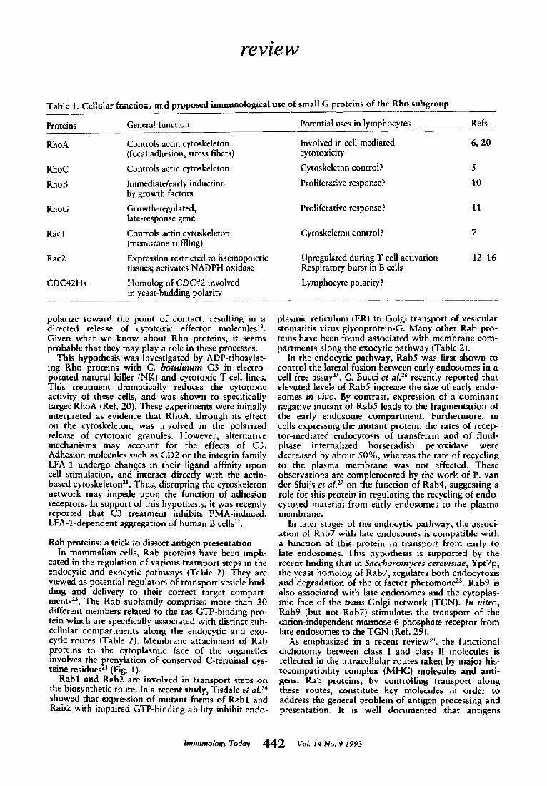
Table 1. Cellular functio~ ard proposed immunological use of small G proteins of the Rho subgroup
Proteins General function Potential uses in lymphocytes Refs
RhoA Controls actin cytoskeleton Involved in cell-mediated 6, 20 (focal adhesion, stress fibers) cytotoxicity
RhoC Controls actin cytoskeleton Cytoskeleton control? 5
RhoB Immediate/early induction Proliferative response? 10 by growth factors
RhoG Growth-regulated, Proliferative response? 11 late-response gene
Racl Controls actin cytoskeleton Cytoskeleton control? 7 (memb:ane ruffling)
Rac2 Expression restricted to haemopoietic Upregulated during T-cell activation 12-16 tissues; activates NADPH oxidase Respiratory burst in B ceils
CDC42Hs Homolog of CDC42 involved Lymphocyte polarity? in yeast-budding polarity
polarize toward the point of contact, resulting in a directed release of cytotoxic effector molecules 19. Given what we know about Rho proteins, it seems probable that they may play a role in these procegses.
This hypothesis was investigated by ADP-rihosylat- ing Rho proteins with C. botul inum C3 in electro- porated natural killer (NK) and cytotoxic T-cell lines. This treatment dramatically reduces the cytotoxic activity of these cells, and was shown to specifically target RhoA (Ref. 20). These experiments were initially interpreted as evidence that RhoA, through its effect on the cytoskeleton, was involved in the polarized release of cytotoxic granules. However, alternative mechanisms may account for the effects of C3. Adhesion molecules such as CD2 or the integrin family LFA-1 undergo changes in their ligand affinity upon cell stimulation, and interact directly with the actin- based cytoskeleton2L Thus, disrupting the cytoskeleton network may impede upon the function of adhesion receptors. In support of this hypothesis, it was recently reported that C3 treatment inhibits PMA-induced, LFA-l-depet,_dent aggregation c,f human B cells :z.
Rab proteins: a trick to dissect antigen presentation In mammalian cells, Rab proteins have been impli-
cated in the regulation of various transport steps in the endocytic and exocytic pathways (Table 2). They are viewed as potential regulators of transport vesicle bud- ding and delivery to their correct target compart- ments zs. The Rab subfamily comprises more than 30 different members related to the ras GTP-binding pro- tein which are specifically associated with distinct sub- cellular compartments along the endocytic apd exo- cytic routes (Table 2). Membrane attachmem of Rab proteins to the cytoplasmic face of the organdies involves the prenylation of conserved C-terminal cys- teine residues 2j (Fig, 1).
Rabl and Rab2 are involved in transport steps on the biosynthetic route. In a recent study, Tisdale e; al.Z 4 showed that expression of mutant forms of R~bl and Rab2 with impaired GTP-binding ability inhibit endo-
plasmic reticulum (ER) to Golgi transport of vesicular stomatitis virus glycoprotein-G. Many other Rab pro- teins have been found associated with membrane com- partments along the exocytic pathway (Table 2).
In the endocytic pathway, Rab5 was first shown to control the lateral fusion between early endosomes in a cell-free assay '-s. C. Bucci et al. 2~ recently reported that elevated levels of Rab5 increase the size of early endo- somes in vivo. By contrast, expression of a dominant negative mutant of Rub5 leads to the fragmentation of the early endosome compartment. Furthermore, in cells expressing the mutant protein, the rates of recep- tor-mediated endocytosis of transferrin and of fluid- phase internalized horseradish peroxidase were decreased by about 50%, whereas the rate of recycling to the plasma membrane was not affected. These observations are complemented by the work of P. van der Slui~s et al. z: on the function of Rab4, suggesting a role for this protein in regulating the recycling of endo- cytosed material from early endosomes to the plasma membrane.
In later stages of the endocytic pathway, the associ- ation of Rab7 with late endosomes is compatible with a function of this protein in transport from early to late endosomes. This hypothesis is supported by the recent finding that in Saccbaromyces cerevisiae, Ypt7p, the yeast homolog of Rab7, regulates both endocytosis and degradation of the 0t factor pheromone 2s. Rab9 is also associated with late endosomes and the cytoplas- mic face of the trans-Golgi network (TGN}. In vitro, Rab9 (but not Rab7) stimulates the transport of the cation-independent mannose-6-phosphate receptor from late endosomes to the TGN (Ref. 29).
As emphasized in a recent review 3°, the functional dichotomy between class l and class I1 molecules is reflected in the intracellular routes taken by major his- tocompatibility complex (MHC) molecules and anti- gens. i/_ab proteins, by controlling transport along these routes, constitute key molecules in order to address the general problem of antigen processing and presentation. It is well documented that antigens
Toa, r 4 4 2 'CoL No. 9 993

review
Table 2. Intracellular locations and functions of the small GTP-binding proteins of the YPT/Sec4/Rab subgroup. Italics refer to the S. cerevisiae Yptl, -7 and Sec4 proteins
Proteins lntracetlular location Transport steps Refs
Rabl Smooth ER and Golgi apparatus ER to Golgi 40
Yptl Golgi apparatus ER to Golgi 35
Rab2 Intermediate compartment ER to Golgi 24, 41 between ER and Golgi
Synaptic vesicles
Early endosomes
Early endosomes, clathrin-coated vesicles, plasma membrane
R~h3a
Rub4
Rab5
Rab6
Rab7
Ypt7
Rab8
Rab9
Sec4p
Golgi: medial- and trans-cisternae
Late endosomes
Late endosomes
Golgi apparatus
trans-Golgi network and late endosomes
Secretory vesicles and plasma membrane
Regulated exocytosis 42, 43
Early endosomes 27, 44
Early endosome lateral 25, 26, 41 fusion, plasma membrane to early endosome
Unknown 45, 46
Unknown 41
Endocytosis and 28 degradation of a-factor
Unknown 47
Late endosomes to 29 trans-Golgi network
Golgi to plasma membrane 34
ER: endoplasmic reticulurn.
captured from the external milieu are processed and associate with class II along the endocytic pathway ~'. However, neither the meeting point of class II mol- ecules and antigenic peptides nor the route of delivery of class I! molecules to the endosomes have been clearly defined. In addition, the transport mechanism of class II-peptide complexes to the cell surface are still unknown. In this section we would like to illustrate how small GTP-binding proteins of the Rub subfamily may help to clarify these different issues.
During the past two years, various morphological studies addressed the co-localization of class II with markers of the endocytic compart.ment~32"3L Discrepancies between those reports probably reflect differences in the distribution of the markers in various antigen-presenting cells (APCs). An important concept emerging from the studies on Rab proteins resides in their s~rict associatk.n will a specific :;abcellular com- partment {Table 2}. Consequently, Rab proteins con- stitute ideal markers of these compartments. For instance, antibodies specific for Rab4, Rab5 and Rab7 associated with early and late endocytic compartments {Table 2} should allow the precise location of the mol- ecules involved in class lI presentation : 'ong the endo- cytic pathway (class II, iuvariant chain and antigens).
As mentioned previously, the expression of domi- nant negative mutant forms of Rub proteins causes a significant inhibition of vesicular transport between two successive compartments and accumulation of transport intermediates :6'zT'J4.3s. In this respect, domi- nant negative mutants that control transport along the endocytic route are of particular inrerest. Indeed, Rab4
and Rab5 that control early endocytic functions (uptake, lateral fusion and recycling) may help to evalu- ate the exact contribution of the early endosome com- partment. For instance, the overexpression of those Rab proteins in antigen-presenting cells (APCs) fol- lowed by a detailed analysis of the steady-state distri- bution of class II molecules should discriminate between a transport of class II to the cell surface followed by rapid internalization versus a direct delivery to the endosomes from the biosynthetic route.
The existence of a class II-bearing compartment specific for APCs has also been suggested from ultra- structural studies3L The existence of tissue-specific Rub proteins ~6-3s favors the possibility that membrane transport to and from such a specific compartment may be regulated by APC-specific Rab protein{s). Cloning strategies based on polymerase chain reaction IPCR) 39 are available to identify novel Rub sequences from professional APCs. Understanding their function should enable us to form a comprehensive view of how antigen maturation and presentation by MHC-class II molecules is achieved.
Perspectives The purpose of this review was largely to speculate
(given our limited knowledge) on the possible roles of Rho and Rab proteins in immunology. Besides p21ras, proteins of the Rho and Rub subfamilies are likely to play a pivotal role in the final steps of the signal trans- duction cascade that elicits the various effector func- tions of lymphocytes. For clarity, these two subfamilies of ras-related proteins were dealt with in separate
roaoy 443 voL No. 9 9o3

review
paragraphs. However, it is quite conceivable that they act in concert to regulate some of the essential lympho- cyte functions. For example, polarized secretion induced by lymphocyte stimulation should require participation of various Rab proteins (at the level of the intracellular vesicles), as well as cytoskeletal con- trol exerted by the Rho proteins. As our understanding of the functions and regulation of small G proteins progresses, it should bring new insights to the function of lymphocytes. In return, since the lymphocyte func- tions are so diverse, and to some extent, easy to manipulate in vitro, an immunological approach of these problems will allow a better understanding of ras-related G proteins. In particular the question of how immune receptor-initiated event.,; could possibly control the activation state of ras-related G proteins should represent a fruitful area of research for the coming years.
Pbitippe Cbavrier and Jean-Pierre Gorvel are at the Centre d'lmmunologie INSERM-CNRS Marseilte Luminy, Ca.,;e 906. 13288 Marseille, Cedex 9, Prance; Jacques Benoglio is at INSERM U333, lnstitut Gustave Roussy, 39 rue Camille Desmoulins, 94805 Villejuif, Cedex, France.
R e f e r e n c e s 1 Hall, A. (1990) Science 249, 635-640 2 Bourne, H.R., Sanders, D.A. and McCormick, F. {1991) Nature 349, 117-127 3 Valencia, A., Chardin, P., Wittinghofer, A. and Sander, C. (1991) Biochemistry 30, 4637-4648 4 Downward, J., Graves, J.D. and Cantrell, D.A. (1992) lmmunoL Today 13, 89-92 5 Chardin, L, Boquet, P., Madaule, P. et aL (1989) EMBO ]. 8, 1087-~092 6 Ridley, A.J. and Hail, A. (1992) Cell 70, 389-399 7 R!d/ey, A.J2 t'aterson,_H_.F:,_Johnston, C.L., Diekmarm, 0. ana ~an, a. !l~y2) Cell 70, 4Ot-410 8 Sertleman, j., Albright, C.F., Foster, L.C. and Weinberg, R.A. (1992) Nature 359, 153-159 9 Shou, C., Farnsworth, C.L., Ned, B.G. and Fog, L.A. (1992) Nature 358, 351-354 10 Jahner, D. and Hunter, T. (1991) ?viol Cell. Biol. 11, 3682-3690 11 Vincent, S., Jeanteur, P. and Fort, P. (1992) Mol. Cell. Biol. 12, 3138-3148 12 Didsbury, J , Webec, R.F., Bokoch, G.M., Evans, T. and Snyderman, R. (1989)]. Biol. Chem. 264, 16378-16382 13 Reibel, L., DorseuiL O., Stancou, R., Bertogl~o. J. and Gacon, G. (1991) Biocbem. Biophys. Res. Commun. 175, 451-458 14 Abo, A., Pick, E., Hall, A. et al. (1991) Nature 353, 668-670 15 Knaus, U.G., Heyworth, P.G., Evans, T., Curnutte, J.T. and Bokoch, G.M. (1991) Science 254, 1512-1515 16 Dorseuil, O., Vazquez, A., Lang, P. et al. (1992)J. Biol. Chem. 267, 20540-20542 17 Nelson, W.J. (!992) Science 258,948-955 18 Poo, W.J., Conrad, L. and Janeway, C.A., Jr ~1988) Nature 332, 378-380 19 Podack, E.R. and Kupfer, A. (1991) Annu. Rev. Cell. Biol. 7, 479-504 20 Lang, p., Guizani, L., Vitte-Mony, I. et al. (1992)]. Biol. Cbem. 267, 11677-11680 21 Hynes, R.O. (1992) Ce!169, 11-25
22 Tomlnaga, T., Sugie, K., Hirata, M. et aL (1993)J. Cell Biol. 120, 1529-1537 23 Pfeffer, S.R. t1992) Trends Cell BioL 2, 41-46 24 Tisdale, E.J., Bourne, J.R., Khosravi-Far~ R., Der, C.J. and Balch, W.E. (1992) J. Cell Biol. 119, 749-761 25 Gorvel, J-P., Chavrier, P., Zerial, M. and Gruenberg, J. (1991) Cell 64, 915-925 26 Bucci, C., Patton, R.G., Mather, I.M..,t aL 11992) Cell 70, 715-728 27 van der Sluiis, P., Hull, M, Webster, P. etal. (1992) Cell 70, 729-740 28 Wichmann, H., Hengst, L. and Gailwitz, D. (1992) Cell 71, 1131-1142 29 Lombardi, D., Soldati, T., Riederer, M.A. et at. (1993) EMBO J. 12, 677-682 30 Neefjes, J.J. and PIoegh, H.L. (1992) lmmunol. Today 13, 179-184 31 Unanue, E.R. (1992) Curt. Opin. Immunol. 4, 63-69 32 Harding, C.V., Unanue, E.R., Slot, J.W., Schwartz, A.L. and Geuze, l-i.j. (i990) Proc. NatlAcad. Sci. USA 87, 5553-5557 33 Peters, P.J., Neefjes, J.J., Oorschot, V., PIoegh, H.L. ar.d Geuze, H.J. (1991) Natvre 349, 668-676 34 Goud, B., Salrninen, A., Walworth, N.C. and Novick, P.J. (1988) Cell 53,753-768 35 Baker, D., Wuestehube, L., Schekman, R. et al. (1990) Proc. Nad Acad. Sci. USA 87, 355-359 36 Mizoguchi, A., Kim, S., Ueda, T. and Takai, Y. 1!989) Biocbem. Biopbys. Res. Commun. 162, 1438-1445 37 Sano, K., Kikuchi, A., Matsui, Y., Teranishi, Y. and Takai, Y. (1989) Biocbem. Biopbys. Res. Commun. 158, 377-385 38 Liitcke, A., Jansson, S., Parton, R.G. et al. J. Cell Biol. (in press) 39 Chavrier, P., Simons, K. ~nd Zerial, M. (1992} Gene 112, 261-264 40 Plutner, H., Cox, A.D., Pin& S. etaL (1991)]. Cell Biol. 115, 31-43 41 Chavrier, P., Parton, R.G., Hauri, H.P., Simons, K. and Zerial, M. (1990) Cell 62, 317-329 42 Fisher von Moltard. G.. Mimaerv. G.A._ Raumort_ K.f et al. (1990) Proc. Nati Acad. Sci. (ISA 87,19ff8"'-1"9'9"2-" 43 Oberhauser, A.F., Monck, J., Balch, W.E. and Fernandez, J.M. (1992) Nature 360, 270-273 44 van der Sluijs, P., Hull, M., Zahraoui, A. et :d. (1991) Proc. Natl Acad. Sci. USA 88, 6313-6317 45 Goud, B., Zahraoui, A., Tavitian, A. and Saraste, J. (1990) Nature 345,553-556 46 Antony, C, Cibert, C., G&aud, G. etal. (1992) J. Cell Sci. 103, 785-796 47 Huber, L.A., Dotti, C., Patton, R. etm. ~1992) Eur. J. Cell Biol. 57 (Suppl. 36), 35
Next month in I T
® Costimulatory molecules and cancer - L ieping Chen
u Natural history of HLA expression during tumour deve!oFment - F. Garrido
• Anti-gal ant ibody in xenotransplantat ion - Uri Galili
• Immune system-blood vessel interactions - Jef lrey Flatt
Im,~.ol~ roaay 4 4 4 rot. 14 No. 9 t99j