an in vitro comparison of the effects of the iron-chelating agents, cp94 and dexrazoxane, on...
TRANSCRIPT

An In Vitro Comparison of the Effects of the Iron-Chelating Agents,CP94 and Dexrazoxane, on Protoporphyrin IX Accumulation forPhotodynamic Therapy and/or Fluorescence Guided Resection
Emma Blake*, James Allen and Alison Curnow
Clinical Photobiology, Peninsula College of Medicine and Dentistry, University of Exeter, Knowledge Spa,Royal Cornwall Hospital, Truro, Cornwall, UK
Received 5 May 2011, accepted 25 July 2011, DOI: 10.1111/j.1751-1097.2011.00985.x
ABSTRACT
Photodynamic therapy (PDT) utilizes the combined interaction
of a photosensitizer, light and molecular oxygen to ablate tumor
tissue. Maximizing the accumulation of the photosensitizer
protoporphyrin IX (PpIX) within different cell types would be
clinically useful. Dermatological PpIX-induced PDT regimes
produce good clinical outcomes but this currently only applies
when the lesion remains superficial. Also, as an adjuvant
therapy for the treatment of primary brain tumors, fluorescence
guided resection (FGR) and PDT can be used to highlight and
destroy tumor cells unreachable by surgical resection. By
employing iron chelators PpIX accumulation can be enhanced.
Two iron-chelating agents, 1,2-diethyl-3-hydroxypyridin-4-one
hydrochloride (CP94) and dexrazoxane, were individually
combined with the porphyrin precursors aminolevulinic acid
(ALA), methyl aminolevulinate (MAL) and hexyl aminolevu-
linate (HAL). Efficacies of the iron-chelating agents were
compared by recording the PpIX fluorescence in human
squamous epithelial carcinoma cells (A431) and human glioma
cells (U-87 MG) every hour for up to 6 h. Coincubation of
ALA ⁄MAL ⁄HAL with CP94 resulted in a greater accumula-
tion of PpIX compared to that produced by coincubation of
these congeners with dexrazoxane. Therefore the clinical
employment of iron chelation, particularly with CP94 could
potentially increase and ⁄ or accelerate the accumulation of
ALA ⁄MAL ⁄HAL-induced PpIX for PDT or FGR.
INTRODUCTION
Photodynamic therapy (PDT) is a clinical treatment resultingfrom the interaction of a photosensitizer, molecular oxygen
and light of a specific wavelength. When all three componentsare combined simultaneously in sufficient amounts they resultin the production of singlet oxygen and other free radicals
causing diseased cells to undergo cell death through necrosis orapoptosis (1). An increased or excessive amount of photosen-sitizer would be clinically useful and so research is taking place
to try to increase photosensitizer accumulation during PDTwith a view to ultimately enhancing clinical efficacy.
The prodrugs aminolevulinic acid (ALA), methyl amino-
levulinate (MAL) and hexyl aminolevulinate (HAL) areporphyrin-inducing precursors of the endogenous photosensi-tizer protoporphyrin IX (PpIX) produced via the heme
biosynthesis pathway in nucleated cells in humans. Usuallyin cells, following PpIX production the next stage in thepathway is the insertion of ferrous iron (Fe2+) under the
action of the ferrochelatase enzyme (2), to convert PpIX intoheme, with the presence of free heme acting as a negativefeedback mechanism limiting the production of any further
ALA (3). However, the exogenous introduction of largeamounts of ALA ⁄MAL ⁄HAL bypasses this negative feedbackloop and there is a resultant accumulation of heme and hemeprecursors in the cell. Notably, the conversion of PpIX to
heme by ferrochelatase is relatively slow, which causes PpIXto temporarily accumulate within cells (4). In addition, indiseased cells some enzymes within the heme biosynthesis
pathway are over or under expressed favoring the continualproduction of PpIX (5–7).
The first results from a clinical trial utilizing ALA-induced
PpIX PDT were reported just over 20 years ago (8) and sincethen PpIX-induced PDT for a variety of cancers has beeninvestigated. Topically it can be used for the treatment of anumber of dermatological indications including the most
common form of skin cancer, basal cell carcinoma (BCC).Although PpIX-induced PDT produces good clinical outcomesand excellent cosmesis for the thinner, superficial BCCs,
efficacy for the treatment of thicker nodular BCCs (nBCC)remains lower than conventional surgical excision (9). Forpatients presenting with nBCC lesions that are large, multiple
and ⁄ or on cosmetically sensitive sites (10), surgical excisioncan be a disfiguring and distressing experience. Therefore,enhancement of PpIX-induced PDT for these thicker lesions
would be beneficial.Another promising application of PpIX accumulation
within cancer cells is within human brain tumor cells. Humanglioblastomas are aggressive primary brain tumors, which
remain the most common form of this malignancy (11) andproblematic to treat given their infiltrative and mobile char-acteristics (12). Conventional treatments for brain tumors
include surgery, radiotherapy and chemotherapy. Although
*Corresponding author email: [email protected] (Emma Blake)� 2011 The AuthorsPhotochemistry and Photobiology � 2011 The American Society of Photobiology 0031-8655/11
Photochemistry and Photobiology, 2011, 87: 1419–1426
1419

these conventional therapies are potentially curative, the5 year survival rates remain below 5%, with approximately80% of reoccurrences occurring within 2 cm of the surgicallyresected margin (13). Activated PpIX, under light of a specific
wavelength, has the ability to fluoresce and Stummer et al.observed a fluorescence ratio of 5:1 between malignant andnormal tissue in vitro (14). Utilizing a technique termed
fluorescence guided resection (FGR) (15), surgeons can exploitPpIX’s natural fluorescent properties enabling greater preci-sion in the removal of brain tumors (16,17). In addition to
FGR, PpIX-induced PDT, as an adjuvant therapy, can be usedto destroy residual tumor tissue unreachable by surgicalresection. Improvement of PpIX accumulation within glio-
blastoma cells for the application of FGR and ⁄ or PDT couldpotentially improve prognosis, prolong time interval to tumorrecurrence and vitally improve quality of life.
It has been shown that PpIX accumulation can be
enhanced by employing iron chelation (18). An iron-chelatingagent can temporarily remove free Fe2+ from the system,inhibiting its insertion into PpIX, thus preventing the
formation of heme and as a result PpIX accumulates in thecell (19). Iron-chelating agents which have been investigatedin the past include ethylenediamine tetra acetic acid (EDTA)
(20) and desferrioxamine (DFO) (21). Liu et al. (2) concludedthat a nonspecific metal chelator (with the potential to inhibitferrochelatase activity), calcium-disodium edentate (CaNa2EDTA) combined with ALA increased PpIX accumulation
and photosensitization in Hep-2 cells. In addition to cellculture tests they treated 12 nonmelanoma skin cancerpatients with ALA combined with CaNa2 EDTA-PDT and
found greater depth of penetration than compared to ALA-PDT alone. From these experiments they postulated that ifCaNa2 EDTA combined with ALA was used clinically for
the treatment of cutaneous cancers the depth of PpIXpenetration and therefore clinical outcome could be improved(2). DFO has greater specificity for iron than EDTA and
research by our group has shown 1,2-diethyl-3-hydroxypyri-din-4-one hydrochloride (CP94) to be significantly superior toDFO in the enhancement of PpIX accumulation using ALAand MAL in fetal lung fibroblasts and squamous carcinoma
cells (19).Dexrazoxane (ICRF-187) is a clinically approved anthra-
cycline-induced cardioprotective agent which has been used
for more than 20 years (22). Dexrazoxane’s proposedmechanism of action is that it readily penetrates cellmembranes, is intracellularly converted into two one-ring
open intermediates followed by the production of ADR-925,which can either remove iron from the iron-anthracyclinecomplex or bind to free iron (22). It has been reported thatall three hydrolysis products are good iron-chelating agents
(23) and dexrazoxane itself is therefore known as an iron-chelating prodrug.
These findings suggest that dexrazoxane may be used
clinically as a successful iron-chelating prodrug to enhancePpIX accumulation and to our knowledge has notpreviously been investigated for this purpose. This study
therefore compares the effects of the iron-chelating agentsCP94 and dexrazoxane, on the accumulation of PpIX withinhuman squamous epithelial cells and human glioma cells
in vitro with ALA, MAL and HAL as the porphyrinprecursors.
MATERIALS AND METHODS
Chemicals and cells. All reagents and chemicals were purchased fromSigma-Aldrich Chemical Company (Poole, UK) unless otherwisestated. U-87 MG (human glioblastoma-astrocytoma, epithelial like)and A431 (human epithelial squamous carcinoma) cells were pur-chased from the European Collection of Cell Cultures (ECACC,Wiltsure, UK). Under aseptic conditions in a class II laminar flowcabinet, cells were cultured in Eagle’s minimum essential medium(EMEM) with 10% fetal calf serum (standardized to give aniron concentration between 450 and 600 lg ⁄ 100 g), 2% (200 mMM)LL-glutamine and 2% (200 U mL)1) penicillin and (200 lg mL)1)streptomycin solution. Stock solutions of ALA ⁄MAL ⁄HAL wereprepared in PBS, adjusted to physiological pH (pH 7.4) using NaOH(0.5 mMM), filter sterilized (0.22 lm Millipore) and stored at )20�C forup to 1 month. Cells were grown in 5% CO2 at 37�C and left to growuntil 70% confluent at which time cells were routinely passaged (every3–5 days).
Iron chelator cytotoxicity. Before investigating PpIX accumulationthe dark toxicity of each iron-chelating agent was determined usingthe XTT (2,3-bis[2-methoxy-4-nitro-5-sulfophenyl]-2H-tetrazololium-5-carboxyanilide inner salt) cell viability assay. This is a colorimetricassay based on the reduction of the XTT tetrazolium salt bymitochondrial dehydrogenase enzymes.
A431 and U-87 MG cells were seeded into multiwell plates at adensity of 1 · 105 cells per mL (1 · 104 cells per well) and left to adhereovernight at 5% CO2 and 37�C. Under low light conditions themedium was aspirated from the wells and the cells washed with PBS inpreparation for the addition of freshly prepared test solutions. Finalconcentrations of dexrazoxane and CP94 were prepared with modifiedEMEM (minus phenol red): dexrazoxane (50 and 150 lm) and CP94(150 lm). A 100 lL volume of each test solution was added to the cellsin triplicate wells. Control wells were also included which containedcells only incubated with modified EMEM. Once the test solutions hadbeen added, cells were incubated for 3 h in 5% CO2 at 37�C.
Following this incubation period, the medium containing the testsolutions was removed from the wells and cells were washed threetimes with PBS. A volume of 120 lL serum free medium per wellcontaining XTT was then added. The plate was returned to theincubator for a further 2 h and following this, absorbance of theresulting color change was measured with a Synergy HT plate reader(BIO-TEK, Germany). Absorbance was read at 450 and 690 nm, withthe 690 nm wavelength being used as a reference range and subtractedfrom the 450 nm reading. Measurements were represented as apercentage of the control cells, incubated with modified EMEM only.
PpIX fluorescence quantification. Cells were seeded into multiwellplates at a density of 1 · 105 cells per mL (1 · 104 cells per well) andleft to adhere overnight at 5% CO2 and 37�C. Under low lightconditions the medium was aspirated from the wells and the cellswashed with PBS in preparation for the addition of freshly preparedtest solutions. Final concentrations of ALA ⁄MAL ⁄HAL were pre-pared with modified EMEM (minus phenol red): ALA (250 lm) ⁄MAL(1000 lm) ⁄HAL (10 lm). To produce solutions of equal iron bindingequivalence dexrazoxane was used at 50 lMM and CP94 at a concen-tration of 150 lMM. To compare equal molar concentrations dexrazox-ane was also used at 150 lMM. All concentration combinations weretested in quintuplicate on each plate in a minimum of three separateexperiments conducted on separate days.
Under dark room conditions PpIX fluorescence quantification wasconducted using a multiwell plate reader (Synergy HT; Bio-Tek, UK).Measurements were taken from the bottom of the wells with a400 ± 30 nm excitation filter and a 645 ± 40 nm emission filter inplace. Readings were taken every hour for a total of 6 h and underdark room conditions to reduce photobleaching of PpIX. After ⁄between each reading, plates were kept in the dark at 5% CO2 and37�C.
Data analysis and statistics. The PpIX fluorescence measurementsfrom the control cells not incubated with any test compound were usedto remove natural cellular PpIX autofluorescence from all the othermeasurements made from the same plate. Following the determinationof the presence of parametric data sets statistical significance betweenindividual groups was determined using the Student’s t-test andANOVA was employed when considering differences between datasets.
1420 Emma Blake et al.

RESULTS
Dark toxicity of CP94 and dexrazoxane
The dark toxicity of the two iron-chelating agents, CP94 anddexrazoxane, incubated alone with squamous carcinoma
(A431) and glioma (U-87 MG) cells was investigated(Fig. 1). It was observed that both iron-chelating agentsproduced similar results in each cell type with the Student’st-test revealing that at the concentrations tested no statistically
significant toxicity was observed with either iron-chelatingagent, when compared to the same cell type incubated withmodified EMEM alone (control group) (Fig. 1).
The effect of CP94 and dexrazoxane on PpIX accumulation in
squamous carcinoma (A431) cells
In the human epithelial carcinoma cells (A431) incubated with
the prodrugs ALA ⁄MAL ⁄HAL with and without the ironchelators CP94 and dexrazoxane, PpIX fluorescence levelsincreased hourly and using an ANOVA with a posttest a
significant linear trend was found (P < 0.01, Fig. 2a,b).Comparison of ALA ⁄MAL ⁄HAL + CP94 with ALA ⁄MAL ⁄HAL alone (Fig. 2a,c) indicated that greater levels ofPpIX fluorescence were detected in cells in the CP94 group and
this difference was found to be statistically significant(ANOVA, P < 0.05) with all three prodrugs. When cellsincubated with ALA ⁄MAL ⁄HAL + dexrazoxane, at either
concentration (50 or 150 lMM), were compared to thoseincubated with ALA ⁄MAL ⁄HAL alone (Fig. 2a,c) using
0
50
100
150
blank control
50 µM dexrazoxane
150 µM dexrazoxane
150 µMCP94
Cel
l via
bilit
y as
% o
f con
trol
U87MG
A431
Figure 1. Dark toxicity of the test compounds, CP94 (150 lMM) anddexrazoxane (50 & 150 lMM), incubated with A431 and U-87 MG cellsalone. Cell viability is presented as a percentage of the blank controlcells incubated with modified EMEM alone. Bars indicate the mean ofthree wells with 1 standard deviation. EMEM = Eagle’s minimumessential medium.
(a) (b)
(c) (d)
0
50
100
150
200
0 1 2 3 4 5 6
PpIX
fluo
resc
ence
as
% o
f con
trol
Time / hours
ALA aloneALA + CP94 (150 µM)ALA + dexrazoxane (50 µM)ALA + dexrazoxane (150 µM)
0
50
100
150
200
0 1 2 3 4 5 6
PpIX
fluo
resc
ence
as
% o
f con
trol
Time / hours
MAL aloneMAL + CP94 (150 µM)MAL + dexrazoxane (50 µM)MAL + dexrazoxane (150 µM)
0
50
100
150
200
0 1 2 3 4 5 6
PpIX
fluo
resc
ence
as
% o
f con
trol
Time / hours
HAL aloneHAL + CP94 (150 µM)HAL + dexrazoxane (50 µM)HAL + dexrazoxane (150 µM)
0123456
Tim
e / h
ours
***
∆
**
**
*
**+
+
*+∆ *
+∆ *
+∆
*+∆ *
+∆
**+
+
*+∆
*+∆
*+∆
*+∆
*+∆
*+
*+
*+ *
+
**
**
∆ *+∆ *
+∆ *
+∆ *
+∆ *
+∆ *
+∆
*+∆
*
**
*+∆
*
*+∆
Figure 2. Protoporphyrin IX (PpIX) fluorescence in A431 cells incubated with (a) 250 lMM ALA, (b) 1000 lMM MAL, (c) 10 lMM HAL alone or incombination with CP94 (150 lMM) or dexrazoxane (50 & 150 lMM), as a function of time. A statistically significant difference in PpIX fluorescencefrom the prodrug only incubated control cells is indicated by * (P < 0.05). A statistically significant difference between cells incubated with CP94and 50 lMM dexrazoxane is indicated by + (P < 0.05) and a statistically significant difference between cells incubated with CP94 and 150 lMM
dexrazoxane is indicated by D (P < 0.05). Times for chelator incubated cells to achieve comparable levels of fluorescence to the ALA ⁄MAL ⁄HALalone treated cells at 6 h, indicated by the hashed lines on panels (a)–(c), are presented in (d). ALA = aminolevulinic acid; MAL = methylaminolevulinate; HAL = hexyl aminolevulinate.
Photochemistry and Photobiology, 2011, 87 1421
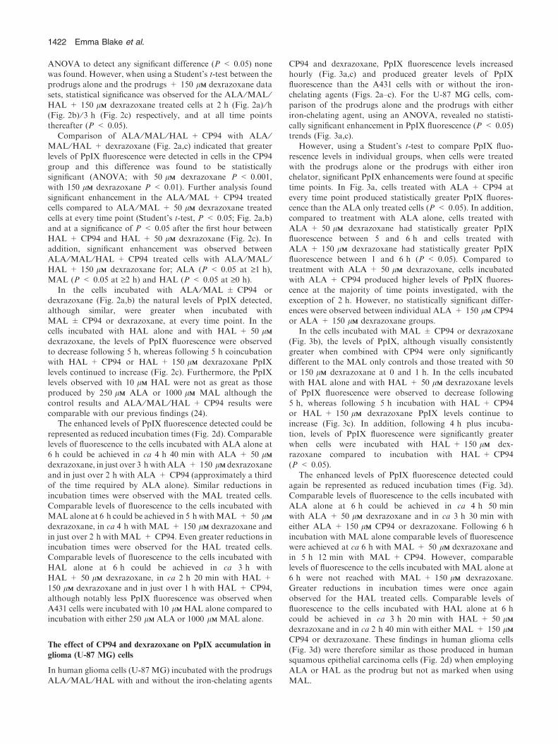
ANOVA to detect any significant difference (P < 0.05) nonewas found. However, when using a Student’s t-test between theprodrugs alone and the prodrugs + 150 lMM dexrazoxane datasets, statistical significance was observed for the ALA ⁄MAL ⁄HAL + 150 lMM dexrazoxane treated cells at 2 h (Fig. 2a) ⁄ h(Fig. 2b) ⁄ 3 h (Fig. 2c) respectively, and at all time pointsthereafter (P < 0.05).
Comparison of ALA ⁄MAL ⁄HAL + CP94 with ALA ⁄MAL ⁄HAL + dexrazoxane (Fig. 2a,c) indicated that greaterlevels of PpIX fluorescence were detected in cells in the CP94
group and this difference was found to be statisticallysignificant (ANOVA; with 50 lMM dexrazoxane P < 0.001,with 150 lMM dexrazoxane P < 0.01). Further analysis found
significant enhancement in the ALA ⁄MAL + CP94 treatedcells compared to ALA ⁄MAL + 50 lMM dexrazoxane treatedcells at every time point (Student’s t-test, P < 0.05; Fig. 2a,b)and at a significance of P < 0.05 after the first hour between
HAL + CP94 and HAL + 50 lMM dexrazoxane (Fig. 2c). Inaddition, significant enhancement was observed betweenALA ⁄MAL ⁄HAL + CP94 treated cells with ALA ⁄MAL ⁄HAL + 150 lMM dexrazoxane for; ALA (P < 0.05 at ‡1 h),MAL (P < 0.05 at ‡2 h) and HAL (P < 0.05 at ‡0 h).
In the cells incubated with ALA ⁄MAL ± CP94 or
dexrazoxane (Fig. 2a,b) the natural levels of PpIX detected,although similar, were greater when incubated withMAL ± CP94 or dexrazoxane, at every time point. In thecells incubated with HAL alone and with HAL + 50 lMM
dexrazoxane, the levels of PpIX fluorescence were observedto decrease following 5 h, whereas following 5 h coincubationwith HAL + CP94 or HAL + 150 lMM dexrazoxane PpIX
levels continued to increase (Fig. 2c). Furthermore, the PpIXlevels observed with 10 lMM HAL were not as great as thoseproduced by 250 lMM ALA or 1000 lMM MAL although the
control results and ALA ⁄MAL ⁄HAL + CP94 results werecomparable with our previous findings (24).
The enhanced levels of PpIX fluorescence detected could be
represented as reduced incubation times (Fig. 2d). Comparablelevels of fluorescence to the cells incubated with ALA alone at6 h could be achieved in ca 4 h 40 min with ALA + 50 lMM
dexrazoxane, in just over 3 hwithALA + 150 lMMdexrazoxane
and in just over 2 h with ALA + CP94 (approximately a thirdof the time required by ALA alone). Similar reductions inincubation times were observed with the MAL treated cells.
Comparable levels of fluorescence to the cells incubated withMAL alone at 6 h could be achieved in 5 h withMAL + 50 lMM
dexrazoxane, in ca 4 h with MAL + 150 lMM dexrazoxane and
in just over 2 h with MAL + CP94. Even greater reductions inincubation times were observed for the HAL treated cells.Comparable levels of fluorescence to the cells incubated withHAL alone at 6 h could be achieved in ca 3 h with
HAL + 50 lMM dexrazoxane, in ca 2 h 20 min with HAL +150 lMM dexrazoxane and in just over 1 h with HAL + CP94,although notably less PpIX fluorescence was observed when
A431 cells were incubated with 10 lMM HAL alone compared toincubation with either 250 lMM ALA or 1000 lMM MAL alone.
The effect of CP94 and dexrazoxane on PpIX accumulation in
glioma (U-87 MG) cells
In human glioma cells (U-87 MG) incubated with the prodrugsALA ⁄MAL ⁄HAL with and without the iron-chelating agents
CP94 and dexrazoxane, PpIX fluorescence levels increasedhourly (Fig. 3a,c) and produced greater levels of PpIXfluorescence than the A431 cells with or without the iron-chelating agents (Figs. 2a–c). For the U-87 MG cells, com-
parison of the prodrugs alone and the prodrugs with eitheriron-chelating agent, using an ANOVA, revealed no statisti-cally significant enhancement in PpIX fluorescence (P < 0.05)
trends (Fig. 3a,c).However, using a Student’s t-test to compare PpIX fluo-
rescence levels in individual groups, when cells were treated
with the prodrugs alone or the prodrugs with either ironchelator, significant PpIX enhancements were found at specifictime points. In Fig. 3a, cells treated with ALA + CP94 at
every time point produced statistically greater PpIX fluores-cence than the ALA only treated cells (P < 0.05). In addition,compared to treatment with ALA alone, cells treated withALA + 50 lMM dexrazoxane had statistically greater PpIX
fluorescence between 5 and 6 h and cells treated withALA + 150 lMM dexrazoxane had statistically greater PpIXfluorescence between 1 and 6 h (P < 0.05). Compared to
treatment with ALA + 50 lMM dexrazoxane, cells incubatedwith ALA + CP94 produced higher levels of PpIX fluores-cence at the majority of time points investigated, with the
exception of 2 h. However, no statistically significant differ-ences were observed between individual ALA + 150 lMM CP94or ALA + 150 lMM dexrazoxane groups.
In the cells incubated with MAL ± CP94 or dexrazoxane
(Fig. 3b), the levels of PpIX, although visually consistentlygreater when combined with CP94 were only significantlydifferent to the MAL only controls and those treated with 50
or 150 lMM dexrazoxane at 0 and 1 h. In the cells incubatedwith HAL alone and with HAL + 50 lMM dexrazoxane levelsof PpIX fluorescence were observed to decrease following
5 h, whereas following 5 h incubation with HAL + CP94or HAL + 150 lMM dexrazoxane PpIX levels continue toincrease (Fig. 3c). In addition, following 4 h plus incuba-
tion, levels of PpIX fluorescence were significantly greaterwhen cells were incubated with HAL + 150 lMM dex-razoxane compared to incubation with HAL + CP94(P < 0.05).
The enhanced levels of PpIX fluorescence detected couldagain be represented as reduced incubation times (Fig. 3d).Comparable levels of fluorescence to the cells incubated with
ALA alone at 6 h could be achieved in ca 4 h 50 minwith ALA + 50 lMM dexrazoxane and in ca 3 h 30 min witheither ALA + 150 lMM CP94 or dexrazoxane. Following 6 h
incubation with MAL alone comparable levels of fluorescencewere achieved at ca 6 h with MAL + 50 lMM dexrazoxane andin 5 h 12 min with MAL + CP94. However, comparablelevels of fluorescence to the cells incubated with MAL alone at
6 h were not reached with MAL + 150 lMM dexrazoxane.Greater reductions in incubation times were once againobserved for the HAL treated cells. Comparable levels of
fluorescence to the cells incubated with HAL alone at 6 hcould be achieved in ca 3 h 20 min with HAL + 50 lMM
dexrazoxane and in ca 2 h 40 min with either MAL + 150 lMM
CP94 or dexrazoxane. These findings in human glioma cells(Fig. 3d) were therefore similar as those produced in humansquamous epithelial carcinoma cells (Fig. 2d) when employing
ALA or HAL as the prodrug but not as marked when usingMAL.
1422 Emma Blake et al.

DISCUSSION
One of the major limitations of PpIX-induced FGR and PDT
is the accumulation and localization of PpIX within tumorcells. Although it has been increasingly reported that PpIXaccumulation can be enhanced with iron chelation (25), the use
of which PpIX precursor and which iron-chelating agent toadminister for different cell types and which to take forwardinto clinical experimentation remains inconclusive.
This in vitro study was carried out to compare the increase
in PpIX fluorescence, over a 6 h-time-period, in humansquamous epithelial carcinoma cells (A431) and human gliomacells (U-87 MG) after exogenous administration of ALA ⁄MAL ⁄HAL with and without the two iron-chelating agentsCP94 and dexrazoxane. CP94 was used because previous invitro studies have shown CP94 to be significantly superior to
the well-known iron chelator DFO (21) at increasing levels ofPpIX (19). In addition, CP94 combined with ALA has beentested in vivo, with the PpIX fluorescence produced in ratcolonic mucosa double that of ALA alone (25). An ongoing
clinical investigation using increasing concentrations of CP94combined with ALA or MAL to carry out PDT on nBCCswhich after 6 weeks are surgically excised, is currently being
undertaken. Clinical assessment prior to excisions has so fardemonstrated that using ALA + 40% CP94 produced asignificantly greater clearance rate in nBCCs than with
ALA-PDT alone (26). The clinical use of CP94, however,remains experimental and nonlicensed at the current time.However, dexrazoxane is already approved and used clinically,exerting its iron-chelating properties successfully as a protec-
tive agent for anthracycline-induced cardiotoxicity. Therefore,comparing the iron-chelating efficacies of CP94 and dexraz-oxane with their ability to increase PpIX accumulation is
useful, and to our knowledge this is the first study toinvestigate the combined interaction of any PpIX precursorwith dexrazoxane.
In the human squamous epithelial carcinoma cells (A431)both CP94 and dexrazoxane treatment groups increased PpIXfluorescence when combined with the three prodrugs ALA ⁄MAL ⁄HAL, compared to each prodrug alone and markedly,
CP94 produced significantly greater levels of PpIX accumula-tion when compared to dexrazoxane for each of the prodrugstested. In addition, in A431 cells treated with CP94 and
dexrazoxane, at both concentrations tested, the same amountof PpIX fluorescence normally produced during 6 h incubationwith ALA ⁄MAL ⁄HAL was produced within shorter time
(a) (b)
(c) (d)
0
100
200
300
400
500
0 1 2 3 4 5 6
PpIX
fluo
resc
ence
as
% o
f con
trol
Time / hours
ALA aloneALA + CP94 (150 µM)ALA + dexrazoxane (50 µM)ALA + dexrazoxane (150 µM)
0
100
200
300
400
500
0 1 2 3 4 5 6
PpIX
fluo
resc
ence
as/ %
of c
ontr
ol
Time / hours
MAL aloneMAL + CP94 (150 µM)MAL + dexrazoxane (50 µM)MAL + dexrazoxane (150 µM)
0
100
200
300
400
500
0 1 2 3 4 5 6
PpIX
fluo
resc
ence
as
% o
f con
trol
Time / hours
HAL aloneHAL + CP94 (150 µM)HAL + dexrazoxane (50 µM)HAL + dexrazoxane (150 µM)
0123456
Tim
e / h
ours
*+
**+
*+
*+
*+
*
**
**
**
* *
*+ * *
+∆ *
+∆
*
+∆
*+
**+
*
*+∆
*
*
*+∆
> 6 hr
*+
**
* *+
Figure 3. Protoporphyrin IX (PpIX) fluorescence in U-87 MG cells incubated (a) 250 lMM ALA, (b) 1000 lMM MAL, (c) 10 lMM HAL alone or incombination with CP94 (150 lMM) or dexrazoxane (50 & 150 lMM), as a function of time. A statistically significant difference in PpIX fluorescencefrom the prodrug only incubated control cells is indicated by * (P < 0.05). A statistically significant difference between cells incubated with CP94and 50 lMM dexrazoxane is indicated by + (P < 0.05) and a statistically significant difference between cells incubated with CP94 and 150 lMM
dexrazoxane is indicated by D (P < 0.05). Times for chelator incubated cells to achieve comparable levels of fluorescence to the ALA ⁄MAL ⁄HALalone treated cells at 6 h, indicated by the hashed lines on panels (a)–(c), are presented in (d). ALA = aminolevulinic acid; MAL = methylaminolevulinate; HAL = hexyl aminolevulinate.
Photochemistry and Photobiology, 2011, 87 1423

periods. Notably, this level of fluorescence was reached threetimes quicker when ALA and MAL were combined with CP94and six times quicker when HAL was combined with CP94.Currently, dermatology PDT clinic drug-light-interval times
are 4–6 h for ALA and 3 h for MAL (27). This time isimportant to allow for optimum levels of PpIX to accumulateand to produce an optimum ratio of photosensitizer accumu-
lation between tumor and nontumor tissues (19). The resultsfrom this study suggest dermatology PDT clinics may benefitmost from inclusion of CP94 in their treatment regimes to
firstly reduce incubation times resulting in a greater number oftreatments being feasible in a day and thus reduced waitingtimes for patients and secondly to increase efficacy in
applications requiring enhancement. However, use of the moreclinically acceptable dexrazoxane may also produce someclinical benefits, and warrants further investigation.
In A431 cells it has been demonstrated here that CP94 is a
greater enhancer of PpIX accumulation than both concentra-tions of dexrazoxane investigated, for each of the PpIXprecursors tested. Notably, dexrazoxane was employed at two
concentrations; 50 lMM and 150 lMM, whereas CP94 was onlyemployed at 150 lMM. This was done to take into account thedifferent iron binding equivalences of the two iron-chelating
agents. Dexrazoxane is hexadentate meaning a single Femolecule is coordinated by one chelator molecule in a 1:1 ratio.CP94 is bidentate meaning it binds iron in the ratio of 3:1. As aresult the concentrations employed were chosen to produce
solutions of equal iron binding equivalence (150 lMM CP94 vs50 lMM dexrazoxane) and fairly compared equimolar concen-trations of each iron-chelating agent (150 lMM) but in each case
CP94 outperformed dexrazoxane.The end hydrolysis product of dexrazoxane, ADR-925, is a
cyclic derivative of EDTA which has been investigated
previously as an enhancer of PpIX-induced PDT (20). Thepolar nature of ADR-925, like that of EDTA prevents it frombeing able to cross cell membranes but this obstacle is
overcome when dexrazoxane is employed instead of EDTAas its likely mode of transport is passive diffusion across cellmembranes, similar to that shown for ICRF-159 (razoxane)(28). As CP94 and dexrazoxane can both readily enter cells one
could postulate the decreased efficacy of dexrazoxane toincrease PpIX accumulation compared to CP94, might beexplained by dexrazoxane’s conversion to its hydrolysis
products. Physiological conditions studies (29,30) have dem-onstrated that dexrazoxane is hydrolyzed to its one-ring openintermediates with a T1 ⁄ 2 of 9.3 h at 37�C and pH 7.4, whereas
the final hydrolysis product ADR-925 was produced with aT1 ⁄ 2 of 23 h. In the present study incubation times of up to 6 honly were employed but PpIX accumulation was still observedto occur at a greater level when each PpIX precursor was
combined with dexrazoxane than incubation with each PpIXprecursor alone. This could be explained by the observationthat dexrazoxane can be enzymatically hydrolyzed, more
rapidly, by dihydropyrimidine amidohydrolase to its one-ringopen intermediates (31,32), which like ADR-925 are also ableto chelate iron. In addition, nonenzymatic metal-ion-promoted
hydrolysis of dexrazoxane has also been reported (23,33) inwhich it was suggested that both ferrous and ferric forms ofiron are able to promote the hydrolysis of dexrazoxane or its
open ring intermediates enhancing their iron-chelating poten-tial.
In the human glioma cells (U-87 MG) it was againdemonstrated that iron chelation with either agent significantlyincreased PpIX fluorescence when combined with the threeprodrugs ALA ⁄MAL ⁄HAL, compared to each prodrug alone.
These results are complementary to previous work conductedby ourselves (24) and also by others (34) who have consideredthe use of PpIX-induced FGR. Valdes et al. (34) suggested
that although the use of ALA for FGR has shown promisingresults in previous clinical trials, due to heterogeneous stainingand variation in PpIX production some tumor margins go
undetected, proposing this may be remedied by combiningFGR with iron chelation. In a recent study by Valdes et al.mice were implanted with xenograft U251-GFP glioma tumor
cells and administered with DFO once a day for 3 days priorto ALA administration producing a 50% increase in PpIXfluorescence relative to the control group (34).
Notably, in our study, at concentrations of equal binding
equivalence (50 lMM dexrazoxane vs 150 lMM CP94) CP94 was astatistically greater enhancer of PpIX accumulation thandexrazoxane, when ALA or HAL were used as the PpIX
precursor. With the prodrug MAL, however, these differenceswere less marked. However, at equimolar concentrations(150 lMM) PpIX accumulation between the two iron-chelating
agents was similar, with the exception of coincubation withHAL where coincubation with dexrazoxane at 150 lMM resultedin greater PpIX accumulation compared to coincubation ofHAL with CP94. Nevertheless, the same amount of PpIX
fluorescence normally produced during 6 h incubation withHAL alone was produced in less than 3 h when HAL wascombined with either CP94 or 150 lMM dexrazoxane. The
in vitro results suggest that HAL coincubated with either ironchelator (CP94 or dexrazoxane) would most likely be the mostfavorable PpIX-induced therapy for the treatment of primary
gliomas through FGR and ⁄ or PDT. However, unlike derma-tology clinics where a reduced prodrug incubation period ishighly favorable, in the surgical resection of brain tumors a
reduced incubation period would take second place to pro-ducing the maximal amount of PpIX possible, given thatsuccessful tumor clearance is directly linked to the level ofPpIX accumulated within malignant cells and is of major
importance.In mice implanted with glioma tumor cells with
subsequent administration of DFO and ALA, 50% more
PpIX fluorescence was recently demonstrated, relative to thecontrol group (34). As previously mentioned CP94 has beenshown to be significantly superior to DFO in enhancing PpIX
accumulation (19) and CP94 has the ability to cross the blood–brain barrier (BBB) (35). In vivo animal studies shouldtherefore investigate the hypothesis that exogenous systemicadministration of ALA ⁄MAL ⁄HAL with CP94 in malignant
glioma tumors could significantly increase PpIX fluorescence.In addition, tumor-to-normal tissue selectivity should also beinvestigated in vivo. In the surgical resection of brain tumors
sparing normal tissue to maximize postoperational quality oflife is an important consideration therefore achieving maximaltumor-to-normal PpIX selectivity is highly sought-after.
Dexrazoxane is hydrophilic and as a result has poor BBBpenetration (36). However, there is a vast (and very desirable)amount of research into assisting larger drug molecules to
cross the BBB. This research includes the coadministration ofa bradykinin agonist which increases the permeability of the
1424 Emma Blake et al.

BBB allowing the drug of interest to pass into the braincompartment (37). An increasingly popular area of research,nanotechnology, may also allow passage of drugs across theBBB and in addition specific brain tumor targeting (38).
Our results in both cell types demonstrate coincubation withCP94 produces the greatest amount of PpIX fluorescence inthe order MAL > ALA > HAL. Markedly, U-87 MG cells
produced much greater levels of PpIX fluorescence than theA431 cells with all three prodrugs. Cell types show variabilityin their PpIX accumulation probably due to differences in their
ferrochelatase activity which varies between cell types (6,39).Ferrochelatase converts PpIX to heme by inserting a Fe2+ intothe protoporphyrin ring. Thus, although not investigated here,
if U-87 MG cells have a lower activity of the ferrochelataseenzyme than A431 cells and a higher metabolic rate, this couldexplain why over the same incubation period U-87 MG cellsproduce greater levels of PpIX fluorescence.
The objective of this study was to compare the effects of twoiron-chelating agents in combination with the porphyrinprecursors ALA ⁄MAL ⁄HAL. For both cell types coincuba-
tion with CP94 produced a greater enhancement of PpIXaccumulation than coincubation with dexrazoxane althoughthis difference was most marked in the dermatological cells
employed. The greatest amount of PpIX fluorescence wasobserved in the order MAL > ALA > HAL and the sameamount of PpIX fluorescence normally produced during 6 hincubation with each prodrug alone was produced in shorter
incubation times, when coincubated with CP94, achieved inthe order HAL < ALA < MAL. In conclusion, although theenhancement of PpIX accumulation for different cell types
may require different PpIX precursors or iron chelatorcombinations, this study has demonstrated that for ALA ⁄MAL ⁄HAL-induced PpIX production, enhancement can be
successfully achieved with iron chelation and provides furtherevidence for the combined clinical investigation of this methodof PDT ⁄FGR enhancement.
REFERENCES
1. Pass, H. I. (1993) Photodynamic therapy in oncology: Mecha-nisms and clinical use. J. Natl Cancer Inst. 85, 443–456.
2. Liu, H. F., S. Z. Xu and C. R. Zhang (2004) Influence of CaNa2EDTA on topical 5-aminolaevulinic acid photodynamic therapy.Chin. Med. J. (Engl.) 117, 922–926.
3. Szeimies, R. M., P. Calzavara-Pinton, S. Karrer, B. Ortel and M.Landthaler (1996) Topical photodynamic therapy in dermatology.J. Photochem. Photobiol. B 36, 213–219.
4. Pye, A., Y. Dogra, J. Tyrrell, P. Winyard and A. Curnow (2009)Redox signaling and regulation in biology and medicine. In Pho-todynamic Therapy with Aminolevulinic Acid and Iron Chelators:A Clinical Example of Redox Signaling (Edited by C. Jacob andP. Winyard), pp. 351–372. Wiley-VCH, Weinheim, Germany.
5. Dailey, H. A. and A. Smith (1984) Differential interaction ofporphyrins used in photoradiation therapy with ferrochelatase.Biochem. J. 223, 441–445.
6. el-Sharabasy, M. M., A. M. el-Waseef, M. M. Hafez and S. A.Salim (1992) Porphyrin metabolism in some malignant diseases.Br. J. Cancer 65, 409–412.
7. Gibson, S. L., D. J. Cupriks, J. J. Havens, M. L. Nguyen and R.Hilf (1998) A regulatory role for porphobilinogen deaminase(PBGD) in delta-aminolaevulinic acid (delta-ALA)-induced pho-tosensitization? Br. J. Cancer 77, 235–242.
8. Kennedy, J. C., R. H. Pottier and D. C. Pross (1990) Photody-namic therapy with endogenous protoporphyrin: IX: Basic prin-ciples and present clinical experience. J. Photochem. Photobiol.B: Biol. 6, 143–148.
9. Rhodes, L. E., M. de Rie, Y. Enstrom, R. Groves, T. Morken, V.Goulden, G. A. Wong, J. J. Grob, S. Varma and P. Wolf (2004)Photodynamic therapy using topical methyl aminolevulinatevs surgery for nodular basal cell carcinoma: Results of amulticenter randomized prospective trial. Arch. Dermatol. 140,17–23.
10. Curnow, A. and A. Pye (2007) Biochemical manipulation via ironchelation to enhance porphyrin production from porphyrin pre-cursors. J. Environ. Pathol. Toxicol. Oncol. 26, 89–103.
11. Muller, P. J. and B. C. Wilson (2006) Photodynamic therapy ofbrain tumors – a work in progress. Lasers Surg. Med. 38, 384–389.
12. Frieboes, H. B., J. S. Lowengrub, S. Wise, X. Zheng, P. Macklin,E. L. Bearer and V. Cristini (2007) Computer simulation of gliomagrowth and morphology. NeuroImage 37, 59–70.
13. Madsen, S. J., C. H. Sun, B. J. Tromberg, V. P. Wallace and H.Hirschberg (2000) Photodynamic therapy of human gliomaspheroids using 5-aminolevulinic acid. Photochem. Photobiol. 72,128–134.
14. Stummer, W., S. Stocker, A. Novotny, A. Heimann, O. Sauer,O. Kempski, N. Plesnila, J. Wietzorrek and H. J. Reulen (1998) Invitro and in vivo porphyrin accumulation by C6 glioma cells afterexposure to 5-aminolevulinic acid. J. Photochem. Photobiol. B 45,160–169.
15. von Beckerath, M., P. Juzenas, L. W. Ma, V. Iani, L. Lofgren andJ. Moan (2001) The influence of UV exposure on 5-aminolevulinicacid-induced protoporphyrin IX production in skin. Photochem.Photobiol. 74, 825–828.
16. Kaneko, S. (2008) Recent advances in PDD and PDT for malig-nant brain tumours. Rev. Laser Eng. 36, 1351–1354.
17. Stummer, W., S. Stocker, S. Wagner, H. Stepp, C. Fritsch, C.Goetz, A. E. Goetz, R. Kiefmann and H. J. Reulen (1998)Intraoperative detection of malignant gliomas by 5-aminolevulinicacid-induced porphyrin fluorescence. Neurosurgery 42, 518–525.Discussion 525–6.
18. Chang, S. C., A. J. MacRobert, J. B. Porter and S. G. Bown (1997)The efficacy of an iron chelator (CP94) in increasing cellularprotoporphyrin IX following intravesical 5-aminolaevulinic acidadministration: An in vivo study. J. Photochem. Photobiol. B 38,114–122.
19. Pye, A. and A. Curnow (2007) Direct comparison of delta-aminolevulinic acid and methyl-aminolevulinate-derived proto-porphyrin IX accumulations potentiated by desferrioxamine orthe novel hydroxypyridinone iron chelator CP94 in culturedhuman cells. Photochem. Photobiol. 83, 766–773.
20. Hanania, J. and Z. Malik (1992) The effect of EDTA and serumon endogenous porphyrin accumulation and photodynamic sen-sitization of human K562 leukemic cells. Cancer Lett. 65, 127–131.
21. Sandeman, D. R., R. Bradford, P. Buxton, S. G. Bown and D. G.Thomas (1987) Selective necrosis of malignant gliomas in miceusing photodynamic therapy. Br. J. Cancer 55, 647–649.
22. Junjing, Z., Z. Yan and Z. Baolu (2010) Scavenging effects ofdexrazoxane on free radicals. J. Clin. Biochem. Nutr. 47, 238–245.
23. Buss, J. L. and B. B. Hasinoff (1995) Ferrous ion strongly pro-motes the ring opening of the hydrolysis intermediates of theantioxidant cardioprotective agent dexrazoxane (ICRF-187).Arch. Biochem. Biophys. 317, 121–127.
24. Blake, E. and A. Curnow (2010) The hydroxypyridinone ironchelator CP94 can enhance PpIX-induced PDT of cultured humanglioma cells. Photochem. Photobiol. 86, 1154–1160.
25. Curnow, A., B. W. McIlroy, M. J. Postle-Hacon, J. B. Porter, A.J. MacRobert and S. G. Bown (1998) Enhancement of 5-amino-laevulinic acid-induced photodynamic therapy in normal rat colonusing hydroxypyridinone iron-chelating agents. Br. J. Cancer 78,1278–1282.
26. Campbell, S. M., C. A. Morton, R. Alyahya, S. Horton, A.Pye and A. Curnow (2008) Clinical investigation of the novel iron-chelating agent, CP94, to enhance topical photodynamic therapyof nodular basal cell carcinoma. Br. J. Dermatol. 159, 387–393.
27. Peng, Q., A. M. Soler, T. Warloe, J. M. Nesland and K. E.Giercksky (2001) Selective distribution of porphyrins in skin thickbasal cell carcinoma after topical application of methyl 5-amino-levulinate. J. Photochem. Photobiol. B 62, 140–145.
Photochemistry and Photobiology, 2011, 87 1425

28. Dawson, K. M. (1975) Studies on the stability and cellular dis-tribution of dioxopiperazines in cultured BHK-21S cells. Biochem.Pharmacol. 24, 2249–2253.
29. Hasinoff, B. B. (1990) The hydrolysis activation of the doxoru-bicin cardioprotective agent ICRF-187 [+)-1,2-bis(3,5-dioxopip-erazinyl-1-yl)propane). Drug Metab. Dispos. 18, 344–349.
30. Hasinoff, B. B. (1994) Pharmacodynamics of the hydrolysis-activation of the cardioprotective agent (+)-1,2-bis(3,5-dioxo-piperazinyl-1-yl)propane. J. Pharm. Sci. 83, 64–67.
31. Hasinoff, B. B., F. X. Reinders and V. Clark (1991) The enzymatichydrolysis-activation of the adriamycin cardioprotective agent(+)-1,2-bis(3,5-dioxopiperazinyl-1-yl)propane. Drug Metab. Dis-pos. 19, 74–80.
32. Hasinoff, B. B. (1993) Enzymatic ring-opening reactions of thechiral cardioprotective agent (+) (S)-ICRF-187 and its (-) (R)-enantiomer ICRF-186 by dihydropyrimidine amidohydrolase.Drug Metab. Dispos. 21, 883–888.
33. Hasinoff, B. B. (1990) The iron(III) and copper(II) complexes ofadriamycin promote the hydrolysis of the cardioprotectiveagent ICRF-187 ((+)-1,2-bis(3,5-dioxopiperazinyl-1-yl)propane).Agents Actions 29, 374–381.
34. Valdes, P. A., K. Samkoe, J. A. O’Hara, D. W. Roberts, K. D.Paulsen and B. W. Pogue (2010) Deferoxamine iron chelation
increases delta-aminolevulinic acid induced protoporphyrin IX inxenograft glioma model. Photochem. Photobiol. 86, 471–475.
35. Fredenburg, A. M., R. K. Sethi, D. D. Allen and R. A. Yokel(1996) The pharmacokinetics and blood–brain barrier permeationof the chelators 1,2 dimethly-, 1,2 diethyl-, and 1-[ethan-1¢ol]-2-methyl-3-hydroxypyridin-4-one in the rat. Toxicology 108, 191–199.
36. Hofland, K. F., A. V. Thougaard, M. Dejligbjerg, L. H. Jensen, P.E. G. Kristjansen, P. Rengtved, M. Sehested and P. B. Jensen(2005) Combining etoposide and dexrazoxane synergizes withradiotherapy and improves survival in mice with central nervoussystem tumors. Clin. Cancer Res. 11, 6722–6729.
37. Inamura, T. and K. L. Black (1994) Bradykinin selectively opensblood–brain barrier in tumors. J. Cereb. Blood Flow Metab. 14,862–870.
38. Brigger, I., J. Morizet, G. Aubert, H. Chacun, M.-J. Terrier-Lacombe, P. Couvreur and G. Vassal (2002) Poly(ethylene glycol)-coated hexadecylcyanoacrylate nanospheres display a combinedeffect for brain tumour targeting. JPET 303, 928–936.
39. Bartosova, J. and Z. Hrkal (2000) Accumulation of protopor-phyrin-IX (PpIX) in leukemic cell lines following induction by5-aminolevulinic acid (ALA). Comp. Biochem. Physiol. C. Toxicol.Pharmacol. 126, 245–252.
1426 Emma Blake et al.