an introduction to cell culture - nano.tbzmed.ac.irnano.tbzmed.ac.ir/uploads/user/40/4- an...
TRANSCRIPT

An Introduction to Cell Culture
PSR
Introduction
This handbook is intended as an introduction to cell
culture basics, covering topics such as getting familiar
with the requirements of a laboratory dedicated to cell
culture experiments, laboratory safety, aseptic technique,
and microbial contamination of cell cultures, as well as
providing basic methods for passaging, freezing, and
thawing cultured cells.
Golchin. A
12/30/2015

16
Research Center for Pharmaceutical Nanotechnology, University Avenue,Tabriz, Iran
URL: nano.tbzmed.ac.ir Email: [email protected]
Contents
Introduction to Cell Culture .......................................................................................................... 19
What is Cell Culture? ................................................................................................................ 19
Classification of Cell Cultures .................................................................................................. 19
Primary Culture ..................................................................................................................... 19
Secondary Culture ................................................................................................................. 19
Cell Line .................................................................................................................................... 19
Types of cell culture .................................................................................................................. 20
Record Keeping ............................................................................................................................ 20
Morphology of Cells in Culture ................................................................................................ 21
Contact Inhibition ...................................................................................................................... 22
Culture Conditions .................................................................................................................... 22
Cell Culture Laboratory ................................................................................................................ 22
Safety Aspects of Cell Culture .................................................................................................. 22
Risk Assessment .................................................................................................................... 22
Operational standards.................................................................................................................... 24
Training ..................................................................................................................................... 24
Access Control and Biosecurity ................................................................................................ 24
PPE ................................................................................................................................................ 24
Shoes ......................................................................................................................................... 24
Lab coats ................................................................................................................................... 24
Face Protection .......................................................................................................................... 24
Gloves........................................................................................................................................ 25
Biohazards ................................................................................................................................. 25
Safety Equipment ...................................................................................................................... 25
Disinfection ............................................................................................................................... 26
Waste Disposal .......................................................................................................................... 26
Cell Culture Equipment ................................................................................................................ 27
Basic Equipment ....................................................................................................................... 27
Expanded Equipment ................................................................................................................ 27
Additional Supplies ................................................................................................................... 27
17
Research Center for Pharmaceutical Nanotechnology, University Avenue,Tabriz, Iran
URL: nano.tbzmed.ac.ir Email: [email protected]
Aseptic Work Area ....................................................................................................................... 28
Materials .................................................................................................................................... 28
Cell Culture Hood ......................................................................................................................... 29
Class I ........................................................................................................................................ 29
Class II....................................................................................................................................... 29
Class II....................................................................................................................................... 29
Class III ..................................................................................................................................... 29
Use of the horizontal laminar-flow clean bench ........................................................................... 29
General principles of handling cell cultures ................................................................................. 30
Sterile Liquid Transfers ............................................................................................................. 31
Working in hoods ...................................................................................................................... 32
Preparation of cell growth medium............................................................................................... 34
Basic Constituents of Media ..................................................................................................... 35
Inorganic Salts ....................................................................................................................... 35
Buffering Systems ................................................................................................................. 35
Carbohydrates ........................................................................................................................ 36
Amino Acids .......................................................................................................................... 36
Vitamins................................................................................................................................. 36
Proteins and Peptides ............................................................................................................. 36
Fatty Acids and Lipids ........................................................................................................... 36
Trace Elements ...................................................................................................................... 36
Freezing and Thawing Cells ......................................................................................................... 36
Adherent Cells ........................................................................................................................... 37
Freeze Procedure ................................................................................................................... 37
Thaw Procedure......................................................................................................................... 37
Suspension Cells ....................................................................................................................... 37
Freeze Procedure ................................................................................................................... 38
Thaw Procedure......................................................................................................................... 38
Key Points ................................................................................................................................. 38
Water ............................................................................................................................................. 39
Incubators ...................................................................................................................................... 39

18
Research Center for Pharmaceutical Nanotechnology, University Avenue,Tabriz, Iran
URL: nano.tbzmed.ac.ir Email: [email protected]
Centrifuges .................................................................................................................................... 40
Storage area ................................................................................................................................... 40
Biological Contamination ............................................................................................................. 40
Bacteria, Molds, and Yeasts ...................................................................................................... 40
Trypsinising/passaging adherent cell lines ................................................................................... 42
Materials required ..................................................................................................................... 42
Method ...................................................................................................................................... 43
Determining cell number .............................................................................................................. 43
Prepare hemacytometer ............................................................................................................. 45
Prepare cell suspension ............................................................................................................. 45
Load hemacytometer ................................................................................................................. 45
Calculate cell number ................................................................................................................ 46
Stain cells with trypan blue to determine cell viability ............................................................. 46
Checking cells ............................................................................................................................... 46

19
Research Center for Pharmaceutical Nanotechnology, University Avenue,Tabriz, Iran
URL: nano.tbzmed.ac.ir Email: [email protected]
Introduction to Cell Culture
What is Cell Culture?
In its simplest form, cell culture involves the dispersal of cells in an artificial environment
composed of nutrient solutions, a suitable surface to support the growth of cells, and ideal
conditions of temperature, humidity, and gaseous atmosphere. In such a system, a researcher can
precisely measure the response of the cell‟s alterations in culture, prospective drugs, the presence
or absence of other kinds of cells, carcinogenic agents, and viruses.
Classification of Cell Cultures
Primary Culture
Cells when surgically or enzymatically removed from an organism and placed in suitable
culture environment will attach and grow are called as primary culture
Primary cells have a finite life span
Primary culture contains a very heterogeneous population of cells
Sub culturing of primary cells leads to the generation of cell lines
Cell lines have limited life span, they passage several times before they become senescent
Cells such as macrophages and neurons do not divide in vitro so can be used as primary
cultures
Lineage of cells originating from the primary culture is called a cell strain
Secondary Culture
When a primary culture is sub-cultured, it becomes known as secondary culture or cell line.
Subculture (or passage) refers to the transfer of cells from one culture vessel to another culture
vessel. This is periodically required to provide fresh nutrients and growing space for
continuously growing cell lines. The process involves removing the growth media and
disassociating the adhered cells (usually enzymatically). Such cultures may be called secondary
cultures.
Cell Line
A cell line or cell strain may be finite or continuous depending upon whether it has limited
culture life span or it is immortal in culture. On the basis of the life span of culture, the cell lines
are categorized into two types:
Finite cell lines - The cell lines which have a limited life span and go through a limited
number of cell generations (usually 20-80 population doublings) are known as finite cell
lines. These cell lines exhibit the property of contact inhibition, density limitation and
anchorage dependence. The growth rate is slow and doubling time is around 24-96 hours.
Continuous cell lines - Cell lines transformed under laboratory conditions or in vitro culture
conditions give rise to continuous cell lines. These cell lines show the property
of ploidy (aneupliody or heteroploidy), absence of contact inhibition and anchorage

20
Research Center for Pharmaceutical Nanotechnology, University Avenue,Tabriz, Iran
URL: nano.tbzmed.ac.ir Email: [email protected]
dependence. They grow either in a monolayer or in suspension (see below). The growth rate
is rapid and doubling time can be 12-24 hours.
Types of cell culture
Fig1: Types of culture. Different modes of culture are represented from left to right. First an
organ culture on a filter disk on a triangular stainless steel grid over a well of medium, seen in
section in the lower diagram. Second, explants cultures in a flask, with section below and with an
enlarged detail in section in the lowest diagram, showing the explants and radial outgrowth under
the arrows. Third, a stirred vessel with an enzymatic disaggregation generating a cell suspension
seeded as a monolayer in the lower diagram. Fourth, a filter well showing an array of cells, seen
in section in the lower diagram, combined with matrix and stromal cells. (Cell culture
basicshandbook, Invitrogen)
Record Keeping
When the sample arrives at the laboratory, it should be entered into a record system and assigned
a number. This record should contain the details of the donor, identified by hospital number
rather than by name, tissue site, and all information regarding collection medium, time in transit,
treatment on arrival, primary disaggregation, and culture details, etc. [Freshney, 2002, 2005].
This information will be important in the comparison of the success of individual cultures, and if
a long-term cell line is derived from the culture, this will be the first element in the cell line‟s
provenance, which will be supplemented with each successive manipulation or experimental
procedure. Such records are best maintained in a computer database where each record can be
derived from duplication of the previous record with appropriate modifications. There may be
issues of data protection and patient confidentiality to be dealt with when obtaining ethical
consent.

21
Research Center for Pharmaceutical Nanotechnology, University Avenue,Tabriz, Iran
URL: nano.tbzmed.ac.ir Email: [email protected]
Morphology of Cells in Culture
On the basis of morphology (shape & appearance) or on their functional characteristics. They are
divided into three:
Epithelial like-attached to a substrate and appears flattened and polygonal in shape
Lymphoblast like- cells do not attach remain in suspension with a spherical shape
Fibroblast like- cells attached to an substrate appears elongated and bipolar

22
Research Center for Pharmaceutical Nanotechnology, University Avenue,Tabriz, Iran
URL: nano.tbzmed.ac.ir Email: [email protected]
Contact Inhibition
When cells contact each other, they cease their growth.
Cells arrest in G0 phase of the cell cycle
Transformed cells will continue to proliferate and pile upon each other
Fig2: cell cycle sample
Culture Conditions
Culture conditions vary widely for each cell type, but the artificial environment in which the
cells are cultured invariably consists of a suitable vessel containing a substrate or medium that
supplies the essential nutrients (amino acids, carbohydrates, vitamins, minerals), growth factors,
hormones, and gases (O2, CO2), and regulates the physicochemical environment (pH, osmotic
pressure, temperature). Most cells are anchorage dependent and must be cultured while attached
to a solid or semi-solid substrate (adherent or monolayer culture), while others can be grown
floating in the culture medium (suspension culture).
Cell Culture Laboratory
Safety Aspects of Cell Culture Risk Assessment
The main aim of risk assessment is to prevent injury, protect property and avoid harm to
individuals and the environment. In many countries the performance of risk assessment is a legal
requirement. Consequently risk assessments must be undertaken prior to starting any activity.
The assessment consists of two elements:

23
Research Center for Pharmaceutical Nanotechnology, University Avenue,Tabriz, Iran
URL: nano.tbzmed.ac.ir Email: [email protected]
1. Identifying and evaluating the risks.
2. Defining ways of avoiding or minimizing the risk.
For animal cell culture the level of risk is dependent upon the cell line to be used and is based on
whether the cell line is likely to cause harm to humans. The different classifications are given
below:
Table1. Classifications of risk levels in cell culture
Low risk Non human/non primate continuous cell lines and some well characterized
human continuous lines.
Medium risk Poorly characterized mammalian cell lines.
High risk
Primary cells derived from human/primate tissue or blood.
Cell lines with endogenous pathogens (the precise categorization is dependent
upon the pathogen) – refer to ACDP guidelines, for details†.
Cell lines used following experimental infection where the categorization is
dependent upon the infecting agent – refer to ACDP guidelines, for details.
For most cell lines the appropriate level of containment is Level 2 requiring a class 2
microbiological safety cabinet. However, this may need to be increased to containment Level 3
depending upon the type of manipulations to be carried out and whether large culture volumes
are envisaged. For cell lines derived from patients with HIV or Human T-Lymphotropic Virus
(HTLV) Level 3 containment is required.
Containment is the most obvious means of reducing risk. Other less obvious measures include
restricting the movement of staff and equipment into and out of laboratories. Good laboratory
practice and good bench techniques such as ensuring work areas are uncluttered, reagents are
correctly labeled and stored, are also important for reducing risk and making the laboratory a
safe environment in which to work. The risk of exposure to aerosols or splashes can be limited
by avoiding rapid pipetting, scraping and pouring. In addition, it is recommended that people
working in laboratories where primary human material is used are vaccinated against Hepatitis
B. Staff training and the use of written standard operating procedures and risk assessments will
also reduce the potential for harm. Cell culture training courses covering the basics of tissue
culture safety.

24
Research Center for Pharmaceutical Nanotechnology, University Avenue,Tabriz, Iran
URL: nano.tbzmed.ac.ir Email: [email protected]
Operational standards
Training
PI is to provide training, First aid and Post Exposure Protocol.
PI is to identify the location of spill control material and safety equipment including eyewash
and shower available on-site.
All personnel must be trained in the Safe Work practices or supervised.
Access Control and Biosecurity
Doors to laboratories where the work is done must not be left open
Restrict entry to laboratory staff, animal handlers, maintenance staff and others on official
business.
A U of M WHIP sign is required with appropriate contact info, Containment level 2 and
biohazard logo.
Refer to Basic Lab Biosecurity Plan in the Biosafety Guide. (available in RCPN website and
laboratories)
PPE Wear the PPE as prescribed by your PI or as found below. Refer also to biosafety Guide.
Shoes
Wear close toed and heel shoes in the lab at all times.
Lab coats
A clean lab coat for changing should be available at all times.
No bare legs and arms are preferred i.e. long pants & socks, lab coat with long sleeves with
cuffs and buttoned up.
If a known or suspected exposure occurs to any clothing, decontaminate clothing before
laundering E.g. autoclaving lab coats or treating spill with bleach. (Unless laundering
facilities are within the containment laboratory and have been proven to be effective in
decontamination).
A back closing gown may be preferred or be required for some work. E.g. Work in a BSC
depending on the specific hazards of your cell line, spill clean-up.
Face Protection
Wear safety glasses for all bench work with the biological materials.
For spill cleanup outside of the BSC wear face protection (full face shield or safety glasses
and mask).

25
Research Center for Pharmaceutical Nanotechnology, University Avenue,Tabriz, Iran
URL: nano.tbzmed.ac.ir Email: [email protected]
Gloves
Wear gloves (e.g., latex, vinyl, co-polymer) for all procedures that might involve direct skin
contact with potentially infectious material.
Inspect gloves for tears and punctures before putting them on.
Have gloves available in sizes required by lab personnel. Nitrile is preferred due to fewer
allergy issues. Nitrile gloves will not maintain their integrity when punctured, thus
identifying a potential exposure sooner.
Do not touch contaminated surfaces with bare hands when removing your gloves.
Wash your hands immediately after removing gloves.
Remove your gloves when leaving the laboratory and before touching clean surfaces in the
lab like phones, computer, light switch, door handles and reference books.
Decontaminate your gloves with other laboratory wastes before disposal.
Wear gloves if you have dermatitis or other lesions on the hands and you have indirect
contact with potentially infectious material.
Biohazards
Viruses pathogenic for humans are one of the most likely biohazards presented by cell cultures.
Where infection with an agent pathogenic for humans is known or suspected, the cell culture
should be handled at a containment level appropriate for the agent concerned. Other potential
biohazards should also be considered. These relate to components of the cell culture medium,
other adventitious agents (e.g. contaminating mycoplasmas), and cell products, some of which
may be biologically active molecules with pharmacological, immunomodulating or sensitizing
properties. In addition, the generation and use of modified cells, for example, hybrids,
transformed cells and cells containing recombinant DNA can be hazardous. These procedures
could potentially result in the appearance of modified or reactivated viruses, novel fusion/hybrid
proteins (especially in cross-species hybrids) and the expression of viral or cellular oncogenes.
Laboratory workers should never culture their own cells. In vitro transformation or genetic
modification could result in malignant disease or expression of an unusual pharmacologically
active protein if they were to be accidentally inoculated into the donor. Therefore, human cells
should be obtained from individuals having no association with the experimental work.
Safety Equipment
Safety equipment in a cell culture laboratory includes primary barriers such as biosafety cabinets,
enclosed containers, and other engineering controls designed to remove or minimize exposure to
hazardous materials, as well as personal protective equipment (PPE) that is often used in
conjunction with the primary barriers. The biosafety cabinet (i.e., cell culture hood) is the most
important equipment to provide containment of infectious splashes or aerosols generated by
many microbiological procedures as well as to prevent contamination of your own cell culture.
For more information, see Cell Culture Hood.

26
Research Center for Pharmaceutical Nanotechnology, University Avenue,Tabriz, Iran
URL: nano.tbzmed.ac.ir Email: [email protected]
Disinfection
Methods designed for the disinfection/decontamination of culture waste, work surfaces and
equipment represent important means for minimizing the risk of harm. Always wear appropriate
personal protective equipment (PPE) such as gloves and eye protection when using concentrated
forms of disinfectants. The selected gloves should protect against the substance being handled.
Manufacturers‟‟ charts will help to identify the best gloves for the work.
The major disinfectants fall into four groups and their relative merits can be summarized as
follows:
Hypochlorites (e.g., Sodium Hypochlorite)
Good general purpose disinfectant
Active against viruses
Corrosive against metals and therefore should not be used on metal surfaces e.g. centrifuges
Readily inactivated by organic matter and therefore should be made fresh daily
Note: When fumigating a cabinet or room using formaldehyde all the hypochlorites must first be
removed as the two chemicals react together to produce carcinogenic products.
Phenolics
Alcohol (e.g. Ethanol, Isopropanol)
Effective concentrations: 70% for ethanol, 60-70% for isopropanol
Their mode of activity is by dehydration and fixation. Effective against bacteria. Ethanol is
effective against most viruses but not non-enveloped viruses
Isopropanol is not effective against viruses
Aldehydes (e.g. Formaldehyde)
Aldehydes are irritants and their use should be limited due to problems of sensitization
should only be used in well ventilated areas.
Formaldehyde is used to fumigate laboratories. The formaldehye is heated in a device so it will
vaporize and all exposed surfaces are coated with the disinfectant.
Generally the use of aldehydes for disinfection and fumigation purposes can be hazardous.
Check local regulations and with your safety advisor.
Waste Disposal
Any employer has a „duty of care‟ to dispose of all biological waste safely in accordance with
national legislative requirements. Given below is a list of ways in which tissue culture waste can
be decontaminated and disposed of safely. One of the most important aspects of the management

27
Research Center for Pharmaceutical Nanotechnology, University Avenue,Tabriz, Iran
URL: nano.tbzmed.ac.ir Email: [email protected]
of all laboratory-generated waste is to dispose of waste regularly and not to allow the amounts to
build up. The best approach is „little and often‟. Different forms of waste require different
treatment.
Tissue culture waste (culture medium) – inactivate for at least 2 hours in a solution of
hypochlorite (10,000ppm) prior to disposal to drain with an excess of water.
Contaminated pipettes should be placed in hypochlorite solution (2500ppm) overnight
before disposal by autoclaving and incineration.
Solid waste such as flasks, centrifuge tubes, contaminated gloves, tissues, etc., should be
placed inside heavy-duty sacks for contaminated waste and incinerated.
If at all possible waste should be incinerated rather than autoclaved.
Waste from specially licensed laboratories e.g. those handling genetically modified level 3
(GM3) organisms require specific treatment and tracking.
Did You Know?
Any employer has a ‘duty of care’ to dispose of all biological waste safely in accordance with
national legislative requirements
Cell Culture Equipment
Basic Equipment
Cell culture hood (i.e., laminar-flow hood or biosafety cabinet)
Incubator (humid CO2 incubator recommended)
Water bath
Centrifuge
Refrigerator and freezer (–20°C)
Cell counter (e.g., CountessR Automated Cell Counter or hemacytometer)
Inverted microscope
Liquid nitrogen (N2) freezer or cryostorage container
Sterilizer (i.e., autoclave)
Expanded Equipment
Aspiration pump (peristaltic or vacuum)
pH meter
Confocal microscope
Flow cytometer
Additional Supplies
Cell culture vessels (e.g., flasks, Petri dishes, roller bottles, multi-well plates)
Pipettes and pipettors

28
Research Center for Pharmaceutical Nanotechnology, University Avenue,Tabriz, Iran
URL: nano.tbzmed.ac.ir Email: [email protected]
Syringes and needles
Waste containers
Media, sera, and reagents
Cells
Aseptic Work Area The human skin harbors a naturally occurring and vigorous population of bacterial and fungal
inhabitants that shed microscopically and ubiquitously. Most unfortunately for cell culture work,
cell culture media and incubation conditions provide ideal growth environments for these
potential microbial contaminants.
Every item that comes into contact with a culture must be sterile. This includes direct contact
(e.g., a pipette used to transfer cells) as well as indirect contact (e.g., flasks or containers used to
temporarily hold a sterile reagent prior to aliquoting the solution into sterile media). Single-use,
sterile disposable plastic items such as test tubes, culture flasks, filters, and pipettes are widely
available and reliable alternatives to the laborious cleaning and sterilization methods needed for
recycling equivalent glass items. However, make certain that sterility of plastic items distributed
in multiunit packages is not compromised by inadequate storage conditions once the package has
been opened.
Flame sterilization is used as a direct, localized means of decontamination in aseptic work at the
open bench. It is most often used (1) to eliminate potential contaminants from the exposed
openings of media bottles, culture flasks, or test tubes during transfers, (2) to sterilize small
instruments such as forceps, or (3) to sterilize wire inoculating loops and needles before and after
transfers. Where possible, flame sterilization should be minimized in laminar-flow environments
as the turbulence generated by the flame can significantly disturb the sterile air stream.
Materials
Antibacterial soap
70% ethanol or other appropriate disinfectant
95% ethanol
Clean, cuffed laboratory coats or gowns
Latex surgical gloves
Clean, quiet work area
Shallow discard pans containing disinfectant
Bunsen burner or pilot-activated burner
1. Frequently disinfect gloved hands with 70% ethanol while doing aseptic work.
2. Dispose of gloves by autoclaving after use. Do not reuse. Bag and autoclave single-use
laboratory coats after use. Bag, autoclave (if necessary), and wash other laboratory coats
within the laboratory facility or send out for cleaning at a laundry certified for handling
biologically contaminated linens.
Never take laboratory clothing home for washing.

29
Research Center for Pharmaceutical Nanotechnology, University Avenue,Tabriz, Iran
URL: nano.tbzmed.ac.ir Email: [email protected]
Cell Culture Hood The cell culture hood provides an aseptic work area while allowing the containment of infectious
splashes or aerosols generated by many microbiological procedures. Three kinds of cell culture
hoods, designated as Class I, II and III,have been developed to meet varying research and clinical
needs. Classes of Cell Culture Hoods
Class I
Cell culture hoods offer significant levels of protection to laboratory personnel and to the
environment when used with good microbiological techniques, but they do not provide cultures
protection from contamination. They are similar in design and air flow characteristics to
chemical fume hoods.
Class II
Cell culture hoods are designed for work involving BSL-1, 2, and 3 materials, and they also
provide an aseptic environment necessary for cell culture experiments.
Class II Biosafety cabinet should be used for handling potentially hazardous materials (e.g., primate-
derived cultures, virally infected cultures, radioisotopes, carcinogenic or toxic reagents).
Class III
Biosafety cabinets are gas-tight, and they provide the highest attainable level of protection to
personnel and the environment. A Class III biosafety cabinet is required for work involving
known human pathogens and other BSL-4 materials.
Use of the horizontal laminar-flow clean bench Laminar-flow cabinets (hoods) are physical containment devices that act as primary barriers
either to protect the material being manipulated within the hood from worker generated or
environmental sources of contamination, or to protect the laboratory worker and laboratory
environment from exposure to infectious or other hazardous materials that are present within the
hood. Cell culture applications utilize two types of laminar flow hoods: (a) the horizontal-flow
clean bench (described here) and (b) the biological safety cabinet (see Alternate Protocol). Both
types of hoods use a high-efficiency particulate air (HEPA) filter and blowers that generate a no
mixing stream of air.
The horizontal laminar-flow clean bench is used to provide a near-sterile environment for the
clean (i.e., no contaminating) handling of nonhazardous material such as sterile media or
equipment. Because the air stream pattern directs the flow of air within the hood directly back to
the hood operator and the room (Fig. 1.3.1), horizontal flow hoods are never to be used with
infectious agents or toxic chemicals.
Materials 70% ethanol or other disinfectant
Horizontal laminar-flow hood, certified for use
Swabs (e.g., cheesecloth, paper towels)

30
Research Center for Pharmaceutical Nanotechnology, University Avenue,Tabriz, Iran
URL: nano.tbzmed.ac.ir Email: [email protected]
Pilot light–activated Bunsen burner
Fig3. The basic layout of a cell culture hood for right-handed workers. Left-handed workers may switch the
positions of the items laid out on the work surface.(Cell culture basicshandbook, Invitrogen)
General principles of handling cell cultures: (Freshney; 2006)
First and foremost, all supplies and reagents that come into contact with the cultures must be
sterile (Phelan, 2007).
Wash hands before and after handling any cell culture. Hand washing stations should be readily
accessible within the laboratory.
Handle only one cell line at a time. There are intrinsic risks of misidentification or cross
contamination between cell cultures when more than one cell line is in use within the laboratory
(Freshney, 2006).
Handle continuous cell lines after the handling of short-term, finite cell cultures.
Quarantine and handle under strict precautions all incoming cell lines until testing concludes the
absence of mycoplasma. Alternatively, purchase cell lines fromrepositories which certify

31
Research Center for Pharmaceutical Nanotechnology, University Avenue,Tabriz, Iran
URL: nano.tbzmed.ac.ir Email: [email protected]
thatmaterials are mycoplasma-free prior to distribution.
Avoid continuous long-term use of antibiotics within cell cultures. The overuse of antibiotics as
prophylaxis may lead to cytotoxicity and pose an increased risk of covert mycoplasma
contamination within the cell lines.
Cultures should be inspected daily for signs of contamination. In addition, testing at regular
intervals for mycoplasma should be conducted to ensure the purity and integrity of the culture.
Promptly discard any contaminated cultures. Retention of these cultures poses a serious threat of
cross contamination to other cultures in the laboratory. If clean-up of the contaminated culture is
attempted, then any work with this culture should be reserved to the very end of the day to
minimize transfer of the contamination.
Sterile Liquid Transfers
Divide sterile solutions into small aliquots whenever possible. For instance, trypsin should be
dispensed in single use quantities of 5 to 10 mL/tube. Ideally, reagents should be prepared in
sufficient amounts to only meet the requirements for the number of samples that are being
processed. Solutions which are left over should be discarded rather than reused. This reduces the
chances for contamination and minimizes the consequences, if it does occur.
Always use separate media bottles for every cell line. This important step reduces both the
possibility of cross contamination with another cell line and limits the spread of contamination if
the bottle of medium becomes contaminated.
Avoid sharing bottles of media or other solutions with coworkers. Cross contamination and lack
of accountability arise from sharing with others.
Do not use the same pipette with different cell lines (Freshney, 2006). Never insert a pipette back
into a bottle of medium after it has been used to feed a culture. This "double pipetting" saves on
pipettes but can easily lead to widespread contamination by other cell lines or mycoplasma.
Do not insert the non-sterile portion of adjustable pipettors into vessels containing cells or sterile
solutions; it is not worth the risk of contamination.
Use filtered pipette tips for aseptic transfers. Using unfiltered pipette tips to transfer cells or
medium can result in contamination of the pipetting device and subsequent pipette tips.
Never mouth pipette, even when nonhazardous substances are being transferred. Using a
pipetting aid not only protects personnel but also reduces the risk of mycoplasma contamination
of cultures with M. orale and M. salivarium.

32
Research Center for Pharmaceutical Nanotechnology, University Avenue,Tabriz, Iran
URL: nano.tbzmed.ac.ir Email: [email protected]
Fig4. Pouring is a risky means of transferring fluids. Try to find safer, more aseptic alternatives such as pumping.
Avoid pouring sterile liquids from one vessel into another (Figure 1). The drop of liquid that
usually remains on the lip of the vessel can easily form a liquid bridge between the no sterile
outside and sterile inside of the vessel. This allows microorganisms from the outside to enter and
contaminate the vessel and its contents, especially when pouring a second time. Pouring also
increases the possibility of aerosol formation (Caputo, 1988). If pouring must be done, remove
any liquid from the threads with a sterile alcohol wipes or gauze pads.
Avoid spills or liquid bridges on the lips of dishes, bottles and flasks. They provide an easy
access point for microorganisms into the vessel. Replace the caps of flasks that have wet threads
or wipe dry with sterile alcohol wipes.
Clean up any spills immediately and swab area with suitable disinfectants.
Working in hoods (Coecke et al., 2005)
Biological safety cabinets and laminar flow hoods provide containment and protection for the
personnel, environment and cell cultures or products from biohazards and cross contamination
during routine procedures. Many different types and classification of safety cabinets and hoods
exist to meet the specific needs of any cell culture laboratory. Product selection will depend on
the nature of the cell culture work and the biosafety level of the materials being used and
processed. Horizontal laminar flow hoods should not be used for cell culture procedures. These
biosafety hoods are designed to protect the work area only; air flow, and any contaminants it
contains, is directed towards the operator (Coecke; 2005).
33
Research Center for Pharmaceutical Nanotechnology, University Avenue,Tabriz, Iran
URL: nano.tbzmed.ac.ir Email: [email protected]
Always have the biosafety cabinets certified at the time of installment and recertify if moved or
repaired. It is also recommended to routinely test the quality of the airflow and filter integrity
every 6 to 12 months.
Biosafety cabinets may be equipped with germicidal UV lights for decontaminating
work surfaces. However, the efficacy of UV lamps has been challenged. The UV light
rays must directly strike a microorganism in order to destroy it. Over time, the UV
output and germicidal capacity from the tube diminishes. Finally, there are safety
concerns related to the exposure to UV light (Phelan, 2007). UV exposure is damaging
to the eyes and skin, therefore, the UV light should never be on while the cabinet is in
use.
Biosafety cabinets and hoods should be turned on 15 minutes prior to use
each day. Alternatively, keep hoods running 24 hours a day during the work
week. Work surfaces should be wiped down with 70% ethanol, or other
suitable disinfectant, before and after each use and between cell lines. Never
use a flammable disinfectant, such as alcohol, if an open flame is in use
nearby.
Wipe down bottles and flasks with 70% ethanol or other suitable disinfectant before being placed
in the cabinet.
Wear a clean lab coat when working in a hood. This coat should be for hood use only and not be
worn anywhere else in the laboratory.
Limit people access to area around the hood while working. This reduces levels of airborne
contaminants, unnecessary distractions and talking.
Avoid unnecessary talking while working in the hood. Talking generates microbial laden
aerosols that can then enter into the hood. Consider wearing a mask if talkingis necessary orif
you have a cold.
Avoid moving materials in or out of the hood while work is in progress.
Keep the hood work area clean and uncluttered. Do not use hoods as storage cabinets. Clutter
makes it very difficult to clean the work surface properly and can disrupt the laminar flow
around the work area.
Do not use open flames, especially Bunsen burners, in laminar flow hoods. The heat plume from
the flame will disrupt the moving curtain of filtered air provided by the hood and increase the
risk of contamination. It is also a major safety issue. Serious hood explosions, fires and injuries
have resulted from gas leaking from Bunsen burners or an open flame igniting alcohol used as a
disinfectant.
34
Research Center for Pharmaceutical Nanotechnology, University Avenue,Tabriz, Iran
URL: nano.tbzmed.ac.ir Email: [email protected]
Doors in the culture area should be kept closed while hood is in use. Opening a door can create a
back draft and disrupts laminar flow in hoods. Consider replacing traditional doors with sliding
doors to eliminate this problem, especially in heavy traffic areas.
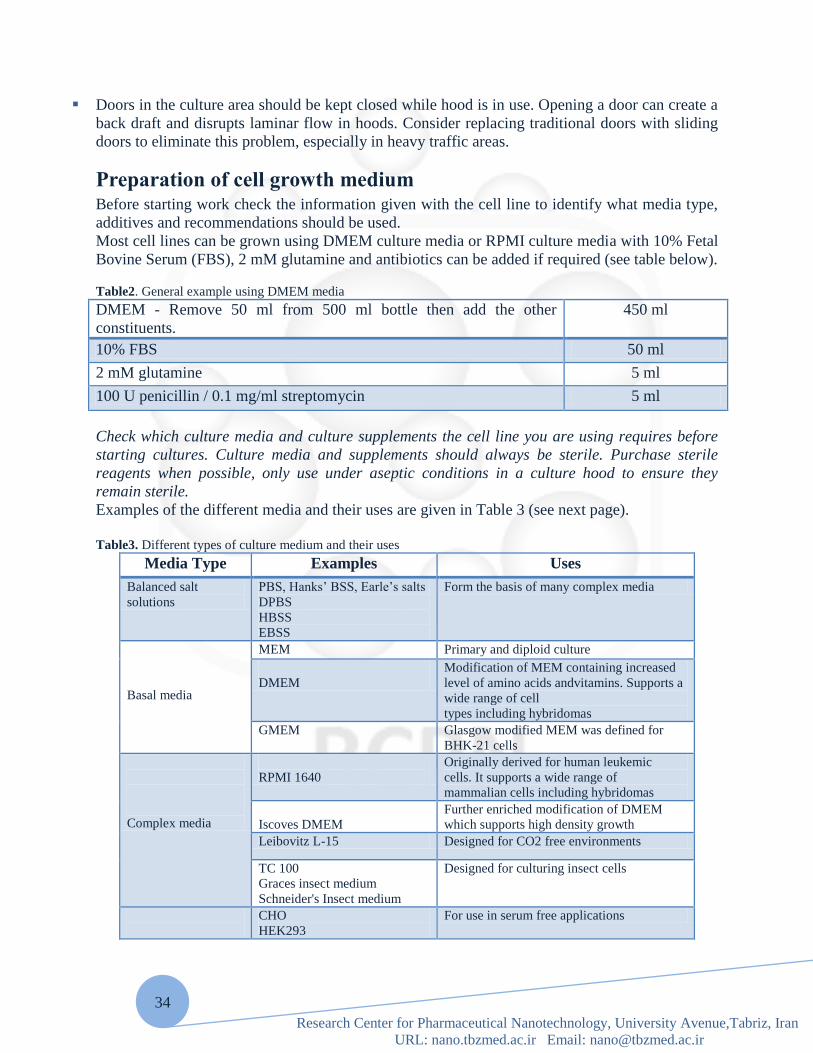
Preparation of cell growth medium Before starting work check the information given with the cell line to identify what media type,
additives and recommendations should be used.
Most cell lines can be grown using DMEM culture media or RPMI culture media with 10% Fetal
Bovine Serum (FBS), 2 mM glutamine and antibiotics can be added if required (see table below).
Table2. General example using DMEM media
DMEM - Remove 50 ml from 500 ml bottle then add the other
constituents.
450 ml
10% FBS 50 ml
2 mM glutamine 5 ml
100 U penicillin / 0.1 mg/ml streptomycin 5 ml
Check which culture media and culture supplements the cell line you are using requires before
starting cultures. Culture media and supplements should always be sterile. Purchase sterile
reagents when possible, only use under aseptic conditions in a culture hood to ensure they
remain sterile.
Examples of the different media and their uses are given in Table 3 (see next page).
Table3. Different types of culture medium and their uses
Media Type Examples Uses
Balanced salt
solutions
PBS, Hanks‟ BSS, Earle‟s salts
DPBS
HBSS
EBSS
Form the basis of many complex media
Basal media
MEM Primary and diploid culture
DMEM
Modification of MEM containing increased
level of amino acids andvitamins. Supports a
wide range of cell
types including hybridomas
GMEM Glasgow modified MEM was defined for
BHK-21 cells
Complex media
RPMI 1640
Originally derived for human leukemic
cells. It supports a wide range of
mammalian cells including hybridomas
Iscoves DMEM
Further enriched modification of DMEM
which supports high density growth
Leibovitz L-15 Designed for CO2 free environments
TC 100
Graces insect medium
Schneider's Insect medium
Designed for culturing insect cells
CHO
HEK293
For use in serum free applications

35
Research Center for Pharmaceutical Nanotechnology, University Avenue,Tabriz, Iran
URL: nano.tbzmed.ac.ir Email: [email protected]
Serum free
media
Ham F10 and derivatives
Ham F12
DMEM/F12
Note: these media must be supplemented
with other factors such as insulin, transferrin
and epidermal growth factor.
These media are usually HEPES buffered
Insect cells
Serum-Free Insect Medium 1
Specifically designed for use with Sf9
insect cells
Basic Constituents of Media
Inorganic salts
Carbohydrates
Amino Acids
Vitamins
Fatty acids and lipids
Proteins and peptides
Serum
Trace Elements
Each type of constituent performs a specific function as outlined below:
Inorganic Salts
The inclusion of inorganic salts in media performs several functions. Primarily they help to
retain the osmotic balance of the cells and help regulate membrane potential by provision of
sodium, potassium and calcium ions. All of these are required in the cell matrix for cell
attachment and as enzyme cofactors.
Buffering Systems
Most cells require pH conditions in the range 7.2-7.4 and close control of pH is essential for
optimum culture conditions. There are major variations to this optimum. Fibroblasts prefer a
higher pH (7.4-7.7) whereas, continuous transformed cell lines require more acid conditions pH
(7.0-7.4).
Regulation of pH is particularly important immediately following cell seeding when a new
culture is establishing and is usually achieved by one of two buffering systems; (i) a “natural”
buffering system where gaseous CO2 balances with the CO3/HCO3 content of the culture
medium and (ii) chemical buffering using a zwitterions called HEPES. Cultures using natural
bicarbonate /CO2 buffering systems need to be maintained in an atmosphere of 5-10% CO2 in
air usually supplied in a CO2 incubator. Bicarbonate/CO2 is low cost, non-toxic and also
provides other chemical benefits to the cells.
HEPES has superior buffering capacity in the pH range 7.2-7.4 but is relatively expensive and
can be toxic to some cell types at higher concentrations. HEPES buffered cultures do not require
a controlled gaseous atmosphere.

36
Research Center for Pharmaceutical Nanotechnology, University Avenue,Tabriz, Iran
URL: nano.tbzmed.ac.ir Email: [email protected]
Most commercial culture media include phenol red as a pH indicator so that the pH status of the
medium is constantly indicated by the color. Usually the culture medium should be
changed/replenished if the color turns yellow (acid) or purple (alkali).
Carbohydrates
The main source of energy is derived from carbohydrates generally in the form of sugars. The
major sugars used are glucose and galactose, however, some media contain maltose or fructose.
The concentration of sugar varies from basal media containing 1g/L to 4.5g/L in some more
complex media. Media containing the higher concentration of sugars are able to support the
growth of a wider range of cell types.
Amino Acids
Amino acids are the building blocks of proteins. „Essential‟ amino acids must be added to culture
media as cells are not able to synthesize these themselves. The concentration of amino acids in
the culture medium will determine the maximum cell density that can be achieved – once
depleted the cells will no longer be able to proliferate.
In relation to cell culture, glutamine, an essential amino acid, is particularly significant. In liquid
media or stock solutions glutamine degrades relatively rapidly. Optimal cell performance usually
requires supplementation of the media with glutamine prior to use.
Adding supplements of non-essential amino acids to media both stimulates growth and prolongs
the viability of the cells in culture.
Vitamins
Serum is an important source of vitamins in cell culture. However, many media are also enriched
with vitamins making them consistently more suitable for a wider range of cell lines. Vitamins
are precursors for numerous co-factors. Many vitamins, especially B group vitamins, are
necessary for cell growth and proliferation and for some lines the presence of B12 is essential.
Some media also have increased levels of vitamins A and E. The vitamins commonly used in
media include riboflavin, thiamine and biotin.
Proteins and Peptides
These are particularly important in serum free media. The most common proteins and peptides
include albumin, transferrin, fibronectin and fetuin and are used to replace those normally
present through the addition of serum to the medium.
Fatty Acids and Lipids
Like proteins and peptides these are important in serum free media since they are normally
present in serum e.g. cholesterol and steroids essential for specialized cells.
Trace Elements
These include trace elements such as zinc, copper, selenium and tricarboxylic acid intermediates.
Selenium is a detoxifier and helps remove oxygen free radicals.\
Freezing and Thawing Cells Comments
Cells should be free of contamination in the form of bacteria, yeast, or fungi.
Mycoplasma testing should be performed prior to freezing.
Freezing media depends on the cell line.
37
Research Center for Pharmaceutical Nanotechnology, University Avenue,Tabriz, Iran
URL: nano.tbzmed.ac.ir Email: [email protected]
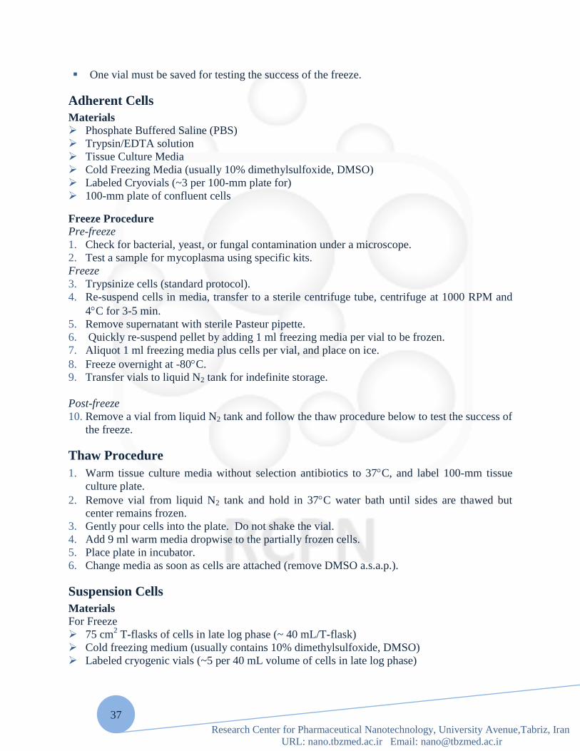
One vial must be saved for testing the success of the freeze.
Adherent Cells
Materials
Phosphate Buffered Saline (PBS)
Trypsin/EDTA solution
Tissue Culture Media
Cold Freezing Media (usually 10% dimethylsulfoxide, DMSO)
Labeled Cryovials (~3 per 100-mm plate for)
100-mm plate of confluent cells
Freeze Procedure
Pre-freeze
1. Check for bacterial, yeast, or fungal contamination under a microscope.
2. Test a sample for mycoplasma using specific kits.
Freeze
3. Trypsinize cells (standard protocol).
4. Re-suspend cells in media, transfer to a sterile centrifuge tube, centrifuge at 1000 RPM and
4C for 3-5 min.
5. Remove supernatant with sterile Pasteur pipette.
6. Quickly re-suspend pellet by adding 1 ml freezing media per vial to be frozen.
7. Aliquot 1 ml freezing media plus cells per vial, and place on ice.
8. Freeze overnight at -80C.
9. Transfer vials to liquid N2 tank for indefinite storage.
Post-freeze
10. Remove a vial from liquid N2 tank and follow the thaw procedure below to test the success of
the freeze.
Thaw Procedure
1. Warm tissue culture media without selection antibiotics to 37C, and label 100-mm tissue
culture plate.
2. Remove vial from liquid N2 tank and hold in 37C water bath until sides are thawed but
center remains frozen.
3. Gently pour cells into the plate. Do not shake the vial.
4. Add 9 ml warm media dropwise to the partially frozen cells.
5. Place plate in incubator.
6. Change media as soon as cells are attached (remove DMSO a.s.a.p.).
Suspension Cells
Materials
For Freeze
75 cm2 T-flasks of cells in late log phase (~ 40 mL/T-flask)
Cold freezing medium (usually contains 10% dimethylsulfoxide, DMSO)
Labeled cryogenic vials (~5 per 40 mL volume of cells in late log phase)

38
Research Center for Pharmaceutical Nanotechnology, University Avenue,Tabriz, Iran
URL: nano.tbzmed.ac.ir Email: [email protected]
For Thaw
Cold tissue culture medium
25 cm2 T-flask
Freeze Procedure
Pre-freeze
1. Check for bacterial, yeast, or fungal contamination under a microscope.
2. Test a sample for mycoplasma using specific kits.
3. When cells have reached late log phase, determine cell density using Coulter counter.
Calculate total number of cells in flask, and determine amount of freeze medium needed.
(Cells should be resuspended in freeze medium at 5,000,000 to 20,000,000 cells/mL.)
Freeze
4. Centrifuge cells in 50 mL Falcon tube at 1000g for 15 minutes.
5. While cells are spinning, make freeze medium (e.g., 90% FBS, 10% DMSO). Label
cryogenic vials with date, cell type, and user‟s initials.
6. Suction away supernatant from centrifuged cells and add freeze medium. Triturate cells until
homogeneous.
7. Quickly aliquot 1 mL of freeze stock per cryogenic vial. Screw each vial closed.
8. Put vials into storage box and place box, insulated with paper towels, into Tupperware®
container. Put entire container into –20°C freezer.
9. After 3 hours, transfer container to –80°C freezer and store overnight.
10. Next day, put cells into appropriate rack in liquid N2 tank.
Post-freeze
11. Remove a vial from liquid N2 tank and follow the thaw procedure below to test the success of
the freeze.
Thaw Procedure
1. Slowly remove appropriate tray rack from liquid N2 tank. Remove long safety pin and take
out one vial from appropriate tray.
2. Put tray back in slot and put safety pin back in place. Return tray rack to liquid N2 tank and
cap tank again.
3. Rapidly thaw vial in 37°C water bath until only a small ice pellet remains. Spray down vial
with ethanol, wipe, and place into hood.
4. Pipette contents of vial (~ 1 mL) into T25 flask.
5. Slowly add 4 mL of cold culture medium, at a rate of about 1 drop every 10 seconds, swirling
occasionally. Add another 5 mL of culture medium.
6. Place flask in appropriate incubator.
7. Since freeze medium contains dimethylsulfoxide (DMSO), spin down cells after 6-12 hours
and resuspend in fresh, prewarmed medium in new T25 flask.
Key Points
1. Most text books recommend washing the thawed cells in media to remove the cryoprotectant.
This is only necessary if the cryoprotectant is known to have an adverse effect on the
particular cell type. For example, some cell types are known to differentiate in the presence

39
Research Center for Pharmaceutical Nanotechnology, University Avenue,Tabriz, Iran
URL: nano.tbzmed.ac.ir Email: [email protected]
of DMSO. In such cases the cells should be washed in media before being added to their final
culture flasks.
2. The addition of the thawed cell suspension to culture medium effectively dilutes the
cryoprotectant (e.g. DMSO) reducing the toxicity of the cryoprotectant. That is why it is
important to add the thawed cell suspension to a larger volume of culture medium
immediately after the ampoule has thawed; do not allow thawed ampoules to sit at room
temperature for long periods.
3. Do not use an incubator or the palm of your hand to thaw cell cultures since the rate of
thawing achieved is too slow resulting in a loss of viability. Use a water bath as described in
the protocol above.
4. If a CO2 incubator is not available gas the flasks for 1-2 minutes with 5% CO2 in 95% air
filtered through a 0.2μm filter.
5. For most cultures it is best practice to subculture before confluence is reached so that the
cells are harvested during their log phase of growth and are at optimum viability ready for
seeding into new flasks. Furthermore there are some specific cell types that must be sub
cultured before confluence is reached in order to maintain their characteristics e.g. the
contact inhibition of NIH 3T3 cells is lost if they are allowed to reach confluence repeatedly.
6. Some hybridomas may be slow to recover post resuscitation therefore start in 20% (v/v) FBS
and 10% (v/v) hybridoma enhancement supplement in the appropriate medium.
Water The water used for making media and washing glassware is a frequent source of chemical
contamination and requires special care to ensure its quality. Traditionally, double or triple glass
distillation was considered to be the best source of high quality water for cell culture media and
solutions. Newer purification systems combining reverse osmosis, ion exchange and ultra
filtration are capable of removing trace metals, dissolved organic compounds and endotoxins and
are increasingly popular. However, these systems must be properly maintained and serviced to
ensure continued water quality. Because of its aggressive solvent characteristics, highly purified
water can leach potentially toxic metal ions from glassware or metal pipes, and plasticizers from
plastic storage vessels or tubing. These contaminants can then end up in media or deposited on
storage vessels and pipettes during washing and rinsing. Water used to generate steam in
autoclaves may contain additives to reduce scale buildup in pipes; these potentially toxic
additives can also end up on glassware.
Incubators Cell cultures require a strictly controlled environment in which to grow.
Specialist incubators are used routinely to provide the correct growth conditions, such as
temperature, degree of humidity and CO2 levels in a controlled and stable manner. Generally,
they can be set to run at temperatures in the range of 28oC (for insect cell lines) to 37oC (for
mammalian cell lines) and set to provide CO2 at the required level (e.g. 5-10%). Some
incubators also have the facility to control the O2 levels. Copper-coated incubators are also now
available. These are reported to reduce the risk of microbial contamination within the incubator
due to the microbial inhibitory activity of copper. The inclusion of a bactericidal agent in the
incubator water trays will also reduce the risk of bacterial and fungal growth. However, there is
no substitute for regular cleaning.

40
Research Center for Pharmaceutical Nanotechnology, University Avenue,Tabriz, Iran
URL: nano.tbzmed.ac.ir Email: [email protected]
Centrifuges Centrifuges are used routinely in tissue culture as part of the subculture routine for most cell
lines and for the preparation of cells for cryopreservation. By their very nature centrifuges
produce aerosols and thus it is necessary to minimize this risk. This can be achieved by
purchasing models that have sealed buckets. Ideally, the centrifuge should have a clear lid so that
the condition of the load can be observed without opening the lid. This will reduce the risk of the
operator being exposed to hazardous material if a centrifuge tube has broken during
centrifugation. Care should always be taken not to over-fill the tubes and to balance them
carefully. These simple steps will reduce the risk of aerosols being generated. The centrifuge
should be situated where it can be easily accessed for cleaning and maintenance. Centrifuges
should be checked frequently for signs of corrosion.
A small bench-top centrifuge with controlled braking is sufficient for most purposes. Cells
sediment satisfactorily at 80 – 150 x g. higher gravitational forces may cause damage and
promote agglutination of the cell pellet.
Storage area There are two main types of liquid-nitrogen storage systems, vapor phase and liquid phase,
which come as wide-necked or narrow-necked storage containers.
Vapor phase systems minimize the risk of explosion with cryostorage tubes, and are required for
storing biohazardous materials, while the liquid phase systems usually have longer static holding
times, and are therefore more economical.
Narrow-necked containers have a slower nitrogen evaporation rate and are more economical, but
wide-necked containers allow easier access and have a larger storage capacity.
Note: Do not store cells in –20°C or –80°C freezers, because their viability quickly decreases
when they are stored at these temperatures.
Biological Contamination Biological contaminants can be subdivided into two groups based on the difficulty of detecting
them in cultures:
Those that are usually easy to detect: Bacteria, molds and yeast;
Those that are more difficult to detect, and as a result potentially more serious culture
problems: Viruses, protozoa, insects, mycoplasmas and other cell lines.
Note: Ultimately, it is the length of time that a culture contaminant escapes detection that will
determine the extent of damage it creates in a laboratory or research project.
Bacteria, Molds, and Yeasts
Bacteria, molds and yeasts are found virtually everywhere and are able to quickly colonize and
flourish in the rich and relatively undefended environment provided by cell cultures.
Because of their size and fast growth rates, these microbes are the most commonly encountered
cell culture contaminants. In the absence of antibiotics, microbes can usually be readily detected
in a culture within a few days of becoming contaminated, either by direct micro- scopic
observation. Or by the effects they have on the culture (pH shifts, turbidity, and cell destruction).

41
Research Center for Pharmaceutical Nanotechnology, University Avenue,Tabriz, Iran
URL: nano.tbzmed.ac.ir Email: [email protected]
However, when antibiotics are routinely used in culture, resistant organisms may develop into
slow growing, low level infections that are very difficult to detect by direct visual observation.
Similar detection problems can occur with naturally slow growing organisms or very small or
intracellular bacteria that are difficult to see during routine microscopic culture observation.
These cryptic contaminants may persist indefinitely in cultures causing subtle but significant
alterations in their behavior. By the time these cryptic contaminants are discovered, many
experiments and cultures may have been compromised. Viruses
Due to their extremely small size, viruses are the most difficult cell culture contaminants to
detect in culture, requiring methods that are impractical for most research laboratories.
Their small size also makes them very difficult to remove from media, sera, and other solutions
of biological origin. However, most viruses have stringent requirements for their original host
species‟ cellular machinery (may also be tissue specific) which greatly limits their ability to
infect cell cultures from other species. Thus, although viruses may be more common in cell
cultures than many researchers realize, they are usually not a serious problem unless they have
cytopathic or other adverse effects on the cultures. Since cytopathic viruses usually destroy the
cultures they infect, they tend to be self-limiting.
Thus, when cultures self-destruct for no apparent reason and no evidence of common biological
contaminants can be found, cryptic viruses are often blamed. They are perfect culprits, unseen
and undetectable; guilty without direct evidence. This is unfortunate, since the real cause of this
culture destruction may be something else, possibly mycoplasma or a chemical contaminant, and
as a result will go undetected to become a more serious problem.
Note: A major concern of using virally infected cell cultures is not their effects on the cultures
but rather the potential health hazards they pose for laboratory personnel. Special safety
precautions should always be used when working with tissues or cells from humans or other
primates to avoid possible transmission of viral infection (HIV, hepatitis B, Epstein-Barr, simian
herpes B virus, among others) from the cell cultures to laboratory personnel. Contact your safety
office for additional assistance if in doubt as to appropriate procedures for working with
potentially hazardous tissues, cultures or viruses.

42
Research Center for Pharmaceutical Nanotechnology, University Avenue,Tabriz, Iran
URL: nano.tbzmed.ac.ir Email: [email protected]
Table 4: Common Problems Associated With Cell Culture
Problem Possible cause Potential solution
Cells difficult to remove
from plastic
Enzyme solution too weak
Inhibitor present in medium (for
example, serum)
Cells too confluent and enzyme cannot
access
cell-substrate interface
Higher concentration needed
Cells require more careful
washing
Cells require trypsinisation at
lower cell density
Cells not adhering readily
to plastic
Cells too heavily treated with trypsin
Insufficient serum or attachment
factors
Dissociating agent (for example, not
inactivated fully)
Mycoplasma contamination
Use less trypsin or treat for
less time
Add more
Add serum or specific
inhibitors
Discard if infected
Suspension cells
clumping together
Mycoplasma contamination
DNA from lysed cells sticking cells
together
Discard if infected
Add DNAse
Poor growth in culture Absence or lower than normal levels of
certain additives
Contamination by bacteria,
mycoplasma or fungi Discard if
infected
Cell density too low
Add missing components
Discard if infected
Increase density
Cell death/low viability Incorrect pH
Faulty media
Correct pH
Correct preparation
Too acidic pH CO2 content too high
Contamination
Modify
If infected, discard
Too basic pH Insufficient CO2
Too few cells
Caps too tight
Increase cell density
Trypsinising/passaging adherent cell lines
Materials required
Trypsin/EDTA
Sterile PBS
Cell culture flasks
10 and 25ml pipettes
5% Virkon
Trypan Blue/counting chamber (optional)
Growth medium

43
Research Center for Pharmaceutical Nanotechnology, University Avenue,Tabriz, Iran
URL: nano.tbzmed.ac.ir Email: [email protected]
Method
1. Put trypsin, growth medium and PBS into 37C incubator for 45-60 minutes to warm before
starting.
2. Swab cabinet with 5% Virkon and 70% ethanol.
3. Decant off spent media from each flask to be split.
4. Add 25ml of warm PBS to each flask and wash monolayer gently. Decant off PBS into 5%
Virkon. Add 5-10ml of trypsin to each flask. Lay the flask flat and wait for 1-2 minutes, or
until most of the cells have rounded up (observe them under a microscope). Decant off
excess trypsin into 5% Virkon, leaving about 1mL in the flask.
5. Incubate flask at 37C for 1-2 minutes.
6. Retrieve flask from incubator and knock the side of the flask against the palm of your hand a
few times to dislodge the cells. The cells should come off easily - if not, reincubate the flask
for a further 1-2 minutes.
7. Resuspend the cells from each flask in 5-10ml of warm growth medium, and divide into
labeled flasks. [If a cell count is to be performed, add 10m l of cell suspension to 90m l of
Trypan Blue and mix well. Add 10m l to a haemocytometer and do cell count. See below]
8. Add 25-50mL of warm growth medium to the cell suspension in each flask, gently mix, and
put flasks in 37C incubator with 5% CO2.
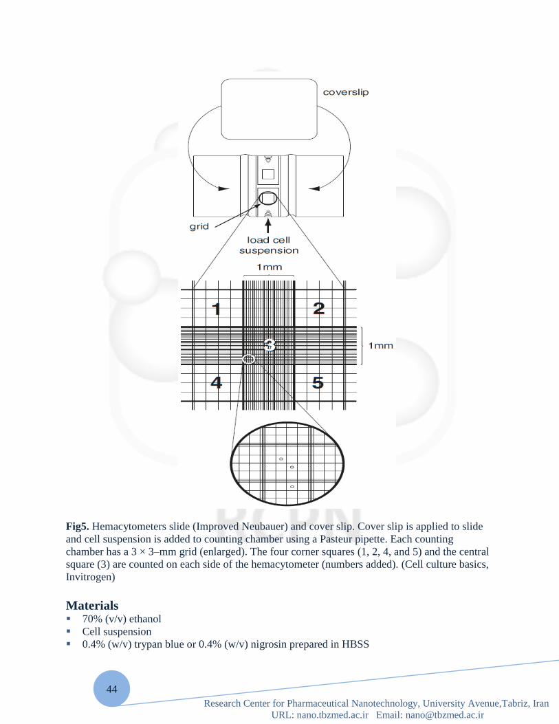
Determining cell number and viability with a Hemacytometer and trypan
blue staining Determining the number of cells in culture is important in standardization of culture conditions
and in performing accurate quantization experiments. A hemacytometer is a thick glass slide
with a central area designed as a counting chamber.
The exact design of the hemacytometer may vary; the one described here is the Improved
Neubauer from Baxter Scientific. The central portion of the slide is the counting platform which
is bordered by a 1-mm groove. The central platform is divided into two counting chambers by a
transverse groove. Each counting chamber consists of a silver footplate on which is etched a 3 ×
3–mm grid. This grid is divided into nine secondary squares, each 1 × 1 mm. The four corner
squares and the central square are used for determining the cell count. The corner squares are
further divided into 16 tertiary squares and the central square into 25 tertiary squares to aid in
cell counting.
Accompanying the hemacytometer slide is a thick, even-surfaced cover slip. Ordinary cover
slips may have uneven surfaces that can introduce errors in cell counting; therefore, it is
imperative that the cover slip provided with the hemacytometer is used in determining cell
number.
Cell suspension is applied to a defined area and counted so cell density can be calculated.
44
Research Center for Pharmaceutical Nanotechnology, University Avenue,Tabriz, Iran
URL: nano.tbzmed.ac.ir Email: [email protected]
Fig5. Hemacytometers slide (Improved Neubauer) and cover slip. Cover slip is applied to slide
and cell suspension is added to counting chamber using a Pasteur pipette. Each counting
chamber has a 3 × 3–mm grid (enlarged). The four corner squares (1, 2, 4, and 5) and the central
square (3) are counted on each side of the hemacytometer (numbers added). (Cell culture basics,
Invitrogen)
Materials 70% (v/v) ethanol
Cell suspension
0.4% (w/v) trypan blue or 0.4% (w/v) nigrosin prepared in HBSS

45
Research Center for Pharmaceutical Nanotechnology, University Avenue,Tabriz, Iran
URL: nano.tbzmed.ac.ir Email: [email protected]
Hemacytometer with cover slip (Improved Neubauer, Baxter Scientific) Hand-held counter
Prepare hemacytometer
1. Clean surface of hemacytometer slide and cover slip with 70% alcohol. Cover slip and slide
should be clean, dry, and free from lint, fingerprints, and watermarks.
2. Wet edge of cover slip slightly with tap water and press over grooves on hemacytometer.
The cover slip should rest evenly over the silver counting area.
Prepare cell suspension
3. For cells grown in monolayer cultures, detach cells from surface of dish using trypsin (see
Basic Protocol, steps 1 to 4).
4. Dilute cells as needed to obtain a uniform suspension. Disperse any clumps.
When using the hemacytometer, a maximum cell count of 20 to 50 cells per
1 × 1 mm square is recommended.
Load hemacytometer
5. Use a sterile Pasteur pipette to transfer cell suspension to edge of hemacytometer counting
chamber. Hold tip of pipette under the cover slip and dispense one drop of suspension.
Suspension will be drawn under the cover slip by capillary action.
The hemacytometer should be considered no sterile. If cell suspension is to be used for cultures,
do not reuse the pipette and do not return any excess cell suspension in the pipette to the original
suspension.
6. Fill second counting chamber. Count cells
7. Allow cells to settle for a few minutes before beginning to count. Blot off excess liquid.
8. View slide on microscope with 100× magnification.
Position slide to view the large central area of the grid; this area is bordered by a set of three
parallel lines. The central area of the grid should almost fill the microscope field. Subdivisions
within the large central area are also bordered by three parallel lines and each subdivision is
divided into sixteen smaller squares by single lines. Cells within this area should be evenly
distributed without clumping. If cells are not evenly distributed, wash and reload hemacytometer.
9. Use a hand-held counter to count cells in each of the four corners and central squares. Repeat
counts for other counting chamber.
Five squares (four corners and one center) are counted from each of the two counting chambers
for a total of ten squares counted.
Count cells touching the 3 middle line of the triple line on the top and left of the squares. Do not
count cells touching the middle line of the triple lines on the bottom or right side of the square.

46
Research Center for Pharmaceutical Nanotechnology, University Avenue,Tabriz, Iran
URL: nano.tbzmed.ac.ir Email: [email protected]
Calculate cell number
10. Determine cells per milliliter by the following calculations:
Cells/ml = average count per square × dilution factor × 104
Total cells = cells/ml × total original volume of cell suspension from which sample was taken.
The number 104 is the volume correction factor for the hemacytometer: each square is 1 × 1 mm
and the depth is 0.1 mm.
Stain cells with trypan blue to determine cell viability
11.Determine number of viable cells by adding 0.5 ml of 0.4% trypan blue, 0.3 ml HBSS, and
0.1 ml cell suspension to a small tube. Mix thoroughly and let stand 5 min before loading
hemacytometer.
Either 0.4% trypan blue or 0.4% nigrosin can be used to determine the viable cell number.
Nonviable cells will take up the dye, while live cells will be impermeable to dye.
12. Count total number of cells and total number of viable (unstained) cells. Calculate Percent
viable cells as follows:
% viable cells =
× 100
13. Decontaminate cover slip and hemacytometer by rinsing with 70% ethanol and then
deionized water. Air dry and store for future use.
Checking cells
1.Cells should be checked microscopically daily to ensure they are healthy and growing as
expected. Attached cells should be mainly attached to the bottom of the flask, round and plump
or elongated in shape and refracting light around their membrane. Suspension cells should look
round and plump and refracting light around their membrane. Some suspension cells may clump.
Media should be pinky orange in color.
2.Discard cells if:
They are detaching in large numbers (attached lines) and/or look shriveled and grainy/dark in
color.
They are in quiescence (do not appear to be growing at all).

47
Research Center for Pharmaceutical Nanotechnology, University Avenue,Tabriz, Iran
URL: nano.tbzmed.ac.ir Email: [email protected]
References
1. Basic Principles of Cell Culture, R. Ian Freshney Centre for Oncology and Applied
Pharmacology, Cancer Research UK Beatson. Laboratories, Garscube Estate, Bearsden,
Glasgow G61 1BD, Scotland, UK.
2. Basic Principles and Best Practices R. Ian Freshney, Centre for Oncology and Applied
Pharmacology, Cancer Research UK Beatson Laboratories, Garscube Estate, Bearsden,
Glasgow G61 1BD, Scotland, UK
3. http://www.sigmaaldrich.com/technical-documents/protocols/biology/resuscitation-of-
frozen.html
4. The Weatherall Institute of Molecular Medicine, University of Oxford, John Radcliffe
Hospital,
http://www.imm.ox.ac.uk/trypsinising-adherent-cell-cultures
5. web.mit.edu/.../Freeze_Thaw_Prot...
6. http://www.biology.arizona.edu/cell_bio/tutorials/cell_cycle/cells2.html
7. Invitrogen cell culture basics handbook
8. Cell culture guidelines. Abcam.com/technical
9. Cancer cell culture. Methods and Protocols. Edited by Simon P. Langdon
10. John Ryan. Understanding and managing cell culture contamination, Technical Bulletin.
Good luck!
Was this document helpful? Tell us on [email protected]