an introduction to viruses
TRANSCRIPT
An Introduction to Viruses
Lecturer
Dr Ashraf Khasawneh
Department of Biomedical Sciences

Virus infections are Universal …….

Introduction to Virology
• A virus is an obligate intracellular
parasite containing genetic material
surrounded by protein
• Virus particles can only be
observed by an electron
microscope
3
Introduction to Virology
• Recognizing the shape, size, and structure
of different viruses is critical to the study
of disease
–Viruses have an inner core of nucleic acid
surrounded by protein coat known as an
envelope
–Most viruses range in sizes from 20 – 450
nanometers
4
Viral Properties
• Viruses are inert (nucleoprotein ) filterable Agents
• Viruses are obligate intracellular parasites
• Viruses cannot make energy or proteins independent of a host cell
• Viral genome are RNA or DNA but not both.
• Viruses have a naked capsid or envelope with attached proteins
• Viruses do not have the genetic capability to multiply by division.
• Viruses are non-living entities
5
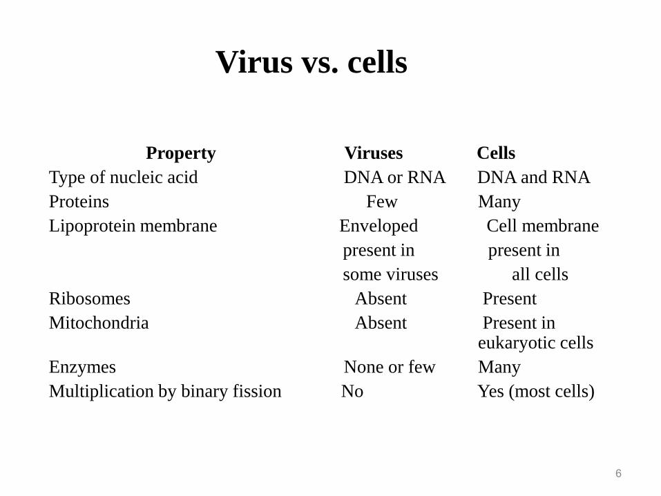
Property Viruses Cells
Type of nucleic acid DNA or RNA DNA and RNA
Proteins Few Many
Lipoprotein membrane Enveloped Cell membrane
present in present in
some viruses all cells
Ribosomes Absent Present
Mitochondria Absent Present in eukaryotic cells
Enzymes None or few Many
Multiplication by binary fission No Yes (most cells)
6
Virus vs. cells

Viruses are Ultramicroscopic
7
Koneman et al. Color Atlas and Textbook of Microbiology 5th Ed. 1997
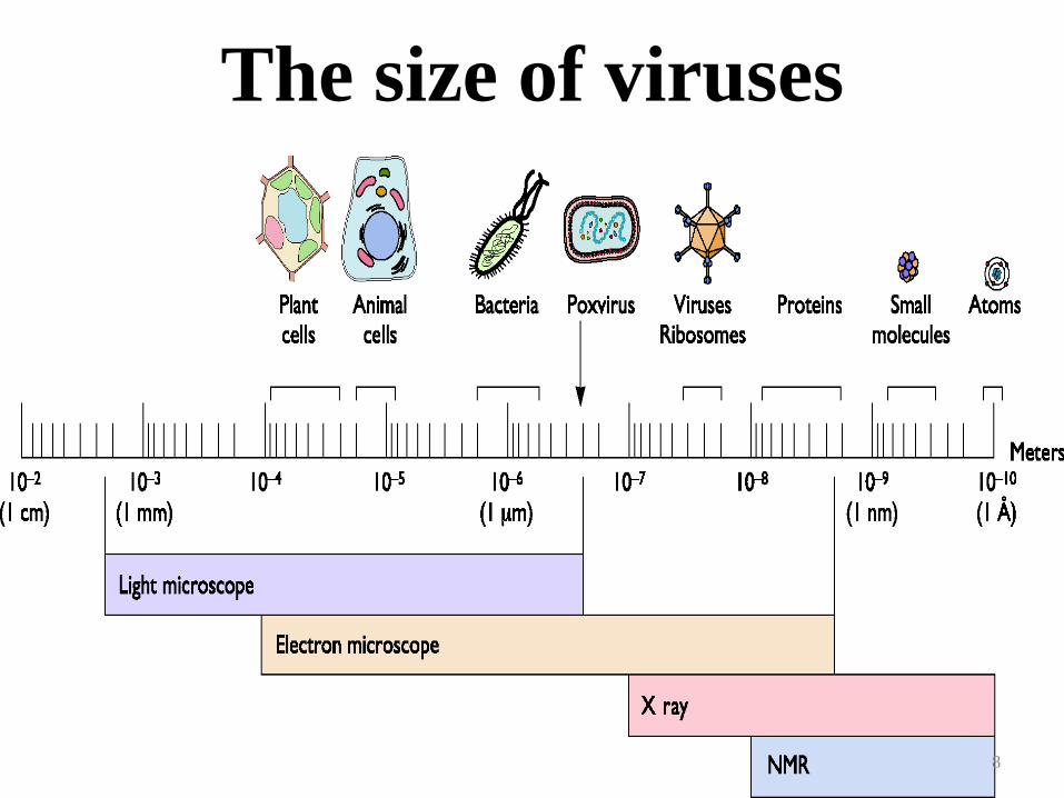
The size of viruses
8

9
VIRAL STRUCTURE – SOME
TERMINOLOGY
• virus particle = virion
• protein which coats the genome =
capsid
• capsid usually symmetrical
• capsid + genome = nucleocapsid
• may have an envelope

Virion
• The complete
infectious unit of
virus particle
• Structurally
mature,
extracellular
virus particles.
10

Viral Structure - Overview
Fig 1. Schematic overview of the structure of animal viruses
** does not exist in all viruses
Nucleic acid
Capsid
Nucleocapsid
Envelope protein
Membrane proteinViral envelope**
Spike protein
11

Distinguishing characteristics of viruses
• Obligate intracellular parasites
• Extreme genetic simplicity
• Contain DNA or RNA
• Replication involves disassembly and
reassembly
• Replicate by "one-step growth”
12
Naming viruses
• No taxa above Family (no kingdom, phylum, etc)
• Classified based on structures, size, nucleic acids, host species, target cells.
• 19 families of animal viruses (6 DNA, 13 RNA)
• Family name ends in – viridae
• Subfamily ends in — virinae
• Genus name ends in – virus
• Species Example
– Family – Herpesviridae
– Subfamily - Herpesvirinae
– Genus – Simplex virus
– Common name – herpes virus (Herpes simplex virus I (HSV-I)
– Disease – fever blisters, cold sores
13

How are viruses named?
• Based on:
- the disease they causepoliovirus, rabies virus
- the type of diseasemurine leukemia virus
- geographic locationsSendai virus, Coxsackie virus
- their discoversEpstein-Barr virus
- how they were originally thought to be contracted
dengue virus (“evil spirit”), influenza virus (the “influence” of bad air)
- combinations of the aboveRous Sarcoma virus
14

15
Virus particle = virion

16
5 BASIC TYPES OF VIRAL STRUCTURE
HELICAL ENVELOPED HELICAL
ENVELOPED ICOSAHEDRAL
COMPLEX
ICOSAHEDRAL
nucleocapsidicosahedral nucleocapsid
nucleocapsid
helical nucleocapsid
lipid bilayer
lipid bilayer
glycoprotein spikes= peplomers
Viral Structure
• Varies in size, shape and symmetry
• 3 types of capsid symmetry:
– Cubic (icosahedral)
• Has 20 faces, each an equilateral triangle. Eg. adenovirus
– Helical
• Protein binds around DNA/RNA in a helical fashion eg.
Coronavirus
– Complex
• Is neither cubic nor helical eg. poxvirus
17

VIRAL STRUCTURE (virion)
18
1. Protect genome during passage
from one cell to another
2. Aid in entry process
3. Package enzymes for early steps
of infection

1. Helical capsid
• Rod-shaped capsomers
• Coil around hollow
center
• Nucleic acid is kept
inside – wound-up
within tube (Helix )
19
Morphological types
CAPSID STRUCTURE
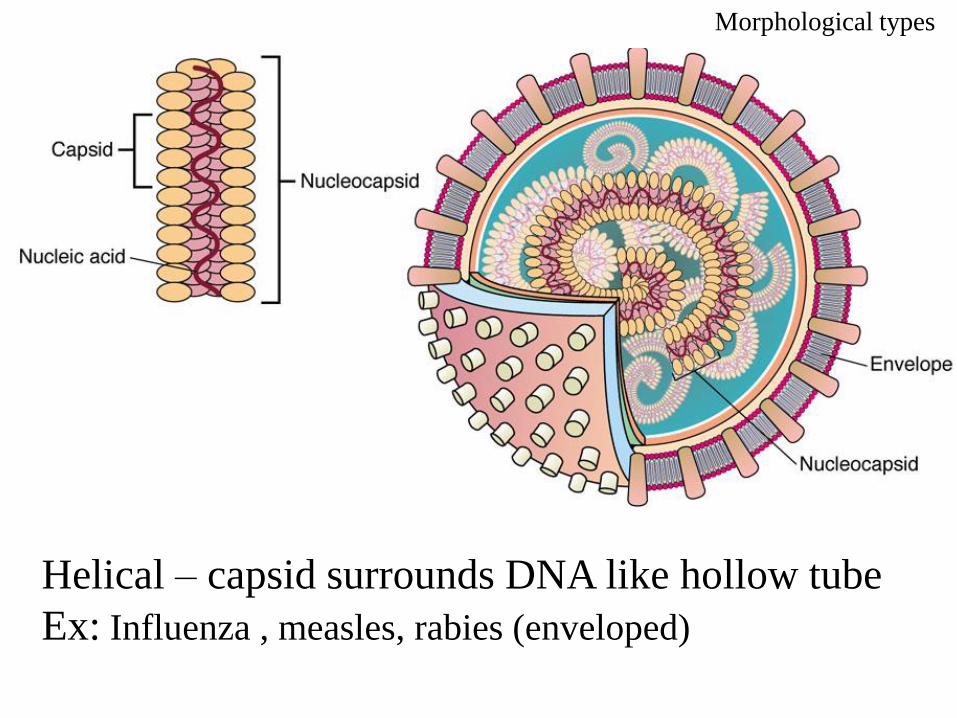
Helical – capsid surrounds DNA like hollow tube
Ex: Influenza , measles, rabies (enveloped)
Morphological types
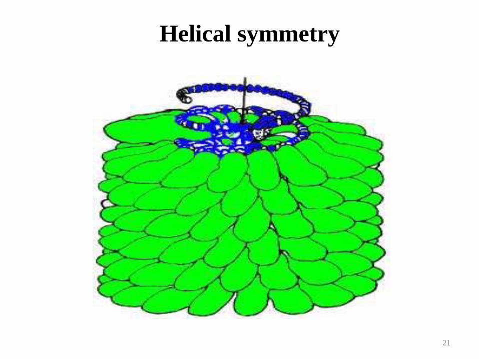
Helical symmetry
21

Helical symmetry
How to
assemble
22
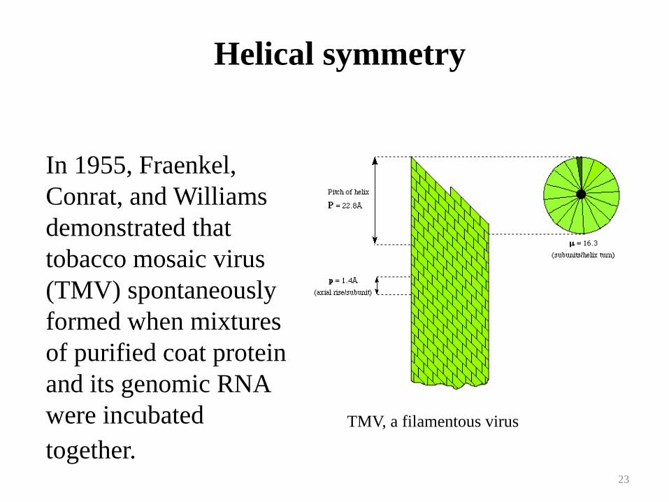
In 1955, Fraenkel,
Conrat, and Williams
demonstrated that
tobacco mosaic virus
(TMV) spontaneously
formed when mixtures
of purified coat protein
and its genomic RNA
were incubated
together.
Helical symmetry
TMV, a filamentous virus
23

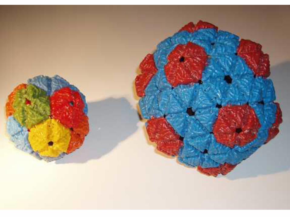
2. icosahedral
• 20-sided with 12 corners
• Vary in the number of
capsomers
• Each capsomer may be
made of 1 or several
proteins
• Some are enveloped
Morphological types
PROTOME
R
25

26

a) Crystallographic structure of a
simple icosahedral virus.b) The axes of symmetry
Icosahedral capsids
27

Cubic or icosahedral symmetry
28

29
ICOSAHEDRAL SYMMETRY

30
ICOSAHEDRAL SYMMETRY

31
ICOSAHEDRAL SYMMETRY

32
ICOSAHEDRAL SYMMETRY

33
Adenovirus

34
Adenovirus

Enveloped helical virus Enveloped icosahedral virus
35

Helical
• California Encephalitis VirusCoronavirusHantavirusInfluenza Virus (Flu Virus)Measles Virus ( Rubeola)Mumps VirusPara influenza VirusRabies VirusRespiratory Syncytial Virus(RSV)
36

Icosahedral
• Adeno-associated Virus (AAV)AdenovirusB19Coxsackievirus - ACoxsackievirus - BCytomegalovirus (CMV)Eastern Equine Encephalitis Virus (EEEV)EchovirusEpstein-Barr Virus (EBV)Hepatitis A Virus (HAV)Hepatitis B Virus (HBV)Hepatitis C Virus (HCV)Hepatitis Delta Virus (HDV)Hepatitis E Virus (HEV)
• Herpes Simplex Virus 1 (HHV1)Herpes Simplex Virus 2 (HHV2)Human Immunodeficiency Virus (HIV)Human T-lymphotrophic Virus (HTLV)Norwalk VirusPapilloma Virus (HPV)Polio virusRhinovirusRubella VirusSaint Louis Encephalitis VirusVaricella-Zoster Virus (HHV3)Western Equine Encephalitis Virus (WEEV)Yellow Fever Virus
37

Complex viruses
• Have additional or special structures
• Examples:
• Poxviruses – lack normal capsid – instead,
layers of lipoprotiens and fibrils on surface
38
cross sectionsurface view

A bacteriophage
• A bacteriophage is any one of a number of
viruses that infect bacteria. They do this by
injecting genetic material, which they carry
enclosed in an outer protein capsid. The
genetic material can be ssRNA, dsRNA,
ssDNA, or dsDNA ('ss-' or 'ds-' prefix denotes
single-strand or double-strand) along with
either circular or linear arrangement.
39

Phage - viruses have a polyhedral head, helical tail and fibers for
attachment.

Classification of viruses
• Nucleic acid
• Capsid
• Presence of envelope
• Replication strategy
41
42
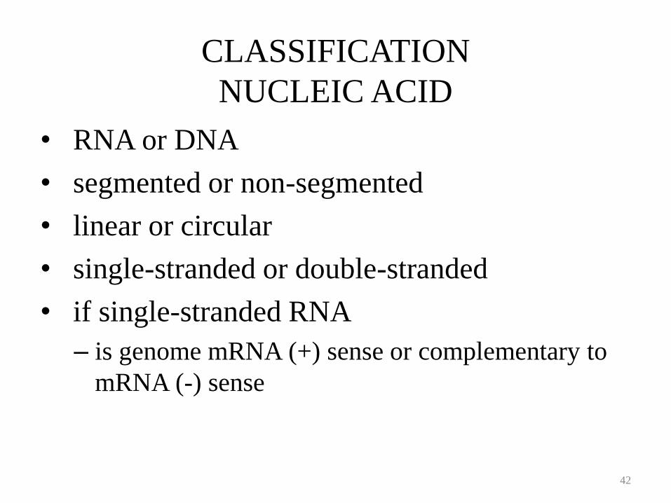
• RNA or DNA
• segmented or non-segmented
• linear or circular
• single-stranded or double-stranded
• if single-stranded RNA
– is genome mRNA (+) sense or complementary to
mRNA (-) sense
CLASSIFICATION
NUCLEIC ACID

43
ENVELOPE
• OBTAINED BY BUDDING THROUGH A CELLULAR MEMBRANE (except poxviruses)
• POSSIBILITY OF EXITING CELL WITHOUT KILLING IT
• CONTAINS AT LEAST ONE VIRALLY CODED PROTEIN
– ATTACHMENT PROTEIN
• LOSS OF ENVELOPE RESULTS IN LOSS OF INFECTIVITY

Properties of naked viruses
• Stable in hostile environment
• Not damaged by drying, acid, detergent, and heat
• Released by lysis of host cells
• Can sustain in dry environment
• Can infect the GI tract and survive the acid and bile
• Can spread easily via hands, dust, fomites, etc
• Can stay dry and still retain infectivity
• Neutralizing mucosal and systemic antibodies are needed
to control the establishment of infection

Naked viruses( Non Enveloped )
• Adeno-associated Virus (AAV)AdenovirusB19Coxsackievirus - ACoxsackievirus - BEchovirusHepatitis A Virus (HAV)Hepatitis E Virus (HEV)Norwalk Virus
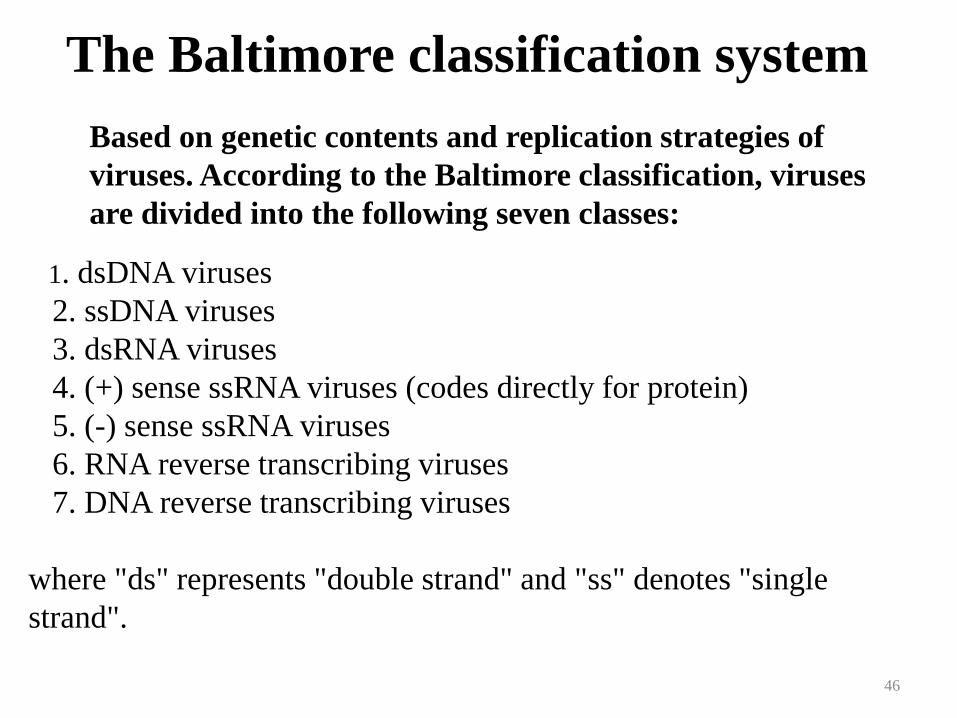
The Baltimore classification system
Based on genetic contents and replication strategies of
viruses. According to the Baltimore classification, viruses
are divided into the following seven classes:
1. dsDNA viruses
2. ssDNA viruses
3. dsRNA viruses
4. (+) sense ssRNA viruses (codes directly for protein)
5. (-) sense ssRNA viruses
6. RNA reverse transcribing viruses
7. DNA reverse transcribing viruses
where "ds" represents "double strand" and "ss" denotes "single
strand".
46

Virus Classification
- the Baltimore classification
• All viruses must produce mRNA, or (+) sense RNA
• A complementary strand of nucleic acid is (–) sense
• The Baltimore classification has + RNA as its central point
• Its principles are fundamental to an understanding of virus classification and genome replication, but it is rarely used as a classification system in its own right
47

Viral genome strategies
• dsDNA (herpes, papova, adeno, pox)
• •ssDNA (parvo)
• •dsRNA (reo, rota)
• •ssRNA (+) (picorna, toga, flavi, corona)
• •ssRNA (-) (rhabdo, paramyxo, orthomyxo,
• bunya, filo)
• •ssRNA (+/-) (arena, bunya)
• •ssRNA (+RTase) (retro, lenti)48
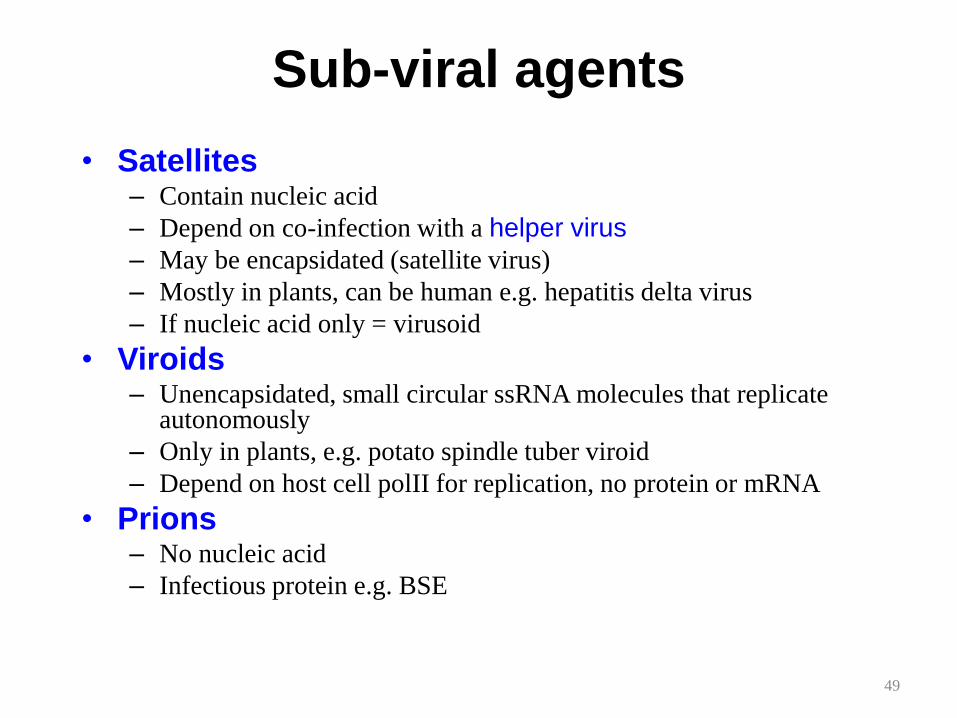
Sub-viral agents
• Satellites– Contain nucleic acid
– Depend on co-infection with a helper virus
– May be encapsidated (satellite virus)
– Mostly in plants, can be human e.g. hepatitis delta virus
– If nucleic acid only = virusoid
• Viroids– Unencapsidated, small circular ssRNA molecules that replicate
autonomously
– Only in plants, e.g. potato spindle tuber viroid
– Depend on host cell polII for replication, no protein or mRNA
• Prions– No nucleic acid
– Infectious protein e.g. BSE
49

Viroids & Prions
• Viroids– ss RNA genome and the smallest known pathogens.– Affects plants
• Prions– Infectious particles that are entirely protein.– No nucleic acid– Highly heat resistant– Animal disease that affects nervous tissue– Affects nervous tissue and results in
• Bovine spongiform encepahltits (BSE) “mad cow disease”, • scrapie in sheep• kuru & Creutzfeld-Jakob Disease (CJD) in humans
50

Viroids
• Viroids are small (200-400nt), circular RNA molecules with a rod-like secondary structure which possess no capsid or envelope which are associated with certain plant diseases. Their replication strategy like that of viruses - they are obligate intracellular parasites.
• Viroids do not encode any proteins and unlike satellites they are not dependent on the presence of another virus
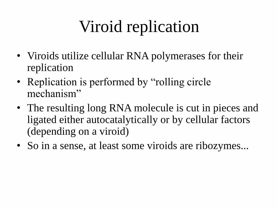
Viroid replication
• Viroids utilize cellular RNA polymerases for their replication
• Replication is performed by “rolling circle mechanism”
• The resulting long RNA molecule is cut in pieces and ligated either autocatalytically or by cellular factors (depending on a viroid)
• So in a sense, at least some viroids are ribozymes...
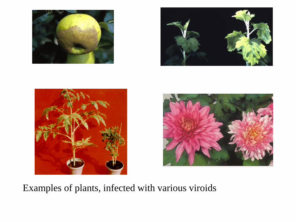
Examples of plants, infected with various viroids

Hepatitis d virus – a chimeric molecule,
half viroid, half satellite
• Viroid like properties
- Rod-like RNA molecule
- Rolling circle replication
- Self-cleaving activty
• Satellite like properties
- Encodes a protein, which is necessary both for encapsidation and replication
- Dependent on presence another virus – HBV
- Genome larger than for viroids (1640 nt)

Prions• Prions are rather ill-defined infectious agents
believed to consist of a single type of protein molecule with no nucleic acid component. Confusion arises from the fact that the prion protein & the gene which encodes it are also found in normal 'uninfected' cells. These agents are associated with diseases such as Creutzfeldt-Jakob disease in humans, scrapie in sheep & bovine spongiform encephalopathy (BSE) in cattle.

Prions
Prions are proteinaceous transmissible pathogens responsible
for a series of fatal neurodegenerative diseases (in humans,
Creutzfeld-Jakob disease and kuru, in animals, bovine
spongioform encephalopathy)
A prion (proteinaceous infectious particle, analogy for virion)
is a type of infectious agent that does not carry the genetic
information in nucleid acid!
Prions are proteins with the pathological conformation that are
believed to infect and propagate the conformational changes of
the native proteins into the the abnormally srtructured form

Disease name Natural host Prion name PrP isoform
Scrapie Sheep, goat Scrapie prion OvPrPSc
Transmissible mink
encephalopathy (TME)
Mink TME prion MkPrPSc
Chronic wasting disease
(CWD)
Elk, mule deer CWD prion MDePrPSc
Bovine spongioform
encephalopathy (BSE)
Cattle BSE prion BovPrPSc
Feline spongioform
encephalopathy (FSE)
Cat FSE prion FePrPSc
Exotic unguale
encephalopathy (EUE)
Greater kudu,
nyala
EUE prion NyaPrPSc
Kuru Human Kuru prion HuPrPSc
Creutzfeldt-Jakob disease
(CJD)
Human CJD prion HuPrPSc
Gerstmann-Straussler-
Scheinker syndrome
(GSS)
Human GSS prion HuPrPSc
Fatal familial insomnia
(FFI)
Human FFI prion HuPrPSc

Prion diseases: rare neurodegenerative disorders (one
person per million)
1. Sporadic (85 %)
In the sixth or seventh decade, rapidly progressive (death in less than a year)
Creutzfeldt-Jakob disease (CJD)
2. Familial (inherited-15%)
Mutations in the PrP gene that favour the transition from the cellular form to the
pathological form of PrP
Gerstmann-Straussler-Scheinker disease (GSS), fatal familial insomnia (FFI)
3. Transmissible (rare; a source of great concern)
Propagation of kuru disease in New Guinea natives (ritualistic cannibalism)
Recently, it has been discovered that BSE had been transmitted to humans in Europe after
consumption of infected beef, producing a variant of the CJD called vCJD

Transmissible spongioform encephalopathy (TSE)=prion disease
A group of progressive conditions that affect the brain and nervous system of humans and animals and are transmitted by prions
The pathology: vacuolar degeneration, neuronal loss, astrocytosis and amyloid plaque formation
The clinical signs: loss of motor functions (lack of coordination, ataxia, involuntary jerking movements), personality changes, depression,
insomnia, confusion, memory problems, dementia, progressive tonic paralysis, death
Definitive diagnostic test: biopsy of brain tissue (histopathologicalexamination and immunostaining for PrPSc)
There is no cure

α-helixβ-sheet
Normal protein
(folded structure)
Disease-associated protein
(misfolded structure)
Conformational change
Aggregation
Gain of toxic
activity
Loss of biological
function

PrPC
The normal protein
is called PrPC (for cellular)
is a transmembrane glycoprotein
(neurons, lymphocytes); its function
is unknown; it binds Cu2+ (regulation
its homeostasis)
has dominant secundary structure α-
helix
is easily soluble
is monomeric and easily digested by
proteases
is encoded by a gene designated
PRNP located on the chromosome 20
PrPSc
The abnormal, disease-producing
protein
is called PrPSc (for scrapie)
has the same amino acid sequence
(primary structure)
has dominant secundary structure β-
sheets
is insoluble
is multimeric and resistant to
digestion by proteases
When PrPSc comes in contact with
PrPC, it converts the PrPC into more of
itself These molecules bind to each
other forming aggregates

Molecular models of the structure
of:
PrPC PrPSc
Predominantly α-helix (3) β-sheets (40%), α-helix (30%)
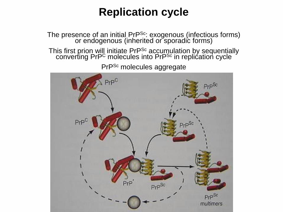
Replication cycle
The presence of an initial PrPSc: exogenous (infectious forms) or endogenous (inherited or sporadic forms)
This first prion will initiate PrPSc accumulation by sequentially converting PrPC molecules into PrPSc in replication cycle
PrPSc molecules aggregate
Summary
The prions are proteins that carry information for self-reproduction (contradict
the central dogma of modern biology)
The prions are expressed in cells of healthy humans and animals; their
abnormal conformations (PrPSc) are insoluble, resistent to digestion and
aggregate
The PrPSc attacks the native prion PrPC, changes its conformation into an
abnormal form and causes an exponential production of insoluble proteins;
they aggregate and form the fibrillar structure
Prion disease are rare fatal degenerative disorders; a portion of them can be
transmitted; this mechanism is not clear (e.g. transmision of BSE to human)
One part of the prion protein can cause apoptosis, or programmed cell death
Prions induce no immune reactions within the human
65
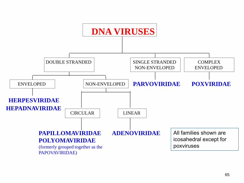
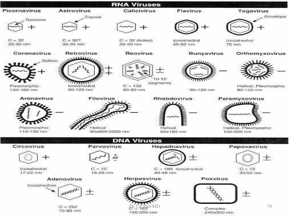
HERPESVIRIDAE
HEPADNAVIRIDAE
ENVELOPED
PAPILLOMAVIRIDAE
POLYOMAVIRIDAE(formerly grouped together as the
PAPOVAVIRIDAE)
CIRCULAR
ADENOVIRIDAE
LINEAR
NON-ENVELOPED
DOUBLE STRANDED
PARVOVIRIDAE
SINGLE STRANDED
NON-ENVELOPED
POXVIRIDAE
COMPLEX
ENVELOPED
DNA VIRUSES
All families shown are
icosahedral except for
poxviruses

DNA viruses
From Principles of
Virology Flint et al
ASM Press
66

67
FLAVIVIRIDAETOGAVIRIDAE
RETROVIRIDAE
ICOSAHEDRAL
CORONAVIRIDAE
HELICAL
ENVELOPED
ICOSAHEDRAL
PICORNAVIRIDAECALICIVIRIDAE
ASTROVIRIDAE
NONENVELOPED
SINGLE STRANDED
positive sense
BUNYAVIRIDAEARENAVIRIDAE
ORTHOMYXOVIRIDAEPARAMYXOVIRIDAE
RHABDOVIRIDAEFILOVIRIDAE
SINGLE STRANDED
negative sense
REOVIRIDAE
DOUBLE STRANDED
RNA VIRUSES
ENVELOPED
HELICAL ICOSAHEDRAL
NONENVELOPED
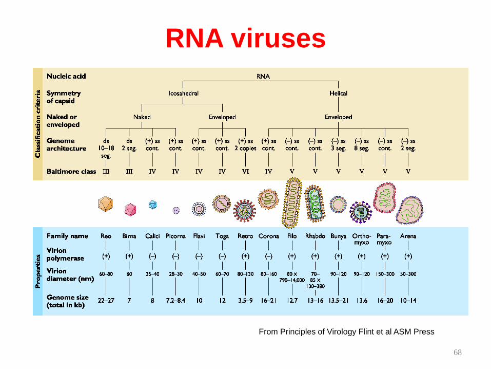
RNA viruses
From Principles of Virology Flint et al ASM Press
68

Dr.T.V.Rao MD 70
71
BASIC STEPS IN VIRAL LIFE
CYCLE
• ADSORPTION
• PENETRATION
• UNCOATING AND ECLIPSE
• SYNTHESIS OF VIRAL NUCLEIC ACID
AND PROTEIN
• ASSEMBLY
• RELEASE