an lc/ms/ms method for stable isotope dilution studies … · an lc/ms/ms method for stable isotope...
TRANSCRIPT
1
An LC/MS/MS method for stable isotope dilution studies of β-carotene
bioavailability, bioconversion and vitamin A status in humans.
Anthony Oxley1, Philip Berry
2, Gordon A Taylor
2, Joseph Cowell
3, Michael J Hall
3, John Hesketh
4,
Georg Lietz1,5
, and Alan V Boddy2
AFFILIATIONS:
1 Human Nutrition Research Centre, Newcastle University, UK.
2 Northern Institute for Cancer Research, Newcastle University, UK.
3 School of Chemistry, Newcastle University, UK.
4 Institute for Cell and Molecular Biosciences, Newcastle University, UK.
CORRESPONDING AUTHOR:
5 Dr Georg Lietz, Human Nutrition Research Centre, School of Agriculture, Food and Rural
Development, Agriculture Building, Newcastle University, Newcastle upon Tyne, NE1 7RU.
Phone: +44 191 222 6893. E-mail: [email protected]
RUNNING TITLE:
LC/MS/MS of [13
C]-β-carotene and [13
C]-vitamin A.
by guest, on October 13, 2018
ww
w.jlr.org
Dow
nloaded from

2
ABSTRACT
Isotope dilution is currently the most accurate technique in humans to determine vitamin A status
and bioavailability/bioconversion of provitamin A carotenoids such as β-carotene. However, limits
of MS detection, coupled with extensive isolation procedures, have hindered investigations of
physiologically-relevant doses of stable isotopes in large intervention trials. Here, a sensitive liquid
chromatography-tandem-mass spectrometry (LC/MS/MS) analytical method was developed to
study the plasma response from co-administered oral doses of 2 mg [13
C10]-β-carotene and 1 mg
[13
C10]-retinyl acetate in human subjects over a 2 week period. A reverse-phase C18 column and
binary mobile phase solvent system separated β-carotene, retinol, retinyl acetate, retinyl linoleate,
retinyl palmitate/retinyl oleate, and retinyl stearate within a 7 min run time. Single reaction-
monitoring (SRM) of analytes was performed under atmospheric-pressure chemical ionisation
(APCI) in positive mode at m/z 537→321 and 269→93 for respective [12
C]-β-carotene and [12
C]
retinoids; m/z 547→330 and 274→98 for [13
C10]-β-carotene and [13
C5] cleavage products; and m/z
279→100 for metabolites of [13
C10]-retinyl acetate. A single one-phase solvent extraction, with no
saponification or purification steps, left retinyl esters intact for determination of intestinally-derived
retinol in chylomicrons versus retinol from the liver bound to retinol-binding protein (RBP). Co-
administration of [13
C10]-retinyl acetate with [13
C10]-β-carotene not only acts as a reference dose for
inter-individual variations in absorption and chylomicron clearance rates, but also allows for
simultaneous determination of an individual's vitamin A status.
SUPPLEMENTARY KEY WORDS:
BCMO1, carotenoid metabolism, retinol metabolism, retinyl esters, tandem mass spectrometry.
ACKNOWLEDGEMENTS:
This research was funded by BBSRC (Grant reference BB/G004056/1) and supported in part by
Cancer Research UK
by guest, on October 13, 2018
ww
w.jlr.org
Dow
nloaded from
3
INTRODUCTION
Vitamin A deficiency (VAD) is a major public health issue in the developing world due to
inadequate intake of both preformed vitamin A and provitamin A carotenoids in the diet (1).
However, detection of subclinical deficiency is problematic since ~85% of vitamin A is stored in
the liver while the level of vitamin A circulating in the blood is under strict homeostatic control and
not indicative of hepatic reserves (2). Increasing the intake of provitamin A carotenoids, primarily
through β-carotene, is seen as a safe way of restoring vitamin A reserves of an individual since high
doses of preformed vitamin A have adverse health effects (3). Although the current vitamin A
equivalency ratio for β-carotene is estimated at 12:1 (by weight) (4), large inter-individual
variations in both absorption and conversion have been observed (5-8).
In the intestinal mucosa, a proportion of absorbed β-carotene undergoes centric cleavage by the β-
carotene 15,15’ monooxygenase (BCMO1) enzyme to produce 2 molecules of retinal which are
further reduced to retinol (vitamin A) (9). For export into the circulation, retinol is esterified to a
long chain fatty acid, typically palmitate, and incorporated, along with intact β-carotene, into
chylomicrons (10). Subsequently, retinyl esters are either stored in hepatic stellate cells or
hydrolysed back to retinol, by the liver, for repartition to other tissue compartments bound to
retinol-binding protein (RBP).
Currently, stable isotopes dilution offers the most accurate determination of β-carotene bioefficacy
and vitamin A status irrespective of high endogenous circulating levels of these micronutrients
(1,2). However, the minimum dose to be administered has been dictated by the detection limit of the
analytical method (2). Furthermore, isolation of carotenoids/retinoids from the plasma matrix for
MS analysis often involves extensive and time-consuming extraction/purification procedures that
have included: saponification, solid-phase extraction (SPE), preparative HPLC and, in the case of
GCMS analysis, further conversion to tert-butyl-dimethylsilyl derivatives (11-18). The aim was to
develop an analytical method that involved a simplified extraction procedure, sensitive MS/MS for
detection of physiological doses of stable isotopes, and short LC runtimes so as to be suitable for
high-throughput of samples from human intervention studies.
by guest, on October 13, 2018
ww
w.jlr.org
Dow
nloaded from
4
MATERIALS AND METHODS
Chemicals.
The following carotenoid and retinoid (>95% all-trans) standards were purchased from Sigma (St.
Louis, MO, USA): β-carotene, lycopene, retinol, retinyl acetate, retinyl palmitate. The [12, 12’, 13,
13’, 14, 14’, 15, 15’, 20, 20’-13
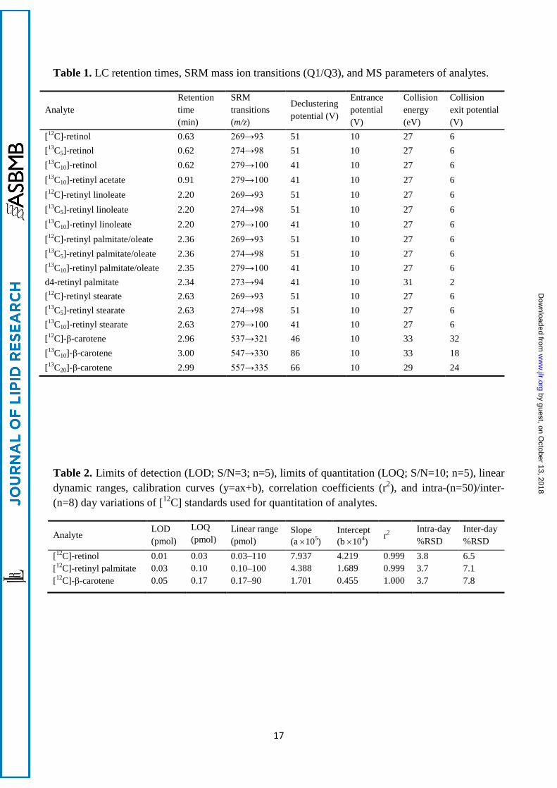
C10]-β-carotene, [8, 9, 10, 11, 12, 13, 14, 15, 19, 20-13
C10]-retinyl
acetate (Figure 1), to be administered to human subjects, were custom synthesised by Buchem BV
(Apeldoorn, The Netherlands) and certified fit for human consumption. Similarly, the [8, 8’, 9, 9’,
10, 10’, 11, 11’, 12, 12’, 13, 13’, 14, 14’, 15, 15’, 19, 19’, 20, 20’-13
C20]-β-carotene, [12, 13, 14, 15,
20-13
C5]-retinol, and [10, 19, 19, 19-d4]-retinyl palmitate stable isotopes (Figure 1) were also
purchased from Buchem BV. Methanol, propan-2-ol, chloroform, ethanol, ethyl acetate, toluene and
acetic acid were all of HPLC-grade and purchased from Fisher Scientific (Loughborough, UK).
BHT, Novozyme 435, and ammonium acetate were obtained from Sigma Aldrich (St. Louis, MO,
USA). Aberlyst A-21, linoleic acid, oleic acid, and stearic acid for synthesis of retinyl esters were
obtained from Alfa Aesar (Heysham, Lancashire, UK).
Synthesis of retinyl esters.
Retinyl esters were synthesised via an enzyme-catalysed transesterification (19) as follows. Into a
dry Schlenk flask, retinyl acetate (33 mg, 0.10 mmol), Novozyme 435 (120 mg), and Aberlyst A-21
(50 mg) were suspended in dry toluene (5 mL). The reaction mixture was stirred under an
atmosphere of N2, and 5 equivalents (0.50 mmol) of the appropriate acid (palmitic, stearic, linoleic
or oleic) was added. After 20 hours at room temperature, the reaction mixture was filtered and
solvent was removed under reduced pressure to give a mixture (approximately 1:4) of the desired
retinyl ester and unreacted acid. The resulting mixtures were used without further purification as
LC/MS/MS standards for the corresponding retinyl esters.
Subjects and blood collection.
Healthy male and female volunteers with an age range of 18-45 years were recruited into the
“ΒetaSNP” dietary intervention study where written informed consent was obtained. Exclusion
criteria were: pregnancy, smoking, high blood pressure, diabetes, BMI >30,
liver/kidney/gastrointestinal disease, lipid metabolic disorders, and consumption of multivitamins
(containing vitamins A, C, E) or β-carotene supplements 3 months prior to the study start. The study
was conducted according to the guidelines set forth in the Declaration of Helsinki, and all
procedures involving human subjects were approved by the Sunderland NRES Committee North
by guest, on October 13, 2018
ww
w.jlr.org
Dow
nloaded from
5
East (REC 09/H0904/20) before registration with the UK Clinical Research Network (UKCRN:
7413). The [13
C10]-β-carotene and [13
C10]-retinyl acetate were prepared for oral administration in
sunflower oil, at respective concentrations of 2 mg ml–1
and 1 mg ml–1
, by sonication in amber
bottles at room temperature for 30 min. Oil solutions were then stored in sterile 1 ml tip-cap amber
oral syringes (Becton Dickinson, Oxford, UK) and used within 1 week of preparation. Fasted
subjects were cannulated via the antecubital vein and blood was drawn into 10 ml EDTA
Vacutainer tubes (Becton Dickinson). Subjects then received the dual isotopic oral dose of 2 mg
[13
C10]-β-carotene and 1 mg [13
C10]-retinyl acetate along with a standardised breakfast meal
consisting of a muffin and yoghurt smoothie. The meal was designed to reflect the same nutrient
content as described by Borel et al. (5) containing 46.3 g of fat (55.5% of total energy intake).
Blood was subsequently collected at 2, 4, 6, 8, 10, 12 hours post-dose via cannulation, and at 24,
48, 168, and 336 hours by simple venepuncture. Each blood sample was immediately centrifuged at
4°C upon collection and the plasma stored at –80°C until analysis.
Plasma extraction and analyte recovery.
An ethanol/ethyl acetate (1:1) solvent extraction was applied to plasma samples to ensure adequate
recovery of all analytes without co-extraction of lipids known to interfere with LC/MS analyses. All
extraction procedures were performed under yellow lighting. To 1 ml of plasma, 10 µl (50 pmol)
each of the [13
C10]-retinyl acetate and [13
C20]-β-carotene internal standards was added before
denaturing with 5 ml of ethanol and 5 ml of ethyl acetate. The sample was then shaken on an orbital
shaker for 10 min and centrifuged at 10,000 rpm for 30 min at 4°C. The supernatant was transferred
to a clean glass tube and the solvent evaporated to dryness under a stream of nitrogen. The residue
was resuspended in 100 µl of ethyl acetate, by vortexing briefly, and transferred to amber glass
vials ready for LC/MS/MS injection.
Due to endogenous levels of [12
C]-β-carotene, retinol, and retinyl palmitate always being present in
‘control’ plasma, recovery of target analytes from the plasma matrix was assessed using the
following stable isotopes: [13
C10]-β-carotene, [13
C5]-retinol, and d4-retinyl palmitate. Blank plasma
was generously provided by the Blood Transfusion Service, Newcastle upon Tyne Hospitals (UK).
For extraction efficiency experiments, 10 µl of [13
C10]-β-carotene, [13
C5]-retinol, and d4-retinyl
palmitate in ethanol were spiked into 1 ml of control plasma at a final concentration of 5 µM.
Plasma was then extracted as described above.
by guest, on October 13, 2018
ww
w.jlr.org
Dow
nloaded from
6
LC/MS/MS analysis.
Chromatographic separation of β-carotene and retinoids was achieved using a Perkin Elmer Series
200 LC (Beckonsfield, UK) equipped with a Gemini C18 column (3µm; 50 mm × 2 mm i.d.) and
SecurityGuard C18 column (4 x 3 mm) both from Phenomenex (Cheshire, UK) maintained at 30°C.
Reverse phase elution of analytes was performed with mobile phases of 0.1M aqueous ammonium
acetate pH5 (A) and 50:50 (w/w) methanol/isopropanol (B). The mobile phase system consisted of
a 1 min linear gradient from 80% to 99% B, held at 99% B for 3 min, then immediately returned to
80% B for 3 min to re-equilibrate. Flow rate was 1.0 ml min-1
with an injection volume of 10µl.
An API4000 triple quadrupole LC/MS/MS (Applied Biosystems, California, USA) was used for
analysis with atmospheric pressure chemical ionisation (APCI) performed in positive ion mode
using nitrogen gas with the following optimum settings: collision gas, 7; curtain gas, 10; ion source
gas 1, 60; ion source gas 2, 15. Temperature of the heated nebulizer was 400oC with an ionspray
voltage of 5500. Optimisation of MS/MS parameters for all analytes was performed by selecting
precursor ions of [M+H]+ for β-carotene, [M+H-18]
+ for retinol, [M+H-256]
+ for retinyl palmitate,
and [M+H-60]+
for retinyl acetate to obtain product ion spectra. Quantitation of analytes was
performed in single reaction monitoring (SRM) mode where mass transitions and optimised
MS/MS parameters are given in Table 1. Analyst® software v1.4.1 (AB SCIEX, Framingham,
USA) was used for SRM, peak integration and analyte quantitation. Peak areas were adjusted
according to internal standard recovery ([13
C10]-retinyl acetate for retinoids and [13
C20]-β-carotene
for carotenes) and quantified against external calibration curves of [12
C]-β-carotene, [12
C]-retinol
and [12
C]-retinyl palmitate (Table 2).
LC/MS/MS validation.
The [12
C] species of β-carotene, retinol and retinyl palmitate were used to assess linear dynamic
ranges, limits of detection (LOD), limits of quantitation (LOQ), intra-/inter-day assay precision, and
to construct external calibration curves. Stock solutions of β-carotene and retinyl palmitate were
prepared in chloroform containing 0.1% BHT at respective concentrations of 0.2 mg ml-1
and 1.0
mg ml-1
. Retinol was dissolved in ethanol, containing 0.1% BHT, at 1.0 mg ml-1
. Stock solutions
were diluted in ethanol for spectrophotometric determination of absolute concentration at λmax
450nm for β-carotene and 325nm for retinol and retinyl palmitate. Concentrations were calculated
from published extinction coefficients (E1%
1cm) for these compounds in ethanol (20,21). A standard
mix of analytes was prepared in ethanol to study linear dynamic range, via serial dilution (11 µM –
by guest, on October 13, 2018
ww
w.jlr.org
Dow
nloaded from
7
5 nM), and for determination of intra- and inter-day assay precision (1 µM) through multiple
injections.
by guest, on October 13, 2018
ww
w.jlr.org
Dow
nloaded from

8
RESULTS
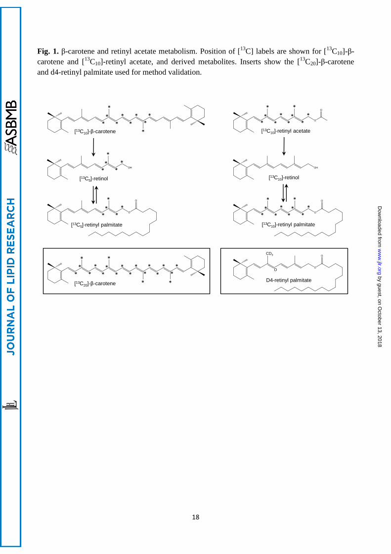
Chemical ionisation (APCI) in positive mode offered greater linear dynamic range for both β-
carotene and retinoids compared to electrospray ionisation (ESI). APCI of retinoids resulted in the
elimination of terminal functional groups to produce identical Q1 precursor ions of [M+H–H20]+ for
retinol, [M+H–CH3CO2H]+ for retinyl acetate, and [M+H–CH3(CH2)14CO2H]
+ for retinyl palmitate.
Consequently, it was necessary to adequately separate retinoids by liquid chromatography (LC)
before selected reaction monitoring (SRM) at m/z 269→93, m/z 274→98, and m/z 279→100 for
respective [12
C], [13
C5], and [13
C10] isotopologues (Table 1). The abundant Q3 product ion for
retinoids was due to cleavage at the C9–C10 double bond where the selected polyene chain fragment
contained all [13
C] labels from m/z 274 and 7 of the [13
C] labels from m/z 279 (Figure 2).
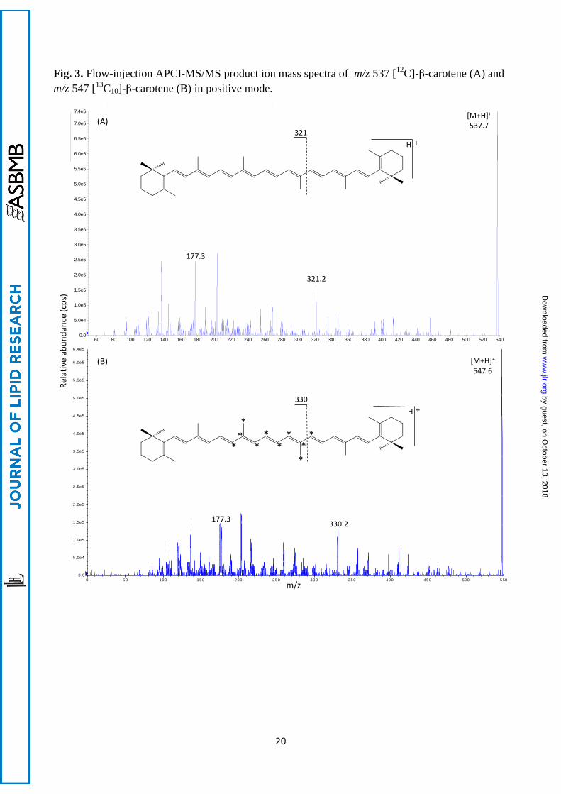
APCI of β-carotene resulted in protonation of the molecule [M+H]+ with an abundant Q3 product
ion at m/z 177 irrespective of isotopic composition (m/z 537→177 [12
C] and m/z 547→177 [13
C];
Figure 3). The geometric isomer of β-carotene, lycopene, also produced a fragment Q3 ion at m/z
537→177 and possessed an identical LC retention time to β-carotene. Furthermore, an unidentified
compound was observed in ‘blank’ plasma at m/z 547→177 which could not be separated from β-
carotene by LC. Therefore, an alternative less abundant fragment of higher m/z was selected for
[13
C]-β-carotene at 330 (Figure 3). This product ion was the result of cleavage at C12–C13 and
contained the majority of the [13
C]-labelling from m/z 547 and also from m/z 557 as internal
standard. The corresponding fragment for [12
C]-β-carotene at m/z 321 was not present for lycopene.
Both trans- and cis- β-carotene isomers produced the same Q3 product ions (Supplemental Fig.I).
Optimised MS/MS parameters and SRM transitions for all analytes are given in Table 1.
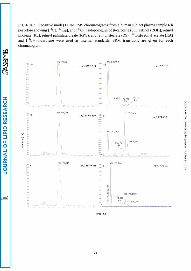
Retinol and retinyl acetate were separated to baseline on a C18 reversed-phase column with a 1 min
linear gradient of 80% to 99% methanol/isopropanol (50:50, w/w); their respective retention times
were 0.63 and 0.91 min (Figure 4). Retinyl palmitate and β-carotene eluted at 2.36 min and 2.96
min respectively under isocratic conditions of 99% methanol/isopropanol. From extracted control
plasma, 2 additional peaks were observed at m/z 269→93 that flanked the retinyl palmitate peak. As
these peaks were suspected to be alternative fatty acid esters of retinol, it was necessary to
synthesize non-commercially available retinyl esters. The presence of the postulated retinyl esters
was confirmed through the use of natural abundance 13
C NMR measured in CDCl3 using a Jeol
ECS-400 MHz. 13
C NMR analysis of the reaction between palmitic acid and retinyl acetate,
revealed a signal at 174.0 ppm which correlates to the carbonyl carbon of retinyl palmitate (in
comparison to commercial standards) and was clearly distinct from retinyl acetate (171.2 ppm) and
by guest, on October 13, 2018
ww
w.jlr.org
Dow
nloaded from
9
palmitic acid (180.4 ppm). Similar 13
C NMR signals were observed for retinyl stearate (174.0 ppm),
retinyl oleate (174.0 ppm) and retinyl linoleate (173.9 ppm) confirming the production of each of
the retinyl esters. Synthetic retinyl palmitate was compared against commercially-available retinyl
palmitate by LC/MS/MS providing the same retention time and mass spectra, further confirming the
formation of the desired retinyl esters. Consequently, LC/MS/MS peaks at 2.20 and 2.63 were
confirmed as retinyl linoleate and retinyl stearate while retinyl oleate co-eluted with retinyl
palmitate at 2.36 min. Total LC run-time was 7 min which included a column re-equilibration
period of 3 min.
From extraction efficiency experiments (n=6), the recoveries of [13
C5]-retinol, d4-retinyl palmitate
and [13
C20]-β-carotene were 39% (± 1.9 SD), 36% (± 2.3 SD), and 30% (± 1.6 SD) respectively.
Although recovery of analytes was relatively low, the mild extraction procedure employed negated
the detrimental effects associated with co-extracted lipids during MS analysis. Furthermore, total
analyte concentrations were calculated using the internal standards [13
C10]-retinyl acetate and
[13
C20]-β-carotene, thus correcting for the low recovery. On-column validation of linear dynamic
range, limit of detection (LOD), and intra and inter-day precision for [12
C] analytes are given in
Table 2. Limits of detection ranged from 10 fmol for retinol to 50 fmol for β-carotene. Linear
dynamic ranges were over 2-3 orders of magnitude with r2 values of >0.999 (Supplemental Fig.II).
Intra- and inter-day precision ranged from 3.7-3.8 % RSD and 6.5-7.8% RSD respectively.
Administered 2 mg [13
C10]-β-carotene could be detected in plasma from 2 h to 2 wk post-dose
(Figure 5). The [13
C10]-β-carotene plasma response exhibited an initial increase to 10 nmol/L at 6 h,
followed by a brief plateau to 8 h, then a steady rise to a maximum of 25 nmol/L at 24 h. The
[13
C10]-β-carotene cleavage product [13
C5]-retinyl palmitate rapidly attained a maximum
concentration of 50 nmol/L at 4 h post-dose, while [13
C5]-retinol started to appear at 3-4 h in plasma
and peaked at 10 h. Metabolites of the 1 mg [13
C10]-retinyl acetate dose reached plasma
concentrations 4- to 6-fold higher than [13
C10]-β-carotene and derived cleavage products. Plasma
kinetics of [13
C10]-retinol and [13
C10]-retinyl palmitate mirrored those observed for [13
C5]-retinol
and [13
C5]-retinyl palmitate. Retinol secreted from the intestine was predominantly esterified to
palmitate and oleate. However, retinyl linoleate levels were higher than retinyl stearate for [13
C5]
cleavage products while retinyl stearate was higher than retinyl linoleate for [13
C10] retinol.
by guest, on October 13, 2018
ww
w.jlr.org
Dow
nloaded from
10
DISCUSSION
In human intervention studies, the size of stable isotope dose given is largely determined by the
limit of detection of the analytical method (1,2). Although carotene absorption and metabolism may
be tracked by the very sensitive method of accelerator mass spectrometry (AMS) (22,23), this
method involves the administration of radiolabelled material, albeit at micro-doses, and requires
laborious sample fractionation to distinguish metabolites, followed by very expensive analysis
using highly specialised equipment that is not widely available. Even if other MS methods such as
gas chromatography/combustion/isotope-ratio (GC/C/IRMS) and electron capture negative
chemical ionisation (ECNCI/MS) allow effective use of physiological doses of retinol (24,25) and
β-carotene (26) tracers, these methods have the disadvantage of requiring extensive sample
preparation, including HPLC purification and derivatisation, before injection into the MS. In
contrast, the application of liquid chromatography mass spectrometry (LC/MS) to the analysis of
retinoid and carotenoid tracers offers the advantages of high sensitivity and selectivity without the
need for hydrolysis and derivatisation (17,27-30). However, isolation of carotenoids and retinoids
from the plasma matrix is frequently carried out individually leading to separate injections, use of
different LC systems, MS ionisation methods (APCI/ESI) and modes (positive/negative) (11-18).
The current method allows for the first time to analyse both [13
C] retinoid and β-carotene tracers
simultaneously using chemical ionisation (APCI) in positive mode. Furthermore, the new method is
more sensitive than comparable LC/MS methods, with detection limits of 10 fmol for retinol and 50
fmol for β-carotene compared to 233 fmol (27) and 672 fmol (29) for retinol and 250 fmol (17), 559
fmol (28) and 57 fmol (27) for β-carotene in previous methods.
The single solvent extraction procedure developed here for both carotenoids and retinoids negated
the effect of interfering plasma lipids (31), without saponification, leaving retinyl esters intact.
Consequently, it was not necessary to prepare triglyceride-rich lipoprotein (TRL) fractions to
discriminate newly-absorbed intestinally-derived retinyl esters from retinol secreted by the liver
bound to retinol binding protein (RBP). However, it is recognised that small amounts (~3%) of
unesterified retinol, derived from administered retinyl acetate and β-carotene, may be present in
lymph chylomicrons (32,33). Although TRL fractions, obtained by ultracentrifugation at a solution
density of <1.006 g ml-1
, contain >83% of retinyl esters in the first 6 hour postprandial period, a
large percentage of plasma retinyl esters is progressively and irreversibly transferred to the denser
LDL fraction resulting in 32% of the plasma retinyl esters localised to the LDL fraction 12 hours
after fat load (34). This transfer of retinyl esters is even more substantial in subjects with familial
hypercholesterolemia (35). Furthermore, inter-individual variation in chylomicron clearance
by guest, on October 13, 2018
ww
w.jlr.org
Dow
nloaded from
11
kinetics, such as delayed chylomicron remnant clearance in subjects with endogenous
hypertriglyceridemia (36), or variation in chylomicron recovery during TRL preparation and
analysis reduces the accuracy of this approach to directly measure the mass of retinyl esters or β-
carotene absorbed (37). Thus, the current method can detect intestinally-derived retinyl esters with
more accuracy compared to methods employing TRL separations (27,37,38).
The current method also allows β-carotene bioefficacy and vitamin A dilution to be studied
concurrently due to differential extrinsic [13
C] labelling of administered compounds. [13
C] isotopes
were selected since deuterated compounds are subject to hydrogen-deuterium exchange and possess
different physicochemical characteristics resulting in altered LC retention times and solvent
extraction efficiencies (2,11,28). Position of [13
C10] labels around the centric 15,15’ double bond on
the β-carotene molecule allowed BCMO1 [13
C5] cleavage products to be distinguished from [13
C10]
metabolites of [13
C10]-retinyl acetate. Although both [13
C10] and [13
C5] metabolites displayed similar
plasma kinetic profiles, concentrations of [13
C5] retinol and retinyl esters were 3-4 fold lower even
though twice the dose of [13
C10]-β-carotene was administered. It is known that intestinal absorption
of synthetic β-carotene is limited although bioavailability is distinctly enhanced when dissolved in
oil (39). Regarding retinyl esters, both [13
C10] and [13
C5] retinol were preferentially esterified to
palmitate and oleate. However subsequent specificities of [13
C5]-retinol for linoleate and [13
C10]-
retinol for stearate were observed which suggests differences in subcellular compartmentalisation
between preformed retinol and retinol from provitamin A sources in the enterocyte before
incorporation in chylomicrons.
Retinyl acetate was co-administered with β-carotene as a reference dose to correct for inter- and
intra-individual variations in intestinal absorption and chylomicron clearance rates (37). The
[13
C10]-retinyl acetate dose can also be used to determine total body vitamin A reserves after a
sufficient period (circa 3 days) of isotope dilution with endogenous pools (1). In some previous
studies, the reference dose was not administered concomitantly with β-carotene to avoid
competition during intestinal absorption (12,14). Single doses of β-carotene have ranged from 5 to
126 mg due to analytical detection limits dictating the minimum dose that can be administered to
human subjects. However, β-carotene bioefficacy is dose-dependent when > 4 mg is ingested (40),
while doses > 6 mg perturb the steady-state equilibrium in the blood (41). The 2 mg utilised in the
current study represents a true physiological dose according to the estimated daily intake of β-
carotene in UK and US populations (39). Although lower doses have been administered daily over a
prolonged period to reach a plateau of isotopic enrichment in the blood (15,16), multiple dosing
cannot establish uptake kinetics.
by guest, on October 13, 2018
ww
w.jlr.org
Dow
nloaded from
12
In summary, this new sensitive analytical method allows for the simultaneous study of β-carotene
bioefficacy and vitamin A status in human subjects at physiological doses for at least 2 weeks. The
simple extraction procedure and single 7 min LCMS run-time for all analytes makes the method
applicable to the high throughput of samples generated in large human intervention studies.
by guest, on October 13, 2018
ww
w.jlr.org
Dow
nloaded from
13
REFERENCES
1. Furr, H. C., M. H. Green, M. Haskell, N. Mokhtar, P. Nestel, S. Newton, J. D. Ribaya-Mercado, G. W.
Tang, S. Tanumihardjo, and E. Wasantwisut. 2005. Stable isotope dilution techniques for assessing
vitamin A status and bioefficacy of provitamin A carotenoids in humans. Public Health Nutrition 8: 596-
607.
2. van Lieshout, M., C. E. West, and R. B. van Breemen. 2003. Isotopic tracer techniques for studying the
bioavailability and bioefficacy of dietary carotenoids, particularly beta-carotene, in humans: a review.
American Journal of Clinical Nutrition 77: 12-28.
3. Haskell, M. J. 2012. The challenge to reach nutritional adequacy for vitamin A: beta-carotene
bioavailability and conversion-evidence in humans. American Journal of Clinical Nutrition 96: 1193S-
1203S.
4. Tang, G. 2010. Bioconversion of dietary provitamin A carotenoids to vitamin A in humans. American
Journal of Clinical Nutrition 91: 1468S-1473S.
5. Borel, P., P. Grolier, N. Mekki, Y. Boirie, Y. Rochette, B. Le Roy, M. C. Alexandre-Gouabau, D.
Lairon, and V. Azais-Braesco. 1998. Low and high responders to pharmacological doses of beta-
carotene: proportion in the population, mechanisms involved and consequences on beta-carotene
metabolism. Journal of Lipid Research 39: 2250-2260.
6. Hickenbottom, S. J., J. R. Follett, Y. M. Lin, S. R. Dueker, B. J. Burri, T. R. Neidlinger, and A. J.
Clifford. 2002. Variability in conversion of beta-carotene to vitamin A in men as measured by using a
double-tracer study design. American Journal of Clinical Nutrition 75: 900-907.
7. Leung, W. C., S. Hessel, C. Meplan, J. Flint, V. Oberhauser, F. Tourniaire, J. E. Hesketh, J. von Lintig,
and G. Lietz. 2009. Two common single nucleotide polymorphisms in the gene encoding beta-carotene
15,15 '-monoxygenase alter beta-carotene metabolism in female volunteers. Faseb Journal 23: 1041-
1053.
8. Lietz, G., A. Oxley, W. Leung, and J. Hesketh. 2012. Single nucleotide polymorphisms upstream from
the beta-Carotene 15,15 '-monoxygenase gene influence provitamin A conversion efficiency in female
volunteers. Journal of Nutrition 142: 161S-165S.
9. Lietz, G., A. Oxley, C. Boesch-Saadatmandi, and D. Kobayashi. 2012. Importance of beta,beta-carotene
15,15 '-monooxygenase 1 (BCMO1) and beta,beta-carotene 9 ',10 '-dioxygenase 2 (BCDO2) in nutrition
and health. Molecular Nutrition & Food Research 56: 241-250.
10. Harrison, E. H. 2012. Mechanisms involved in the intestinal absorption of dietary vitamin A and
provitamin A carotenoids. Biochimica Et Biophysica Acta-Molecular and Cell Biology of Lipids 1821:
70-77.
11. Dueker, S. R., A. D. Jones, G. M. Smith, and A. J. Clifford. 1994. Stable-isotope methods for the study
of beta-carotene-d(8) metabolism in humans utilizing tandem mass-spectrometry and high-performance
liquid-chromatography. Analytical Chemistry 66: 4177-4185.
by guest, on October 13, 2018
ww
w.jlr.org
Dow
nloaded from
14
12. Lin, Y. M., S. R. Dueker, B. J. Burri, T. R. Neidlinger, and A. J. Clifford. 2000. Variability of the
conversion of beta-carotene to vitamin A in women measured by using a double-tracer study design.
American Journal of Clinical Nutrition 71: 1545-1554.
13. Novotny, J. A., S. R. Dueker, L. A. Zech, and A. J. Clifford. 1995. Compartmental analysis of the
dynamics of beta-carotene metabolism in an adult volunteer. Journal of Lipid Research 36: 1825-1838.
14. Tang, G., J. Qin, G. G. Dolnikowski, and R. M. Russell. 2000. Vitamin A equivalence of beta-carotene
in a Woman as determined by a stable isotope reference method. European Journal of Nutrition 39: 7-
11.
15. van Lieshout, M., C. E. West, Muhilal, D. Permaesih, Y. Wang, X. Y. Xu, R. B. van Breemen, A. F. L.
Creemers, M. A. Verhoeven, and J. Lugtenburg. 2001. Bioefficacy of beta-carotene dissolved in oil
studied in children in Indonesia. American Journal of Clinical Nutrition 73: 949-958.
16. Van Loo-Bouwman, C. A., T. H. J. Naber, R. B. van Breemen, D. Zhu, H. Dicke, E. Siebelink, P. J. M.
Hulshof, F. G. M. Russel, G. Schaafsma, and C. E. West. 2010. Vitamin A equivalency and apparent
absorption of beta-carotene in ileostomy subjects using a dual-isotope dilution technique. British Journal
of Nutrition 103: 1836-1843.
17. Wang, Y., X. Y. Xu, M. van Lieshout, C. E. West, J. Lugtenburg, M. A. Verhoeven, A. F. L. Creemers,
and R. B. van Breemen. 2000. A liquid chromatography-mass spectrometry method for the
quantification of bioavailability and bioconversion of beta-carotene to retinol in humans. Analytical
Chemistry 72: 4999-5003.
18. Zhu, D., Y. Wang, Y. Pang, A. Liu, J. Guo, C. A. Bowman, C. E. West, and R. B. van Bremen. 2006.
Quantitative analyses of beta-carotene and retinol in serum and feces in support of clinical bioavailability
studies. Rapid Communications in Mass Spectrometry 20: 2427-2432.
19. Boaz, N. W., and S. K. Clendennen. June 10, 2006. Preparation of retinyl esters. United States Patent
Application 20080085534.
20. Barua, A. B., and H. C. Furr. 1998. Properties of retinoids - Structure, handling, and preparation.
Molecular Biotechnology 10: 167-182.
21. Craft, N. E., and J. H. Soares. 1992. Relative solubility, stability, and absorptivity of lutein and beta-
carotene in organic-solvents. Journal of Agricultural and Food Chemistry 40: 431-434.
22. Dueker, S. R., Y. M. Lin, B. A. Buchholz, P. D. Schneider, M. W. Lame, H. J. Segall, J. S. Vogel, and
A. J. Clifford. 2000. Long-term kinetic study of beta-carotene, using accelerator mass spectrometry in an
adult volunteer. Journal of Lipid Research 41: 1790-1800.
23. Ho, C. C., F. F. de Moura, S.-H. Kim, B. J. Burri, and A. J. Clifford. 2009. A minute dose of C-14-beta-
carotene is absorbed and converted to retinoids in humans. Journal of Nutrition 139: 1480-1486.
24. Tang, G. W., J. Qin, and G. Dolnikowski. 1998. Deuterium enrichment of retinol in humans determined
by gas chromatography electron capture negative chemical ionization mass spectrometry. Journal of
Nutritional Biochemistry 9: 408-414.
by guest, on October 13, 2018
ww
w.jlr.org
Dow
nloaded from

15
25. Tanumihardjo, S. A. 2000. Vitamin A status assessment in rats with C-13(4)-retinyl acetate and gas
chromatography/combustion/isotope ratio mass spectrometry. Journal of Nutrition 130: 2844-2849.
26. Parker, R. S., J. E. Swanson, B. Marmor, K. J. Goodman, A. B. Spielman, J. T. Brenna, S. M. Viereck,
and W. K. Canfield. 1993. Study of beta-carotene metabolism in humans using C-13-beta-carotene and
high-precision isotope ratio mass-spectrometry. Annals of the New York Academy of Sciences 691: 86-
95.
27. Fleshman, M. K., K. M. Riedl, J. A. Novotny, S. J. Schwartz, and E. H. Harrison. 2012. An LC/MS
method for d8-beta-carotene and d4-retinyl esters: beta-carotene absorption and its conversion to vitamin
A in humans. Journal of Lipid Research 53: 820-827.
28. Pawlosky, R. J., V. P. Flanagan, and J. A. Novotny. 2000. A sensitive procedure for the study of beta-
carotene-d8 metabolism in humans using high performance liquid chromatography-mass spectrometry.
Journal of Lipid Research 41: 1027-1031.
29. van Breemen, R. B., D. Nikolic, X. Y. Xu, Y. S. Xiong, M. van Lieshout, C. E. West, and A. B.
Schilling. 1998. Development of a method for quantitation of retinol and retinyl palmitate in human
serum using high-performance liquid chromatography atmospheric pressure chemical ionization mass
spectrometry. Journal of Chromatography A 794: 245-251.
30. van Breemen, R. B., L. Dong, and N. D. Pajkovic. 2012. Atmospheric pressure chemical ionization
tandem mass spectrometry of carotenoids. International Journal of Mass Spectrometry 312: 163-172.
31. Hagiwara, T., T. Yasuno, K. Funayama, and S. Suzuki. 1998. Determination of lycopene, alpha-carotene
and beta-carotene in serum by liquid chromatography atmospheric pressure chemical ionization mass
spectrometry with selected-ion monitoring. Journal of Chromatography B 708: 67-73.
32. Huang, H. S., and D. S. Goodman. 1965. Vitamin A and carotenoids. I. Intestinal absorption and
metabolism of 14C-labelled vitamin A alcohol and beta-carotene in the rat. The Journal of biological
chemistry 240: 2839-2844.
33. Goodman, D. S., R. Blomstrand, B. Werner, H. S. Huang, and T. Shiratori. 1966. The intestinal
absorption and metabolism of vitamin A and beta-carotene in man. The Journal of clinical investigation
45: 1615-1623.
34. Krasinski, S. D., J. S. Cohn, R. M. Russell, and E. J. Schaefer. 1990. Postprandial plasma vitamin-A
metabolism in humans - a reassessment of the use of plasma retinyl esters as markers for intestinally
derived chylomicrons and their remnants. Metabolism-Clinical and Experimental 39: 357-365.
35. Rubinsztein, D. C., J. C. Cohen, G. M. Berger, D. R. Vanderwesthuyzen, G. A. Coetzee, and W. Gevers.
1990. Chylomicron remnant clearance from the plasma is normal in familial hypercholesterolemic
homozygotes with defined receptor defects. Journal of Clinical Investigation 86: 1306-1312.
36. Cortner, J. A., P. M. Coates, N. A. Le, D. R. Cryer, M. C. Ragni, A. Faulkner, and T. Langer. 1987.
Kinetics of chylomicron remnant clearance in normal and in hyperlipoproteinemic subjects. Journal of
Lipid Research 28: 195-206.
by guest, on October 13, 2018
ww
w.jlr.org
Dow
nloaded from
16
37. Edwards, A. J., C. S. You, J. E. Swanson, and R. S. Parker. 2001. A novel extrinsic reference method for
assessing the vitamin A value of plant foods. American Journal of Clinical Nutrition 74: 348-355.
38. van Vliet, T., W. H. P. Schreurs, and H. Vandenberg. 1995. Intestinal beta-carotene absorption and
cleavage in men - response of beta-carotene and retinyl esters in the triglyceride-rich lipoprotein fraction
after a single oral dose of beta-carotene. American Journal of Clinical Nutrition 62: 110-116.
39. Grune, T., G. Lietz, A. Palou, A. C. Ross, W. Stahl, G. Tang, D. Thurnham, S.-a. Yin, and H. K.
Biesalski. 2010. Beta-carotene is an important vitamin A source for human. Journal of Nutrition 140:
2268S-2285S.Brubacher, G. B., and H. Weiser. 1985. The vitamin A activity of beta-carotene.
International Journal for Vitamin and Nutrition Research 55: 5-15.
40. Brubacher, G. B., and H. Weiser. 1985. The vitamin A activity of beta-carotene. International Journal
for Vitamin and Nutrition Research 55: 5-15.
41. Tang, G. 2012. Techniques for measuring vitamin A activity from beta-carotene. American Journal of
Clinical Nutrition 96: 1185S-1188S.
by guest, on October 13, 2018
ww
w.jlr.org
Dow
nloaded from
17
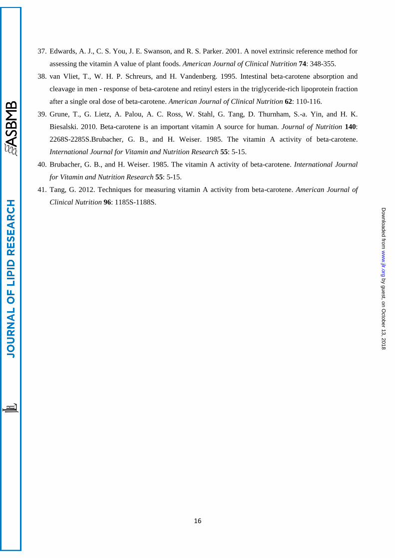
Table 1. LC retention times, SRM mass ion transitions (Q1/Q3), and MS parameters of analytes.
Analyte
Retention
time
(min)
SRM
transitions
(m/z)
Declustering
potential (V)
Entrance
potential
(V)
Collision
energy
(eV)
Collision
exit potential
(V)
[12
C]-retinol 0.63 269→93 51 10 27 6
[13
C5]-retinol 0.62 274→98 51 10 27 6
[13
C10]-retinol 0.62
279→100 41 10 27 6
[13
C10]-retinyl acetate 0.91 279→100 41 10 27 6
[12
C]-retinyl linoleate 2.20 269→93 51 10 27 6
[13
C5]-retinyl linoleate 2.20 274→98 51 10 27 6
[13
C10]-retinyl linoleate 2.20 279→100 41 10 27 6
[12
C]-retinyl palmitate/oleate 2.36 269→93 51 10 27 6
[13
C5]-retinyl palmitate/oleate 2.36 274→98 51 10 27 6
[13
C10]-retinyl palmitate/oleate 2.35 279→100 41 10 27 6
d4-retinyl palmitate 2.34 273→94 41 10 31 2
[12
C]-retinyl stearate 2.63 269→93 51 10 27 6
[13
C5]-retinyl stearate 2.63 274→98 51 10 27 6
[13
C10]-retinyl stearate 2.63 279→100 41 10 27 6
[12
C]-β-carotene 2.96 537→321 46 10 33 32
[13
C10]-β-carotene 3.00 547→330 86 10 33 18
[13
C20]-β-carotene 2.99 557→335 66 10 29 24
Table 2. Limits of detection (LOD; S/N=3; n=5), limits of quantitation (LOQ; S/N=10; n=5), linear
dynamic ranges, calibration curves (y=ax+b), correlation coefficients (r2), and intra-(n=50)/inter-
(n=8) day variations of [12
C] standards used for quantitation of analytes.
Analyte LOD
(pmol)
LOQ
(pmol)
Linear range
(pmol)
Slope
(a 105)
Intercept
(b 104)
r2
Intra-day
%RSD
Inter-day
%RSD
[12
C]-retinol 0.01 0.03 0.03–110 7.937 4.219 0.999 3.8 6.5
[12
C]-retinyl palmitate 0.03 0.10 0.10–100 4.388 1.689 0.999 3.7 7.1
[12
C]-β-carotene 0.05 0.17 0.17–90 1.701 0.455 1.000 3.7 7.8
by guest, on October 13, 2018
ww
w.jlr.org
Dow
nloaded from
18
Fig. 1. β-carotene and retinyl acetate metabolism. Position of [13
C] labels are shown for [13
C10]-β-
carotene and [13
C10]-retinyl acetate, and derived metabolites. Inserts show the [13
C20]-β-carotene
and d4-retinyl palmitate used for method validation.
* ** **
**
*
* **
*
* **
**
**
*
*
***
*
[13C10]-β-carotene
[13C5]-retinol
[13C5]-retinyl palmitate
[13C10]-retinyl acetate
[13C10]-retinyl palmitate
[13C10]-retinol
[13C20]-β-caroteneD4-retinyl palmitate
CD3
D
*
***
* * ** **
**
*
* *
**
**
*
*
**** *
*
* **
**
**
* by guest, on October 13, 2018
ww
w.jlr.org
Dow
nloaded from
19
Fig. 2. Flow-injection APCI-MS/MS product ion mass spectra of m/z 269 [12
C]-retinol (A); m/z 274
[13
C5]-retinol (B); and m/z 279 [13
C10]-retinyl acetate (C) in positive mode.
m/z
Rel
ativ
e ab
un
dan
ce (
cps)
+MS2 (269.30) CE (25): 20 MCA scans from Sample 6 (C12 Retinol) of figures.wiff (Heated Nebulizer) Max. 1.3e7 cps.
0 20 40 60 80 100 120 140 160 180 200 220 240 260 280m/z, Da
0.0
5.0e5
1.0e6
1.5e6
2.0e6
2.5e6
3.0e6
3.5e6
4.0e6
4.5e6
5.0e6
5.5e6
6.0e6
6.5e6
7.0e6
7.5e6
8.0e6
8.5e6
9.0e6
9.5e6
1.0e7
1.1e7
1.1e7
1.2e7
1.2e7
1.3e7
1.3e7
In
te
ns
ity
, c
ps
+MS2 (274.10) CE (25): 20 MCA scans from Sample 7 (5xC13 Retinol) of figures.wiff (Heated Nebulizer) Max. 4.9e6 cps.
0 20 40 60 80 100 120 140 160 180 200 220 240 260 280m/z, Da
0.0
2.0e5
4.0e5
6.0e5
8.0e5
1.0e6
1.2e6
1.4e6
1.6e6
1.8e6
2.0e6
2.2e6
2.4e6
2.6e6
2.8e6
3.0e6
3.2e6
3.4e6
3.6e6
3.8e6
4.0e6
4.2e6
4.4e6
4.6e6
4.8e6
In
te
ns
ity
, c
ps
+MS2 (279.40) CE (27): 20 MCA scans from Sample 4 (10xC13RetAcetate) of figures.wiff (Heated Nebulizer) Max. 2.5e6 cps.
0 20 40 60 80 100 120 140 160 180 200 220 240 260 280m/z, Da
0.0
1.0e5
2.0e5
3.0e5
4.0e5
5.0e5
6.0e5
7.0e5
8.0e5
9.0e5
1.0e6
1.1e6
1.2e6
1.3e6
1.4e6
1.5e6
1.6e6
1.7e6
1.8e6
1.9e6
2.0e6
2.1e6
2.2e6
2.3e6
2.4e6
2.5e6
In
te
ns
ity
, c
ps
[M+H-H20]+
269.1
92.5
97.5
99.5
[M+H-H20]+
274.1
[M+H–CH3CO2H]+
279.1
(A)
(B)
(C)
93
98 H +**
**
*
100
***
**
*
*
***
H +
H +
by guest, on October 13, 2018
ww
w.jlr.org
Dow
nloaded from
20
Fig. 3. Flow-injection APCI-MS/MS product ion mass spectra of m/z 537 [12
C]-β-carotene (A) and
m/z 547 [13
C10]-β-carotene (B) in positive mode.
+MS2 (537.60): 20 MCA scans from Sample 5 (alltransBC_3) of figures.wiff (Heated Nebulizer) Max. 7.4e5 cps.
60 80 100 120 140 160 180 200 220 240 260 280 300 320 340 360 380 400 420 440 460 480 500 520 540m/z, Da
0.0
5.0e4
1.0e5
1.5e5
2.0e5
2.5e5
3.0e5
3.5e5
4.0e5
4.5e5
5.0e5
5.5e5
6.0e5
6.5e5
7.0e5
7.4e5
In
te
ns
ity
, c
ps
[M+H]+
537.7
321.2
+M S2 (547 .60 ) C E (33): 20 MC A scans from S am p le 11 (10xc13bcar2 ) o f figu res.w if f (H eated N ebu lize r) Max. 6 .4e5 cps.
0 50 100 150 200 250 300 350 400 450 500 550
m/z, D a
0 .0
5 .0e4
1 .0e5
1 .5e5
2 .0e5
2 .5e5
3 .0e5
3 .5e5
4 .0e5
4 .5e5
5 .0e5
5 .5e5
6 .0e5
6 .4e5
In
te
ns
ity
, c
ps
(A)321
177.3
[M+H]+
547.6(B)
330.2
330
*
H +
177.3
*
* * ** *
*
**
H +
Rel
ativ
e ab
un
dan
ce (
cps)
m/z
by guest, on October 13, 2018
ww
w.jlr.org
Dow
nloaded from
21
Fig. 4. APCI (positive mode) LC/MS/MS chromatograms from a human subject plasma sample 6 h
post-dose showing [12
C], [13
C10], and [13
C5] isotopologues of β-carotene (βC), retinol (ROH), retinyl
linoleate (RL), retinyl palmitate/oleate (RPO), and retinyl stearate (RS). [13
C10]-retinyl acetate (RA)
and [13
C20]-β-carotene were used as internal standards. SRM transitions are given for each
chromatogram.
XIC of +MRM (8 pairs): 557.5/335.2 Da ID: 20xC13 Bcar from Sample 35 (70F) of 120320_recovery_IS_validation.wiff (Heated Nebulizer), Smoot... Max. 3026.7 cps.
0.5 1.0 1.5 2.0 2.5 3.0 3.5 4.0 4.5 5.0 5.5 6.0 6.5Time, min
0
200
400
600
800
1000
1200
1400
1600
1800
2000
2200
2400
2600
2800
3000
In
te
ns
ity
, c
ps
2.98
1.521.25
XIC o f +M RM (8 pa irs): 537 .5 /321 .1 D a ID : C12 Bca r from S amp le 34 (70E) o f 120320_recove ry_IS _va lida tion .w iff (H ea ted Nebu lize r), Sm oo thed M ax. 1 .3e4 cps.
0 .5 1 .0 1 .5 2 .0 2 .5 3 .0 3 .5 4.0 4 .5 5 .0 5 .5 6 .0 6 .5
T im e , min
0 .00
500 .00
1000 .00
1500 .00
2000 .00
2500 .00
3000 .00
3500 .00
4000 .00
4500 .00
5000 .00
5500 .00
6000 .00
6500 .00
7000 .00
7500 .00
8000 .00
8500 .00
9000 .00
9500 .00
1 .00e4
1 .05e4
1 .10e4
1 .15e4
1 .20e4
1 .25e4
1 .29e4
In
te
ns
ity
, c
ps
2.96
XIC o f +M RM (8 pa irs): 557 .5 /335 .2 Da ID : 20xC 13 B ca r from S am p le 34 (70E ) o f 120320_rec ov e ry_ IS _va lida tion .w iff (Hea ted Nebu lize r), S m oo t... M ax. 2395 .4 cps.
0 .5 1 .0 1 .5 2 .0 2 .5 3 .0 3 .5 4.0 4 .5 5 .0 5 .5 6 .0 6 .5
T im e , min
0
100
200
300
400
500
600
700
800
900
1000
1100
1200
1300
1400
1500
1600
1700
1800
1900
2000
2100
2200
2300
2395
In
te
ns
ity
, c
ps
2 .96
1.52 1 .701 .39 2 .00 3 .53
XIC o f +M RM (8 pa irs): 269 .1 /93 .1 Da ID : Re tino l from S am ple 34 (70E) o f 120320_recove ry_ IS_va lida tion .w iff (H eated Nebu liz er) M ax. 3 .4e5 cps.
0 .5 1 .0 1 .5 2 .0 2 .5 3 .0 3 .5 4.0 4 .5 5 .0 5 .5 6 .0 6 .5
T im e , min
0 .0
2 .0e4
4 .0e4
6 .0e4
8 .0e4
1 .0e5
1 .2e5
1 .4e5
1 .6e5
1 .8e5
2 .0e5
2 .2e5
2 .4e5
2 .6e5
2 .8e5
3 .0e5
3 .2e5
3 .4e5
In
te
ns
ity
, c
ps
0 .63
2 .36
XIC o f +M RM (8 pa irs): 274 .1 /98 .1 Da ID : 5 X C13 Re tino l from Sam ple 34 (70E) o f 120320_recove ry_ IS_va lida tion .w if f (H ea ted Nebu li ze r) M ax. 1 .1e4 cps.
0 .5 1 .0 1 .5 2 .0 2 .5 3 .0 3 .5 4.0 4 .5 5 .0 5 .5 6 .0 6 .5
T im e , min
0 .00
500 .00
1000 .00
1500 .00
2000 .00
2500 .00
3000 .00
3500 .00
4000 .00
4500 .00
5000 .00
5500 .00
6000 .00
6500 .00
7000 .00
7500 .00
8000 .00
8500 .00
9000 .00
9500 .00
1 .00e4
1 .05e4
1 .08e4
In
te
ns
ity
, c
ps
2 .35
0 .62
2 .63
4 .464 .141 .340 .19
XIC o f +M RM (8 pa irs): 279 .4 /100 .2 Da ID : 10XC13 Re tino l from S amp le 34 (70E) o f 120320_recove ry_ IS_va lida tion .w if f (H eated Nebu liz er) M ax. 5 .4e4 cps.
0 .5 1 .0 1 .5 2 .0 2 .5 3 .0 3 .5 4.0 4 .5 5 .0 5 .5 6 .0 6 .5
T im e , min
0 .0
5000 .0
1 .0e4
1 .5e4
2 .0e4
2 .5e4
3 .0e4
3 .5e4
4 .0e4
4 .5e4
5 .0e4
5 .4e4
In
te
ns
ity
, c
ps
0 .91
2 .35
0 .62
2 .63
3.00 [13C10]-βC
2.96 [12C]-βC
2.99 [13C20]-βC
m/z 537→ 321
m/z 547→ 330
m/z 557→ 335
0.63 [12C]-ROH
[12C]-RPO2.36
[12C]-RS2.63
[12C]-RL 2.20
m/z 269→93
0.62 [13C5]-ROH
2.36 [13C5]-RPO
2.63 [13C5]-RS[13C5]-RL
2.20
m/z 274→98
m/z 279→1000.91 [13C10]-RA
2.35 [13C10]-RPO
2.63 [13C10]-RS
[13C10]-RL2.20
(A)
(B)
(C)
(D)
(E)
(F)
0.62
[1
3C
10]-
RO
H
Time (min)
Inte
nsi
ty (
cps) by guest, on O
ctober 13, 2018w
ww
.jlr.orgD
ownloaded from

22
Fig. 5. Quantitative LC/MS/MS analysis of mean plasma responses from 45 human subjects (±
SEM) over the whole 14 day study period (A, C) and during the first 48 hours (B, D). Administered
[13
C10]-β-carotene (βC) and resulting [13
C5] cleavage products (ROH, retinol; RE, total retinyl
esters; RL, retinyl linoleate; RPO, retinyl palmitate/retinyl oleate; RS, retinyl stearate) are shown in
(A) and (B). [13
C10] metabolites of administered [13
C10]-retinyl acetate are shown in (C) and (D).
0
10
20
30
40
50
60
70
80
0 50 100 150 200 250 300 350
nm
ol/
L
Time (h)
[13C10]βC
[13C5]ROH
[13C5]RE
0
10
20
30
40
50
60
0 10 20 30 40 50
nm
ol/
LTime (h)
[13C5]RPO
[13C5]RL
[13C5]RS
(A)(B)
0
50
100
150
200
250
300
350
0 50 100 150 200 250 300 350
nm
ol/
L
Time (h)
[13C10]ROH
[13C10]RE
0
50
100
150
200
250
0 10 20 30 40 50
nm
ol/
L
Time (h)
[13C10]RPO
[13C10]RL
[13C10]RS
(C) (D)
by guest, on October 13, 2018
ww
w.jlr.org
Dow
nloaded from