an overview on interactive medical image segmentation
TRANSCRIPT
ZHAO AND XIE: OVERVIEW ON INTERACTIVE MEDICAL SEGMENTATION 1Annals of the BMVA Vol. 2013, No. 7, pp 1–22 (2013)
An Overview on Interactive MedicalImage SegmentationFeng Zhao and Xianghua Xie
Department of Computer Science,Swansea University, Swansea SA2 8PP, UK⟨[email protected], [email protected]⟩
Abstract
Image segmentation is often described as partitioning an image into a finite number ofsemantically non-overlapping regions. In medical applications, it is a fundamental pro-cess in most systems that support medical diagnosis, surgical planning and treatments.Generally, this process is done manually by clinicians, which may be time-consumingand tedious. To alleviate the problem, a number of interactive segmentation methodshave been proposed in the literature. These techniques take advantage of automaticsegmentation and allow users to intervene the segmentation process by incorporatingprior-knowledge, validating results and correcting errors, thus potentially lead to accu-rate segmentation results. In this paper, we present an overview on interactive segmen-tation techniques for medical images.
1 Introduction
Due to the restrictions imposed by image acquisition, pathology, and biological variation,the medical images captured by various imaging modalities such as X-ray computed to-mography (CT) and magnetic resonance imaging (MRI) are generally of high complexityand ambiguity. Image segmentation is typically used to locate objects of interest and theirboundaries to make the representation of a volumetric image stack more meaningful andeasier for analysis. Traditionally, this process is manually done slice by slice, which requiresexpert knowledge to obtain accurate boundary information for the regions of interest. Thisediting process may take a lot of time as well. A number of computer-aided segmentationtechniques have been developed for medical images, which can usually be distinguished asautomatic (unsupervised), interactive (semi-supervised), and supervised methods.
Supervised segmentation methods [Hansen and Higgins, 1997, Reyes-Aldasoro and Bhalerao,2007, Olivier et al., 2008, Schaap et al., 2011] require manually labelled training data for de-tecting specific objects in images, which may limit the scope of these methods. Unsupervised(automatic) methods (e.g., thresholding [Smith et al., 2007], watershed [Grau et al., 2004],edge detection [Mondal et al., 2011], morphological operation [Kubota et al., 2011], neural
c⃝ 2013. The copyright of this document resides with its authors.It may be distributed unchanged freely in print or electronic forms.
2 ZHAO AND XIE: OVERVIEW ON INTERACTIVE MEDICAL SEGMENTATIONAnnals of the BMVA Vol. 2013, No. 7, pp 1–22 (2013)
network [Pitiot et al., 2002], region growing [Wu et al., 2008a], and shape analysis [Diciottiet al., 2011]) provide segmentation results without prior-knowledge about the images anddo not require user interaction. These methods are usually applicable for the segmentationof well-circumscribed objects. When applied to a stack of medical images, they are able togenerate rough segmentation results. These results can be further refined by the interventionof human experts. In computer-aided diagnosis, therapy planning and treatment, interac-tive segmentation [Kass et al., 1988, Boykov and Jolly, 2001, Yeo et al., 2011] has becomemore and more popular in recent years, as the combination of human experts and machineintelligence can provide improved segmentation accuracy and efficiency with minimal userintervention [Lee et al., 2008]. The improved segmentation results can be used to reconstructthe 3D structures of tissues and enhance the real-time visualisation on the screen for clin-icians to navigate through the data. This can provide great benefits to many applicationsincluding locating tumours, measuring tissue volumes, surgery, and diagnosing diseases.
In this overview, we will focus on the interactive segmentation methods popular formedical image analysis. Our goal is to better understand the implications of user interactionfor the design of interactive segmentation methods and how they affect the segmentationresults. The remainder of this paper is organised as follows. In Section 2, we present theinteractive segmentation methodologies including fundamental approaches, learning-basedapproaches, and energy minimisation-based approaches. The variety of user interactions inmedical image segmentation are given in Section 3. In Section 4, we explain the criteria forthe evaluation of the overall segmentation quality and give examples for the comparison ofthe segmentation results by different methods. We finally conclude this paper in Section 5.
2 Interactive Segmentation Methodologies
Interactive segmentation [Olabarriaga and Smeulders, 1997, Smeulders et al., 1997] plays animportant role in the segmentation of medical images, where user intervention is suggestedas an additional source of information. This technique leverages the expert knowledge ofusers to produce accurate segmentation of anatomical structures, which facilitates measure-ment and diagnosis of various diseases. Many approaches have been taken in interactivesegmentation, which can be broadly classified into the following categories.
2.1 Fundamental approaches
In this section, we will review some common techniques (e.g., level set, region growing) thatare used in interactive segmentation of medical data (see Fig. 1).
Edge-based and region-based level set segmentation methods provide a direct way toestimate the geometric properties of anatomical structures. They are popular as a generalframework for many applications of medical image analysis [Baillard and Barillot, 2000, Cre-mers et al., 2007b], such as brain MRI images and 3D CT of carotid arteries. Region grow-ing [Adams and Bischof, 1994] is a simple region-based interactive segmentation method.Several variants of this technique have been proposed for medical image segmentation, e.g.,the adaptive region growing algorithm introduced in [Wu et al., 2008b]. They perform wellwith respect to noise and usually produce good segmentation results. However, these tech-niques may result in holes or over-segmentation due to noise or variation of intensity.

ZHAO AND XIE: OVERVIEW ON INTERACTIVE MEDICAL SEGMENTATION 3Annals of the BMVA Vol. 2013, No. 7, pp 1–22 (2013)
Figure 1: The hierarchical taxonomy of fundamental interactive segmentation methods.
Statistical approach [Hao and Li, 2007] is also applied to identify different tissue struc-tures from medical images, which involves manual interaction to segment images in or-der to obtain a sufficiently large set of training samples. This technique is mainly applica-ble for problems with sufficient prior knowledge about the shape or appearance variationsof the relevant structures [Chung and Noble, 1999, Kim et al., 2005]. Mortensen and Bar-rett [Mortensen and Barrett, 1998] developed an effective graphical tool (intelligent scissors)for performing 2D segmentation by providing immediate feedback for boundary selection asthe mouse moves, which gives the user constant awareness of what belongs to the current se-lection. Other graph-based segmentation tools include region-based intelligent paint [Reeseand Barrett, 2002] and 3D live surface [Armstrong et al., 2007].
2.2 Learning-based approaches
As illustrated in Fig. 2, this interactive strategy can react dynamically to the user based onthe input priors (e.g., shape and appearance), and then refine the segmentation results forthe user. In this framework, the user only needs to label the foreground and background ona single volumetric data, the algorithm learns the correlation between them adaptively, andcompletes the segmentation on other volumetric data automatically. The goal is to improvethe performance of the computational part and possibly reduce the need for future userintervention, leading to interaction efficiency.
In the method described by Elliot et al. [Elliot et al., 1992], the segmentation result ob-tained with user interaction is compared to the result obtained when the default parametersettings are used. The difference between the two is used to calibrate the parameters for thecomputational part, which are used as default values in future segmentation sessions. Inslice-by-slice segmentation of 3D images, the information obtained with interaction in oneslice can be propagated to the next in different ways. In [Sijbers et al., 1996], all the pixelsinside the resulting object are propagated as seeds for region growing in the next slice. Inthe active paintbrush [Maes, 1998], selected points inside and outside the resulting objectare propagated as ‘hint’ that indicates regions in the next slice where the object should (orshould not) be located. The interactive method described in [Cagnoni et al., 1999] uses a setof reference contours drawn by the user to find the optimal parameters for an elastic-contourmodel using a genetic algorithm. The optimised parameters are used in all the other slices inthe same or another dataset. In Yu’s method [Yu and Yla-Jaaski, 1996], the resulting bound-ary itself is propagated as the initial contour for deformation in the next slice. In the methodby Wink et al. [Wink et al., 1997], the contour in the next slice is estimated on the basis oflocal similarity measures of the image intensity pattern at the resulting boundary.

4 ZHAO AND XIE: OVERVIEW ON INTERACTIVE MEDICAL SEGMENTATIONAnnals of the BMVA Vol. 2013, No. 7, pp 1–22 (2013)
Figure 2: Learning-based interactive segmentation methods.
To overcome the application dependency, Bhanu and Fonder [Bhanu and Fonder, 2000]proposed a learning-based interactive segmentation approach, in which the user can selectsets of examples and counter-examples to interactively train the segmentation. The imagesegmentation is guided by a genetic algorithm that learns the appropriate subset and spatialcombination of a collection of discriminative functions, associated with image features. Thegenetic algorithm encodes the discriminative functions into a functional template represen-tation, which can be applied to the input image to produce a segmentation result. In [Veer-araghavan and Miller, 2011], Veeraraghavan and Miller combined SVM-based active learn-ing with GrowCut interactive segmentation to achieve a robust segmentation despite uservariability with a comparable accuracy to a fully user guided segmentation with half numberof user interactions on average. In [Santner et al., 2009], Santner et al. applied an online Ran-dom Forests (RFs) [Breiman, 2001, Saffari et al., 2009] to predict the labels of foreground andbackground pixels by learning complex pixel descriptors extracted from Gaussian smoothedimages. The RF classifier is a set of multiple random decision trees. Each tree is trained bythe training set randomly selected from the entire training set, and it consists of two typesof nodes: split nodes and leaf nodes. In order to find an appropriate decision function fora split node, a set of random functions is generated and evaluated on every sample at thatnode. The one maximising the gain of information is selected as the best classifier at thatnode. The process continues recursively at each split node until reaching a leaf node. A leafnode is created when the tree depth is reached or no gain of information is achieved if a splithappens. Associated with every leaf node is the probabilities for all the classes at that node.
The lack of labelled multimodal medical image data is a major obstacle for devisinglearning-based interactive segmentation tools. Transductive learning (TL) or semi-supervisedlearning offers a workaround by leveraging unlabelled and labelled data to infer labels forthe test set given a small portion of label information. Lee et al. [Lee et al., 2009] proposeda novel algorithm for interactive segmentation using TL and inference in conditional mix-ture naive Bayes models (T-CMNB) with spatial regularisation constraints. T-CMNB is anextension of the transductive naive Bayes algorithm to the semi-nonparametric case, andthe naive conditional independence assumption allows efficient inference of marginal andconditional distributions for large scale learning and inference.
2.3 Energy minimisation-based approaches
This class of segmentation methods partitions an image into different regions based on en-ergy minimisation. Here, energy is a cost function measuring the variation within a labellingand the disagreement between the labelling and the observed data. Among many other ap-proaches, graph cut-based methods and deformable model-based methods are particularlypopular in medical image segmentation. These techniques aim to find a global or local solu-tion for the boundary and region segmentation of objects in images and their performance
ZHAO AND XIE: OVERVIEW ON INTERACTIVE MEDICAL SEGMENTATION 5Annals of the BMVA Vol. 2013, No. 7, pp 1–22 (2013)
can be efficiently improved by involving users in the process at the time minimising userinput.
2.3.1 Graph cut-based approaches
Based on combinatorial optimisation, graph cut [Shi and Malik, 2000, Boykov and Jolly,2001] approaches the segmentation problem by minimising an energy function defined on acombination of both region and boundary terms. In this approach, a graph is composed ofvertices representing image pixels or voxels, and edges connecting the vertices. The graphedges are assigned some nonnegative weights or costs, and a cut is a subset of edges that par-tition the vertices into disjoint sets. The cost function consists of both regional and boundaryinformation, which needs to be well defined to provide a globally optimal solution. Manycurrent techniques use graph cut for image segmentation. It has been shown to be effectivein the segmentation of images [Li et al., 2004, Rother et al., 2004] and volumes [Armstronget al., 2007]. The use of graph cut for segmentation of 3D surfaces has been extensively vali-dated for medical image volumes [Li et al., 2006b]. However, the execution time can be tensof minutes to cut volumes of 2-8M voxels. To accelerate the process, a single layer of overseg-mentation regions has been used in the place of voxels for medical volumes which reducesthe computation time to tens of seconds [Yuan et al., 2005]. Lombaert et al. [Lombaert et al.,2005] used a resolution pyramid to perform coarse-to-fine refinement, enabling computa-tion on the order of tens of seconds as well. In these techniques, the users are involved in theprocess by roughly marking out the objects of interest and the background before applyingthe graph cut-based segmentation. By using instant feedback, additional user interaction isspecified to refine the results.
2.3.2 Deformable model-based approaches
Based on variational framework, deformable modelling [Kass et al., 1988, Caselles et al.,1993, Malladi et al., 1995, Eviatar and Somorjai, 1996] segments images by minimising anenergy function defined on a continuous contour or surface. It can adapt to complex shapevariations and incorporate priors to regularise segmentation. Deformable modelling hasbeen widely applied in applications such as shape extraction and object tracking, in whichcurves or surfaces evolve under the influence of both internal and external forces to extractthe object boundaries.
Explicit models such as active contour models (snakes) [Kass et al., 1988, Eviatar andSomorjai, 1996] represent contours or surfaces in their parametric form during deformation,which have the ability to track the points on the curves or surfaces across time, and are suit-able for real-time applications. However, they generally have difficulties in handling topo-logical changes due to the parameterisation of the curves or surfaces. To address these limita-tions, McInerney and Terzopoulos [McInerney and Terzopoulos, 1999] developed topologyadaptive deformable models by formulating deformable surfaces in terms of an affine cellimage decomposition to deal with topological changes usually existing in medical imagevolumes. This explicit model requires a periodic reparameterisation mechanism to managecomplex shapes and changes in topology. This technique can effectively segment complexanatomic structures from medical volume images. However, it only performs well whenthe model is required to inflate or deflate everywhere, which limits its applications. Otherapproaches [Delingette and Montagnat, 2001, Bredno et al., 2003, Lauchaud and Taton, 2005]

6 ZHAO AND XIE: OVERVIEW ON INTERACTIVE MEDICAL SEGMENTATIONAnnals of the BMVA Vol. 2013, No. 7, pp 1–22 (2013)
have been proposed to handle topological changes. These techniques generally involve a setof heuristic algorithms to detect self-intersections and handle splitting and merging of thedeforming grid, which can be computationally expensive. In addition, they may not workwell on structures consisting of complex topologies.
To address the limitations of explicit deformable models, implicit deformable models [Caselleset al., 1993, Malladi et al., 1995] are introduced, based on the theory of curve evolution andthe level set method [Osher and Sethian, 1988, Sethian, 1999]. In the implicit models, theevolution of curves or surfaces is implicitly represented as a level set of a higher dimen-sional scalar function and the deformation of the models is based on geometric measuressuch as the unit normal and curvature. Thus, the evolution is independent of the parameter-isation and topological changes such as splitting and merging can be handled automatically.Implicit deformable models have been widely used in the segmentation of anatomical struc-tures from 3D medical images [Baillard and Barillot, 2000, Holtzman-Gazit et al., 2006, Lawand Chung, 2009].
Deformable models often vary in the object boundary representation and external forcefield used. Previous approaches can be distinguished as gradient-based methods [Malladiet al., 1995, Xu and Prince, 1998b, Paragios et al., 2004, Li et al., 2005, Xie and Mirmehdi,2008], region-based methods [Chan and Vese, 2001, Paragios and Deriche, 2002, Vese andChan, 2002, Kim et al., 2005, Cremers et al., 2007b], and hybrid methods [Kimmel, 2003, Xieand Mirmehdi, 2004]. Gradient-based techniques have been found useful when there is lim-ited prior knowledge and image gradients are reasonable indications of object boundaries.However, they require careful initialisation and it may be difficult for them to achieve initial-isation invariance and robust convergence. This is especially true when segmenting objectswith complex geometries and shapes in 3D images. Notably, Xie in [Xie, 2010] presentedan initialisation-invariant edge-based active contour model, which provides great freedomin contour initialisation. Region-based techniques have been widely applied to image seg-mentation as well. In the popular approach [Chan and Vese, 2001], Chan and Vese assumedthe image consists of regions of approximately piecewise-constant intensities, and then ex-tracted the objects based on the average intensities inside and outside the contour. Thismethod is useful for the extraction of objects with smoothly varying boundaries. However,it has difficulties dealing with image regions with intensity inhomogeneity. Other region-based approaches also assumed that the image objects are composed of distinct regionalfeatures. This is usually not true for real images due to intensity inhomogeneity and multi-modal nature. In the hybrid approach [Kimmel, 2003], Kimmel used image gradient vectordirections as an alignment measure, combined with the geodesic active contour and mini-mal variance criterion [Chan and Vese, 2001]. The alignment measure is used to optimise theorientation of the curve with respect to the image gradients. This measure, together with thegradient-based geodesic measure and the region-based minimal variance criterion is thenused to push or pull the contour towards the image boundary. However, this hybrid tech-nique requires careful tuning of the different parameters associated with various measuresin order to efficiently bridge the image gradient and regional information. In addition, onlylocal edge information is used in the alignment measure, while edge information of pixelslocated away from the contour is not considered.
As shown in Fig. 3, the geometric active contour model [Caselles et al., 1993, Malladiet al., 1995] and subsequent geodesic active contour model [Caselles et al., 1997, Siddiqi et al.,1998] are two early deformable models for image segmentation. However, they have diffi-culties in handling the boundary concavities, weak edges and image noise. The generalised
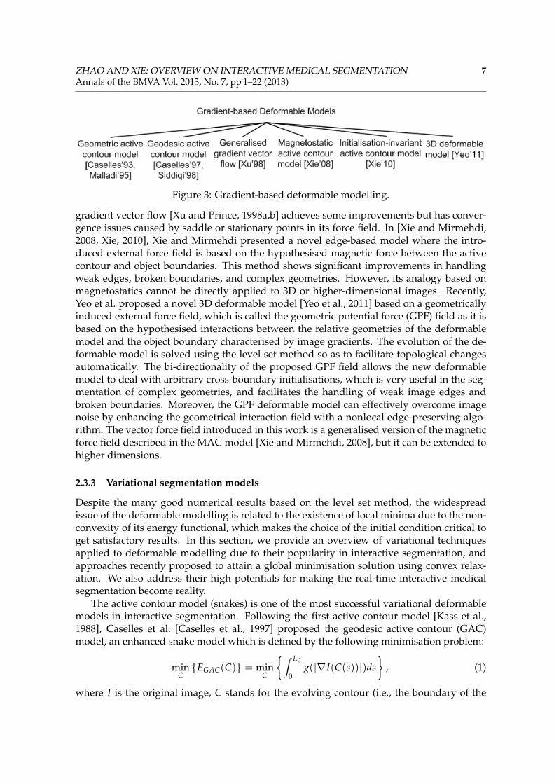
ZHAO AND XIE: OVERVIEW ON INTERACTIVE MEDICAL SEGMENTATION 7Annals of the BMVA Vol. 2013, No. 7, pp 1–22 (2013)
Figure 3: Gradient-based deformable modelling.
gradient vector flow [Xu and Prince, 1998a,b] achieves some improvements but has conver-gence issues caused by saddle or stationary points in its force field. In [Xie and Mirmehdi,2008, Xie, 2010], Xie and Mirmehdi presented a novel edge-based model where the intro-duced external force field is based on the hypothesised magnetic force between the activecontour and object boundaries. This method shows significant improvements in handlingweak edges, broken boundaries, and complex geometries. However, its analogy based onmagnetostatics cannot be directly applied to 3D or higher-dimensional images. Recently,Yeo et al. proposed a novel 3D deformable model [Yeo et al., 2011] based on a geometricallyinduced external force field, which is called the geometric potential force (GPF) field as it isbased on the hypothesised interactions between the relative geometries of the deformablemodel and the object boundary characterised by image gradients. The evolution of the de-formable model is solved using the level set method so as to facilitate topological changesautomatically. The bi-directionality of the proposed GPF field allows the new deformablemodel to deal with arbitrary cross-boundary initialisations, which is very useful in the seg-mentation of complex geometries, and facilitates the handling of weak image edges andbroken boundaries. Moreover, the GPF deformable model can effectively overcome imagenoise by enhancing the geometrical interaction field with a nonlocal edge-preserving algo-rithm. The vector force field introduced in this work is a generalised version of the magneticforce field described in the MAC model [Xie and Mirmehdi, 2008], but it can be extended tohigher dimensions.
2.3.3 Variational segmentation models
Despite the many good numerical results based on the level set method, the widespreadissue of the deformable modelling is related to the existence of local minima due to the non-convexity of its energy functional, which makes the choice of the initial condition critical toget satisfactory results. In this section, we provide an overview of variational techniquesapplied to deformable modelling due to their popularity in interactive segmentation, andapproaches recently proposed to attain a global minimisation solution using convex relax-ation. We also address their high potentials for making the real-time interactive medicalsegmentation become reality.
The active contour model (snakes) is one of the most successful variational deformablemodels in interactive segmentation. Following the first active contour model [Kass et al.,1988], Caselles et al. [Caselles et al., 1997] proposed the geodesic active contour (GAC)model, an enhanced snake model which is defined by the following minimisation problem:
minC
EGAC(C) = minC
∫ LC
0g(|∇I(C(s))|)ds
, (1)
where I is the original image, C stands for the evolving contour (i.e., the boundary of the
8 ZHAO AND XIE: OVERVIEW ON INTERACTIVE MEDICAL SEGMENTATIONAnnals of the BMVA Vol. 2013, No. 7, pp 1–22 (2013)
evolving region in I), LC =∫ LC
0 ds denotes the Euclidean length of the curve C, and g isan edge indicator function (e.g., g(|∇I|) = 1/(1 + β|∇I|2), β is a positive constant) thatvanishes at object boundaries of I. The GAC energy function in (1) is actually a new lengthobtained by weighting the Euclidean element of length ds by the function g which containsinformation concerning the boundaries of objects.
In the GAC model, the existence of local minima in EGAC can prevent the segmentationof meaningful objects in the images. Such a drawback necessitates the definition of a seg-mentation model that can provide correct results independently of the initial condition, i.e.,a global minimum of a convex functional. In [Bresson et al., 2007], Bresson et al. proposeda fast global minimisation framework based on the unification of three variational models,namely the active contour model [Kass et al., 1988, Caselles et al., 1997], the Rudin-Osher-Fatemi (ROF) denoising model [Rudin et al., 1992] and the Mumford-Shah segmentationmodel [Mumford and Shah, 1989]. In the unified approach of image segmentation and ROFimage denoising models, the convex energy functional is defined based on the dual formu-lation of the total variation (TV) norm [Chan et al., 1999, Chambolle, 2004, Aujol et al., 2006]:
minu
ETVg(u, λ)
= min
u∫
Ωg(x)|∇u|dx︸ ︷︷ ︸
TVg(u)
+λ∫
Ω|u − f |dx, (2)
where Ω represents the image domain, u is a characteristic function, f is a given (possiblynoisy) image, λ is a positive parameter controlling the scale of observation of the solution,and TVg(u) is the weighted total variation norm of the function u. The introduction of theweight function g(x) in the TV-norm gives the link between the snakes/GAC model and theproposed functional ETVg(u, λ). Bresson et al. [Bresson et al., 2007] proved that EGAC(C) in(1) and ETVg(u) in (2) describe the same energy, when g(x) is an edge indicator function, u isa characteristic function 1ΩC of a closed set ΩC ⊂ Ω whose boundary is denoted by C, and uis allowed to vary continuously in the interval [0, 1]. The advantage of the energy functionaldefined in (2) over the one defined in (1) is its (non-strict) convexity, making it possible toderive a global optimal solution for u.
Applying the weighted TV minimisation algorithm [Bresson et al., 2007], Unger et al. [Ungeret al., 2008] proposed the interactive TVSeg framework by minimising the following varia-tional image segmentation model:
minu∈[0,1]
ETVSeg(u, λ(x))
= min
u∈[0,1]
∫Ω
g(x)|∇u|dΩ +∫
Ωλ(x)|u − f |dΩ
, (3)
where f ∈ [0, 1] is provided by the user using the sampling brushes, indicating foreground( f = 1) and background ( f = 0) seed regions. The spatially varying parameter λ(x) isresponsible for the interpretation of the information contained in f . The incorporation ofdifferent constraints (hard constraints: λ(x) → ∞, weak constraints: 0<λ(x) < ∞, and noconstraints: λ(x) = 0) enables the user to interact with the algorithm.
TVSeg is fast and easy to use, but exploits the colour histograms information only. Toimprove the segmentation quality, Santner et al. in [Santner et al., 2009] applied the Ran-dom Forests to learn the foreground and background labels from complex pixel descriptorsconsisting of colour, patch, and histograms of oriented gradients (HOGs) information. Toregularise these labels for the final segmentation results, they employed a variational model

ZHAO AND XIE: OVERVIEW ON INTERACTIVE MEDICAL SEGMENTATION 9Annals of the BMVA Vol. 2013, No. 7, pp 1–22 (2013)
that combines the weighted TV-norm with a more flexible pointwise data term:
minu∈[0,1]
Ep(u, λ)
= min
u∈[0,1]∫
Ωg(x)|∇u|dΩ︸ ︷︷ ︸
TVg(u)
+λ∫
Ωu f dΩ︸ ︷︷ ︸
Region
, (4)
where the weighting g(∇I) = exp(−α|∇I|β) is an edge indicator function, u is the binarylabelling results from the Random Forests classification, f is a function of probability (e.g.,f = log(pbg/p f g), a probability ratio of the background and foreground regions), and thepositive parameter λ controls the influence of the TVg term and the Region data term onthe segmentation results. As shown in [Bresson et al., 2007], the energy functional in (4)becomes convex by letting u vary continuously between [0, 1]. Compared with TVSeg, thesimple product in the data term makes the global minimisation easier and faster. Usingthe recent primal-dual algorithms [Zhu et al., 2008, Zhu and Chan, 2008], a global optimalsolution can be quickly obtained [Santner et al., 2009].
A great advantage of the above methods [Unger et al., 2008, Santner et al., 2009] is theirhigh parallelisation potential. By implementing them on massively parallel processors suchas the graphics processing unit (GPU), the real-time interactive segmentation becomes feasi-ble, which is critical for the clinical applications.
2.4 Comparative studies
Medical images usually contain complex geometries and topologies, noise and weak edges.To generate good segmentation results, careful initialisation is commonly required. It isalso worth noting that the method performs well on some particular dataset collected by acertain imaging technology (e.g., CT, MRI, or ultrasound) may not produce decent resultson other datasets of the same or another imaging modality. In this section, we present acomparative evaluation of some interactive segmentation techniques discussed above withrespect to their accuracy, given specific medical datasets and initialisation configurations.
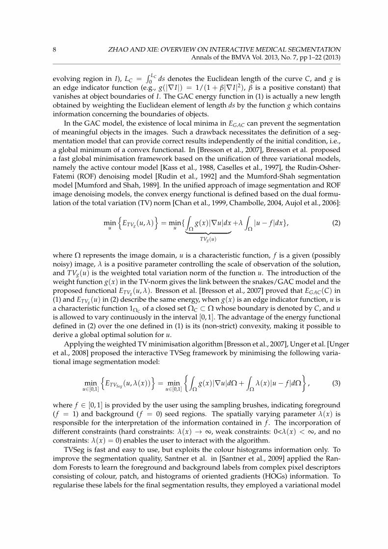
As presented in Fig. 4, the segmentation results of the intelligent scissors (a fundamentalapproach) [Mortensen and Barrett, 1998] and the united snakes (a deformable model-basedapproach) [Liang et al., 2006] are compared on several medical images in terms of accuracy.We can see that with only a few seed points, the united snakes outperforms the intelligentscissors and its segmentation boundaries are comparable to the ‘ideal’ boundaries used asreferences in the intelligent scissors [Mortensen and Barrett, 1998].
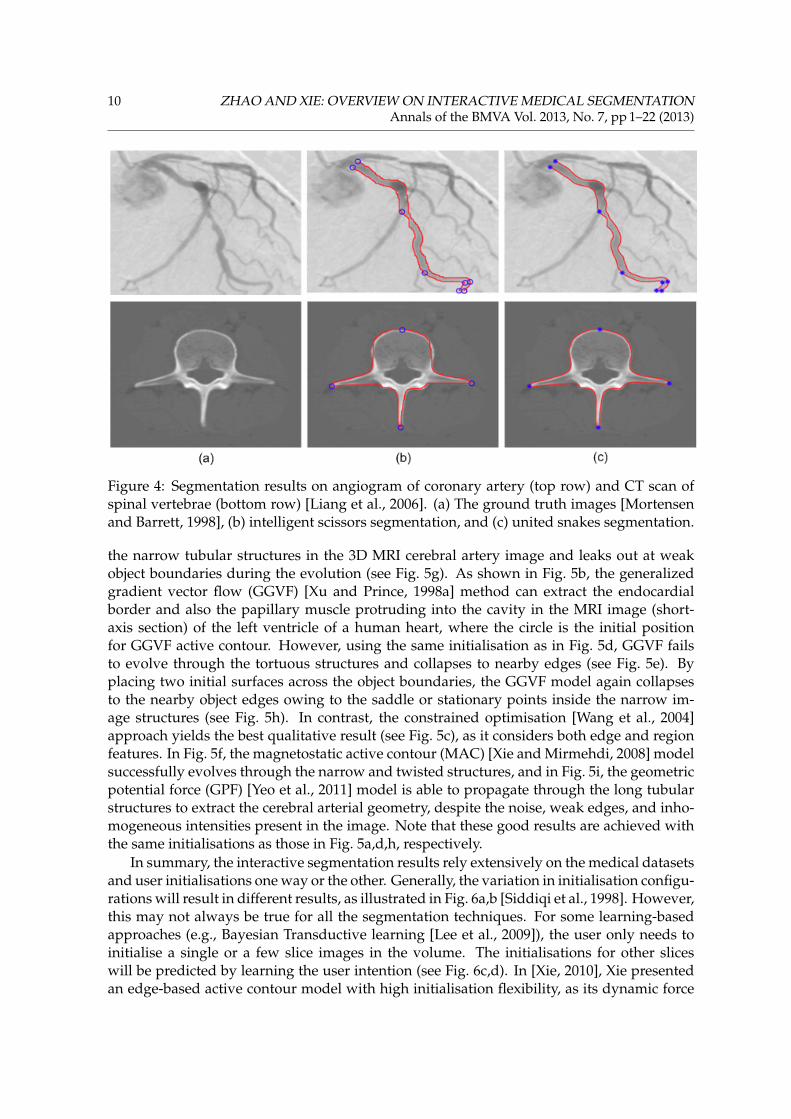
In [He et al., 2008], various deformable contour methods were compared on represen-tative images selected from several medical datasets including CT, MRI, ultrasound, andmicroscopy images. As an example of complex contour shape with deep concavities, sharpprotrusions, and inhomogeneous interior intensities, seven initial contours (formed as circlesaround the user selected locations) were tried on a coronal MRI brain image. As depictedin Fig. 5a, the best result for the geodesic active contour (GAC) [Caselles et al., 1997] showsthat GAC produces incomplete contour and it cannot acquire small sharp protrusions in thecontour segments at the lower left and right sides of the brain. Applying the same methodto another MRI brain image containing weak edges and complex topologies with acute con-cavities, it steps across the weak edges but fails to localise the boundaries (see Fig. 5d), withthe initialisation across both the left and right hemispheres. With two initial surfaces be-ing placed inside the object of interest, the GAC model usually cannot propagate through
10 ZHAO AND XIE: OVERVIEW ON INTERACTIVE MEDICAL SEGMENTATIONAnnals of the BMVA Vol. 2013, No. 7, pp 1–22 (2013)
Figure 4: Segmentation results on angiogram of coronary artery (top row) and CT scan ofspinal vertebrae (bottom row) [Liang et al., 2006]. (a) The ground truth images [Mortensenand Barrett, 1998], (b) intelligent scissors segmentation, and (c) united snakes segmentation.
the narrow tubular structures in the 3D MRI cerebral artery image and leaks out at weakobject boundaries during the evolution (see Fig. 5g). As shown in Fig. 5b, the generalizedgradient vector flow (GGVF) [Xu and Prince, 1998a] method can extract the endocardialborder and also the papillary muscle protruding into the cavity in the MRI image (short-axis section) of the left ventricle of a human heart, where the circle is the initial positionfor GGVF active contour. However, using the same initialisation as in Fig. 5d, GGVF failsto evolve through the tortuous structures and collapses to nearby edges (see Fig. 5e). Byplacing two initial surfaces across the object boundaries, the GGVF model again collapsesto the nearby object edges owing to the saddle or stationary points inside the narrow im-age structures (see Fig. 5h). In contrast, the constrained optimisation [Wang et al., 2004]approach yields the best qualitative result (see Fig. 5c), as it considers both edge and regionfeatures. In Fig. 5f, the magnetostatic active contour (MAC) [Xie and Mirmehdi, 2008] modelsuccessfully evolves through the narrow and twisted structures, and in Fig. 5i, the geometricpotential force (GPF) [Yeo et al., 2011] model is able to propagate through the long tubularstructures to extract the cerebral arterial geometry, despite the noise, weak edges, and inho-mogeneous intensities present in the image. Note that these good results are achieved withthe same initialisations as those in Fig. 5a,d,h, respectively.
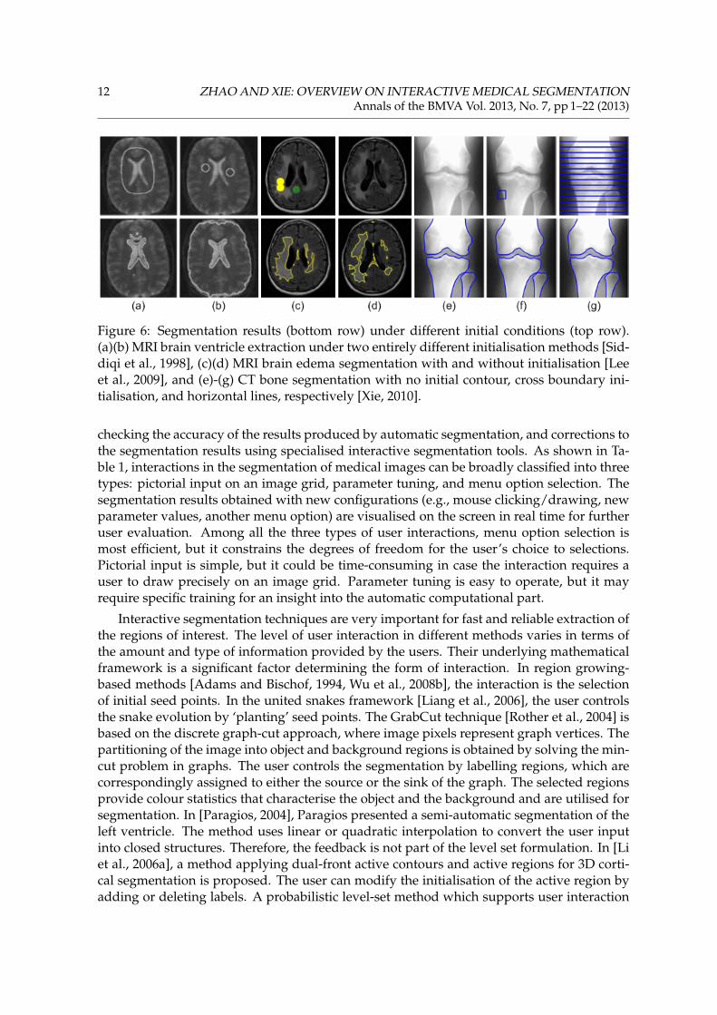
In summary, the interactive segmentation results rely extensively on the medical datasetsand user initialisations one way or the other. Generally, the variation in initialisation configu-rations will result in different results, as illustrated in Fig. 6a,b [Siddiqi et al., 1998]. However,this may not always be true for all the segmentation techniques. For some learning-basedapproaches (e.g., Bayesian Transductive learning [Lee et al., 2009]), the user only needs toinitialise a single or a few slice images in the volume. The initialisations for other sliceswill be predicted by learning the user intention (see Fig. 6c,d). In [Xie, 2010], Xie presentedan edge-based active contour model with high initialisation flexibility, as its dynamic force
ZHAO AND XIE: OVERVIEW ON INTERACTIVE MEDICAL SEGMENTATION 11Annals of the BMVA Vol. 2013, No. 7, pp 1–22 (2013)
Figure 5: Segmentation results on MRI images of the (a,c) brain [He et al., 2008], (b) leftventricle [Xu and Prince, 1998a], (d-f) brain [Xie and Mirmehdi, 2008], and (g-i) cerebralartery [Yeo et al., 2011] using different methods: GAC [Caselles et al., 1997], GGVF [Xuand Prince, 1998a], constrained optimisation [Wang et al., 2004], (MAC) [Xie and Mirmehdi,2008], and GPF [Yeo et al., 2011].
field, unique bidirectionality, and constrained diffusion-based level set evolution providegreat freedom in contour initialisation. As shown in Fig. 6e-g, no discernable difference canbe seen from the segmentation results under three completely different initialisations.
3 Interactions in Medical Image Segmentation
In an interactive segmentation framework, user intervention is tightly coupled with an au-tomatic segmentation algorithm leveraging the user’s high-level anatomical knowledge andthe automated method’s computational capability. Real-time visualisation on the screen en-ables the user to quickly validate and correct the automatic segmentation results in a sub-domain where the variational model’s statistical assumptions do not agree with the user’sexpert knowledge. The user intervention mainly includes initialisation of the methods,
12 ZHAO AND XIE: OVERVIEW ON INTERACTIVE MEDICAL SEGMENTATIONAnnals of the BMVA Vol. 2013, No. 7, pp 1–22 (2013)
Figure 6: Segmentation results (bottom row) under different initial conditions (top row).(a)(b) MRI brain ventricle extraction under two entirely different initialisation methods [Sid-diqi et al., 1998], (c)(d) MRI brain edema segmentation with and without initialisation [Leeet al., 2009], and (e)-(g) CT bone segmentation with no initial contour, cross boundary ini-tialisation, and horizontal lines, respectively [Xie, 2010].
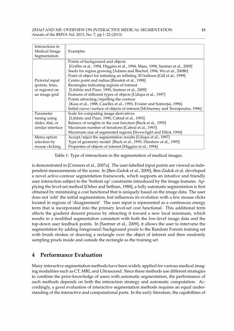
checking the accuracy of the results produced by automatic segmentation, and corrections tothe segmentation results using specialised interactive segmentation tools. As shown in Ta-ble 1, interactions in the segmentation of medical images can be broadly classified into threetypes: pictorial input on an image grid, parameter tuning, and menu option selection. Thesegmentation results obtained with new configurations (e.g., mouse clicking/drawing, newparameter values, another menu option) are visualised on the screen in real time for furtheruser evaluation. Among all the three types of user interactions, menu option selection ismost efficient, but it constrains the degrees of freedom for the user’s choice to selections.Pictorial input is simple, but it could be time-consuming in case the interaction requires auser to draw precisely on an image grid. Parameter tuning is easy to operate, but it mayrequire specific training for an insight into the automatic computational part.
Interactive segmentation techniques are very important for fast and reliable extraction ofthe regions of interest. The level of user interaction in different methods varies in terms ofthe amount and type of information provided by the users. Their underlying mathematicalframework is a significant factor determining the form of interaction. In region growing-based methods [Adams and Bischof, 1994, Wu et al., 2008b], the interaction is the selectionof initial seed points. In the united snakes framework [Liang et al., 2006], the user controlsthe snake evolution by ‘planting’ seed points. The GrabCut technique [Rother et al., 2004] isbased on the discrete graph-cut approach, where image pixels represent graph vertices. Thepartitioning of the image into object and background regions is obtained by solving the min-cut problem in graphs. The user controls the segmentation by labelling regions, which arecorrespondingly assigned to either the source or the sink of the graph. The selected regionsprovide colour statistics that characterise the object and the background and are utilised forsegmentation. In [Paragios, 2004], Paragios presented a semi-automatic segmentation of theleft ventricle. The method uses linear or quadratic interpolation to convert the user inputinto closed structures. Therefore, the feedback is not part of the level set formulation. In [Liet al., 2006a], a method applying dual-front active contours and active regions for 3D corti-cal segmentation is proposed. The user can modify the initialisation of the active region byadding or deleting labels. A probabilistic level-set method which supports user interaction
ZHAO AND XIE: OVERVIEW ON INTERACTIVE MEDICAL SEGMENTATION 13Annals of the BMVA Vol. 2013, No. 7, pp 1–22 (2013)
Interactions inMedical Image ExamplesSegmentation
Points of background and objects[Griffin et al., 1994, Higgins et al., 1994, Maes, 1998, Santner et al., 2009]Seeds for region growing [Adams and Bischof, 1994, Wu et al., 2008b]Point of object for initiating an inflating 3D balloon [Gill et al., 1999]
Pictorial input Centre point and radius [Bzostek et al., 1998](points, lines, Rectangles indicating regions of interestor regions) on [Lifshitz and Pizer, 1990, Santner et al., 2009]an image grid Features of different types of objects [Udupa et al., 1997]
Points attracting/repelling the contour[Kass et al., 1988, Caselles et al., 1993, Eviatar and Somorjai, 1996]Initial curve/surface of objects of interest [McInerney and Terzopoulos, 1996]
Parameter Scale for computing image derivativestuning using [Lifshitz and Pizer, 1990, Cabral et al., 1993]slider, dial, or Balance of weights in the cost function [Buck et al., 1995]similar interface Maximum number of iterations [Cabral et al., 1993]
Maximum size of segmented regions [Sivewright and Elliot, 1994]Menu option Accept/reject the segmentation results [Udupa et al., 1997]selection by Type of geometry model [Buck et al., 1995, Hinshaw et al., 1995]mouse clicking Properties of objects of interest [Higgins et al., 1994]
Table 1: Type of interactions in the segmentation of medical images.
is demonstrated in [Cremers et al., 2007a]. The user-labelled input points are viewed as inde-pendent measurements of the scene. In [Ben-Zadok et al., 2009], Ben-Zadok et al. developeda novel active-contour segmentation framework, which supports an intuitive and friendlyuser interaction subject to the ‘bottom up’ constraints introduced by the image features. Ap-plying the level-set method [Osher and Sethian, 1988], a fully automatic segmentation is firstobtained by minimising a cost functional that is uniquely based on the image data. The userdoes not ‘edit’ the initial segmentation, but influences its evolution with a few mouse clickslocated in regions of ‘disagreement’. The user input is represented as a continuous energyterm that is incorporated into the primary level-set cost functional. This additional termaffects the gradient descent process by attracting it toward a new local minimum, whichresults in a modified segmentation consistent with both the low-level image data and thetop-down user feedback points. In [Santner et al., 2009], it allows the user to intervene thesegmentation by adding foreground/background pixels to the Random Forests training setwith brush strokes or drawing a rectangle over the object of interest and then randomlysampling pixels inside and outside the rectangle as the training set.
4 Performance Evaluation
Many interactive segmentation methods have been widely applied for various medical imag-ing modalities such as CT, MRI, and Ultrasound. Since these methods use different strategiesto combine the prior-knowledge of users with automatic segmentation, the performance ofsuch methods depends on both the interaction strategy and automatic computation. Ac-cordingly, a good evaluation of interactive segmentation methods requires an equal under-standing of the interactive and computational parts. In the early literature, the capabilities of
14 ZHAO AND XIE: OVERVIEW ON INTERACTIVE MEDICAL SEGMENTATIONAnnals of the BMVA Vol. 2013, No. 7, pp 1–22 (2013)
interactive segmentation methods can be demonstrated in terms of accuracy, efficiency, andrepeatability [Olabarriaga and Smeulders, 2001]. These criteria are highly related, particu-larly accuracy and efficiency are interdependent since a user usually produce more accuratesegmentation results given more time. Meanwhile, it is important to note that the perfor-mance evaluation may be considered reasonable for one application but not acceptable foranother.
Accuracy is the most common criterion for performance evaluation, indicating the de-gree of similarity between the segmentation results and their respective ground truth. Itcan be assessed subjectively by a human expert by ranking the results [Udupa et al., 1997]or objectively by comparing the results with the ground truth using different distance mea-sures [de Graaf et al., 1992, Chalana and Kim, 1997, Bello and Colchester, 1998]. The groundtruth adopted is a golden standard usually generated by a human expert using manual seg-mentation tools. Due to manual processing, the golden standard may incorporate variationand subjectivity, which should be taken into account in the global evaluation of accuracy.
Efficiency of an interactive segmentation can be assessed with respect to the total com-putation time [Mortensen and Barrett, 1998] or the amount of user’s effort to complete thesegmentation task. This effort is determined mostly by the amount and the nature of userinteractions. The amount of interaction is often estimated in terms of the number of mouseclicks [Vehkomäi et al., 1997, Bzostek et al., 1998], while the nature of interaction correlates tothe complexity of the task performed by the user [Hastreiter and Ertl, 1998, Mortensen andBarrett, 1998]. Task complexity involves several issues, including the effort to accomplish re-quired mouse operation, the type of knowledge needed to input data during interaction, andthe predictability of the impact of user input. It is reasonable to conclude that the efficiencyof a method is high if the computational part is fast, highly autonomous and predictable,and the user interaction is simple, few and quick.
Repeatability is to evaluate the extent to which the same result can be generated in dif-ferent segmentation sessions with the same user intention. In such a case, the same objectsof interest are segmented several times by a user (intra-operator) and the results are com-pared. The same procedure is followed to assess the inter-operator repeatability. The differ-ences indicate the intra-operator or inter-operator variability of results [Udupa et al., 1997,Mortensen and Barrett, 1998, Gill et al., 1999].
In [McGuinness and O’Connor, 2010], McGuinness and O’Connor presented a compar-ative evaluation of four popular interactive segmentation algorithms: seeded region grow-ing [Adams and Bischof, 1994], interactive graph cuts [Boykov and Jolly, 2001], simple in-teractive object extraction [Friedland et al., 2005], and interactive segmentation using binarypartition trees [Salembier and Garrido, 2000]. In a series of user-experiments, the partici-pants were asked to extract 100 objects using different interactive segmentation algorithmsby simply marking areas of foreground and background with a mouse, constrained withina time limit of 2 minutes for each object. The updated segmentation masks were storedalong with the elapsed time after each participant performed a new interaction. Everyrecorded mask was evaluated against the corresponding manually segmented ground truthto measure the average segmentation accuracy over time, using two benchmarks: the well-known Jaccard index [Ge et al., 2007] for measuring object accuracy, and the proposed fuzzymetric [McGuinness and O’Connor, 2010] for measuring boundary accuracy. As reportedin [McGuinness and O’Connor, 2010], the overall average boundary and object accuracies ofthe interactive graph cuts and the binary partition trees are higher than those of the othertwo methods.
ZHAO AND XIE: OVERVIEW ON INTERACTIVE MEDICAL SEGMENTATION 15Annals of the BMVA Vol. 2013, No. 7, pp 1–22 (2013)
It is worth noting that quantitative assessment is generally difficult for real medical im-ages, as they contain complex anatomical structures and the manual segmentation by a hu-man expert may be unavailable. Mostly, qualitative results are provided instead.
5 Conclusions
In this paper, we review the interactive image segmentation techniques that are widely usedin many medical applications. This review gives us some insights into the state-of-the-artsegmentations and user interventions. Even though the research on interactive segmentationis expanding rapidly, there are still many challenges to be faced. Based on this review, wemake the following observations.
1. Interactive segmentation takes advantage of automatic segmentation and allows usersto intervene the segmentation process, which are very important for fast and reliablemedical image segmentation.
2. User intervention (e.g., initialisation, validating results, correcting errors) provides ad-ditional source of information for image segmentation, thus potentially produces ac-curate segmentation results.
3. A significant amount of reported works are based on energy minimisation, especiallythe variational deformable modelling approaches. Specific parameter tuning and/orcareful initialisation are usually involved in these methods. Their segmentation accu-racy and efficiency may vary on different datasets and also rely on the initialisationconfigurations. Techniques that can efficiently segment anatomical structures withhigh accuracy are still a challenge for interactive medical image segmentation. A goodthing is that these methods are not mutually exclusive, so they can be incorporated tohandle practical problems.
4. The desired object boundary might be unclear or even missing in many medical im-ages. Thus, it is important to impose soft or hard constraints to capture the intricatedetails and bridge gaps along object boundaries in practical image segmentation.
5. Interactive segmentation aims to achieve interaction efficiency by incorporating intel-ligence with automatic segmentation, leading to the ability of learning user intentionand dealing with new volumetric images. A possible direction for future work couldbe how to efficiently learn the intention of the user so as to reduce the number of userinteractions.
6. To be viable for practical applications, an interactive segmentation approach should(i) minimise user interaction, (ii) minimise segmentation variability among users and(iii) be computationally fast to allow quick user editing. These concerns can be ad-dressed by combining the machine learning techniques with interactive segmentationalgorithms. Such a combined approach could provide a promising direction for accu-rate segmentation of medical images.
7. Real-time interactive segmentation of multimodal volumetric medical images is highlydesirable for clinical applications. Owing to the advancement in high performancecomputing such as GPU, the real-time response becomes feasible. Thus, algorithms

16 ZHAO AND XIE: OVERVIEW ON INTERACTIVE MEDICAL SEGMENTATIONAnnals of the BMVA Vol. 2013, No. 7, pp 1–22 (2013)
with high parallelisation potential that can be easily implemented on the GPU are de-sirable.
8. There is a clear need of some benchmark medical datasets and well-defined perfor-mance evaluation protocols for comparative studies.
References
R. Adams and L. Bischof. Seeded region growing. IEEE Trans. Pattern Anal. Mach. Intell., 16(6):641–647, 1994.
C. J. Armstrong, B. L. Price, and W. A. Barrett. Interactive segmentation of image volumeswith live surface. Computers & Graphics, 31(2):212–229, 2007.
J. F. Aujol, G. Gilboa, T. Chan, and S. Osher. Structure-texture image decomposition - mod-eling, algorithms, and parameter selection. Int. J. Comput. Vis., 67(1):111–136, 2006.
C. Baillard and C. Barillot. Robust 3D segmentation of anatomical structures with level sets.In Proc. Int. Conf. Medical Image Computing and Computer Assisted Intervention, pages 236–245, 2000.
F. Bello and A. C. F. Colchester. Measuring global and local spatial correspondence usinginformation theory. In Proc. Int. Conf. Medical Image Computing and Computer Assisted Inter-vention, pages 964–973, 1998.
N. Ben-Zadok, T. Riklin-Raviv, and N. Kiryati. Interactive level set segmentation for image-guided therapy. In Proc. Int. Sym. Biomedical Imaging: From Nano to Macro, pages 1079–1082,2009.
B. Bhanu and S. Fonder. Learning based interactive image segmentation. In Proc. Int. Conf.Pattern Recognit., pages 299–302, 2000.
Y. Boykov and M. P. Jolly. Interactive graph cuts for optimal boundary and region segmen-tation of objects in N-D images. In Proc. IEEE Int. Conf. Comput. Vis., pages 105–112, 2001.
J. Bredno, T. M. Lehmann, and K. Spitzer. A general discrete contour model in two, three, andfour dimensions for topology-adaptive multichannel segmentation. IEEE Trans. PatternAnal. Mach. Intell., 25(5):550–563, May 2003.
L. Breiman. Random forests. Machine Learning, 45:5–32, 2001.
X. Bresson, S. Esedoglu, P. Vandergheynst, J. P. Thiran, and S. J. Osher. Fast global minimiza-tion of the active contour/snake model. J. Math. Imaging Vis., 28(2):151–167, 2007.
T. A. Buck, H. H. Ehricke, W. Strasser, and L. Thurfjel. 3D segmentation of medical structuresby integration of ray-casting with anatomic knowledge. Computers & Graphics, 19(3):441–449, 1995.
A. Bzostek, G. Ionescu, L. Carrat, C. Barbe, O. Chavanon, and J. Troccaz. Isolating movinganatomy in ultrasound without anatomical knowledge: Application to computer-assistedpericardial punctures. In Proc. Int. Conf. Medical Image Computing and Computer AssistedIntervention, pages 1041–1048, 1998.
ZHAO AND XIE: OVERVIEW ON INTERACTIVE MEDICAL SEGMENTATION 17Annals of the BMVA Vol. 2013, No. 7, pp 1–22 (2013)
J. E. Cabral, K. S. White, Y. Kim, and E. L. Effmann. Interactive segmentation of brain tumorsin MR images using 3D region-growing. In Proc. SPIE Conf. Medical Imaging, pages 171–181, 1993.
S. Cagnoni, A. B. Dobrzeniecki, R. Poli, and J. C. Yanch. Genetic algorithm-based interactivesegmentation of 3D medical images. Image and Vision Computing, 17(12):881–895, 1999.
V. Caselles, F. Catte, T. Coll, and F. Dibos. A geometric model for active contours in imageprocessing. Numer. Math., 66(1):1–31, 1993.
V. Caselles, R. Kimmel, and G. Sapiro. Geodesic active contours. Int. J. Comput. Vis., 22(1):61–79, 1997.
V. Chalana and Y. Kim. A methodology for evaluation of boundary detection algorithms onmedical images. IEEE Trans. Med. Imag., 16(5):642–652, 1997.
A. Chambolle. An algorithm for total variation minimization and applications. J. Math.Imaging Vis., 20(1-2):89–97, 2004.
T. Chan and L. Vese. Active contours without edges. IEEE Trans. Image Process., 10(2):266–277, Feb. 2001.
T. Chan, G. Golub, and P. Mulet. A nonlinear primal-dual method for total variation-basedimage restoration. SIAM J. Sci. Comput., 20(6):1964–1977, 1999.
A. C. S. Chung and J. A. Noble. Statistical 3D vessel segmentation using a Rician distribution.In Proc. Int. Conf. Medical Image Computing and Computer Assisted Intervention, pages 82–89,1999.
D. Cremers, O. Fluck, M. Rousson, and S. Aharon. A probabilistic level set formulation forinteractive organ segmentation. In Proc. SPIE Medical Imaging, 2007a.
D. Cremers, M. Rousson, and R. Deriche. A review of statistical approaches to level setsegmentation: Integrating color, texture, motion and shape. Int. J. Comput. Vis., 72(2):195–215, 2007b.
C. N. de Graaf, A. S. E. Koster, K. L. Vincken, and M. A. Viergever. A methodology for thevalidation of image segmentation methods. In Proc. Annual Sym. Computer-Based MedicalSystems, pages 17–24, 1992.
H. Delingette and J. Montagnat. Shape and topology constraints on parametric active con-tours. J. Comput. Vis. Image Understand., 83(2):140–171, 2001.
S. Diciotti, S. Lombardo, M. Falchini, G. Picozzi, and M. Mascalchi. Automated segmentationrefinement of small lung nodules in CT scans by local shape analysis. IEEE Trans. Biomed.Eng., 58(12):3418–3428, 2011.
P. J. Elliot, J. M. Knapman, and W. Schlegel. Interactive image segmentation for radiationtreatment planning. IBM Systems Journal, 31(4):620–634, 1992.
H. Eviatar and R. L. Somorjai. A fast simple active contour algorithm for biomedical images.Pattern Recognition Letters, 17:969–974, 1996.

18 ZHAO AND XIE: OVERVIEW ON INTERACTIVE MEDICAL SEGMENTATIONAnnals of the BMVA Vol. 2013, No. 7, pp 1–22 (2013)
G. Friedland, K. Jantz, and R. Rojas. SIOX: Simple interactive object extraction in still images.In Proc. IEEE Int. Sym. Multimedia, pages 253–260, 2005.
F. Ge, S. Wang, and T. Liu. New benchmark for image segmentation evaluation. J. ElectronicImaging, 16(3):033011, 2007.
J. D. Gill, H. M. Ladak, D. A. Steinman, and A. Fenster. Development and evaluation of asemi-automatic 3D segmentation of the carotid arteries from 3D ultrasound images. InProc. SPIE Conf. Medical Imaging, pages 214–221, 1999.
V. Grau, A. U. J. Mewes, M. Alcaniz, R. Kikinis, and S. K. Warfield. Improved watershedtransform for medical image segmentation using prior information. IEEE Trans. Med.Imag., 23(4):447–458, Apr. 2004.
L. D. Griffin, A. C. F. Colchester, S. A. Röll, and C. S. Studholme. Hierarchical segmentationsatisfying constraints. In Proc. British Mach. Vis. Conf., pages 135–144, 1994.
M. W. Hansen and W. E. Higgins. Relaxation methods for supervised image segmentation.IEEE Trans. Pattern Anal. Mach. Intell., 19(9):949–962, 1997.
J. Hao and M. Li. A supervised bayesian method for cerebrovascular segmentation. WSEASTrans. Signal Process., 3(12):487–495, 2007.
P. Hastreiter and T. Ertl. Fast and interactive 3D segmentation of medical volume data. InProc. Conf. Image and Multi-dimensional Digital Signal Processing, pages 41–44, 1998.
L. He, Z. Peng, B. Everding, X. Wang, C. Y. Han, K. L. Weiss, and W. G. Wee. A comparativestudy of deformable contour methods on medical image segmentation. Image and VisionComputing, 26(2):141–163, 2008.
W. E. Higgins, J. M. Reinhardt, and W. L. Sharp. Semi-automatic construction of 3D medicalimage-segmentation processes. In Proc. SPIE Conf. Visualization in Biomedical Computing,pages 59–71, 1994.
K. P. Hinshaw, R. B. Altman, and J. F. Brinkley. Shape-based models for interactive segmen-tation of medical images. In Proc. SPIE Conf. Medical Imaging, pages 771–780, 1995.
M. Holtzman-Gazit, R. Kimmel, N. Peled, and D. Goldsher. Segmentation of thin structuresin volumetric medical images. IEEE Trans. Image Process., 15(2):354–363, Feb. 2006.
M. Kass, A. Witkin, and D. Terzopoulos. Snakes: Active contour models. Int. J. Comput. Vis.,1(4):321–331, 1988.
J. Kim, J. W. Fisher, A. Yezzi, M. Cetin, and A. S. Willsky. A nonparametric statistical methodfor image segmentation using information theory and curve evolution. IEEE Trans. ImageProcess., 14(10):1486–1502, Oct. 2005.
R. Kimmel. Geometric Level Set Methods in Imaging, Vision, and Graphics, chapter Fast edgeintegration, pages 59–77. Berlin, Germany: Springer-Verlag, 2003.
T. Kubota, A. K. Jerebko, M. Dewan, M. Salganicoff, and A. Krishnan. Segmentation ofpulmonary nodules of various densities with morphological approaches and convexitymodels. Medical Image Analysis, 15(1):133–154, 2011.

ZHAO AND XIE: OVERVIEW ON INTERACTIVE MEDICAL SEGMENTATION 19Annals of the BMVA Vol. 2013, No. 7, pp 1–22 (2013)
J.-O. Lauchaud and B. Taton. Deformable model with a complexity independent from imageresolution. J. Comput. Vis. Image Understand., 99(3):453–475, 2005.
M. Law and A. Chung. A deformable surface model for vascular segmentation. In Proc. Int.Conf. Medical Image Computing and Computer Assisted Intervention, pages 59–67, 2009.
N. Lee, R. T. Smith, and A. F. Laine. Interactive segmentation for geographic atrophy inretinal fundus images. In Proc. 42nd Asilomar Conf. Signals, Systems and Computers, pages655–658, 2008.
N. Lee, J. Caban, S. Ebadollahi, and A. Laine. Interactive segmentation in multimodal med-ical imagery using a bayesian transductive learning approach. In Proc. Medical Imaging:Computer-Aided Diagnosis, pages 72601W–1–10, 2009.
C. Li, J. Liu, and M. Fox. Segmentation of edge preserving gradient vector flow: An approachtoward automatically initializing and splitting of snakes. In Proc. IEEE Conf. Comput. Vis.Pattern Recognit., pages 162–167, 2005.
H. Li, A. Yezzi, and L. D. Cohen. 3D brain segmentation using dual-front active contourswith optional user interaction. Int. J. Biomedical Imaging, 2006:1–17, 2006a.
K. Li, X. Wu, D. Z. Chen, and M. Sonka. Optimal surface segmentation in volumetric images-A graph-theoretic approach. IEEE Trans. Pattern Anal. Mach. Intell., 28(1):119–134, 2006b.
Y. Li, J. Sun, C.-K. Tang, and H.-Y. Shum. Lazy snapping. In Proc. ACM SIGGRAPH, pages303–308, 2004.
J. Liang, T. McInerney, and D. Terzopoulos. United snakes. Medical Image Analysis, 10(2):215–233, 2006.
L. M. Lifshitz and S. M. Pizer. Multiresolution hierarchical approach to image segmentationbased on intensity extrema. IEEE Trans. Pattern Anal. Mach. Intell., 12(6):529–540, 1990.
H. Lombaert, Y. Sun, L. Grady, and C. Xu. A multilevel banded graph cuts method for fastimage segmentation. In Proc. IEEE Int. Conf. Comput. Vis., pages 259–265, 2005.
F. Maes. Segmentation and registration of multimodal images: From theory, implementation andvalidation to a useful tool in clinical practice. PhD thesis, Katholieke Universiteit Leuven,Leuven, BE., 1998.
R. Malladi, J. A. Sethian, and B. C. Vemuri. Shape modeling with front propagation: A levelset approach. IEEE Trans. Pattern Anal. Mach. Intell., 17(2):158–175, Feb. 1995.
K. McGuinness and N. E. O’Connor. A comparative evaluation of interactive segmentationalgorithms. Pattern Recognition, 43(2):434–444, 2010.
T. McInerney and D. Terzopoulos. Deformable models in medical image analysis: A survey.Medical Image Analysis, 1(2):91–108, 1996.
T. McInerney and D. Terzopoulos. Topology adaptive deformable surfaces for medical imagevolume segmentation. IEEE Trans. Med. Imag., 18(10):840–850, Oct. 1999.
20 ZHAO AND XIE: OVERVIEW ON INTERACTIVE MEDICAL SEGMENTATIONAnnals of the BMVA Vol. 2013, No. 7, pp 1–22 (2013)
T. Mondal, A. Jain, and H. K. Sardana. Automatic craniofacial structure detection on cephalo-metric images. IEEE Trans. Image Process., 20(9):2606–2614, 2011.
E. N. Mortensen and W. A. Barrett. Interactive segmentation with intelligent scissors. Graph-ical Models and Image Processing, 60(5):349–384, 1998.
D. Mumford and J. Shah. Optimal approximations of piecewise smooth functions and asso-ciated variational problems. Commun. Pure Appl. Math., 42(5):577–685, 1989.
S. D. Olabarriaga and A. W. M Smeulders. Setting the mind for intelligent interactive seg-mentation: Overview, requirements, and framework. In Proc. Int. Conf. Information Process-ing in Medical Imaging, pages 417–422, 1997.
S. D. Olabarriaga and A. W. M. Smeulders. Interaction in the segmentation of medical im-ages: A survey. Medical Image Analysis, 5(2):127–142, 2001.
J. Olivier, C. Mocquillon, J. J. Rousselle, R. Boné, and H. Cardot. A supervised texture-basedactive contour model with linear programming. In Proc. IEEE Int. Conf. Image Processing,pages 1104–1107, 2008.
S. J. Osher and J. A. Sethian. Fronts propagation with curvature dependent speed: Algo-rithms based on Hamilton-Jacobi formulations. J. Comp. Phys., 79:12–49, 1988.
N. Paragios. Variational methods and partial differential equations in cardiac image analysis.In Proc. Int. Sym. Biomedical Imaging: From Nano to Macro, pages 17–20, 2004.
N. Paragios and R. Deriche. Geodesic active regions and level set methods for supervisedtexture segmentation. Int. J. Comput. Vis., 46(3):223–247, 2002.
N. Paragios, O. Mellina-Gottardo, and V. Ramesh. Gradient vector flow geometric activecontours. IEEE Trans. Pattern Anal. Mach. Intell., 26(3):402–407, 2004.
A. Pitiot, A. W. Toga, N. Ayache, and P. Thompson. Texture based MRI segmentation with atwo-stage hybrid neural classifier. In Proc. World Congress Computational Intelligence/INNS-IEEE Int. Joint Conf. Neural Networks, pages 2053–2058, 2002.
L. J. Reese and W. A. Barrett. Image editing with intelligent paint. In Proc. Eurographics,pages 714–724, 2002.
C. C. Reyes-Aldasoro and A. Bhalerao. Volumetric texture segmentation by discriminantfeature selection and multiresolution classification. IEEE Trans. Med. Imag., 26(1):1–14, Jan.2007.
C. Rother, V. Kolmogorov, and A. Blake. Grabcut - Interactive foreground extraction usingiterated graph cuts. In Proc. ACM SIGGRAPH, pages 309–314, 2004.
L. I. Rudin, S. Osher, and E. Fatemi. Nonlinear total variation based noise removal algo-rithms. Phys. D, 60(1-4):259–268, 1992.
A. Saffari, C. Leistner, J. Santner, M. Godec, and H. Bischof. On-line random forests. In Proc.ICCV Workshop On-line Computer Vision, pages 1393–1400, 2009.
ZHAO AND XIE: OVERVIEW ON INTERACTIVE MEDICAL SEGMENTATION 21Annals of the BMVA Vol. 2013, No. 7, pp 1–22 (2013)
P. Salembier and L. Garrido. Binary partition tree as an effcient representation for imageprocessing, segmentation, and information retrieval. IEEE Trans. Image Process., 9(4):561–576, 2000.
J. Santner, M. Unger, T. Pock, C. Leistner, A. Saffari, and H. Bischof. Interactive texturesegmentation using random forests and total variation. In Proc. British Mach. Vis. Conf.,2009.
M. Schaap, T. van Walsum, L. Neefjes, C. Metz, E. Capuano, M. de Bruijne, and W. Niessen.Robust shape regression for supervised vessel segmentation and its application to coro-nary segmentation in CTA. IEEE Trans. Med. Imag., 30(11):1974–1986, 2011.
J. A. Sethian. Level Set Methods and Fast Marching Methods: Evolving Interfaces in ComputationalGeometry, Fluid Mechanics, Computer Vision, and Material Science. Cambridge UniversityPress, 1999.
J. Shi and J. Malik. Normalized cuts and image segmentation. IEEE Trans. Pattern Anal. Mach.Intell., 22(8):888–905, Aug. 2000.
K. Siddiqi, Y. Lauzière, A. Tannenbaum, and S. Zucker. Area and length minimizing flowsfor shape segmentation. IEEE Trans. Image Process., 7(3):433–443, 1998.
J. Sijbers, A. Van der Linden, P. Scheunders, J. Van Audekerke, D. Van Dyck, and E. Ra-man. Volume quantization of the mouse cerebellum by semi-automatic 3D segmentationof magnetic resonance images. In SPIE Conf. Medical Imaging, pages 553–560, 1996.
G. J. Sivewright and P. J. Elliot. Interactive region and volume growing for segmentingvolumes in MR and CT images. Medical informatics, 19(1):71–80, 1994.
A. W. M. Smeulders, S. D. Olabarriaga, R. Van den Boomgaard, and M. Worring. Designconsiderations for interactive segmentation. In Proc. Conf. Visual Information Systems, pages5–12, 1997.
C. M. Smith, J. Smith, S. K. Williams, J. J. Rodriguez, and J. B. Hoying. Automatic thresh-olding of three-dimensional microvascular structures from confocal microscopy images. J.Microscopy, 225(3):244–257, 2007.
J. K. Udupa, L. Wei, S. Samarasekera, Y. Miki, M. A. van Buchem, and R. I. Grossman. Multi-ple sclerosis lesion quantification using fuzzy-connectedness principles. IEEE Trans. Med.Imag., 16(5):598–609, 1997.
M. Unger, T. Pock, W. Trobin, D. Cremers, and H. Bischof. TVSeg - Interactive total variationbased image segmentation. In Proc. British Mach. Vis. Conf., 2008.
H. Veeraraghavan and J. V. Miller. Active learning guided interactions for consistent imagesegmentation with reduced user interactions. In Proc. Int. Sym. Biomedical Imaging: FromNano to Macro, pages 1645–1648, 2011.
T. Vehkomäi, G. Gerig, and G. Székely. A user-guided tool for efficient segmentation ofmedical data. In Proc. Conf. Computer Vision, Virtual Reality and Robotics in Medicine andMedical Robotics and Computer-Assisted Surgery, pages 685–694, 1997.

22 ZHAO AND XIE: OVERVIEW ON INTERACTIVE MEDICAL SEGMENTATIONAnnals of the BMVA Vol. 2013, No. 7, pp 1–22 (2013)
L. Vese and T. Chan. A multiphase level set framework for image segmentation using themumford and shah model. Int. J. Comput. Vis., 50(3):271–293, 2002.
X. Wang, L. He, and W. G. Wee. Deformable contour method: A constrained optimizationapproach. Int. J. Comput. Vis., 59(1):87–108, 2004.
O. Wink, K. J. Zuiderveld, and M. A. Viergever. Interactive volume segmentation using localsimilarity measurements. In Proc. Computer Assisted Radiology Conf., page 996, 1997.
J. Wu, S. Poehlman, M. D. Noseworthy, and M. V. Kamath. Texture feature based automatedseeded region growing in abdominal MRI segmentation. In Proc. Int. Conf. BioMedicalEngineering and Informatics, volume 2, pages 263–267, 2008a.
J. Wu, F. Ye, J. Ma, X. Sun, J. Xu, and Z. Cui. The segmentation and visualization of humanorgans based on adaptive region growing method. In Proc. Int. Conf. Comp. Inf. Tech., pages439–443, 2008b.
X. Xie. Active contouring based on gradient vector interaction and constrained level setdiffusion. IEEE Trans. Image Process., 19(1):154–164, 2010.
X. Xie and M. Mirmehdi. RAGS: Region-aided geometric snake. IEEE Trans. Image Process.,13(5):640–652, May 2004.
X. Xie and M. Mirmehdi. MAC: Magnetostatic active contour model. IEEE Trans. PatternAnal. Mach. Intell., 30(4):632–647, 2008.
C. Xu and J. L. Prince. Generalized gradient vector flow external forces for active contours.Signal Process., 71(2):131–139, 1998a.
C. Xu and J. L. Prince. Snakes, shapes, and gradient vector flow. IEEE Trans. Image Process.,7(3):359–369, Mar. 1998b.
S. Y. Yeo, X. Xie, I. Sazonov, and P. Nithiarasu. Geometrically induced force interaction forthree-dimensional deformable models. IEEE Trans. Image Process., 20(5):1373–1387, May2011.
X. Yu and J. Yla-Jaaski. Interactive surface segmentation for medical images. In Proc. Int.Conf. Signal Processing, pages 1178–1181, 1996.
X. Yuan, N. Zhang, M. X. Nguyen, and B. Chen. Volume cutout. Visual Computer (SpecialIssue of Pacific Graphics), 21(8-10):745–754, 2005.
M. Zhu and T. Chan. An efficient primal-dual hybrid gradient algorithm for total variationimage restoration. Technical Report 08-34, UCLA CAM, 2008.
M. Zhu, S. Wright, and T. Chan. Duality-based algorithms for total-variation-regularizedimage restoration. Technical Report 08-33, UCLA CAM, 2008.