analysing nanostructures michael rappolt school of food science and nutrition faculty of mathematics...
TRANSCRIPT

Analysing Nanostructures
Michael Rappolt
School of Food Science and NutritionFACULTY OF MATHEMATICS AND PHYSICAL SCIENCES
1. BSc Student Lecture: Physics with Introduction to Modern Physics
2nd of June 2014, Faculty of Electrical Engineering, University of Ljubljana
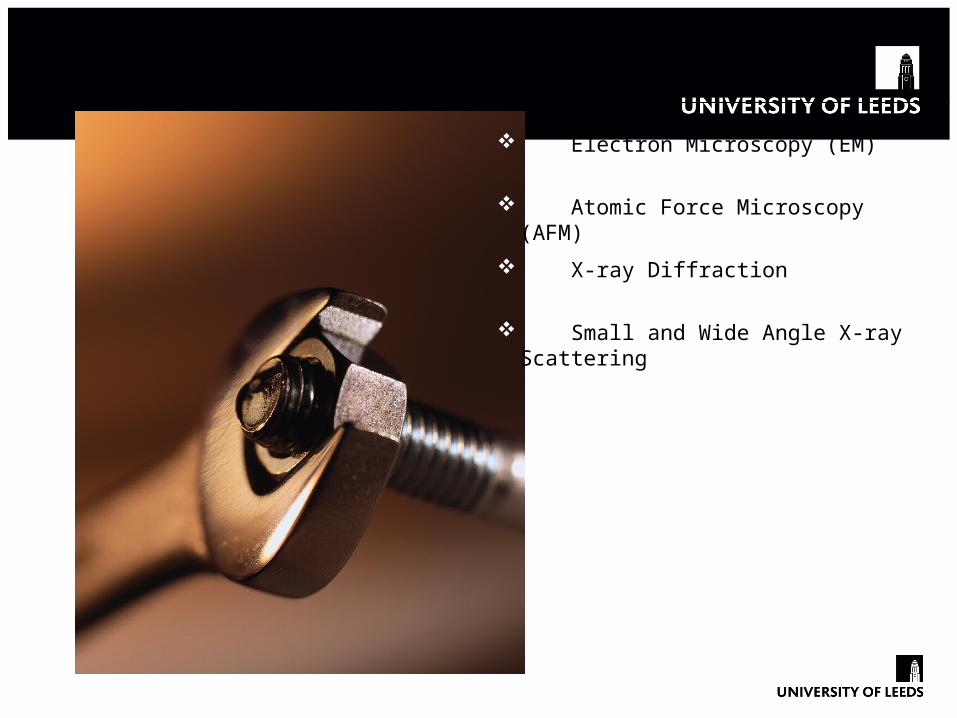
Tools for Analysing the Nano-World
Electron Microscopy (EM)
X-ray Diffraction
Atomic Force Microscopy (AFM)
Small and Wide Angle X-ray Scattering

Discovery of the DNA Structure
Rosalind Franklin's
X-ray diffraction photograph
of DNA and Watson and Crick in front of their DNA model, 1953

Electron Microscopy: DNA Origami
Bai, X. -C.; Martin, T. G.; Scheres, S. H. W.; Dietz, H. (2012). PNAS 109: 20012–20017
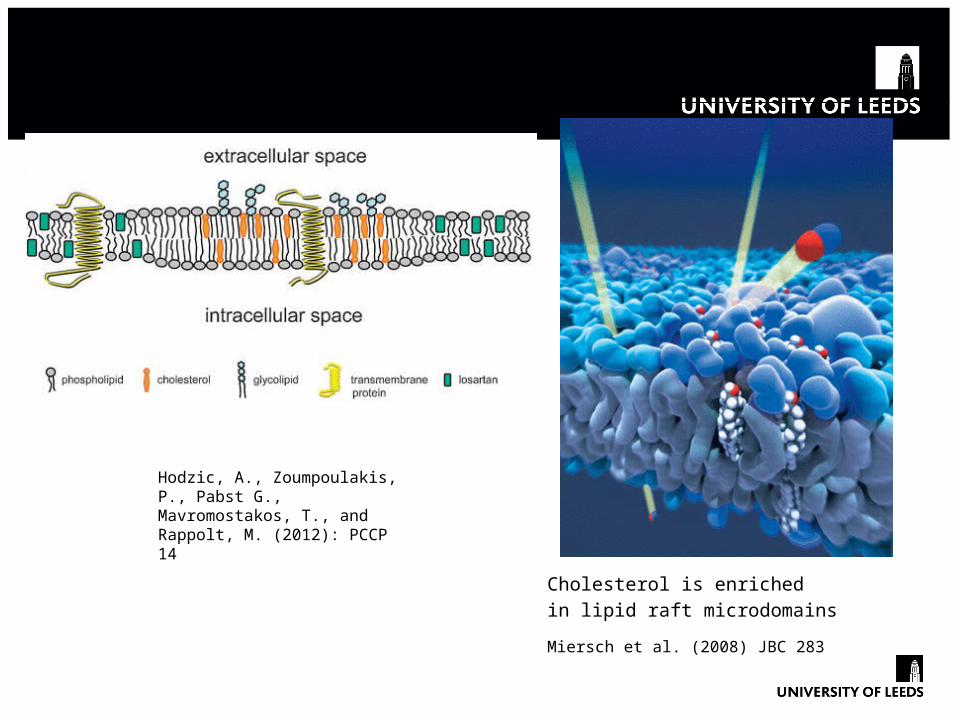
Membrane Rafts = Protein Platforms
Cholesterol is enriched
in lipid raft microdomains
Miersch et al. (2008) JBC 283
Hodzic, A., Zoumpoulakis, P., Pabst G., Mavromostakos, T., and Rappolt, M. (2012): PCCP 14
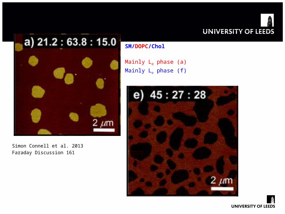
AFM: Looking at Membrane Rafts
Simon Connell et al. 2013
Faraday Discussion 161
SM/DOPC/Chol
Mainly Ld phase (a)
Mainly Lo phase (f)
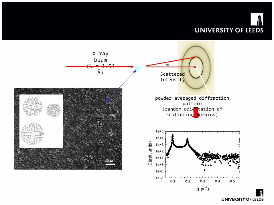
X-ray Scattering Set-Up
X-ray beam(l = 1.54 Å)
2q
Detector
ScatteredIntensity
powder averaged diffraction pattern(random orientation of scattering domains)
q (Å-1)
0.1 0.2 0.3 0.4 0.5
I (a
rb.
un
its)
1e-2
1e-1
1e+0
1e+1
1e+2
1e+3
1e+4
1e+5
profile
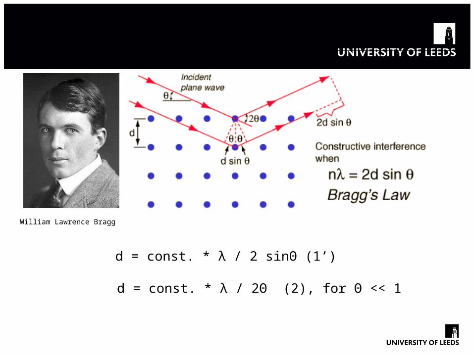
Bragg’s Law from 1912
d = const. * λ / 2 sinΘ (1’)
d = const. * λ / 2Θ (2), for Θ << 1
William Lawrence Bragg
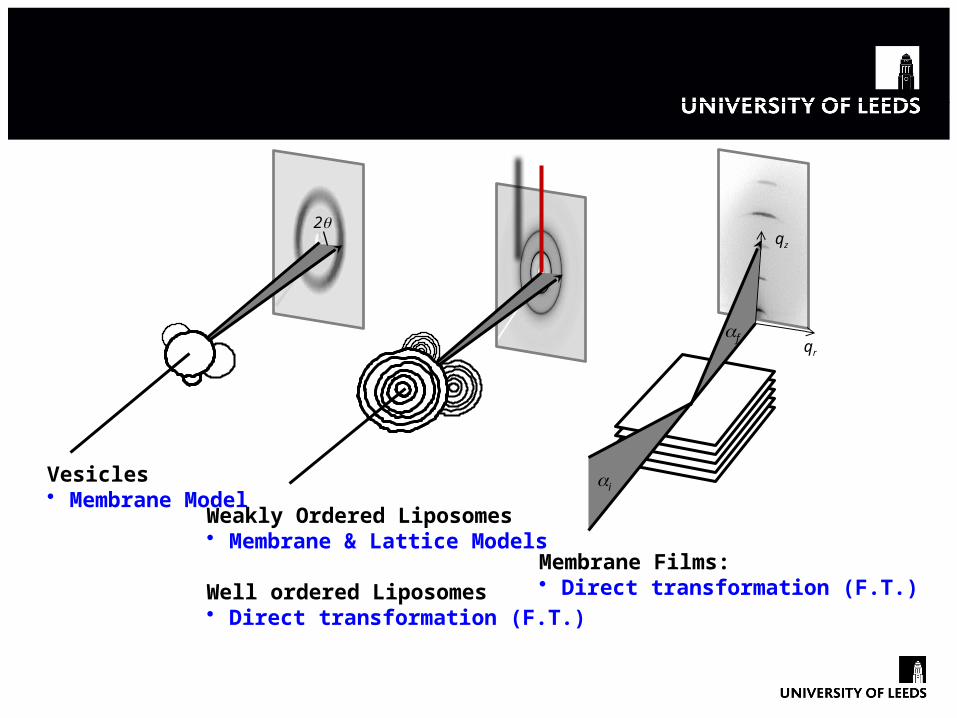
Diffuse Scattering and Diffraction
2q
ai
afqr
qz
Vesicles• Membrane Model
Weakly Ordered Liposomes • Membrane & Lattice Models
Well ordered Liposomes • Direct transformation (F.T.)
Membrane Films:• Direct transformation (F.T.)

Fourier Transform & Modelling
Rappolt, M. et al. (2003): Biophys. J. 84, 3111
2
1
Pabst, G., Rappolt ,M. et al. (2000): PRE 62, 4000Rappolt, M. et al. (2004):Recent Res. Devel. ,Vol.3, 363Heftberger, P. et al. (2013): J. Appl.Cryst., in press
Direct transformation (F.T.)

Protein Crystallography

Protein Structure, e.g. Ribosome
More than 20 years of work are in this stucture!

SAXS and WAXS
1-100 nm 0.1-0.9 nm
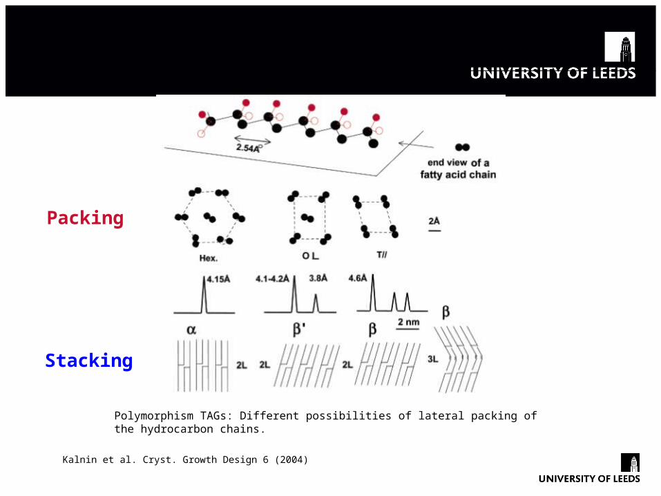
Packing & Stacking with Triacylglycerides (TAGs)
Polymorphism TAGs: Different possibilities of lateral packing of the hydrocarbon chains.
Packing
Stacking
Kalnin et al. Cryst. Growth Design 6 (2004)

Cacao Butter
SAXS and WAXS patterns: I(γ), II(α), IV(β’), V(β2), and VI(β1) polymorphic phases of cocoa butter.
SAXS WAXS
Silva et al. Cryst. Growth Design 9 (2009)
q = 2π/d (Å-1)
0.125 (2L)
0.138 (2L)
0.141 (2L)
0.095 (3L)

Starch Granule Structure
Semi-crystalline region
9 nm
repeat
Double helix
(amylose)Sarkar & Pérez BMC Bioinformatics 13 (2012)
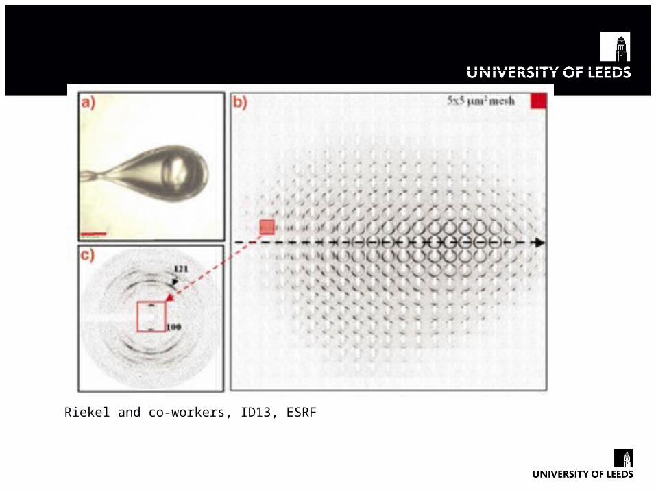
Starch Granule: Fibre Diffraction & SAXS
Riekel and co-workers, ID13, ESRF

Aligned and Non-Aligned Regions
Riekel and co-workers, ID13, ESRF

Thank You For Your Attention!
New camera SAXSpace in the Lipid Biophysics Lab
School of Food Science & Nutrition, University of Leeds, UK