analysis of a 43-kda glycoprotein from the … · 18460 trichinella spiralis 43-kda glycoprotein...
TRANSCRIPT
THE JOURNAL 0 1992 by The American Society for Blochemistry
OF BIOLOGICAL CHEM!STRY and Molecular Biology, Inc.
Vol , 267. No. 26. Issue of September 15, , pp. 18459-18465,1992 Printed in U. S. A.
Analysis of a 43-kDa Glycoprotein from the Intracellular Parasitic Nematode Trichinella spiralis”
(Received for publication, March 11, 1992)
Demetris K. VassilatisSQ, Dickson Despommierll, David E. MisekSII , Ramona I. Polverell, Allen M. Gold**, and Lex H. T. Van der PloegStSS From the $Department of Genetics and Development, the 7Division of Tropical Medicine, School of Public Health, and the **Department of Biochemistry and Molecular Biophysics, Columbia University, New York, New York 10032
The L1 larvae of the parasitic nematode Trichinella spiralis invade skeletal muscle and initiate a process that has been interpreted to represent skeletal muscle dedifferentiation. In this process, the infected region of the muscle cell is converted into a unique structure, called the Nurse cell. The nematode T. spiralis can survive for tens of years within the cytoplasm of the Nurse cell and secretes proteins into the cytoplasm that are believed to play a role in mediating the Nurse cell formation or maintenance. We have cloned a cDNA encoding the T. spiralis-derived, 43-kDa secreted pro- tein. Structural analysis of the predicted 344-amino acid sequence revealed an N terminally located signal peptide and a potential helix-loop-helix motif in the main body of the protein. Antibodies raised against the 43-kDa recombinant protein were used in immunocy- tolocalizations of T. spiralis-infected skeletal muscle sections. These antibodies strongly stained the Nurse cell nuclei and the nematode itself. Specific, though slightly weaker staining also occurred in the Nurse cell cytoplasm. In Western blots, the antibodies react with the 43-kDa protein but also detected at least two other T. spiralis-secreted proteins. DNA hybridizations re- veal at least one additional 43-kDa-related sequence encoded in the T. spiralis genome. We conclude that either the 43-kDa protein and/or a closely related 43- kDa family member is secreted into the muscle and translocates to the muscle-derived nuclei. This model may provide insights into the mechanisms involved in Nurse cell formation.
The parasitic nematode Trichinella spiralis is infective to a wide range of hosts including mice, rats, swine, and humans. Host infection begins upon ingestion of raw or undercooked
*This work was supported by a grant from the John D. and Catherine T. MacArthur Foundation (to L. H. T. V. D. P.), by a grant from the Burroughs Wellcome Fund (to L. H. T. V. D. P.), and by National Institutes of Health Grant R01-AI-10627-21 (to D. D.). The costs of publication of this article were defrayed in part by the payment of page charges. This article must therefore be hereby marked “aduertisement” in accordance with 18 U.S.C. Section 1734 solely to indicate this fact.
The nucleotide sequence(s) reported in this paper has been submitted
M95499. to the GenBankTM/EMBL Data Bank with accession number($
8 Current address: Dept. of Genetics and Molecular Biology, RY- 8OY-255, Merck Sharp & Dohme Research Laboratories, Rahway, NJ 07065.
1) Current address: Dept. of Signal Transduction, Parke-Davis Pharmaceutical Research Division, 2800 Plymouth Rd., Ann Arbor, MI 48105.
$3 To whom correspondence should be addressed. A Burroughs Wellcome Scholar in Molecular Parasitology.
meats containing infective L1 (pre-adult) larvae. After pas- sage through the stomach, the L1 larvae migrate to the small intestine, where they invade the columnar epithelium and undergo four molts over a 30-hour period to reach sexual maturity (1-4). The adults mate within the intestinal epithe- lium (5), and larvae are born 5-10 days later. The newborn larvae exit the epithelium from the basolateral surface and migrate through the bloodstream and lymphatics (6,7). Tran- sient infections of various organs including the heart, brain, retina choroid in the eye, and liver have been reported, but their primary destination is skeletal muscle. When a newborn larva enters a skeletal muscle syncitium it induces permanent alterations, leading to the formation of the Nurse cell, in which it can survive for up to 30 years (8-11). T. spiralis lives in the cytoplasm of the Nurse cell. Infection results in loss of myofiber proteins (day 4-8 postintramuscular invasion (11, 12)), muscle-specific gene inactivation (13), enlargement and proliferation of muscle cell nuclei (day 6 postintramuscular invasion (14)), and alterations in the levels of certain enzy- matic activities (such as phosphofructokinase), as well as secretion of collagen (day 10 postintramuscular invasion (11, 15)) and the possible secretion of angiogenic factors (day 12 postintramuscular invasion (11)). Growth and differentiation of T. spiralis and the formation of the Nurse cell are virtually complete within 20 days postintramuscular invasion (16). Induction of Nurse cell formation depends upon the T. spiralis larvae, and hence it is reasonable to hypothesize that the worm directs the process via secretion of proteins and metab- olites.
T. spiralis contains specialized secretory cells called stichio- cytes, in which at least 20-30 proteins are synthesized and stored in vesicles (17-19). These proteins are enriched in the S3 (large particle) fraction from L1, muscle-derived larvae. Pooled fractions of S3 proteins further purified by preparative isoelectric focussing have been shown to induce protection against subsequent infection (18). Although these proteins have been characterized in terms of their apparent molecular weights, glycosylation patterns, and isoelectric points, their structures and potential functions in the larvae or in Nurse cell formation remain unknown.
An abundant 43-kDa glycoprotein is present in the S3 fraction and secretion of the L1 larva. Antibodies raised against the purified 43-kDa protein recognized numerous protein bands and stained proteins that localized to the nuclei and to the cytoplasm of infected muscle cells (20). These antibodies reacted almost exclusively with the carbohydrate moieties of the protein (21) and hence the identity of the T. spiralis-secreted protein(s) that translocate to the nuclei of the infected muscle cell remained undetermined.
We have cloned the cDNA encoding the 43-kDa glycopro- tein. Structural analysis of the deduced 344-amino acid se-
18459

18460 Trichinella spiralis 43-kDa Glycoprotein
quence revealed a possible helix-loop-helix domain that shares 46% amino acid identity with the Drosophila protein emc and 38% with the myogenic program negative regulator protein Id in the first 13-amino acid helix (22,23). Immunolocalizations using antibodies raised against a fusion protein containing the 43-kDa amino acid sequence gave strong staining in muscle cell-derived nuclei in infected cells. Our data indicate that the 43-kDa protein or a related family member(s) is secreted by T. spiralis while living in the cytoplasm of skeletal muscle after which the protein(s) translocate(s) to the nuclei of the infected muscle cell.
MATERIALS AND METHODS
T. spiralis Infections-L1 muscle larvae were isolated from stock infected mice and rats by peptic digestion according to Despommier et al. (24). Oral infections were administered to anesthetized mice or rats with the aid of a 1-ml tuberculin syringe fitted with an 18-gauge blunted needle. Mice routinely received 500 viable larvae, whereas rats received 5000 viable larvae. Infections were allowed to proceed for a minimum of 35 days before animals were sacrificed.
Synthesis of Degenerate Anti-sense Oligonucleotides-Anti-sense oligonucleotides were synthesized on an Applied Biosystems model 380A DNA Synthesizer based upon sequence data obtained from N- terminal and internal peptides of the 43-kDa protein (21). First, degenerate oligonucleotides were synthesized based on the partial protein sequence from the N terminus of the purified, reduced, pyridylethylated 43-kDa protein (21). The following N-terminal amino acid sequence was chosen: Asp-Asp-Thr-Glu-Trp-Phe. The anti-sense sequence chosen was 5' A/GAACCAC/TTCA/G/TGTG/ ATCG/ATC 3' (oligonucleotide pool 43k-2). The 43k-3 oligonucleo- tide pool, derived from the amino acid sequence Met-Phe-Gly-Asn-
5' GTC/TTCA/GTTG/T/CGCCG/AAACAT 3'. Glu-Thr of a CNBr fragment of 43 kDa had the nucleotide sequence
Preparation of RNA and Poly(A+) RNA from T. spiralis Ll Larvae and Adult Worms-L1 muscle larvae and adult worms were harvested as previously described (24), flash-frozen in liquid nitrogen, and then stored at -80 "C until used. Approximately 2 g of frozen worms were suspended in 4 M guanidinium isothiocyanate, 14% P-mercaptoetha- nol, 100 mM Tris-HC1, pH 7.5, dissociated by mixing with a Polytron (on average, 3 X 30 s), and then centrifuged for 5 min at 12,000 X g in order to pellet insoluble debris. The solution was overlaid on a 3- ml cesium chloride cushion ( r = 1.67 g/ml) in SW41 tubes and centrifuged at 36,000 rpm for 18 h. The RNA pellet was resuspended in 10 mM Tris-HC1, pH 7.5, containing 100 pg/ml proteinase K, extracted with phenol, chloroform, isoamyl alcohol, precipitated with ethanol, and stored in ethanol at -20 "C (25, 26).
Poly(A+) RNA was purified from total RNA by oligo(dT) fraction- ation.
Preparation of Genomic DNA from T. spiralis Ll Larvae-Approx- imately 2 g of frozen T. spiralis L1 larvae were placed in a mortar (on dry ice) and ground until all larvae were broken (as determined by light microscopy). Broken larvae were placed in a small Erlenmeyer flask and lysis buffer (100 mM Tris-HC1, pH 7.5; 200 mM NaCl, 100 mM EDTA, 1% 0-mercaptoethanol, 1.5% SDS') was added. The solution was incubated at 37 "C until larvae were lysed (normally about 2 h) and then extracted with phenol twice, carefully to avoid shearing the genomic DNA. The DNA was ethanol-precipitated and lightly spooled onto a Pasteur pipette, after which it was washed in 70% ethanol and resuspended in 10 mM Tris-HC1, pH 7.5, 1 mM EDTA (TE) containing 50 pglml RNase A followed by incubation at 37 "C for 30 min. Proteinase K was added to 100 pglml, and the solution was incubated for an additional 15 min at 37 "C. The DNA was extracted with phenol twice, precipitated with ethanol, and resuspended in 4 ml of TE.
In order to separate DNA from glycogen, ethidium bromide was added to the DNA to a final concentration of 40 pg/ml. A solution of cesium chloride was prepared so that 1 part of DNA added to 4 parts of cesium chloride solution would give a final density of 1.67 g/ml. This solution was centrifuged in a SW50.1 rotor at 30,000 rpm for 40 h. The banded DNA was visualized using a hand-held UV light, carefully removed from the tube using a sterile Pasteur pipette, extracted twice with isoamyl alcohol, ethanol-precipitated, resus-
The abbreviations used are: SDS, sodium dodekyl sulfate; kb, kilobase(s); nt, nucleotide(s).
pended in TE, and stored at -20 "C until used. Preparation of cDNA Libraries-T. spiralis cDNA libraries were
prepared according to the method of Gubler and Hoffman (27) using the cDNA Synthesis System Plus kit (Amersham Corp.), following the protocol included in the kit. The newly synthesized cDNA was methylated using EcoRI methylase (New England Biolabs, Beverly, MA). Dephosphorylated EcoRI linkers (New England Biolabs) were phosphorylated with [32P/ATP, and then ligated onto newly synthe- sized cDNA. Linkers were cut back with the restriction enzyme EcoRI, and cDNA was separated from free linkers by running the mixture over a Sepharose CL-4B column equilibrated in 10 mM Tris-HCl, pH 7.5,O.l mM EDTA, 300 mM NaC1. The cDNA was ligated onto X gt l l (Stratagene, La Jolla, CA), packaged in Gigapack Gold packaging extract (Stratagene), and amplified in Y1090 Escherichia coli (Pro- mega Biotec) grown in LB medium containing 10 mM MgSO, and 100 pg/ml ampicillin.
Screening of cDNA Libraries-T. spiralis cDNA libraries were titered, plated, and screened with modifications of published proto- cols. Briefly, a T. spiralis X gtll cDNA library was plated on Y1090 E. coli, and approximately 120,000 plaques were screened in duplicate using degenerate 32P-labeled oligonucleotide probes described above. Hybridizations were done at 25 "C in 6 X NET (1 X N E T 150 mM NaCl, 1 mM EDTA, 15 mM Tris-HC1, pH 7.5,l X Denhardt's solution, 2% dextran sulfate, 0.02% sodium pyrophosphate, 0.1% SDS) with 40 pg of tRNA/ml (Sigma) added as carrier. The filters were washed at 44 "C in 6 X SSC, 0.5% Triton X-100 with several buffer changes and exposed at -80 "C on Kodak XR-5 x-ray film. Several washes at higher stringencies (up to 55 "C) were performed to compare and improve signal-to-noise ratios. All positive clones that could be ob- tained were rescreened until single plaques could be isolated.
Positive clones were picked and resuspended in 1 ml of sterile SM buffer (50 mM Tris-HC1, pH 7.5, 100 mM NaC1, 10 mM MgS04, 2% gelatin) and stored with 50 pl of chloroform at 4 "C.
Preparation of DNA from Positive X Clones-X phage from positive clones was amplified overnight in Y1090 bacteria grown in LB me- dium (containing 10 mM MgSO, and 100 pg ampicillin/ml). Bacterial debris was removed by centrifugation, and fresh Y1090 bacteria were added. The phage was again allowed to amplify overnight. Bacterial debris was removed by centrifugation, and then DNase I (25 pg/ml) and RNase A (75 pg/ml) were added, and the solution was incubated at 37 "C for 1 h. Phage was precipitated with polyethylene glycol, resuspended in SM buffer, and extracted twice with chloroform after which EDTA was added to a final concentration of 10 mM. The sample was extracted with phenol three times, precipitated with ethanol, and resuspended in 10 mM Tris-HC1, pH 7.5.
Subcloning of Insert DNA from X Clones-Purified X DNA was digested with a 4-fold excess of the restriction enzyme EcoRI and insert DNA band isolated from 1.0% low melting point agarose gels (GIBCO-BRL). The insert was subcloned into pucl8 and used to transform E. coli DH5a bacteria.
Northern Blots, Southern Blots, and DNA Sequencing-Restriction enzyme fragments, purified from the digestedplasmid, were subcloned into M13 mp19 and used for sequencing according to the dideoxy chain termination method (28). In order to generate overlapping clones, DNA was cleaved with specific restriction endonucleases and ligated into the appropriate M13 mp19 vector.
T. spiralis genomic DNA and plasmid DNA were digested to completion by restriction endonucleases, and the digestion products were fractionated on agarose gels. After transfer to nitrocellulose filters (29), hybridizations with 32P-labeled probes were performed as described (30). After hybridization, filters were washed to 0.1 X SSC, 0.1% SDS at 65 "C prior to exposure using Kodak XAR-5 x-ray film.
NET overnight, and washed at 55 "C in 6 X SSC, 0.1% SDS. The Low stringency Southern hybridizations were done at 50 "C in 6 X
filters were exposed on Kodak XAR-5 x-ray film. Northern analysis of T. spiralis total and poly(A+) RNA was
performed according to the procedure of Boedtker (31). Briefly, 1-2 pg of poly(A+) RNA or 5-10 pg of total RNA was run in each lane of a 1.0% agarose gel containing 2.2 M formaldehyde. After transfer to nitrocellulose, the filters were prehybridized and hybridized with 32P- labeled probes (30). Posthybridization washes were performed at 0.1 X SSC, 0.1% SDS at 65 "C prior to exposure of Kodak XAR-5 x-ray film.
Expression and Isolation of Recombinant Proteins 313 and C-95- A 0.9-kb Fnu4HI fragment of the 43-kDa cDNA, encoding the se- creted form of the protein minus its 9 C-terminal amino acids, was subcloned into the blunt-ended Sal1 restriction site of the bacterial expression vector pGEMEX-1 (Promega Biotec). In this vector, the

Trichinella spiralis 43-kDa Glycoprotein 18461 cDNA is expressed as a fusion protein with gene 10 of the T7 bacteriophage. The plasmid was used to transform E. coli strain JM109 (DE) (Promega Biotec). Transformed cells were grown in 100 ml of LB medium with ampicillin at 37 "C until an Amnm of 0.2-0.5, after which isopropyl-1-thio-8-D;-galactopyranoside was added to a final concentration of 0.5 mM. The cells were further incubated at 37 "C for 3 h. The fusion protein was separated by preparative SDS- polyacrylamide gel electrophoresis and visualized by negatively stain- ing the gel with Progreen Staining System (Integrated Separation Systems). The band containing the fusion protein was cut from the gel, and the protein was electroeluted in Tris-glycine-SDS buffer (190 mM glycine, 25 mM Tris, 0.1.% SDS).
For expression of the 95 C-terminal amino acids, a FspZ/Sau3A 400-base pair fragment of the 43-kDa cDNA was subcloned into the blunt-ended EcoRI/BamHI restriction enzyme sites of the expression vector Path-111. The 95 C-terminal amino acids of 43-kDa protein would be expressed under the control of the Trp promoter and fused to the C terminus of the TrpE protein. The plasmid was used to transform E. coli strain HB101. The cells were grown at 37 "C in M9 minimal media and were induced by the addition of 0-indoleacrylic acid (20 pg/ml). The TrpE-C-95 fusion protein was purified by the method described for the 313 fusion protein.
Preparation of Antisera-Two adult New Zealand White rabbits were injected intradermally with approximately 100 pg of the fusion protein mixed with complete Freund's adjuvant. Four additional boosting injections of 100 pg of protein in incomplete Freund's adjuvant were given to the animals at 2-week intervals. The first boosting injection was given 6 weeks after the first immunization. Preimmune serum was collected prior to the immunizations. Sera were kept at 4 'C after the addition of 0.1% sodium azide. IgG/IgA fractions were isolated using ammonium sulfate precipitation (32).
Immunocytolocalizatwns-Slides with paraffin-embedded sections of 15-day-, 35-day-, and 6-month-old infected mouse tongue, thigh, and diaphragm muscle were used in immunocytolocalizations. The paraffin was removed from the sections by three 3-min emersions in xylenes, followed by serial hydration in ethanol/water baths (loo%, 95%, 90%, 80%, 7075, 50%, 30%, and sterile water). Sections were blocked with 10% horse serum. Antisera or purified IgG/IgA were incubated at 1:lOO to 1:lOOO dilutions in 0.1 M PBS, pH 7.2, at 4 "C overnight. A horseradish peroxidase-conjugated goat anti-rabbit sec- ondary antibody (Sigma) was reacted at a 1:2000 dilution in 0.1 M PBS, pH 7.2, at room temperature for 1 h. The reactions were developed with 0.5 mg/ml DAB (Sigma) and 0.003% H202 (Sigma) in 0.01 M PBS, pH 7.6. Sections were examined by light microscopy and photographed on Kodak Kodacolor slide film or Kodak Tri-X black- and-white film.
Western Blots-Samples were subjected to SDS-polyacrylamide gel electrophoresis fractionation and electrotransferred to nitrocellulose sheets as described by Towbin et al. (33). A horseradish peroxidase- conjugated goat anti-rabbit secondary antibody was used for detec- tion. The remainder of the procedure was carried out as described by Tsang et al. (34).
RESULTS
Isolation and Characterization of T. spiralis 43-kDa cDNA- I n order to isolate cDNA clones encoding the 43-kDa protein, two sets of degenerate antisense oligonucleotide pools (43k-2 and 43k-3; see "Materials and Methods" for details) were synthesized based on the partial peptide sequence of the 43- kDa protein (21). Since data indicating preferential codon usage of T. spiralis are unavailable, oligonucleotides with all possible codons for the above amino acids were incorporated into each pool. The only consideration that was applied to decrease the degeneracy of the pools was the predicted infre- quent occurrence of -CG- dinucleotides in many eukaryotic genomes. Hybridization of these synthetic oligonucleotide probes to RNA in Northern blots revealed that both synthetic oligonucleotide pools (43k-2 and 43k-3) detected a similarly sized 1.3-kb mRNA in T. spiralis L1 larvae poly(A+) RNA (Fig. 1, hybridizations for 43 k-2 pool only shown).
A T. spiralis cDNA library consisting of 120,000 individual recombinant clones was screened, using 32P-labeled 43k-2 as a probe. We selected five putative positively hybridizing clones for further screening. Of these, three proved to be
300c 350c
1.3 kb -
400c
FIG. 1. RNA analysis of T. spiralis L1 larvae poly(A+) RNA (2 pgllane) hybridized at 26 O C in 6 X NET with the '*P-end- labeled 43k-2 oligonucleotide. The different panels represent posthybridizational washes in 6 X SSC at varying temperatures. The washing temperatures are indicated above each Northern blot.
positive upon rescreening. One clone hybridizing to both 43k- 2 and 43k-3 oligonucleotide pools was chosen, and its nucleo- tide (nt) sequence was determined (Fig. 2). The cDNA meas- ured 1250 nt and encoded a 344-amino acid open reading frame and a 70-nucleotide poly(A) tail. The size of this cDNA therefore closely matches that of the 1.3-kb mRNA identified in the Northern blot in Fig. 1, and the nucleotide sequence obtained must represent almost the complete mRNA.
The predicted 344-amino acid open reading frame starts with a translation initiation codon (35; Fig. 2). The nucleotide sequence of the cDNA extended another 6 n t 5', and addi- tional ATG translation initiation codons could not be identi- fied in this region (nucleotide sequence 5' TTCGTT 3'). Since the N-terminal amino acid sequence, obtained from the purified 43-kDa peptide, started with an alanine (codon start- ing at nucleotide position 1 in Fig. 2), it is reasonable to assume that we have indeed identified the ATG translation initiation codon and an immediately adjacent signal peptide, which had been cleaved from the purified protein.
The predicted amino acid sequence of the 43-kDa cDNA was compared with the peptide sequence obtained from the purified 43-kDa protein. All of the six peptides that had been sequenced (21), representing a total of 112 amino acids, were present in the predicted amino acid sequence of the 43-kDa cDNA (amino acids according to Gold et al. (21) are depicted in boldface in Fig. 2). Only a single discrepancy existed a t amino acid position 81 (amino acid 1 is the translation initi- ator Met), where the peptide sequence gave a threonine, whereas the cDNA sequence showed a methionine codon. We assume that this results from misinterpretation of the peptide sequencing due to low yield, since the proposed threonine was the last amino acid of the sequenced peptide (Fig. 2). In vitro expression of the 43-kDa cDNA in rabbit reticulocyte lysates showed a single polypeptide band of the expected size in SDS- polyacrylamide gel electrophoresis (data not shown). We con- clude that the cDNA clone indeed encoded the 43-kDa pro- tein.
The predicted amino acid sequence of the 43-kDa protein revealed several interesting features. First, the translation initiation methionine codon is followed by a basic amino acid, Arg, and a stretch of hydrophobic amino acids indicating the presence of a signal peptide. This finding is consistent with the fact that the 43-kDa protein is secreted by the nematode. Second, there are two potential N-linked glycosylation sites at amino acids 24 and 117 (Fig. 2). These two N-glycosylation

18462 Trichinella spiralis 43-kDa Glycoprotein -60 -50 -40 -30 -a0 -10
ATG '32A ATA TAC ATT TlT CR ACT GCT TK TCC GTC ATC TIG CAC M C TOT TIC CM M R I ~ I P L S A I W V I L R N C L Q
1 10 ao 30 40 SO
A T F U T CU GCT M C TCT ACA TCC AGA ACT CCT ACA U T U T ACA CM 1ui TlT T T A CIT I n A I ' A g c * T c n L A T d D T L o u r L
60 70 80 90 100 110
~ M A ~ G T A C C T C T A T T A M C C C T M C A T A A T T M ~ C C T M T C C T C C T T c c G C A ~ K ~ V C L L X A K I I ~ P A N A ~ ~ A
n o 130 140 150 160 170
I D O A N M ~ T D S G E A L V Q T L A ~ M T GAT Gw, CCA M T A l C M C ACC U T CCC CAC CCT TIC G?? CM ACG CIT GCT CU
180 190 aoo 210 220 a 3 0
TGG A m GCG CU ATA CIT GAT GAC A% ACA C.3 CR CGC TAT AGC M C ACG CCT CCA MA U Y G P I L D D I T A L G Y S N T P P K
ado as0 a60 a70 aao 290
s T I T s Q T T s s x c I L M r c m L T TCT ACG ATT ACA TCT CAC ACT ACT Tu M A M CCT ATT ¶ T A A X TIT CCA U T G M ACT
300 310 310 330 340 350
ACG GAT CGA TlT TGC TTA CTC CAC ACT TlT CM '32A CU TlT CU M C AGC GTC CCC Tcc T D G ~ U L L ~ T ~ E R A ~ P ~ S V A ~
360 170 3ao 390 400 410
T C A T c c C C C T C A M C T l T A C T T C A C U C C T C A C S Y P S K P T S L G H
4ao 430 440 450 460 470
V P L ~ V P A L O Y Q E V V I Y I G CU CTA ATA FIT CCT CU CIT CM TAT CAC D M GTA GTA ATF TAT TlT GG
480 490 500 510 520 530
CMmTuTucuMCoc..oQy Q V S S L K A T E ? A D L 4 c " l L I
ORYZUA TCAWAU
540 550 560 570 580 590
~ C U A C A A T A A C A C U C C A C T ~ T G C M C C A G C M A C T A T T A ~ A C C C T A M T M G ~ ~ P T I T P P L Y N Q Q T I T T L N S A
600 610 620 630 640 650
ClK Tu ACA GlT GTA TAT TCC AM ACA FCT Tu TCC CCI T F A G M A% TAT CCT AGC TK L S T V V Y S K T S S S R L E M y Q S r
660 670 680 690 700 710
CTC GCT MA GlT A% GTA GTC M T ATC CGC ATC TGC CCT CTA ACA GAT M T ACA CK C M L A K V M V V N M R I U A V T D N T L Q
710 730 740 750 760 770
A C A A C A T O T C C T ~ M A A T T ~ ~ ~ M A G T C ~ M A ~ C C A G T A A C C A T T G A T T T C O D K I D T V X V V K S P V T I D
7ao 790 aoo a10 oao 830
CGC ACC CM M T U T AGA ACC MA GAC MA M CM TGC GCA CTI ATA GAT GAC MA CCT G T Q N D R S K D K S Q Y A V I D D K P
840 050 860 870 880 890
GTG TIC TCC TlT ACA ACA U T CCT TIC M ACT MA CAG AGA ACX CTA CCT CGA ACT GCT V ~ C P T T N G Y S T K Q R T V A G S A
900 910 920 930 940 950
ACA TGC ATT ACT CM CM GTA mC AGC M T TIG TlT CCT ACT FCT GCT GCA M T TIT A'i7 T C I T Q Q V V S N L I A T S A A N F I
960 970 980 990 1000 1010
Cff TGC CCA TAC ACC T M TAG M G M T AGC TlT CM CM T U MA TAT M T ACA M T TIC P C P Y S -
loa0 1030 1040 1050 1060 1070
'i7A CIT T U ATA TAT T U T M TAG CIT TCC GlT TCC T M U T CM XJI ATT CCA C I T M
1080 1090 1100 1110 1120 1130
MA AGA M T TSC AT-MA T U Fpc ATC ATC MA MA MA AM MA MA MA MA MA
FIG. 2. Nucleotide sequence and deduced amino acid se- quence of the 43-kDa cDNA. The one-letter amino acid code is used. Numbering of nucleotides starts with the first nucleotide of the first amino acid (alanine) found at the N terminus of the secreted form of the 43-kDa protein. The amino acids corresponding to pre- viously sequenced peptides are indicated in boldface. Two potential N-linked glycosylation sites are boxed. The putative helix-loop-helix motif is boxed, and the two potential amphipathic helices are shaded. The potential poly(A) addition signal AATAAA is underlined.
sites are consistent with the two-step deglycosylation ob- served following treatment of the peptide with endoglycosi- dase F (21). In the sequenced peptides, the putatively glyco- sylated asparagines could not be identified, indicating that most likely they are both glycosylated. Third, structural analysis of the peptide backbone revealed the presence of two strong amphipathic helices, which conform to potential helix- loop-helix motifs (Fig. 3A). Interestingly, these motifs have been found in several proteins, among which are several that are involved in control of muscle gene expression (36). Helix 1 shares 46% amino acid identity with the negative regulator of Drosophila neuronal development emc (22). In addition, helix 1 shares 38% amino acid identity with the muscle
""""" ~~
Helix ; L
Cl".C....d
B ".I,. I L m P Hd(r1l
:a*.. ~~~~~~~~ twn;;&;wI;@~ *YOEWIT(PGQYIS TEFA?)LT*LI SL
l d LY L6E'
FIG. 3. A, schematic presentation of the two amphipathic helices in the putative helix-loop-helix domain of the 43-kDa protein. The second helix is in the reverse direction of the first. The hydrophobic amino acids are indicated in boldface letters, and the polar or charged amino acids are outlined. The clustered hydrophobic amino acids of both helices are boxed. The order of the amino acids in each of the helices is indicated numerically in the center circle of each helix. B, alignment of putative homologous regions of the 43-kDaprotein helix- loop-helix motif with that of the Drosophila protein ernc and the myogenic inhibitor Id. The helices and the loop regions are indicated at the top. Identical amino acids between the 43-kDa protein and either emc or Id are boxed.
A B 1 2 3 4 1 2 3 4
23Kb -
9.4 Kb - 6.6 Kb - 4.4 Kb -
WKb - 20Kb -
,,. .
.- !. '* I
FIG. 4. Southern blot of T. spiralis genomic DNA (5 pgl lane) digested with restriction enzymes. Lanes 1-4, EcoRI, PuuII, ScaI, and BstEII, respectively. Size standards are shown on the left. The Southern blot was hybridized with the 43-kDa cDNA and washed at 65 "C and 0.1 X SSC (panel A ) or at 55 "C and 6 X SSC (panel B) . The low intensity hybridizing bands are indicated with arrowheads in the ScaI restriction enzyme digestion only in panel A, whereas additional bands in panel B are indicated with asterisks. A t this exposure, the additional bands identified in panel A are not yet visible in panel B .
differentiation negative regulator protein Id (23; Fig. 3B). Helix 2 has no obvious homologies with known helix-loop- helix proteins. Another feature of the 43-kDa predicted amino acid sequence is that the four C-terminal amino acids (CPYS) are consistent with the CXXX isoprenylation motif (37).
After we had analyzed the 43-kDa cDNA clone, a similar T. spiralis-derived cDNA sequence was reported by Su et al. (38). The predicted amino acid sequence of this cDNA, which did not include the predicted signal peptide sequence, revealed two amino acid substitutions when compared with the pre- dicted amino acid sequence described here. Other sequences with significant homology could not be detected in searches through the data base.
Genomic Organization of the 43-kDa Gene-Hybridization of the 43-kDa cDNA with T. spiralis genomic DNA at high stringency revealed a single restriction enzyme fragment only, indicative of a single gene encoding the 43-kDa protein (Fig. 4A). Additional weakly hybridizing bands can also be seen in some digests (bands labeled with arrowheads, Fig. 4A, lane 3).
Trichinella spiralis 43-kDa Glycoprotein 18463
A B l a l a
A 1 2 3 B 1 2 3
13Kb - rn 0.7Kb
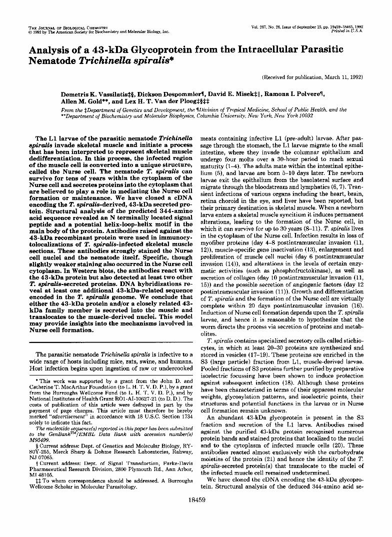
FIG. 5. A, Northern blots of size-separated t o t a l T. spiralis L1 larvae RNA ( l a n e I ) or adult ( l a n e a) RNA (10 pgllane) hybridized with the 43-kDa cDNA. Expression of the 43-kDa gene is observed only in the L1 larvae. The approximate size of the hybridizing band is indicated on the left of the panel. B, the panel in A was rehybridized with 7'. spiralis-derived cDNA clone 17.2, which detects a 700-base pair RNA, to confirm that approximately equal amounts of RNA had been loaded in each lane.
The intensity of the additional bands does not increase when hybridizations are performed at lower stringencies, indicating that these fragments encode a small region of absolute identity that could be a small exon. Alternatively, since our laboratory strain of T. spiralis is not cloned, the possibility also exists that a different strain, with polymorphisms at the 43-kDa locus, contaminates the population.
In hybridizations at reduced stringencies (6 x NET, 50 "C), an additional band of almost equal intensity also hybridized with the 32P-labeled 43-kDa cDNA (Fig. 4B; bands are indi- cated with asterisks). The additional hybridizing band indi- cates the possible presence of another gene in the genome of T. spiralis, with homology to the 43-kDa cDNA.
Developmental Expression of the 43-kDa Gene-The expres- sion of the 43-kDa gene was restricted to the pre-adult L1 larvae of T. spiralis. Northern blots of total RNA from adult worms (Fig. 5A, lane a) and L1 larvae (6 months old; Fig. 5A, lane 1 ) were hybridized with 32P-labeled 43-kDa cDNA, and the expected 1.3-kb mRNA was detected in the L1 larvae but not in adult worms. The presence of RNA in the lane with RNA from adult worms was verified by rehybridization of the blot with another T. spiralis cDNA (Fig. 5B)? The precise time of onset of the 43-kDa gene expression is not known. Immunocytolocalization studies on synchronously infected muscle cells using antiglycosylated 43-kDa sera show the first appearance of staining in the stichiocyte cells of larvae at day 7 postintramuscular infection (20). However, these antisera exhibit extensive cross-reactivity with several other stichio- cyte-derived proteins, and the exact onset of 43-kDa gene expression is therefore still obscure.
Several Proteins Are Detected in Western Blots, with Anti- serum Raised against Recombinant 43-kDa Proteins-Pre- vious studies to determine the localization of the 43-kDa protein in infected skeletal muscle suffered from a lack of specificity of the antibodies, which detected numerous bands in Western blots (Fig. 6A, lane 3 ) . Most of these bands had been attributed to cross-reacting carbohydrate epitopes (21). We therefore raised antibodies against recombinant 43-kDa proteins purified from E. coli. These antibodies should have a markedly improved specificity when compared with antibod- ies raised against the purified glycosylated 43-kDa protein and should be diagnostic for the cellular location of the 43- kDa protein. We expressed two fusion proteins in E. coli. One
* D. Misek, unpublished results.
431
321
< -
< -
(t 91 't
- "
"
Ip I
" " L "
C
43k-
32k-
I : Irn
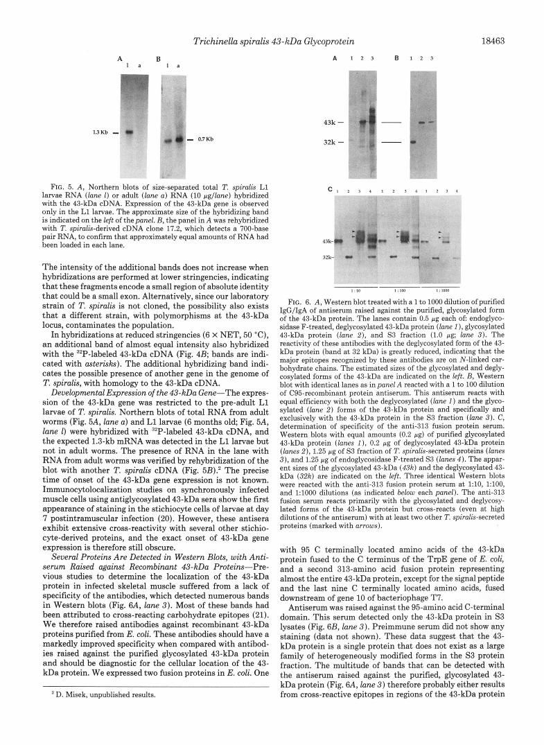
FIG. 6. A, Western blot treated with a 1 to 1000 dilution of purified IgG/IgA of antiserum raised against the purified, glycosylated form of the 43-kDa protein. The lanes contain 0.5 pg each oE endoglyco- sidase F-treated, deglycosylated 43-kDa protein (lane I ) , glycosylated 43-kDa protein (lane 2 ) , and S3 fraction (1.0 pg; lune 3 ) . The reactivity of these antibodies with the deglycosylated form of the 43- kDa protein (band at 32 kDa) is greatly reduced, indicating that the major epitopes recognized by these antibodies are on N-linked car- bohydrate chains. The estimated sizes of the glycosylated and degly- cosylated forms of the 43-kDa are indicated on the left. B, Western blot with identical lanes as in panel A reacted with a 1 to 100 dilution of C95-recombinant protein antiserum. This antiserum reacts with equal efficiency with both the deglycosylated (lane 1) and the glyco- sylated (lane 2) forms of the 43-kDa protein and specifically and exclusively with the 43-kDa protein in the S3 fraction (lane 3). C, determination of specificity of the anti-313 fusion protein serum. Western blots with equal amounts (0.2 pg) of purified glycosylated 43-kDa protein (lanes I ) , 0.2 pg of deglycosylated 43-kDa protein (lunes 2 ) , 1.25 pg of S3 fraction of T. spiralis-secretedproteins (lanes 3 ) , and 1.25 pg of endoglycosidase F-treated S3 (lanes 4 ) . The appar- ent sizes of the glycosylated 43-kDa (43k) and the deglycosylated 43- kDa (32k) are indicated on the left. Three identical Western blots were reacted with the anti-313 fusion protein serum at l : l O , 1:100, and 1:lOOO dilutions (as indicated below each panel). The anti-313 fusion serum reacts primarily with the glycosylated and deglycosy- lated forms of the 43-kDa protein but cross-reacts (even at high dilutions of the antiserum) with at least two other T. spiralis-secreted proteins (marked with arrows).
with 95 C terminally located amino acids of the 43-kDa protein fused to the C terminus of the TrpE gene of E. coli, and a second 313-amino acid fusion protein representing almost the entire 43-kDa protein, except for the signal peptide and the last nine C terminally located amino acids, fused downstream of gene 10 of bacteriophage T7.
Antiserum was raised against the 95-amino acid C-terminal domain. This serum detected only the 43-kDa protein in S3 lysates (Fig. 6B, lane 3 ) . Preimmune serum did not show any staining (data not shown). These data suggest that the 43- kDa protein is a single protein that does not exist as a large family of heterogeneously modified forms in the S3 protein fraction. The multitude of bands that can be detected with the antiserum raised against the purified, glycosylated 43- kDa protein (Fig. 6A, lane 3 ) therefore probably either results from cross-reactive epitopes in regions of the 43-kDa protein

18464 Trichinella spiralis 43-kDa Glycoprotein
other than the 95 C-terminal amino acids or from cross- reactive carbohydrate epitopes shared with other proteins.
Antibodies raised against the 313-amino acid fusion protein (anti-313 fusion) also detected the purified 43-kDa glycopro- tein (Fig. 6C, lanes 1 ) and the deglycosylated form of the purified 43-kDa protein (Fig. 6C, lanes 2). Immunoadsorbed anti-313 fusion antiserum (adsorbed on E. coli-bacteriophage T7-gene 10 lysates immobilized on nitrocellulose filters to remove gene 10-specific antibodies) gave the same results, whereas preimmune serum did not show reactivity (data not shown). The anti-313 fusion sera primarily reacted with the 43-kDa protein in the S3 fraction of L1 larvae (Fig. 6, lanes 3), as shown by the effect of increased dilution of the anti- serum (Fig. 6C). However, this serum cross-reacts with at least two other T. spiralis proteins contained in the S3 fraction (Fig. 6C, lanes 3, bands identified with arrowheads). Degly- cosylation of the S3 proteins reduced their size, but did not result in the production of a single reactive protein band (Fig. 6C, lanes 4 ) . We, therefore, assume that the antiserum raised against the recombinant 313 fusion protein detects either several forms of the 43-kDa protein that differ in the process- ing of their C-terminal end or additional proteins bearing immunological similarities in their peptide backbone.
Cytolocalizatwn of the 43-kDa Protein in Infected Skeletal Muscle-Immunocytolocalizations using the anti-313 fusion serum, or purified IgG/IgA fractions, on sections obtained 15 days after synchronous infection or 35 days and 6 months after oral infections, showed staining of the cytoplasm and the muscle-derived nuclei of the Nurse cells (Fig. 7, B and C, data for the 35-day infections shown only; several nuclei have been highlighted with arrows; please note that significant staining of the nematode is only observed in sections that include the stichiocytes; compare with Fig. 8A for preimmune serum; due to the fixation, the worm is separated from the cytoplasm). Immunoadsorbed anti-313 fusion protein serum (adsorbed on E. coli bacteriophage T7 gene 10 lysates) gave identical results (data not shown). We have been unsuccessful, so far, in obtaining well defined staining of the Nurse cell nuclei and cytoplasm with the antiserum raised against the fusion protein encoding the 95 C-terminal amino acids. This could be due to the lower titer of this antiserum or masking of the epitope(s) present in the 95 C-terminal amino acids during the fixation techniques. Alternatively, a processed form of the 43-kDa protein or the product of another gene (see Fig. 4B) may locate to the cytoplasm and/or nuclei of the Nurse cell.
We tentatively conclude that antibodies raised against the 43-kDa recombinant protein identify a T. spiralis-derived protein(s) that is/are secreted into the cytoplasm of the in- fected muscle cell, at least one of which translocates to the muscle-derived nuclei of the Nurse cell.
DISCUSSION
We have cloned a cDNA that encodes the 43-kDa glycopro- tein of the nematode T. spiralis. The gene for the 43-kDa protein is expressed in the L1 larvae of infected skeletal muscle (this paper), and the 43-kDa protein can be purified from in vitro secretions and the S3 fraction of L1 larvae that have been isolated from skeletal muscle (21). We, therefore, consider it reasonable to assume that the 43-kDa protein is indeed synthesized by the L1 larvae in the Nurse cell and that i t is secreted into the cytoplasm of infected skeletal muscle.
The predicted 344-amino acid sequence of the 43-kDa gene indicates that this cDNA encodes a secreted glycoprotein with a typical signal peptide and two potential N-linked carbohy- drate moieties. Structural analysis of the predicted amino acid
b
FIG. 7. Immunocytolocalizations using rabbit anti-313 re- combinant IgG/IgA. Mouse muscle sections obtained 35 days post- infection with 7’. spiralis were used. A horseradish peroxidase-conju- gated goat anti-rabbit antiserum was used for detection of antibody. Staining was performed with diaminobenzidine and HZ02. A, preim- mune IgG/IgA at 1:500 dilution. The uninfected muscle, the Nurse cell, and the worm do not stain. Staining of a region on the right is due to clustered red blood cells that have endogenous peroxidase activity. B, anti-BlB-IgG/IgA at a 1:500 dilution. The Nurse cell cytoplasm and the muscle cell-derived Nurse cell nuclei (indicated by arrows) stain strongly. In this particular section, the worm does not stain since there are no stichiocytes in this region of the nematode. C, region of another Nurse cell from the same experiment as shown in panel €3, but at a higher magnification. The stained nuclei are indicated by arrows. In this section, T. spiralis stichiocytes also stain, whereas cytoplasmic staining in this region of the Nurse cell is almost absent.
sequence revealed a possible helix-loop-helix motif with ho- mology to the negative regulator of myogenic differentiation Id and the Drosophila protein em. Antibodies raised against the recombinant 43-kDa protein detect protein(s) in the nu- clei and cytoplasm of infected skeletal muscle. However, these antibodies detected up to three bands, including the 43-kDa protein, any one of which may be localized to the skeletal muscle nuclei; the identity of the nuclearly located protein therefore remains uncertain. We also cannot exclude the less likely possibility that the T. spiralis infection induced expres- sion of a nuclear protein encoded by skeletal muscle, which shares epitopes with the T. spiralis-derived 43-kDa protein. Because of these uncertainties, we tentatively conclude that either the 43-kDa protein itself, or the product of a related gene, is localized to the nuclei of infected muscle cells.
This research was initiated because of our interest in the mechanisms by which the intracellular nematode T. spiralis induces formation of the Nurse cell. The possibility that the 43-kDa protein, or a closely related family member of this protein, locates to the nucleus of the affected myocyte and the homology of the 43-kDa protein with helix-loop-helix

Trichinella spiralis 43-kDa Glycoprotein 18465
proteins provide us with the intriguing option that these secreted proteins might function in modulating gene expres- sion of the affected muscle cell.
Acknowledgments-We thank Dr. S. Le Blancq for critical reading of the manuscript and S. Buck for helpful discussion.
REFERENCES 1. Villella, J. B. (1958) J. Parasitol. 44% 41 2. Ali Khan, 2. (1966) J. Purasitol. 62, 248-259 3. Kozek. W. J. (1971) J. Purasitol. 67. 1015-1028 4. Kozek; W. J. (1971j J. Purasitol. 67; 1029-1038 5. Gardiner, C. H. (1976) J. Purasitol. 62,865-870 6. Basten, A., and Beeson, P. B. (1970) J. Exp. Med. 131, 1288-1304
8. Despommier, D. D. (1976) Musculature: Ecological Aspects of Parasitology 7. Harley, J. P., and Gallicchico, V. (1971) Exp. Purasitol. 30, 11-12
9. Ribas-Mujal, D., and Rivera-Pomar, J. M. (1968) Virchows Arch. A 346,
~ ~~~~~~~
(Kennedy, C. R., ed) pp. 270-285, Elsevier-North Holland, Amsterdam
154-168 10. Stewart, G. L. (1973) Thesis, Rice University, Houston 11. Despommier, D. D. (1975) Am. J. Pathol. 78,477-496 12. Jasmer. D. P., Bohnet, S.. and Prieur, D. J. (1991) Exp. Parasitol. 72,321-
331 13. Jasmer, D. P. (1990) Exp. Parasitol. 70,452-465 14. Despommier, D., Symmans, W. F., and Dell, R. (1991) J. Parasitol. 77,
15. Teppema, J. S., Robinson, J. E., and Ruitenberg, E. J. (1973) Parasitology 290-295
66, 291-296
16. Des ommier, D. D., Aron, L., and Turgeon, L. (1975) Exp. Parasitol. 37,
17. Despommler, D. D., and Muller, M. (1976) J. Parasitol. 62,775-785 18. Despommier, D. D., and Laccetti, A. (1981) Ex . Parasitol. 61,279-295
20. Despommier, D. D., Gold, A. M., Buck, S. W., Capo, V., and Silberstein, D. 19. Despommier, D. D., and Laccetti, A. (1981) J. fkasitol. 67,332-339
21. Gold, A. M., Despommier, D. D., and Buck, S. W. (1990) Mol. Biochem.
198-116
(1990) Exp. Parasztol. 71,27-38
22. Garrell, J. and Modollel, J. (1990) Cell 61, 39-48 23. Benezra, k., Davis, R. L., Lockshon, D., Turner, D. L., and Weintraub, H. 24. Despommier, D. D., Campbell, W. E., and Blair, L. S. (1977) Parasitology
Parasitol. 41, i87-196
(1990) Cell 61,49-59
25. 26.
27. 28.
29. 30. 31. 32.
33.
34.
74,109-119 Auffray C , and Rougeon F. (1980) Eur. J. Biochem, 107,,303-314 Gl@hL+.,'Crkvenjakov, k., and Byus, C. (1974) Bzochemlstry 13, 2633-
Gubler, U., and Hoffman B. J. (1983) Gem Amst ) 26 263 269 Sanger F., Coulsen, A. k., Barrell, B. G., Qmith, A. j. H.: and Roe, B.
Southern E. (1975) J. Mol. Biol. 98,503-517 Jeffre s k. J. and Flavell, R. A. (1977) Cell 12, 429-439 Boedtkr, H. (l971),Bioehim. Biophys. Acta 240, 448-453 Harboe H. and In Ild A (1973) In A Manual o Quantztatiue Immunoelec-
trop&re&: Met%& and Ap lications (Axefsen, N. H. Kroll, J., and Weeke B., eds) p 161-164, eniversitetforla et, Oslo, dorway
Towbin, k., Staeheyin, T., and Gordon, J. (1959) Proc. Natl. Acad. Sci. U. S. A. 76,4350-4354
Tsang, V. C. W., Peralta, J. M., and Simons, A. R. (1983) Methods Enzymol.
2637
(1986) J. Mol. Biol. 143, 161-178