analysis of bioactive chemical compounds of aspergillus ... · full length research paper analysis...
TRANSCRIPT

Vol. 7(x), pp. xx-xx, xxx 2015
DOI: 10.5897/JPP2015.0254
Article Number: 3149CE853451
ISSN 2141-2502
Copyright © 2015
Author(s) retain the copyright of this article
http://www.academicjournals.org/JPP
Journal of Pharmacognosy and Phytotherapy
Full Length Research Paper
Analysis of bioactive chemical compounds of Aspergillus niger by using gas chromatography-mass
spectrometry and fourier-transform infrared spectroscopy
Imad Hadi Hameed*, Lena Fadhil Hamza and Sabreen A. Kamal
Department of Molecular Biology, Babylon University, Hilla City, Iraq.
Received 9 June 2015; Accepted 9 July 2015.
Bioactives are chemical compounds often referred to as secondary metabolites. Thirty five bioactive compounds were identified in the methanolic extract of Aspergillus niger. The identification of bioactive chemical compounds is based on the peak area, retention time molecular weight and molecular formula. Gas chromatography-mass spectrometry (GC-MS) analysis of Aspergillus niger revealed the existence of the 6-Acetyl-ß-d-mannose, 4-[Dichloromethyl]-2-[[2-[1-methyl-2-pyrrolidinyl]ethyl]amino-6-trichloro, 2-Furan-carboxaldehyde,5-methyl, 2,2,2-Trifluoro-N-[2-(1-hydroxy-2,2,6,6-tetramethyl-piperidin-4-yl), 2,4-Dihydroxy-2,5-dimethyl-3(2H)-furan-3-one, HEPES (4-(2-hydroxyethyl)-1-piperazineethanesulfonic acid), Tetraacetyl-d-xylonic nitrile, Eicosanoic acid, phenylmethyl ester, Dodecanoic acid, 3-hydroxy, Desulphosinigrin, Glycyl-dl-serine, 2,5-Dimethyl-4-hydroxy-3(2H)-furanone, 2,5-Furandicarboxaldehyde, 2H-Oxecin-2-one,3,4,7,8,9,10- hexahydro-4-hydroxy-10-methyl, 6-Acetyl-ß-d-mannose, DL-Leucine, N-glycyl, 4H-Pyran-4-one,2,3-dihydro-3,5-dihydroxy-6-methyl, I-Gala-l-ido-octonic lactone, 2H-Pyran,tetrahydro-2-(12-pentadecynyloxy), 5-Hydroxymethylfurfural, Strychane,1-acetyl-20α-hydroxy-16-methylene, α-D-Glucopyranoside, O-α-D-glucopyranosyl-(1.fwdarw.3)ß-D-fru, Boroxin, tris(2,3-dimethylbut-2-yl), 16-Nitrobicyclo[10.4.0]hexadecane-1-ol-13-one, 3-[3-Bromophenyl]-7-chloro-3,4-dihydro-10-hydroxy-1,9(2H,10H)-a, Uric acid, 1,2,4-Trioxolane-2-octanoic acid ,5-octyl-,methyl ester, Tetraacetyl-d-xylonic nitrile, 1,2-Cyclopentanedicarboxylic acid, 4-(1,1-dimethylethyl)-,dimethyl, 2-Bromotetradecanoic acid, i-Propyl 11,12-methylene-octadecanoate, 1H-2,8a-Methanocyclopenta[a]cyclopropa [e]cyclodecan-11-one, and Octadecanoic acid. The FTIR analysis of A. niger proved the presence of aromatic rings, alkenes, aliphatic fluoro compounds, tetiary amine, C-N stretch, aromatic nitro compounds, ammonium ions and organic nitrate which shows major peaks at 696.30, 744.52, 821.68, 844.82, 900.76, 931.62, 1026.13, 1145.72, 1207.44, 1234.44, 1261.45, 1315.45, 1359.82, 1377.17, 1413.82, 1452.40, 1631.78, 1741.72, 2924.09, 3118.90, 3217.27 and 3271.27. Datura stramonium was very active against A. niger. Methanolic extract of bioactive compounds of A. niger were assayed for in vitro antibacterial activity against Pseudomonas aerogenosa, Escherichia coli, Proteus mirabilis, Staphylococcus aureus and Klebsiella pneumonia by using the diffusion method in agar. The zones of inhibition were compared with different standard antibiotics. The diameters of inhibition zones ranged from 0.46±0.1 to 6.52±0.61 mm for all treatments. Key words: Aspergillus niger, bioactive compounds, gas chromatography-mass spectrometry, fourier-transform infrared spectroscopy.
INTRODUCTION Aspergillus spp are ubiquitous opportunistic moulds that cause both allergic and invasive syndromes. The genus comprises approximately 180 species, of which 33 have been associated with human disease (Segal et al., 1998; Perfect et al., 2001). Aspergillus niger is the third most common species associated with invasive pulmonary aspergillosis (Bellini et al., 2003; Anupama et al., 2007). A. niger has a great economical and biotechnological interest and is extensively used for production of extracellular enzymes and organic acids such as citric acid (Baker, 2006; Perrone et al., 2007; Mogensen et al., 2010). It also
produces fumonisin B2 (FB2) along with OTA. 9, 19, 27. Fumonisins are suspected to cause human and animal toxicoses, and are regarded as carcinogenic (Susca et al., 2010; Chacko et al., 2012; Gebreselema et al., 2013). A culture yielding Aspergillus spp, in addition to enabling a diagnosis of invasive aspergillosis, may further define therapeutic options via susceptibility testing or the isolation of a species possessing inherent antifungal resistance; examples of the latter include Aspergillus terreus and Aspergillus nidulans, which are both resistant to amphotericin B (Walsh, 2004). The main disadvantage of culture is that it is relatively slow (the process takes days), is relatively insensitive, and requires specialized expertise for species determination.
In common with other pathogenic fungi, the ability to grow at 37°C distinguishes Aspergillus spp from other nonpathogenic environmental moulds. Aspergillus spp can be recovered on most routine solid and liquid microbiological media (example, blood agar, chocolate agar, brain heart infusion broth). A fungal-specific medium example, sabouraud dextrose agar should be included at the time of initial specimen set-up in clinical scenarios in which Aspergillus spp (or other moulds) are considered possible pathogens, because of superior yield (Horvath and Dummer, 1995). The addition of antibiotics example, chloramphenicol and gentamicin to the medium is required for the recovery of Aspergillus spp from specimens obtained from nonsterile sites, since they prevent bacterial
overgrowth. Cycloheximide, a eukaryotic protein synthesis inhibitor, is frequently added to fungal media to inhibit the overgrowth of cultures by non-pathogenic environmental moulds; however, on occasion, cycloheximide may inhibit the growth of Aspergillus spp. The aim of this study were analysis of the secondary metabolites and the evaluation of antibacterial and antifungal activity . MATERIALS AND METHODS
Collection and growth condition
A. niger was isolated from dried fruit and the pure colonies were
selected, isolated and maintained in potato dextrose agar slants (Usha and Masilamani, 2013). After the species were identified by the identification key, spores were grown in a liquid culture of potato
dextrose broth (PDB) and incubated at 25°C in a shaker for 16 days at 130 rpm.
Production, extraction and determination of metabolites
The metabolites were determined and extracted for gas chromatography (GC) analysis using the method of Siddiquee et al.
(2012) with some modifications. The extraction was performed by adding 25 ml methanol to 100 ml liquid culture in an Erlenmeyer flask after the infiltration of the culture. The mixture was incubated at 4°C for 10 min, and then shook for 10 min at 130 rpm. Metabolites was separated from the liquid culture and evaporated to dryness with a rotary evaporator at 45ºC. The residue was dissolved in 1 ml methanol, filtered through a 0.2 μm syringe filter, and stored at 4ºC for 24 h before being used for gas chromatography–mass spectrometry (GC-MS) (Imad et al., 2014a).
The identification of the components was based on comparison of their mass spectra with those of NIST mass spectral library as well as on comparison of their retention indices either with those of authentic compounds or with literature values.
GC-MS analysis
Bioactive compound were examined for the chemical composition using GC-MS (Agilent 789N) equipped with a DB-5MS column (30 m×0.25 mm i.d., 0.25 um film thickness, J&W Scientific, Folsom, CA). The oven temperature was programmed as for the previous analysis (Imad et al., 2015a; Muhanned et al., 2015). Helium was used as the carrier gas at the rate of 1.0 ml/min. Effluent of the GC column was introduced directly into the source of the MS via a transfer line (250°C). Ionization voltage was 70 eV and ion source temperature was 230°C. Scan range was 41 to 450 amu. The constituents were identified after being compared with available data in the GC-MS library in the literatures (Imad et al., 2015b; Mohammed et al., 2013).
Fourier transform infrared spectrophotometer (FTIR)
The powdered sample of the A. niger specimen was treated for
fourier transform infrared spectroscopy (Shimadzu, IR Affinity 1, Japan). The sample was run at infrared region between 400 nm and 4000 nm.
Determination of antibacterial activity of crude fraction of A. niger compounds
The test pathogens (E. coli, Pseudomonas aeruginosa, Klebsiella
pneumoniae and Staphylococcus aureus) were swabbed in Muller Hinton agar plates. 90μl of fungal extracts was loaded on the bored wells. The wells were bored in 0.5cm in diameter. The plates were incubated at 37°C for 24 h and examined. After the incubation the diameter of inhibition zones around the discs was measured.
*Corresponding author. E-mail: [email protected].
Author(s) agree that this article remain permanently open access under the terms of the Creative Commons Attribution
License 4.0 International License

A B
Figure 1. Morphological characterization of Aspergillus niger. (B) Microscopic observation (A) colony.
Determination of antifungal activity
A. niger isolate was suspended in potato dextrose broth and diluted
to approximately 105 colony forming unit (CFU) per ml. They were “flood inoculated onto the surface of Potato dextrose agar and then dried. Standard agar well diffusion method was followed (Perez et al., 1990; Perez et al., 1999; Erdemogllu et al., 2003; Bagamboula et al., 2004). Five-millimeter diameter wells were cut from the agar using a sterile cork-borer, and 25 μl of the samples solutions (Nerium olender, Ricinus communis, Datura stramonium, Linum
usitatissimum, Anastatica hierochuntica and Gramineae poaceae) were delivered into the wells. The plates were incubated for 48 h at room temperature (Huda et al., 2015a; Ameera et al., 2015; Imad et al., 2015c). Antimicrobial activity was evaluated by measuring the zone of inhibition against the test microorganisms. Methanol was used as solvent control. Amphotericin B and fluconazole were used as reference antifungal agent (Anesini and Perez, 1993; Rukayadi et al., 2006; Huda et al., 2015b). The tests were carried out in triplicate. The antifungal activity was evaluated by measuring the inhibition-zone diameter observed after 48 h of incubation.
Statistical analysis
Data were analyzed using analysis of variance (ANOVA), and differences among the means were determined for significance at P < 0.05 using Duncan’s multiple range test (by statistical package for the social sciences (SPSS) software) Version 9.1 (Imad et al., 2014b).
RESULTS AND DISCUSSION Isolation of fungi from dried fruit The fungi were isolated from dried fruit by serial dilution method. Based on morphological, characteristics of fungi was isolated in selective media of potato dextrose agar
media. Morphological, Microscopical and microscopical characteristics of fungal strains were determined using specific media light and compound microscope Figure 1.
Production and Identification of secondary metabolites from the methanolic crude extract of A. niger by gas chromatography and mass spectrometry
and fourier-transform infrared spectroscopy
The 400 ml of fermentation broth (PDA broth) which contain 200 μl of the standardized fugal suspensions were used to inoculate the flasks and incubated at 37°C on a shaker at 90 rpm for 7 days. After fermentation, the secondary metabolites were produced by isolated microorganisms.
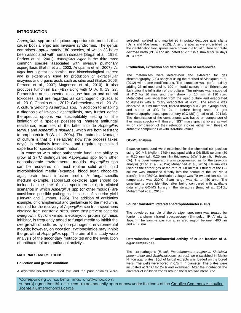
Gas chromatography and mass spectroscopy analysis of compounds was carried out in methanolic extract of A. niger, as shown in Table 1. The GC-MS chromatogram of the seventeen peaks of the compounds detected was shown in Figure 2. Chromatogram GC-MS analysis of the methanol extract of A. niger showed the presence of twenty major peaks and the components corresponding to the peaks were determined as follows. The First set up peak were determined to be 6-Acetyl-ß-d-mannose (Figure 3). The second peak indicated to be 4-[Dichloromethyl]-2-[[2-[1-methyl-2-pyrrolidinyl]ethylamino-6-trichloro (Figure 4). The next peaks considered to be 2-Furan-carboxaldehyde, 5-methyl, 2,2,2-Trifluoro-N-[2-(1-hydroxy-2,2,6,6-tetramethyl-piperidin-4-yl), 2,4-Dihydroxy-2,5-dimethyl-3(2H)-furan-3-one, HEPES, Tetraacetyl-d-xylonic nitrile, eicosanoic acid, phenylmethyl ester, dodecanoic acid , 3-hydroxy, Desulphosinigrin,
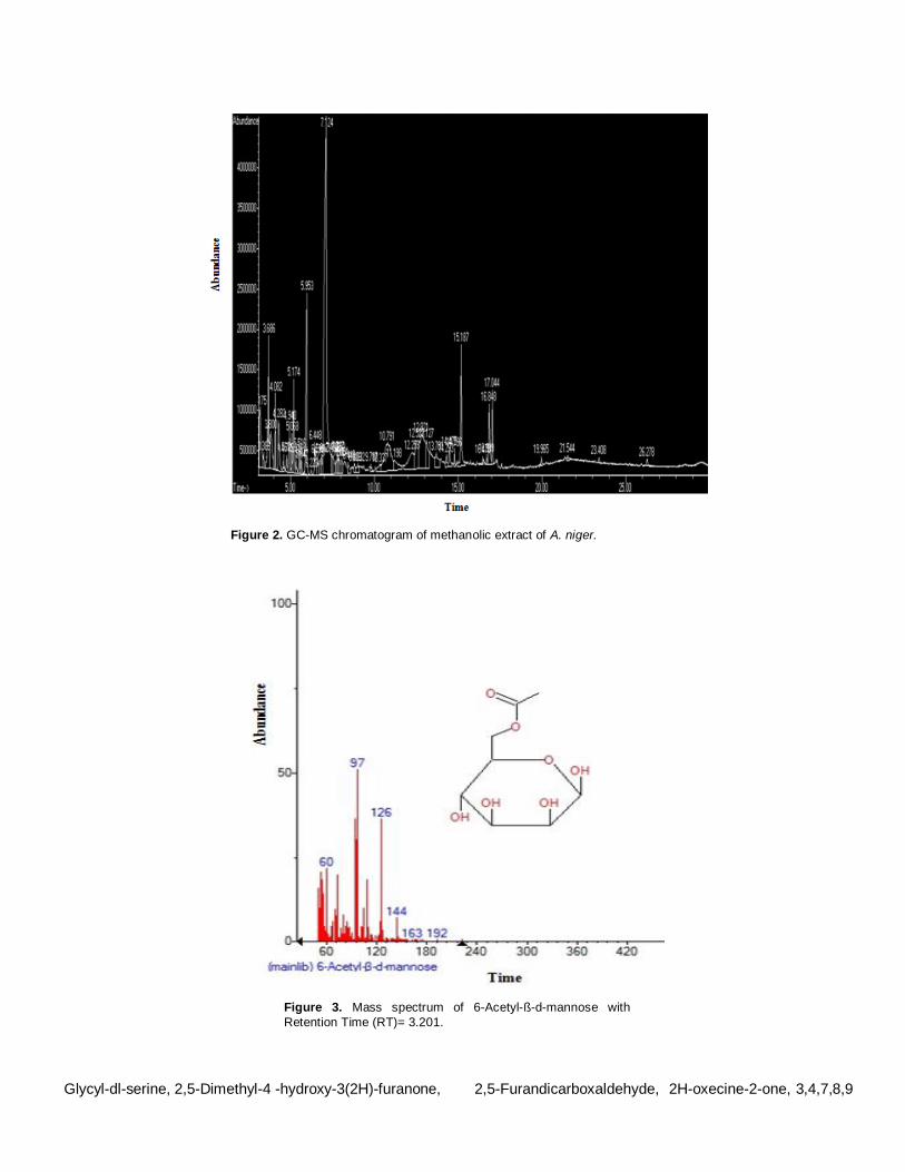
Figure 2. GC-MS chromatogram of methanolic extract of A. niger.
Figure 3. Mass spectrum of 6-Acetyl-ß-d-mannose with
Retention Time (RT)= 3.201.
Glycyl-dl-serine, 2,5-Dimethyl-4 -hydroxy-3(2H)-furanone, 2,5-Furandicarboxaldehyde, 2H-oxecine-2-one, 3,4,7,8,9
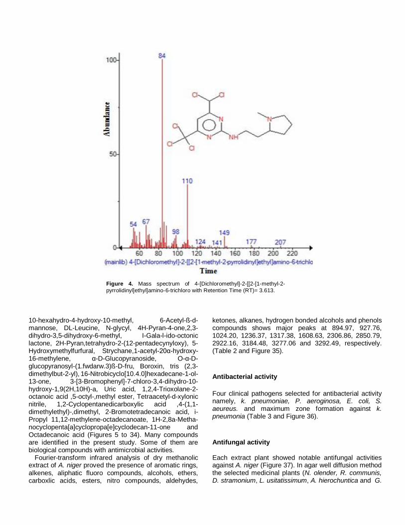
Figure 4. Mass spectrum of 4-[Dichloromethyl]-2-[[2-[1-methyl-2-
pyrrolidinyl]ethyl]amino-6-trichloro with Retention Time (RT)= 3.613.
10-hexahydro-4-hydroxy-10-methyl, 6-Acetyl-ß-d- mannose, DL-Leucine, N-glycyl, 4H-Pyran-4-one,2,3-dihydro-3,5-dihydroxy-6-methyl, I-Gala-l-ido-octonic lactone, 2H-Pyran,tetrahydro-2-(12-pentadecynyloxy), 5-Hydroxymethylfurfural, Strychane,1-acetyl-20α-hydroxy-16-methylene, α-D-Glucopyranoside, O-α-D-glucopyranosyl-(1.fwdarw.3)ß-D-fru, Boroxin, tris (2,3-dimethylbut-2-yl), 16-Nitrobicyclo[10.4.0]hexadecane-1-ol-13-one, 3-[3-Bromophenyl]-7-chloro-3,4-dihydro-10-hydroxy-1,9(2H,10H)-a, Uric acid, 1,2,4-Trioxolane-2-octanoic acid ,5-octyl-,methyl ester, Tetraacetyl-d-xylonic nitrile, 1,2-Cyclopentanedicarboxylic acid ,4-(1,1-dimethylethyl)-,dimethyl, 2-Bromotetradecanoic acid, i-Propyl 11,12-methylene-octadecanoate, 1H-2,8a-Metha-nocyclopenta[a]cyclopropa[e]cyclodecan-11-one and Octadecanoic acid (Figures 5 to 34). Many compounds are identified in the present study. Some of them are biological compounds with antimicrobial activities.
Fourier-transform infrared analysis of dry methanolic extract of A. niger proved the presence of aromatic rings, alkenes, aliphatic fluoro compounds, alcohols, ethers, carboxlic acids, esters, nitro compounds, aldehydes,
ketones, alkanes, hydrogen bonded alcohols and phenols compounds shows major peaks at 894.97, 927.76, 1024.20, 1236.37, 1317.38, 1608.63, 2306.86, 2850.79, 2922.16, 3184.48, 3277.06 and 3292.49, respectively. (Table 2 and Figure 35).
Antibacterial activity
Four clinical pathogens selected for antibacterial activity namely, k. pneumoniae, P. aeroginosa, E. coli, S. aeureus. and maximum zone formation against k. pneumonia (Table 3 and Figure 36).
Antifungal activity Each extract plant showed notable antifungal activities against A. niger (Figure 37). In agar well diffusion method the selected medicinal plants (N. olender, R. communis, D. stramonium, L. usitatissimum, A. hierochuntica and G.
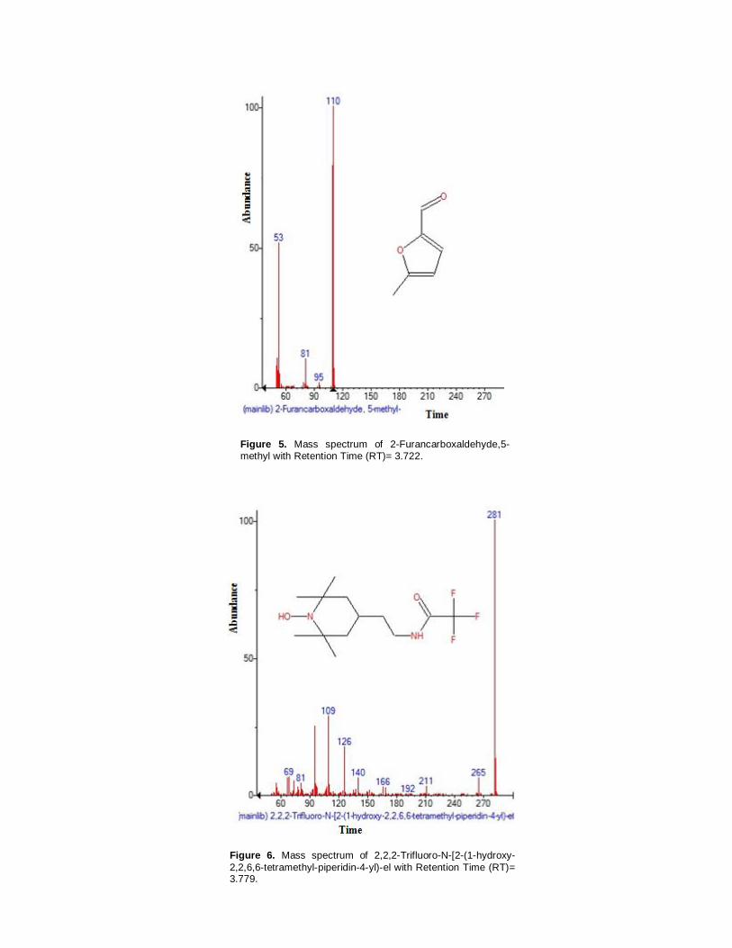
Figure 5. Mass spectrum of 2-Furancarboxaldehyde,5-
methyl with Retention Time (RT)= 3.722.
Figure 6. Mass spectrum of 2,2,2-Trifluoro-N-[2-(1-hydroxy-
2,2,6,6-tetramethyl-piperidin-4-yl)-el with Retention Time (RT)= 3.779.

Figure 7. Mass spectrum of 2,4-Dihydroxy-2,5-dimethyl-
3(2H)-furan-3-one with Retention Time (RT)= 4.076.
Figure 8. Mass spectrum of 2 HEPES with Retention Time
(RT)= 4.271.

Figure 9. Mass spectrum of Tetraacetyl-d-xylonic nitrile with Retention Time (RT)= 4.465.
Figure 10. Mass spectrum of eicosanoic acid , phenylmethyl ester with retention time (RT)= 4.546.

Figure 11. Mass spectrum of dodecanoic acid , 3-hydroxy
with retention time (RT)= 4.574.
Figure 12. Mass spectrum of desulphosinigrin with retention time (RT)= 4.654.

Figure 13. Mass spectrum of Glycyl-dl-serine with retention
time (RT)= 4.763.
Figure 14. Mass spectrum of 2,5-Dimethyl-4-hydroxy-3(2H)-furanone with retention time (RT)= 4.929.
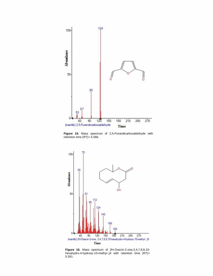
Figure 15. Mass spectrum of 2,5-Furandicarboxaldehyde with
retention time (RT)= 5.066.
Figure 16. Mass spectrum of 2H-Oxecin-2-one,3,4,7,8,9,10-
hexahydro-4-hydroxy-10-methyl-,[4 with retention time (RT)= 5.261.

Figure 17. Mass spectrum of DL-Leucine , N-glycyl with retention time (RT)= 5.616.
Figure 18. Mass spectrum of 4H-Pyran-4-one,2,3-dihydro-3,5-
dihydroxy-6-methyl with retention time (RT)= 5.942.
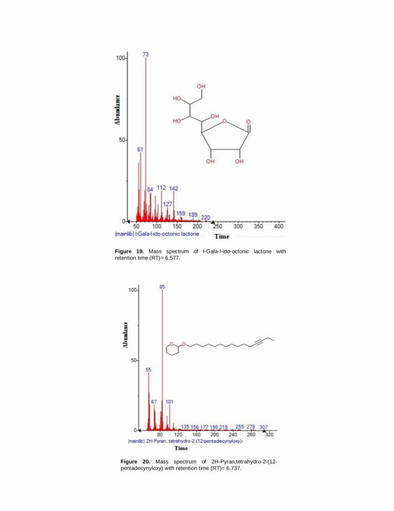
Figure 19. Mass spectrum of I-Gala-l-ido-octonic lactone with
retention time (RT)= 6.577.
Figure 20. Mass spectrum of 2H-Pyran,tetrahydro-2-(12-
pentadecynyloxy) with retention time (RT)= 6.737.

Figure 21. Mass spectrum of 5-Hydroxymethylfurfural with retention time
(RT)= 7.120.
Figure 22. Mass spectrum of Strychane,1-acetyl-20α-hydroxy-16-
methylene with retention time (RT)= 8.053.

Figure 23. Mass spectrum of α-D-Glucopyranoside ,O-α-D-
glucopyranosyl-(1.fwdarw.3)ß-D-fru with retention time (RT)= 7.836.
Figure 24. Mass spectrum of Boroxin , tris(2,3-dimethylbut-2-yl)
with retention time (RT)= 8.442.
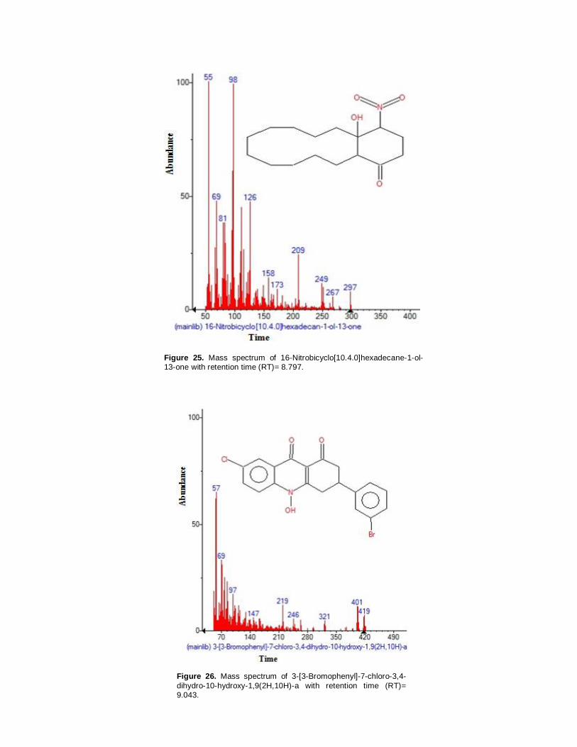
Figure 25. Mass spectrum of 16-Nitrobicyclo[10.4.0]hexadecane-1-ol-13-one with retention time (RT)= 8.797.
Figure 26. Mass spectrum of 3-[3-Bromophenyl]-7-chloro-3,4-
dihydro-10-hydroxy-1,9(2H,10H)-a with retention time (RT)= 9.043.

Figure 27. Mass spectrum of uric acid with retention time
(RT)= 9.672.
Figure 28. Mass spectrum of 1,2,4-Trioxolane-2-octanoic acid ,5-octyl-,methyl ester with retention time (RT)= 11.320.

Figure 29. Mass spectrum of Tetraacetyl-d-xylonic nitrile with retention time (RT)= 12.276.
Figure 30. Mass spectrum of 1,2-Cyclopentanedicarboxylic acid ,4-(1,1-
dimethylethyl)-,dimethyl with retention time (RT)= 13.975.

Figure 31. Mass spectrum of 2-Bromotetradecanoic acid with retention time (RT)=
14.771.
Figure 32. Mass spectrum of i-Propyl 11,12-methylene-octadecanoate with retention time
(RT)= 15.022.

Figure 33. Mass spectrum of 1H-2,8a-
Methanocyclopenta[a]cyclopropa[e]cyclodecan-11-one with retention time (RT)= 17.214.
Figure 34. Mass spectrum of octadecanoic acid with retention time (RT) =
17.048.
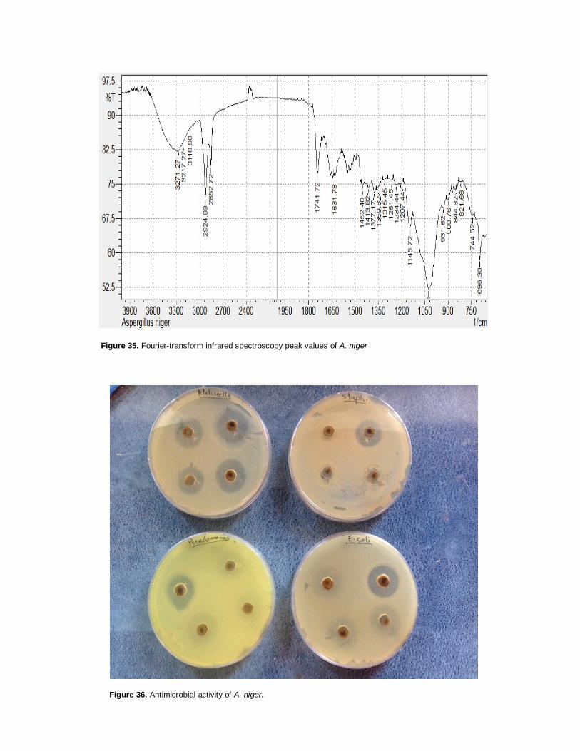
Figure 35. Fourier-transform infrared spectroscopy peak values of A. niger
Figure 36. Antimicrobial activity of A. niger.

Table 1. Major bioactive chemical compounds identified in methanolic extract of Aspergillus niger.
MS Fragment- ions Chemical structure Exact mass Molecular weight Formula RT (min) Phytochemical compound S/N
60, 97, 126, 144, 163, 192
222.073953 222 C6H14O7 3.201 6-Acetyl-ß-d-mannose 1
54, 67, 84, 98, 110, 124, 141, 149, 177, 207
403.989586 403
3.613 4-[Dichloromethyl]-2-[[2-[1-methyl-2-pyrrolidinyl]ethyl]amino-6-trichloro
2
53, 81, 95, 110
110.0367794 110 C6H6O2 3.722 2-Furancarboxaldehyde,5-methyl 3
69, 81, 109, 126, 140, 166, 192, 211, 265, 281
296.171164 296
3.779 2,2,2-Trifluoro-N-[2-(1-hydroxy-2,2,6,6-tetramethyl-piperidin-4-yl)
4
poaceae) were effective against A. niger (Table 4). D. stramonium was very highly active against A. niger. A. niger was found to be sensitive to all
test medicinal plants, and mostly comparable to the standard reference antifungal drug amphotericin B and fluconazole to some extent.
CONCLUSION The results of this study showed that A. niger

Table 1. Contd.
55, 73, 84, 101,144
144.042258 144 C6H8O4 4.076 2,4-Dihydroxy-2,5-dimethyl-3(2H)-furan-3-one 5
55, 65, 84, 99, 112, 143, 157, 174, 207, 237
238.098728 238 4.271 HEPES 6
60, 67, 73, 95, 112, 133, 176, 197, 215, 233, 251, 270
343.090332 343 C14H17NO9 4.465 Tetraacetyl-d-xylonic nitrile 7.
57, 71, 85, 91, 108, 126, 147, 167, 207, 281
402.349781 402 C27H46O2 4.546 Eicosanoic acid , phenylmethyl ester 8

Table 1. Contd.
55, 69, 83, 96, 112, 138, 151, 180, 200
216.125445 216 C12H24O3 4.574 Dodecanoic acid , 3-hydroxy 9
60, 73, 85, 103, 127, 145, 163, 213, 262
279.077658 279
4.654 Desulphosinigrin 10
60, 74, 85, 114, 126
162064056 162
4.763 Glycyl-dl-serine 11
57, 72, 85, 94,128
128.047344 128 C6H8O3 4.929 2,5-Dimethyl-4-hydroxy-3(2H)-furanone 12

Table 1. Contd.
53, 67, 95, 124
124.016044 124 C6H4O3 5.066 2,5-Furandicarboxaldehyde 13
55, 70, 81, 95, 112, 124, 142, 166, 184
184.109944 184 C10H16O3 5.261 2H-Oxecin-2-one,3,4,7,8,9,10- hexahydro-4-hydroxy-10-methyl-,[4
14
55, 86, 114, 132, 157, 188
188.116093 188
5.616 DL-Leucine , N-glycyl 16
55, 72, 85, 101, 115, 144
144.042258 144 C6H8O4 5.942 4H-Pyran-4-one,2,3-dihydro-3,5-dihydroxy-6-methyl 18

Table 1. Contd.
61, 73, 84,112, 127, 142, 159, 189, 220
238.068868 238 C8H14O8 6.577 I-Gala-l-ido-octonic lactone 19
55, 67, 85, 101, 135, 156, 177, 198, 219, 255, 279, 307
308.27153 308 C20H36O2 6.737 2H-Pyran,tetrahydro-2-(12-pentadecynyloxy) 20
53, 69, 81, 97, 126
126.031694 126 C6H6O3 7.120 5-Hydroxymethylfurfural 21
57, 70, 88, 97, 130, 166, 196, 224, 239, 253, 281, 295, 338
338.199429 338
8.053 Strychane,1-acetyl-20α-hydroxy-16-methylene 22

Table 1. Contd.
60, 73, 85, 97,113, 126, 145, 187
504.169035 504 C18H32O16 7.836 α-D-Glucopyranoside ,O-α-D-glucopyranosyl-(1.fwdarw.3)ß-D-fru
23
55, 69, 84, 95, 115, 137, 157, 181, 209, 251, 292, 321
336.317837 336
8.442 Boroxin , tris(2,3-dimethylbut-2-yl) 24
55, 69, 81, 98, 126, 158, 173, 209, 249, 267, 297
297.194008 297
8.797 16-Nitrobicyclo[10.4.0]hexadecane-1-ol-13-one 25

Table 1. Cont’d.
57, 69, 97, 147, 219, 246, 321, 401, 419
416.976732 416
9.043 3-[3-Bromophenyl]-7-chloro-3,4-dihydro-10-hydroxy-1,9(2H,10H)-a
26
54, 69, 82, 97, 125, 140, 168
168.02834 168
9.672 Uric acid 27
56, 69, 83, 98, 111, 143, 155, 185, 215, 259, 281, 311
344.256275 344 C19H36O5 11.320 1,2,4-Trioxolane-2-octanoic acid ,5-octyl-,methyl ester
28
60, 73, 112, 133, 164, 197, 228, 270
343.090332 343
12.276 Tetraacetyl-d-xylonic nitrile 29
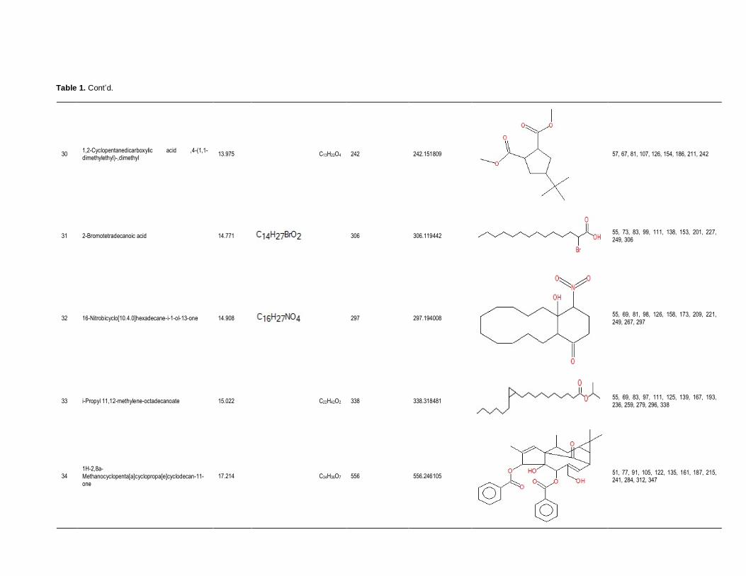
Table 1. Cont’d.
57, 67, 81, 107, 126, 154, 186, 211, 242
242.151809 242 C13H22O4 13.975 1,2-Cyclopentanedicarboxylic acid ,4-(1,1-dimethylethyl)-,dimethyl
30
55, 73, 83, 99, 111, 138, 153, 201, 227, 249, 306
306.119442 306
14.771 2-Bromotetradecanoic acid 31
55, 69, 81, 98, 126, 158, 173, 209, 221, 249, 267, 297
297.194008 297
14.908 16-Nitrobicyclo[10.4.0]hexadecane-i-1-ol-13-one 32
55, 69, 83, 97, 111, 125, 139, 167, 193, 236, 259, 279, 296, 338
338.318481 338 C22H42O2 15.022 i-Propyl 11,12-methylene-octadecanoate 33
51, 77, 91, 105, 122, 135, 161, 187, 215, 241, 284, 312, 347
556.246105 556 C34H36O7 17.214 1H-2,8a-Methanocyclopenta[a]cyclopropa[e]cyclodecan-11-one
34

Table 1. Cont’d.
60, 73, 83, 97, 115, 129, 143, 157, 171,
185, 199, 227, 241 ,255, 284
284.27153 284 C18H36O2 17.048 Octadecanoic acid 35
Table 2. Fourier-transorm infrared spectroscopy peak values of A. niger.
S/N Peak (Wave number cm-ˡ) Intensity Bond Functional group assignment Group frequency
1 696.30 58.479 C-H Aromatic rings 690-900
2 744.52 68.028 C-H Alkenes 675-995
3 821.68 75.498 C-H Alkenes 675-995
4 844.82 74.141 C-H Alkenes 675-995
5 900.76 71.557 C-H Alkenes 675-995
6 931.62 69.887 C-H Alkenes 675-995
7 1026.13 52.098 C-F stretch Aliphatic fluoro compounds 1000-10150
8 1145.72 65.416 C-F stretch Aliphatic fluoro compounds 1000-10150
9 1207.44 75.125 C-H Tetiary amine, C-N stretch 1150-1207
10 1234.44 74.798 - Unknown -
11 1261.45 75.761 - Unknown -
12 1315.45 75.890 - Aromatic nitro compounds 1310-1390
13 1359.82 74.081 - Aromatic nitro compounds 1310-1390
14 1377.17 73.205 - Aromatic nitro compounds 1310-1390
15 1413.82 74.198 - Ammonium ions 1390-1430
16 1452.40 73.841 -CH3 Methyl-CH. asym 1430-1470
17 1631.78 76.752 - Organic nitrate 1620-1640
18 1741.72 77.128 - Unknow -
19 2852.72 78.925 - Methylene-CH. asym 2840-2860
20 2924.09 72.033 - Methylene-CH. asym 2915-2935
21 3118.90 87.299 - Unknown -
22 3217.27 83.936 O-H Normal polymeric O-H stretch 3200-3400
23 3271.27 82.140 O-H Normal polymeric O-H stretch 3200-3400

Table 3. Zone of inhibition (mm) of test bacterial strains to A. niger bioactive compounds and standard antibiotics.
Fungal products Antibiotics
Bacteria
Klebsiella
pneumonia
Pseudomonas
eurogenosa
Staphylococcus
aureus
Proteus
mirabilis
Escherichia
coli
Fungal products 6.52±0.61 4.71±0.52 6.16±0.42 5.51±0.62 6.30±0.43
Rifambin 1.12±0.1 1.10±0.1 1.21±0.5 0.60±0.1 0.81±0.2
Streptomycin 1.25±0.3 1.11±0.3 1.30±0.5 1.73±0.2 1.34±0.6
Kanamycin 0.82±0.3 0.53±0.4 0.60±0.2 0.46±0.1 0.92±0.1
Cefotoxime 1.29±0.5 1.50±0.1 1.27±0.1 1.22±0.6 1.25±0.3
Table 4. Zone of inhibition (mm) of test different bioactive compounds and standard
antibiotics of plants to A. niger.
S/N Plant Zone of inhibition (mm)
1 Nerium olender (Alkaloids) 4.19±0.25
2 Ricinus communis (Alkaloids) 4.70
3 Datura stramonium (Alkaloids) 7.81±0.61
4 Linum usitatissimumا(Crude) 7.60±0.50
5 Anastatica hierochuntica (Crude) 3.52±0.09
6 Gramineae poaceae (Crude) 7.50±0.13
7 Amphotericin B 5.0±0.20
8 Fluconazol 13.0±0.00
9 Control 0.00
Figure 37. Antifungal activity of extract plant on A. niger.

produce many important secondary metabolites with high biological activities. Based on the significance of employing bioactive compounds in pharmacy to produce drugs for the treatment of many diseases, the purification of compounds produced by A. niger can be useful. ACKNOWLEDGEMENTS The author’s sincere thanks go to Dr. Ali Al-Marzuqi for providing them with the opportunity to work on this study. They also would like to thank Zainab Al-Habubi from the Department of Biology for her guidance and help in the laboratory work. Conflict of interest Authors have none to declare. REFERENCES Ameera OH, Imad HH, Huda J, Muhanned AK (2015). Determination of
Alkaloid Compounds of Ricinus communis by gas chromatography-
mass spectroscopy (GC-MS). J. Med. Plant. Res. 9(10):349-359. Anesini C, Perez C (1993). Screening of plants used in Argentine folk
medicine for antimicrobial activity. J. Ethnopharmacol. 39:119-128. Anupama M, Narayana KJ, Vijayalakshmi M (2007). Screening of
streptomyces perpuofucus for antimicrobial metabolites. Res. J.
Microbiol. 2:992-994. Baker SE (2006). Aspergillus niger genomics: past, present and into the
future. Med. Mycol. 44:17–21.
Bellini C, Antonini P, Ermanni S (2003). Malignant otitis externa due to Aspergillus niger. Scand. J. Infect. Dis. 35(4):284-288.
Chacko S, Vijay S, Ernest D (2012). A comparative study on selected
marine actinomycetes from pulicat, Muttukadu, and Ennore estuaries. Asian Pac. J. Trop. Biomed. 2(3):S1827-S1834.
Gebreselema G, Feleke M, Samuel S, Nagappan R (2013). Isolation
and characterization of potencial antibiotic producing actinomycetes from water and sediments of lake Tana, Ethiopia. Asian Pac. J. Trop. Biomed. 3(6):426-435.
Horvath JA, Dummer S (1996). The use of respiratory-tract cultures in the diagnosis of invasive pulmonary aspergillosis. Am. J. Med. 100:171-178.
Huda J, Ameera OH, Imad HH, Muhanned AK (2015a). Characterization of alkaloid constitution and evaluation of antimicrobial activity of Solanum nigrum by using (GC-MS). J.
Pharmacogn. Phytother. 7(4):56-72. Huda J, Imad HH, Muhanned AK (2015b). Analysis of alkaloid
phytochemical compounds in the ethanolic extract of Datura
stramonium and evaluation of antimicrobial activity. Afr. J. Biotechnol.
14(19):1668-1674. Imad H, Ameer I, Mohammed A, Cheah Y, Aamera J (2014a).
Haplotypes and variable position detection in the mitochondrial DNA coding region encompassing nucleotide positions 10,716–11,184. Mitochondrial DNA 26(4):544-9.
Imad H, Mohammed A, Aamera J, Ameer I, Cheah Y (2014b).
Haplotype data of mitochondrial DNA coding region encompassing nucleotide positions 11,719–12,184 and evaluate the importance of
these positions for forensic genetic purposes in Iraq. Mitochondrial DNA 4:1-4.
Imad HH, Huda J, Muhanned AK, Ameera OH (2015c). Alkaloid constitution of Nerium oleander by using gas chromatography- mass
specroscopy (GC-MS). J. Med. Plant. Res. 9(9):326-334. Imad HH, Israa A, Hawraa J (2015a). Gas chromatography mass
spectrum and fouriertransform infrared spectroscopy analysis of methanolic extract of Rosmarinus oficinalis leaves. J. Pharmacogn.
Phytother. 7(6):90-106.
Imad HH, Mohammed AJ, Muhanned AK (2015b). Forensic analysis of mitochondrial DNA hypervariable region HVII (encompassing nucleotide positions 37 to 340) and HVIII (encompassing nucleotide
positions 438-574) and evaluate the importance of these variable positions for forensic genetic purposes. Afr. J. Biotechnol. 14(5):365-375.
Mogensen JM, Frisvad JC, Thrane U, Nielsen KF (2010). Production of fumonisin B2 and B4 by Aspergillus niger on grapes and raisins. J Agric. Food Chem. 58:954-8.
Mohammed A, Imad H (2013). Autosomal STR: From locus information to next generation sequencing technology. Res. J. Biotechnol. 8(10):92-105.
Muhanned AK, Ameer IA, Imad HH, Mohammed AJ (2015). A New Polymorphic Positions Discovered in Mitochondrial DNA Hypervariable Region HVIII From Central and North-Central of Iraq.
Mitochondrial DNA 23:1-5. Perfect JR, Cox GM, Lee JY (2001). The impact of culture isolation of
Aspergillus species: a hospital-based survey of aspergillosis. Clin.
Infect Dis. 33:1824-1833. Perrone G, Susca A, Cozzi G, Ehrlich K, Varga J, Frisvad JC (2007).
Biodiversity of Aspergillus species in some important agricultural
products. Stud. Mycol. 59:53-66. Rukayadi Y, Yong D, Hwang JK (2006). In vitro anticandidal activity of
xanthorrhizol isolated from Curcuma xanthorrhiza Roxb. J.
Antimicrob. Chemother. 57:1231-1234. Segal BH, DeCarlo ES, Kwon-Chung KJ, Malech HL, Gallin JI, Holland
SM (1998). Aspergillus nidulans infection in chronic granulomatous
disease. Medicine (Baltimore) 77:345-54. Susca A, Proctor RH, Mule G, Stea G, Ritieni A, Logrieco A (2010).
Correlation of mycotoxin fumonisin B2 production and presence of
the fumonisin biosynthetic gene fum8 in Aspergillus niger from grape. J. Agric. Food Chem. 58:9266-7922.
Usha NS, Masilamani SM (2013). Bioactive compound produced by
streptomycin strain. Int. J. Pharm. Pharm. Sci. 5(1):0975-14. Walsh TJ (2004). Detection of galactomannan antigenemia by enzyme
immunoassay for the diagnosis of invasive aspergillosis: variables that affect performance. J. Infect. Dis. 190:641-49.