analysis of demographic & clinical characteristics and
TRANSCRIPT

IOSR Journal of Pharmacy and Biological Sciences (IOSR-JPBS)
e-ISSN:2278-3008, p-ISSN:2319-7676. Volume 12, Issue 6 Ver. I (Nov. – Dec. 2017), PP 76-88
www.iosrjournals.org
DOI: 10.9790/3008-1206017688 www.iosrjournals.org 76 | Page
Analysis of demographic & clinical characteristics and
management of patients presenting with ureteric colic in the
emergency department
Dr. Layth Salim Hadi1, Dr.Samir Ali Muter(FICMS)
2, Dr.Fatin Farooq Salih
3,
Dr. Salah M. Tajer F.I.C.M.S4, Dr.Mohammed Bassil Ismail(CABMS)
5,
1emergrncy department, Medical City Complex, Baghdad, Iraq. 2Urology Department, Medical City Complex, Baghdad, Iraq.
3college of adminstratio and economics, university of Baghdad
4C.A.B.S, General surgery Department, Medical City Complex, Baghdad, Iraq.
5Urology Department, Medical City Complex, Baghdad, Iraq.
Abstract Background:Ureteric colic is commonly diagnosed and treated in the emergency department
[1] .It is a large
worldwide [2]
. It is caused by acute partial or complete ureteric obstruction due to a calculus in the vast
majority of cases [3]
. Many factors have been suggested to contribute to the occurrence of renal & ureteric
stones, including diet, water hardness, geographical location, race, occupation, sex, and climatic conditions [7–
9] .
Aim of the study:to evaluate the demographic , clinical characteristics and management of patients presented
with ureteric colic per one year .
Subjects & Methods:This study was carried out in the surgical emergency department of Baghdad Teaching
Hospital of Medical City through one year ( from 01/12/2011 to 30/11/2012 ). Any patient older than 13 years
of age presented to the surgical emergency department complaining of ureteric colic were evaluated by history
, clinical examination & investigations diagnosed to have stone, may be renal &/or ureteral, will be included in
the study. Age , sex , chief complaint , clinical details of history & examination . also the variables include the
investigations & drugs given ,will be assessed . The study exclude any patient ≤ 13 years of age & the pregnant
female patients , as these patients were usually received by the emergency department of the pediatric &
gynecological emergency departments , respectively. The trauma patients of any age who presented with
ureteric colic due to trauma also excluded because of their complaining of abdominal pain mostly related to
trauma .
Results: A total of 371 patients with ureteric colic & 264 patients diagnosed as urolithiasis were enrolled in
this study . The results showed that the most common cause of ureteric colic was stone (71.16%) with males
(75.38%) affected more than females (24.62%).The most common age period of presentation was (21-30) .
Clinically most commonly the patients presented with loin pain (82.9%) , with mostly right side
(49.25%).Ultrasound is the most investigation used (88.14%) , followed by general urine examination (78.17%)
showing that crystals is the most common its finding (91.29%) .The best conservative management used is the
combination of tramadol & diclofenac .Most patients discharged well after receiving management & getting
improvement of their emergent condition with advice to be followed by a uro-surgery outpatient clinic , while
(11.56%) need admission , mostly due to unresolved symptoms in spite of receiving good analgesic
management .
Conclusion: Urolithiasis is the most common cause of ureteric colic ,while other causes include
musculoskeletal pain , pyelonephritis , bowel diseases , tumors , abscesses , pneumonia & others are less
common .Males affected more than females .Twenty to thirty years of age is the most common age period of
presentation .Increasing weather temperature associated with increasing presentation of ureteric colic patients
.Loin pain is the most clinical presentation . Ultrasound is the most investigation used in our hospital .Tramadol
& diclofenac is the best treatment used.
Keywords: urolithiasis, ureteric colic, emergency department
--------------------------------------------------------------------------------------------------------------------------------------- Date of Submission: 21-09-2017 Date of acceptance: 16-11-2017
---------------------------------------------------------------------------------------------------------------------------------------
I. Subjects and Methods Design and Settings:This study was carried out in the surgical emergency department of Baghdad Teaching
Hospital of Medical City through one year from ( 1st of December 2011) to ( 30
th of November 2012 ) .

Analysis of demographic & clinical characteristics and management of patients presenting with ..
DOI: 10.9790/3008-1206017688 www.iosrjournals.org 77 | Page
Sample and sampling technique: Any patient older than 13 years of age presented to the surgical emergency
department complaining of ureteric colic & by history , clinical examination & investigations diagnosed to have
stone, may be renal &/or ureteral, will be included in the study . The total number(no.) . of these patients will be
assessed as a no. & percentages in relation to the total no. of patients attending the surgical emergency
department .
Definition of variables : For each patient , the following variables were assessed: age , sex , chief complaint ,
clinical details of history & examination including the pain & its site , onset , radiation , associated symptoms of
nausea , vomiting , fever , dysuria & change color of urine in addition to any aggravating or relieving factors .
Also the variables include the investigations & drugs given to the patient in the emergency department . The day
of presentation will be assessed in relation to its temperature & season .
Methods and data collection: At presentation to the emergency department the patient usually complaining of
severe pain that prevent his compliance & cooperation with the physician , so after rapidly excluding the life
threatening conditions , an analgesia will be given to the patient , then complete data of history & clinical
examination will be collected , followed by investigations including : laboratory ( general urine examination &
blood investigations for white blood cells &/or biochemical ) & Radiological ( ultrasound , X-ray &/or CT
scan). The response to the treatment will be assessed & collected. The total no. of patients presented to the
surgical emergency department will be collected from the emergency department records per one year & the no.
of the renal colic patients & no. of patients with urolithiasis & their percentages will be assessed .The
temperature data & its changes over one year were collected from the Iraqi Meteorological Organization &
Seismology .
Limitations of the study : The study exclude any patient ≤ 13 years of age , the pregnant female patients & the
trauma patients of any age who presented with renal colic due to trauma .Not all patients presented with ureteric
colic had complete investigations may be due to the refuse of the patients to complete their management &
discharge on their responsibilities or some investigations as ultrasound & CT scan may be not available
especially at night .The study including the total no. of patients presented to emergency department complaining
of any surgical diseases but this having exceptions during certain conditions as explosions where the victims
patients would be excluded & their no. would not be involved in the total no. of patients .
Statistical analysis: The data analysed using computer facilities. The statistical analyses were completed using
SPSS-16 (Statistical Packages for Social Sciences- version 16). Statistical analysis included the descriptive
measures of frequency and percentages and the application of analytic test of significance, the Pearson chi-
square test, for testing the significance of difference between percentages for the qualitative variables was used
with a P value of <0.05 as cut-off level of significance.
II. Introduction Ureteric Colic:
Acute severe colicky pain in the flank region is termed as renal colic , which is commonly diagnosed
and treated in the emergency department [1]
. It is caused by acute partial or complete ureteric obstruction due to
a calculus in the vast majority of cases. In approximately 5% of the patients, ureteric colic may be caused by
abnormalities of the urinary tract unrelated to a stone disease such as pyelonephritis and pelviureteric
junction(PUJ)obstruction. A proportion of up to 10% of patients with ureteric colic may have extrinsic ureteral
obstruction by a variety of other conditions including intestinal, gynecological, retroperitoneal and vascular
lesions [3]
.
The worldwide occurrence of urolithiasis is estimated at around 1–6.6% with the likelihood to form
stones depending on the geographical location [4-6]
. Many factors have been suggested to contribute to the
occurrence of renal stones, including diet, water hardness, geographical location, race, occupation, sex, and
climatic conditions [7–9]
.
The traditional explanation for renal colic has been that the ureteric obstruction causes a direct increase
in intraluminal pressure of the collecting system, physically stretching it, and stimulating nerve endings in the
lamina propria. In response to this distension, the smooth muscle in the wall of the ureter contracts as it tries to
move the stone. [10]
.
Effects of Acute , Complete & Prolonged Ureteral Obstruction :-
The effects of ureteral obstruction on renal blood flow (RBF), endoluminal pressure and glomerular filtration
rate (GFR) were described by Moody et al. in 1975. He found three phases: [11-13]
(1) In the first 90 min there is an increase in intraluminal pressure and RBF, with a minimum decrease of GFR
(to about 80% of pre-obstruction values).
(2) From the 90th minute to the 5th hour of obstruction, there is an initial decline in RBF and GFR, while
ureteral pressure reaches a plateau.

Analysis of demographic & clinical characteristics and management of patients presenting with ..
DOI: 10.9790/3008-1206017688 www.iosrjournals.org 78 | Page
(3) After the 5th hour of obstruction, a progressive decrease is observed in ureteralpressure, RBF and GFR (to
40–50% of pre-obstructive values)
Diagnosis:
- History & clinical examination :
Clinical examination and history-taking are the most commonly used diagnostic methods in acute abdominal
pain. associated with gastrointestinal symptoms because of reflex stimulation of the celiac ganglion and because
of the proximity of adjacent intraperitoneal organs. Thus, renal pain may be confused with pain of
intraperitoneal origin[14,15]
. Atypical presentation of renal colic may also occur in patients with horseshoe
kidneys or renal ectopia [10]
.
- Laboratory investigations
Blood tests : Blood tests including a full blood count may reveal typically a mild leucocytosis seen as part of an acute phase
response in patients with ureteric stones ..Urea & creatinine and electrolytes are mandatory to assess renal
function.[16]
Dipstick urinalysis :
Dipstick urinalysis will be positive for blood in 85% of cases and may also demonstrate leucocytes and nitrites
if there is infection present , pH may be indicative of the type of stone, acidic urine suggests uric acid stones
whilst alkaline urine is suggestive of infection. [16]
Stone analysis : Stone analysis should be performed if available, as this may direct treatment in patients who suffer from
recurrent stone disease .[17,18]
- Imaging :
In the past decades alternatives to Intra-Venous-Pyelography (I.V.P) have been proposed for the study of
patients with ureteric colic .[19,20].
In 1992 it was suggested to replace (I.V.P) with plain film of the abdomen (
Kidney – Ureter – Bladder : KUB ) & ultrasonography. [24]
.In 1995 , it was suggested to replace urography with
unenhanced helical CT ( UHCT )[25].
. The use of NCCT & the introduction of DUS , has changed the strategy
of diagnosis of renal colic , while MRI and radionuclide renal study are less commonly used methods for the
diagnosis of renal [26,27)
Management:
The goals of treatment of renal colic are to relieve the pain and maximally preserve renal function by
release of ureteric obstruction. The first step in the treatment for acute renal colic caused by obstructing ureteral
stones is medical relief of symptoms.[28].
When a drug therapy does not resolve the symptoms, the placement of
a ureteral catheter or a nephrostomy tube has routinely represented the next step[29]
.They are usually followed by
ureteroscopy (URS) or extracorporeal shockwave lithotripsy (ESWL), which currently represents the mainstay
of treatment for symptomatic ureteral stones[30]
.
- Conservative Management
Ureteral stones with a diameter less than 5 mm will pass in up to 68% of cases; however [31]
. Overall, stone size
and position, degree of impaction and of obstruction at the initial presentation are factors influencing the
likelihood of and the time to spontaneous passage [33]
. pain relief still remains as the most urgent step in patients
with an acute stone episode. More recently the NSAIDs have gained increasing use , and opiate analgesics are
still required as rescue analgesia [34,35]
.
-Medical Expulsive Therapy
Medical expulsive therapy (MET) has recently emerged as an appealing option for the initial management of
ureteral stones[36]
. Both α-antagonists and calcium channel blockers have been shown to inhibit the contraction
of ureteral muscle responsible for ureteral spasms while allowing antegrade stone progression[37,38]
.
- Active Stone Removal Active stone removal should be considered for stones with a diameter ≥ 7 mm and when adequate pain relief
cannot be achieved.Stone obstruction is associated with infection & there is a risk of urosepsis in single kidneys
with obstruction or in cases of bilateral obstruction[29]
. When an active ureteral stone treatment is warranted, the
best procedure to choose is dependent on several factors, besides stone size and location, including operators'
experience, patient preference, available equipment and related costs[30]
.

Analysis of demographic & clinical characteristics and management of patients presenting with ..
DOI: 10.9790/3008-1206017688 www.iosrjournals.org 79 | Page
Aim of study
This study is designed to evaluate the demographic , clinical characteristics and management of patients
presented to the emergency department complaining of ureteric colic and diagnosed as urolithiasis with their
seasonal variation per one year.
III. Results The total no. of patients presented to the surgical emergency department of Baghdad Teaching Hospital
per one year ( from 01/12/2011 to 30/11/2012 ) had been recorded 17941 with 11027 (61.46) for males & 6914
(38.54%) for females .The no. of patients presented with ureteric colic ( right or left side abdominal pain ) was
1722 ( 9.6 % ) , of them 1063 (61.73%)patients were males while females were 659 (38.27%) .Just 371
(21.54%) patients of these 1722 , complete their assessment of history , clinical examination , investigations &
receiving full management for their complains , 227 (61.19%) were males & 144 (38.81%) were females with
male to female ratio ( 1.58:1 ) & revealing that only 264 (71.16%) patients diagnosed as urolithiasis ( renal
&/or ureteral ) , 199 (75.38%) were males & 65 (24.62%) were females with male to female ratio (3.06:1) .
Then, the patients with urolithiasis no.= 264 (71.16% ) of the patients with complete assessment 371 ,
had been categorized according to their ages & gender into groups of age periods with the no. of patients & their
percentages in each period ,as shown in table -2- , with their mean & range of ages , revealing that patients with
ages ranging between (21 - 30) years of age were the more presenting patients with urolithiasis 96 (36.36%) ,
while the least common presentation is for patients with 60 years of age & more = 13 patients (4.93%) for both
males & females .Patients ages range between (15 – 75) with mean age of (34.02) & (SD = ±13.09)
History & clinical examination are very important tools in the diagnosis & assessment of patients with
urolithiasis ,so in our study we categorized the patients according to the previous positive history of stone for
both males & females which shows that the percentage of females with positive past history was ( 29.23% )
which is more than that of males ( 24.12% ) , the history of previous similar attack within the previous 5 years
from the time of presentation showing that about (69.7%) of patients having previous similar attacks for at least
one time within 5 years , & (28.79%) of patients with renal colic as their 1st attack ,the family history of
urolithiasis revealing that about (21.97%) of patients having positive family history .
Also the clinical data show that the most common presenting symptom is loin pain for both males (
84.2%) & females (78.9%) , while the presentation with anuria is the least common symptom also for both
males (3.4%) & females (1.7%)
The patients with urolithiasis usually presented with unilateral pain of the right side (49.24 %) more
than of the left side (46.21 %) & bilateral for (4.5%) .
The results of laboratory investigations show that the general urine examination being the most one
used (78.17%) ,followed by Complete Blood Count (CBC) (54.17%) ,especially white blood cells (WBC) , the
other blood tests (23.18%) , the biochemical tests ,as : blood urea , serum creatinine ,& random blood sugar
were done to assess the renal function in certain conditions including the old age & diabetic patients , while the
radiological investigations show that the ultrasound is the most commonly used 327 patients ( 88.14% )
followed by X-rays (KUB) Kidney-Ureter-Bladder 69 patients (18.6%) , 60 patients of them(18.35%) having
combined U/S & KUB , while 38 patients having CT-scan (10.24%).
The results of general urine examination show that most patients with urolithiasis having crystals in
their urine (91.29%) ,(78.03%) having RBC , &(58.33%)having pus ≥ one plus (+) .The complete blood count
used in urolithiasis patients is mainly for the leukocyte count (white blood cells WBC) &they are classified into
patients with leukocyte count < 10.000 & with ≥ 10.000 , the results show that 196 patients with urolithiasis of
the total 264 (i.e.74.24%) having WBC count <10.000. The PH of urine & the composition of stone by stone
analysis , although beneficial in directing the management , usually not available in the emergency department
lab. , because need more facilities , so not included .
The clinical assessment & radiological investigations , show that most patients having renal
stone(74.24 %) more than ureteric(25.76 %) & usually with hydronephrosis(71.59% ).
For treatment used in the emergency department, the patients with urolithiasis (264) receive analgesia ,
i.v. hydration , &/or antiemetic .The most common analgesic drug used , after assuring that patients having no
allergy & no contraindications to these drugs , is the diclofenac (by intramuscular injection of 3 ml / 75 mg
ampoule ) , followed by tramadol ( by intramuscular or intravenous injection of 2 ml / 100 mg ampoule ) , of
these (264) patients (247) response to analgesia (93.56%), assessed by resolving their pain in ≤30 minutes
(mainly for patients treated by tramadol) or in >30 minutes following the drug injection , while (17) patients
(6.44%) not responded to analgesic drugs used, which also show the recurrence rate of pain in ≤ 8 hours after
response to analgesia , that may occur during the period of patient observation or in short time after discharge
from the emergency department .

Analysis of demographic & clinical characteristics and management of patients presenting with ..
DOI: 10.9790/3008-1206017688 www.iosrjournals.org 80 | Page
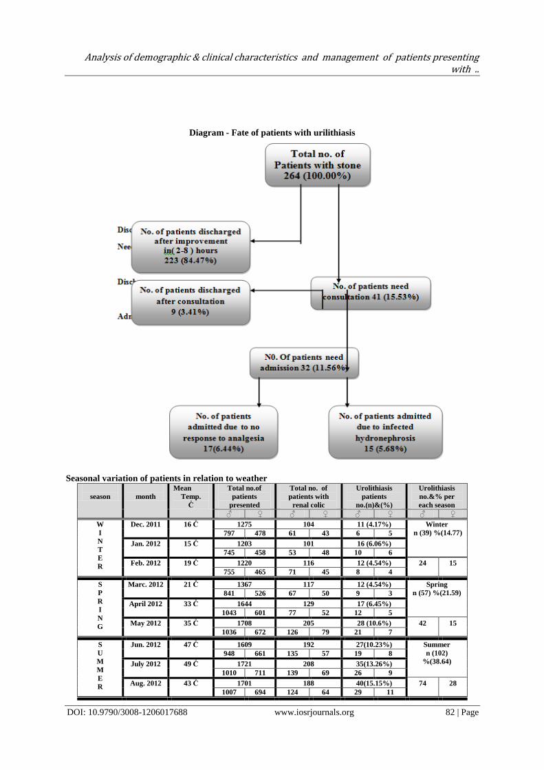
Most patients 223 ( 84.47% ) discharge from the emergency room within (2hours to 8hours), i.e. after
complete resolve of their complains ,but 41 patient (15.53% ) need consultations with other disciplines & 32
patients of them need hospitalization (11.9% of the whole sample) , the admission of these patients was due to :
persistent pain (17 patients) despite analgesic management (6.4%) ; & (15 patients) due to infected
hydronephrosis (5.5% ) .
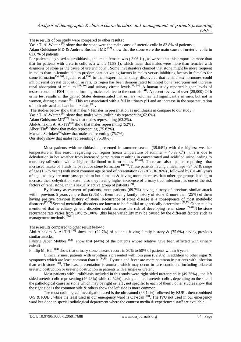
These results were analyzed in relation to weather temperature & season.We saw that the highest no.
of patients presentation was in summer season for total patients & for patients with urolithiasis (38.64%) with
the highest weather temperature all over the year (mean temperature of summer = 46.33 Co) ,while the least no.
of presentation was in winter (14.77%) with the least weather temperature (mean temperature of winter = 16.7).
The total no. of patients not proved as urolithiasis were107 (28.84 %) , the males were 56 (52.34%) &
the females were 51 (47.66%) with male to female ratio (1.2:1) , these different diagnoses & their percentages
are shown in table -11- , from which we can see that musculoskeletal pain is the most common probable
cause(24.3%) as a total for whole patients , for males it is followed by pyelonephritis (23.21%) , but for females,
pyelonephritis is the most common cause (23.53%) followed by gynecological diseases (19.61%).
The total no. of patients per year in relation of gender to assessment Total no.
Assessment
Total n (%) 17941
(100%) Male n (%)
11027 (61.46%) Female n(%) 6914
(38.54%)
P value
Male to
female
ratio
Patients with renal colic 1722 1063 659
0.8101
1.61:1 100% 61.73% 38.27%
Renal colic patients complete their assess.
371 227 144
0.7567
1.58:1 100% 61.19% 38.81%
Patients with diagnosis other than
stone 107 56 51
0.4942
1.10:1 100% 52.34% 47.66%
Patients diagnosed with stone 264 199 65
< 0.0001
3.06:1 100% 75.38% 24.62%
The relation of patients with stone to age period of presentation
Age
13-20 21-30 31-40 41-50 51-60 ≥ 60
Total
Male
n 10 73 57 30 19 10 199
% 5.03% 36.68% 28.64% 15.08% 9.54% 5.03% 100.00%
Female
n 10 23 16 8 5 3 65
% 15.38% 35.38% 24.62% 12.13% 7.69% 4.62% 100.00%
Total
n 20 96 73 38 24 13 264
% 7.58% 36.36% 27.65% 14.39% 9.09% 4.93 100.00%
Chi-squar (X2) = 7.714 df = 5 P = 0.1727
Patients ages range between (15 – 75) with mean age of (34.02) & (SD = ±13.09)
History of patients with urolithiasis
Positive History
Total n (%) 264 (100%) Male n (%)
199 (100%)
Female n (%) 65 (100%)
Previous historyof stone 67 (25.38%)
48 (24.12%)
19 (29.23%)
Previous similar attack within 5 years 184
(69.7%)
138
(69.35%)
46
(70.7%)
Positive family history of stone 58 (21.97%)
45 (22.61%)
13 (20.0%)
1st attack presentation of renal colic pain 76
(28.79%)
55
(72.37%)
21
(27.63%)
Clinical presentation of patients with urolithiasis Clinically Total n (%) 264 Male n (%) 199 Female n (%) 65 P value
Loin pain 218 (82.9%) 167 (84.2%) 51 (78.9%) 0.314
Pain Radiation 76 (28.8%) 31 (40.7%) 45 (59.3%) <0.001
Abdominal cramps 49 (18.7%) 27 (14.0%) 22 (33.3%) <0.001
Back pain 22 (8.5%) 15 (7.9%) 7 (10.5%) 0.4131
Nausea & vomiting 22 (8.5%) 14 (7.3%) 8 (12.3%) 0.1818
Color change of urine 18 (6.6%) 15 (7.9%) 3 (3.5%) 0.4171
Dysuria 16 (5.9%) 11 (5.6%) 5 (7.6%) 0.5254

Analysis of demographic & clinical characteristics and management of patients presenting with ..
DOI: 10.9790/3008-1206017688 www.iosrjournals.org 81 | Page
Fever 15 (5.7%) 10 (5.0%) 5 (7.6%) 0.420
Groin pain 9 (3.4%) 7 (3.4%) 2 (3.5%) 0.865
Anuria 8 (2.9%) 7 (3.4%) 1 (1.7%) 0.491
Side of renal colic presentation Side of renal colic Males n ( )
% ( )
Females n ( )
% ( )
Total n ( )
% ( )
Right – sided
98
(49.25 %) 32
(49.23 %) 130
(49.24 %)
Left – sided
92
(46.23 %) 30
(46.15 %) 122
(46.21 %)
Bilateral
9
(4.52 %) 3
(4.62 %) 12
(4.55 %)
Total n ( )
% ( )
199
(100 %)
65
(100 %)
264
(100 %)
Chi-square ( X2 ) = 0.001 df = 2 P value = 0.9995
Laboratory investigations ordered to patients with urolithiasis
The investigation
Total no. of patients 371
No. of patients have investigation Their %
General urine examination 291 (78.17%)
Complete Blood Count 201 (54.17%)
Other Biochemical Blood Tests 86 (23.18%)
Radiological investigations ordered to patients with urolithiasis
The investigation
Total no. of patients 371
No. of patients have investigation Their %
Ultrasound 327 (88.14%)
X-ray ( KUB ) 69 (18.6%)
U/S & KUB 60 (18.35%)
CT-scan 38 (10.24%)
Results of GUE & CBC Constituents No. of Patients Their %
Crystals of GUE 241 91.29%
RBC of GUE 206 78.03%
Pus ≥ (+) of GUE 154 58.33%
WBC of CBC <10.000 196 74.24%
Site of the stone in relation to hydronephrosis
Pathology
No. of patients & % Site of stone
Renal Ureteric
Stone with hydronephrosis &/or hydroureter 189
(71.59 %) 138 51
Stone with out hydronephrosis or hydroureter 75
(28.41 %)
58 17
Total 264
(100.00 %) 196
(74.24 %) 68
(25.76 %)
Response to analgesia & recurrence after response
Drug (analgesic)
Used
Total
N ( 247 )
% ( )
Response to analgesia
Recurrence of pain
in 8 hour Pain start to resolve
in≤30minutes
Pain start to resolve
in>30minutes
Diclofenac 102 41 (40.2%) 61 (59.8%) 5 (4.9%)
(41.3%)
Tramadol 94 79 (84.1%) 15 (15.9%) 9 (9.6%)
(38.1%)
Tramadol & diclofenac 51 48 (94.2%) 3 (5.8%) ---
(20.6%)
Analysis of demographic & clinical characteristics and management of patients presenting with ..
DOI: 10.9790/3008-1206017688 www.iosrjournals.org 82 | Page
Diagram - Fate of patients with urilithiasis
Seasonal variation of patients in relation to weather
season
month
Mean
Temp.
C̊
Total no.of
patients
presented
Total no. of
patients with
renal colic
Urolithiasis
patients
no.(n)&(%)
Urolithiasis
no.&% per
each season
♂ ♀ ♂ ♀ ♂ ♀ ♂ ♀
W
I
N
T
E
R
Dec. 2011 16 C̊ 1275 104 11 (4.17%) Winter
n (39) %(14.77) 797 478 61 43 6 5
Jan. 2012 15 C̊ 1203 101 16 (6.06%)
745 458 53 48 10 6
Feb. 2012 19 C̊ 1220 116 12 (4.54%) 24 15
755 465 71 45 8 4
S
P
R
I
N
G
Marc. 2012 21 C̊ 1367 117 12 (4.54%) Spring
n (57) %(21.59) 841 526 67 50 9 3
April 2012 33 C̊ 1644 129 17 (6.45%)
1043 601 77 52 12 5
May 2012 35 C̊ 1708 205 28 (10.6%) 42 15
1036 672 126 79 21 7
S
U
M
M
E
R
Jun. 2012 47 C̊ 1609 192 27(10.23%) Summer
n (102)
%(38.64) 948 661 135 57 19 8
July 2012 49 C̊ 1721 208 35(13.26%)
1010 711 139 69 26 9
Aug. 2012 43 C̊ 1701 188 40(15.15%) 74 28
1007 694 124 64 29 11

Analysis of demographic & clinical characteristics and management of patients presenting with ..
DOI: 10.9790/3008-1206017688 www.iosrjournals.org 83 | Page
A
U
T
U
M
N
Sep. 2012 39 C̊ 1677 137 31(11.74%) Autumn
n (66) %(25.0) 1046 631 83 54 23 8
Oct. 2012 37 C̊ 1510 128 21(7.95%)
943 567 70 58 15 6
Nov. 2012 23 C̊ 1306 97 14(5.31%) 47 19
856 450 57 40 9 5
Total no. 17941 1722 264 Each total no.
represent 100% Male female n
%
11027
61.46
6941
38.54
1063
61.73
659
38.27
199
75.38
65
24.62
Chi-squar (X2) = 9.21 df = 3 p = 0.0266
Figure showing the percentages of patients presented in relation to season
Patients with diagnoses not proved as urolithiasis
Diagnosis
Males
n & (%)
Females
n & (%)
Total
n & (%)
Musculoskeletal pain 17 (30.36%) 9 (17.65%) 26 (24.3%)
Pyelonephritis 13 (23.21%) 12 (23.53%) 25 (23.4%)
Bowel disease 6 (10.71%) 7 (13.73%) 13 (12.15%)
Appendicitis 4 (7.14%) 4 (7.84%) 8 (7.48%)
Cholecystitis 3 (5.36%) 3 (5.89%) 6 (5.61%)
Pancreatitis 2 (3.57%) 1 (1.96%) 3 (2.8%)
Gynecological diseases - 10 (19.61%) 10 (9.35%)
Tumor / Abscess 4 (7.14%) 2 (3.92%) 6 (5.61%)
Congenital diseases 3 (5.36%) 1 (1.96%) 4 (3.74%)
Pneumonia 2 (3.57%) 2 (3.92%) 4 (3.74%)
Zoster infection 2 (3.57%) - 2 (1.87%)
Total 56 (52.34%) 51 (47.66%) 107 (100%)
Chi-squar (X2)= 68.49 df = 9 P = < 0.0001
IV. Discussion The history, clinical examination & investigations show that 264 of 371 (71.16%) patients with
ureteric colic presented to the surgical emergency department diagnosed as urolithiasis , so the most common
cause of these patients with ureteric colic is stone,renal &/or ureteral .Urolithiasis affects 5-15% of the
population worldwide [45]
& there are increase in urolithiasis cases which may be attributed to societal and
lifestyle factors, including equality in the workforce; increased athletic activity with the risk of dehydration;
dietary changes such as high-fat, high-protein diets, increased intake of soft drinks, and increased sodium intake
from processed, preserved, and fast food consumption; and increasing rates of obesity and metabolic syndrome [46-48]
.Other researchers suggest environmental and geographic factors, theorizing that global warming and
higher population density in hot, sunny, arid areas account for the increase in incidence [49,50]
.
0
5
10
15
20
25
30
35
40
45
50
Winter 14.77 Spring 21.59 Summer 38.64 Autumn 25
% 0
f p
atie
nts
wit
h u
rolit
hia
sis
% of patients with urolithiasis per season
Analysis of demographic & clinical characteristics and management of patients presenting with ..
DOI: 10.9790/3008-1206017688 www.iosrjournals.org 84 | Page
These results of our study were compared to other results :
Yasir T. Al-Wattar [51]
show that the stone were the main cause of ureteric colic in 83.8% of patients .
Adam Goldstone MD & Andrew Bushnell MD [52]
show that the stone were the main cause of ureteric colic in
63.6 % of patients .
For patients diagnosed as urolithiasis , the male:female was ( 3.06:1 ) , as we see that this proportion more than
that for patients with ureteric colic as a whole (1.58:1), which mean that males were more than females with
diagnosis of stone as the cause of ureteric colic . Some investigators claimed that stone might be more frequent
in males than in females due to predominant activating factors in males versus inhibiting factors in females for
stone formation[54, 55]
. Iguchi et al.[56]
, in their experimental study, discovered that female sex hormones could
inhibit renal crystal deposition in rats. Estrogen has been demonstrated to inhibit bone resorption and increase
renal absorption of calcium [59, 60]
and urinary citrate levels[57, 58]
. A human study reported higher levels of
testosterone and FSH in stone forming males relative to the controls [61]
. A recent review of over (28,000) 24 h
urine test results in the United States demonstrated that urinary volumes fall significantly in men, but not in
women, during summer [62]
. This was associated with a fall in urinary pH and an increase in the supersaturation
of both uric acid and calcium oxalate [62]
.
The studies below show that males > females in presentation as urolithiasis in compare to our study :
Yasir T. Al-Wattar [51]
show that males with urolithiasis representing(62.6%).
Adam Goldstone MD[52]
show that males representing (63.3%).
Abd-Alhakim A. Al-Ta'i[53]
show that males representing (52%) .
Albert Tiu[63]
show that males representing (75.82%).
Mustafa Serinken[64]
show that males representing (75.7%).
Our study show that males representing ( 75.38%) .
Most patients with urolithiasis presented in summer season (38.64%) with the highest weather
temperature in this season regarding our region (mean temperature of summer = 46.33 Co) , this is due to
dehydration in hot weather from increased perspiration resulting in concentrated and acidified urine leading to
more crystallization with a higher likelihood to form stones [65-67]
. There are also papers reporting that
increased intake of fluids helps reduce stone formation [68-70]
.These patients having a mean age =34.02 & range
of age (15-75 years) with most common age period of presentation (21-30) (36.36%) , followed by (31-40) years
of age , as they are more susceptible to hot climates & having more exercises than other age groups leading to
increase their dehydration state , also they having higher incidence of urinary tract infection , as one of the risk
factors of renal stone, in this sexually active group of patients [71]
.
By history assessment of patients, most patients (69.7%) having history of previous similar attack
within previous 5 years , more than (20%) of them having family history of stone & more than (25%) of them
having positive previous history of stone .Recurrence of stone disease is a consequence of most metabolic
disorders[73,74]
.Several metabolic disorders are known to be familial or genetically determined[73,75]
.Other studies
mentioned that hereditary genetic disorder could increase the risk of developing renal stone [76-78]
.The stone
recurrence rate varies from 10% to 100% ,this large variability may be caused by the different factors such as
management methods [79-81]
.
These results compared to other result below :
Abd-Alhakim A. Al-Ta'I [53]
show that (22.7%) of patients having family history & (75.6%) having previous
similar attacks.
Fakhria Jaber Muhbes [82]
show that (44%) of the patients whose relative have been afflicted with urinary
calculi.
Phillip M. Hall [83]
show that urinary stone disease recurs in 30% to 50% of patients within 5 years.
Clinically most patients with urolithiasis presented with loin pain (82.9%) in addition to other signs &
symptoms which are least common than it [84,85]
. Dysuria and fever are more common in patients with infection
than with stone [86]
. The least presentation is anuria , which may occur in rare conditions including bilateral
ureteric obstruction or ureteric obstruction in patients with a single & ureter .
Most patients with urolithiasis included in this study were right sided ureteric colic (49.25%) , the left
sided ureteric colic representing (46.23%) while (4.52%) having bilateral ureteric colic , depending on the site of
the pathological cause as stone which may be right or left , not specific to each of them , other studies show that
the right side is the common side & others show the left side is more common .
The most radiological investigation used is the ultrasound (88.14%) followed by KUB , then combined
U/S & KUB , while the least used in our emergency ward is CT-scan [89]
. The IVU not used in our emergency
ward but done in special radiological department where the contrast media & experienced staff are available .

Analysis of demographic & clinical characteristics and management of patients presenting with ..
DOI: 10.9790/3008-1206017688 www.iosrjournals.org 85 | Page
Cupisti et al. [90]
reported that ultrasound is the first line diagnostic study in patients suspected to have
stone disease and ureteric colic and have not recommended tomography , in spite of its high sensitivity &
specificity , as the first choice as for financial reasons. On the other hand, Pfister et al. [91]
advocated that
tomography had a better economic outcome in patients with ureteric colic since it has high diagnostic accuracy,
effectiveness, speed , lower risk , and slightly more costs when compared to intravenous urography.Another
study recommended unenhanced spiral computed tomography in the initial radiological evaluation of stone
disease and ureteric colic for its high diagnostic accuracy[92,93]
.
Other laboratory investigations used were mainly GUE & CBC .GUE showing that most patients with
urolithiasis having crystals (91.29%) , RBC (78.3%) ,& Pus ≥ (+) (58.33%)..The urinalysis is the investigation
most commonly ordered in patients with presumptive diagnosis of ureteric colic and reveals hematuria in 90%
of patients with stones [95]
. One-tenth of patients with ureteric colic would not exhibit hematuria [96]
. One
retrospective study found that 67% of patients with ureterolithiasis had more than (5) RBC per HPF and (89%)
of patients had more than zero RBC/HPF on microscopic examination of the urine [66]
.
The CBC show that (74.24%) of patients having WBC <10.000 .Other laboratory investigations (as
blood urea & creatinine) are usually used for old patients & patients with comorbid diseases with suspected
renal impairments for the assessment of their renal function(94)
.
In our study , most patients discharged after receiving treatment & resolve of their complain usually in
(2-8) hours with advise to be followed by a urosurgery outpatient clinic. Patients with urolithiasis may need
admission as in patients with no response to treatment or infected hydronephrosis.
Providing relief from the debilitating pain caused by obstructing calculi is a therapeutic challenge.
Singh et al [97]
reported that the medical therapy of acute renal colic should be started with NSAIDs. They also
claimed that supplementary agents should be added only if excruciating pain is persistent [97]
. In our study ,most
analgesic drug used is the diclofenac used in (41.3%) of patients by intramuscular injection of (3ml/75 mg)
while other analgesic drug used is tramadol used in (38.1%) of patients by intramuscular or intravenous
injection of (2ml/100mg) . The results show that the best treatment used is the combination of (diclofenac &
tramadol) with rapid response & less frequency of recurrence .
Mustafa Serinken [64]
show that NSAIDs used in (86.4%) of patients , while opiates used in
(32.3%).Tunji A Lasoye [98]
show that NSAIDs used in (85.8%) of patients , while opiates used in (9.4%).
V. Conclusions The most common clinical presentation is the loin pain .
The most common cause of ureteric colic is the stone (urolithiasis) may be renal &/or ureteral.
The males more affected than females.
The middle age patients (20th
& 30th
) are the most common patients presented with urolithiasis.
The weather temperature affects the incidence of urolithiasis ,increasing weather temperature associated
with increasing the incidence .
The history & family history should be assessed by the emergency physician for patients with urolithiasis as
> 1/5 of patients having positive family history.
The U/S is the most used & acceptable investigation used in our emergency ward for ureteric colic patients.
The best treatment used for relieving the complain of patients with ureteric colic is the combination of
centrally acting analgesic drugs & NSAID.
References [1] Haddad MC, Sharif HS , Shahed MS , et al . Renal Colic : diagnosis & outcome . Radiology 1992;184:83-88 .
[2] Labrecque M, Dostaler L, Rousselle R, et al: Efficacy of nonsteroidal anti-inflammatory drugs in the treatment of acute renal colic.
Arch Intern Med 1994;154:1381–1387. [3] Dalrymple NC, Verga M, Anderson KR, et al: The value of unenhanced helical computerized tomography in the management of
acute flank pain. J Urol 1998; 159:735–740.
[4] Laerum E: Urolithiasis in general practice: an epidemiological study from a Norwegian district. Scand J Urol Nephrol 1983; 17: 313–319.
[5] Lee YH, Huang WC, Tsai JY, et al . Epidemiological studies on the prevalence ofupper urinary calculi in Taiwan. Urol Int 2002;
68: 172–177. [6] Stamatelou KK, Francis ME, Jones CA, et al . Time trends in reported prevalence of kidney stones in the United States: 1976–1994.
Kidney Int 2003; 63: 1817–1823.
[7] Wickham JEA, Buck AC: Renal Tract Stone: Metabolic Basis and Clinical Practice. NewYork, Churchill Livingstone, 1990. [8] Elliott JP Jr, Gordon JO, Evans JW, Platt L: A stone season. A 10-year retrospective study of 768 surgical stone cases with respect
to seasonal variation. J Urol 1975; 114: 574–577.
[9] Borghi L, Guerra A, Meschi T, et al . Relationship between supersaturation and calcium oxalate crystallization in normals and idiopathic calcium oxalate stone formers. Kidney Int 1999; 55: 1041–1050.
[10] Clark AJ, Norman RW: ‘Mirror pain’ as an unusual presentation of renal colic. Urology 1998;51:116–118.

Analysis of demographic & clinical characteristics and management of patients presenting with ..
DOI: 10.9790/3008-1206017688 www.iosrjournals.org 86 | Page
[11] Moody TE, Vaughan ED Jr, Gillenwater JY: Relationship between renal blood flow and ureteral pressure during 12 hours of total
unilateral ureteral occlusion. Invest Urol 1975;13:246– 251. [12] Lanzone JA, Gulmi FA, Chou SY, et al . Renal hemodynamics in acute unilateral ureteral obstruction: Contribution of endothelium-
derived relaxing factor. J Urol 1995:153:2055–2059.
[13] Klahr S: New insight into the consequences and mechanisms of renal impairment in obstructive nephropathy. Am J Kidney Dis 1991;18:689–699.
[14] de Dombal FT: Acute abdominal pain – An OMGE survey. Scand J Gastroenterol 1979; 14(suppl 56):29–43.
[15] de Dombal FT: The OMGE acute abdominal pain survey. Progress report 1986. Scand J Gastroenterol 1988;23(suppl 144):35–42. [16] Christopher R Chapple : Renal and Urology I : Surgery (Oxford) : (July 2010) :Volume 28, Issue 7 : Pages 307-368 .
[17] Dalla Palma L, Staccul F, Bazzocchi M , et al. (1993) Ultrasonography and plain film versus intravenous urography in ureteric colic
. Clin Radiol 47: 333-336 . [18] Smith RC ,Essen macher KR , Rosen field AT et al .(1995) Acute flank pain : comparison of non-contrast-enhanced CT and
intravenous urography . Radiology 194: 789-794 .
[19] Eskelinen M, Ikonen J, Lipponen P. Usefulness of history-taking, physical examination and diagnostic scoring in acute renal colic. Eur Urol 1998;34:467–473.
[20] Mutagi A, Williams JW, Nettleman M: Renal colic: Utility of the plain abdominal roentgenogram.Arch Intern Med 1991;151:1589–1592.
[21] Platt JF, Rubin JM, Ellis JH: Acute renal obstruction:Evaluation with intra-renal duplex Doppler and conventional US. Radiology
1993;186:685–688. [22] Hill MC, Rich JL, Mardiat JG, et al: Sonography vs. excretory urography in acute flank pain. AJR Am J Roentgen 1985;144:1235–
1238.
[23] Sinclair D, Wilson S, Toi A, et al: The evaluation of suspected renal colic: Ultrasound scan versus excretory urography. Ann Emerg Med 1989;18:556–559.
[24] Platt JF, Rubin JH, Ellis JH, Dipietro MA: Duplex Doppler US of the kidney: Differentiation of obstructive from non-obstructive
dilatation. Radiology 1989;17:515–517. [25] Burge HJ, Middleton WD, McClennan BL, Hildebolt CF: Ureteral jets in healthy subjects and in patients with unilateral ureteral
calculi: Comparison with color Doppler US. Radiology 1991;180:437–442.
[26] Smith RC, Rosenfield AT, Choe KA, et al:Acute flank pain: Comparison of non- contrastenhanced CT and intravenous urography. Radiology 1995;194:789–794.
[27] Regan F, Bohlman ME, Khazan R, et al: MR urography using HASTE imaging in the assessment of ureteric obstruction. AJR Am J
Roentgen 1996;167:115–120. [28] Gutman M, Braf Z, Kaver I, et al: The role of the radionuclide renal study in the management of renal colic. Br J Urol 1993;71:530–
534.
[29] Preminger GM, Tiselius HG, Assimos DG et al .Guideline for the management of ureteral calculi. American Urological Association Education and Research, Inc; European Association of Urology. Eur Urol . 2007 ; 52:1610–1631
[30] Autorino R, Osorio L, Lima EA. Rapid extracorporeal shock wave lithotripsy for proximal ureteral calculi in colic versus noncolic
patients. Eur Urol 2007;52:1264-5. [31] Lotan Y, Gettman MT, Roehrborn CG, et al. Management of ureteral calculi: A cost comparison and decision making analysis. J
Urol 2002;167:1621-9.
[32] Miller OF, Kane CJ. Time to stone passage for observed ureteral calculi: A guide for patient education. J Urol 1999;162:688-91. 2013
[33] Cooper JT, Stack GM, Cooper TP. Intensive medical management of ureteral calculi. Urology 2000;56:575-8.
[34] Francesca F, Bader P, Echtle D, et al . EAU guidelines on pain management. Eur Urol 2003;44:383-9. [35] Worster A, Richards C. Fluids and diuretics for acute ureteric colic. Cochr Data Syst Rev 2005;3:CD004926.
[36] Hollingsworth JM, Rogers MA, Kaufman SR, et al. Medical therapy to facilitate urinary stone passage: A meta-analysis. Lancet
2006;368:1171-9. [37] Davenport K, Timoney AG, Keeley FX. A comparative in vitro study to determine the beneficial effect of calcium-channel and
alpha1-adrenoceptor antagosim on human ureteric activity. BJU Int 2006;98:651-5.
[38] Troxel SA, Jones AW, Magliola L, Benson JS. Physiologic effect of nifedipine and tamsulosin on contractility of distal ureter. J
Endourol 2006;20:565-8.
[39] Singh A, Alter HJ, Littlepage A. A systematic review of medical therapy to facilitate passage of ureteral calculi. Ann Emerg Med
2007;50:552-67. [40] Sigala S, Dellabella M, Milanese G, et al. Evidence for the presence of alpha1 adrenoceptor subtypes in the human ureter.
Neurourol Urodyn 2005;24:142-8.
[41] Joshi HB, Obadeyi OO, Rao PN. A comparative analysis of nephrostomy, JJ stent and urgent in situ extracorporeal shock wave lithotripsy for obstructing ureteric stones. BJU Int 1999;84:264-9.
[42] Preminger GM, Kettelhut MC, Elkins SL, Seger J, Fetner CD. Ureteral stenting during extracorporeal shockwave lithotripsy: Help
or hindrance. J Urol 1989;142:32-6. [43] Doublet JD, Tchala K, Tligui M, et al . In situ extracorporeal shock wave lithotripsy for acute renal colic due to obstructing ureteral
stones. Scan J Urol Nephrol 1997;31:137-9.
[44] Anagnostou T, Tolley D. Management of ureteric stones. Eur Urol 2004;45:714-21. [45] Moe OW. Kidney stones: pathophysiology and medical management. Lancet 2006;367:333-44.
[46] Scales CD Jr, Curtis LH, Norris RD, et al. Changing gender prevalence of stone disease. J Urol. 2007;177(3):979-982.
[47] Del Valle EE, Negri AL, Spivacow FR, et al . Metabolic diagnosis in stone formers in relation to body mass index. Urol Res. June 10, 2011.
[48] Siener R. Impact of dietary habits on stone incidence.Urol Res. 2006;34(2):131-133.
[49] Soucie JM, Coates RJ, McClellan W, et al . Relation between geographic variability in kidney stones prevalence and risk factors for stones. Am J Epidemiol. 1996;143(5):487-495.
[50] Fakheri RJ, Goldfarb DS. Ambient temperature as a contributor to kidney stone formation: implications of global warming. Kidney Int. 2011;79(11):1178-1185.
[51] Yasir T. Al-Wattar.The validity of U/S and KUB in detecting urinary stones in patients with renal colic. A Dissertation submitted to
the Scientific Council of Urology in partial fulfillment for the degree of Fellowship of the Iraqi Commission For Medical Specialization In Urology (1998).

Analysis of demographic & clinical characteristics and management of patients presenting with ..
DOI: 10.9790/3008-1206017688 www.iosrjournals.org 87 | Page
[52] Adam Goldstone MD, Andrew Bushnell MD. Does diagnosis change as a result of repeat renal colic computed tomography scan in
patients with a history of kidney stones?. American Journal of Emergency Medicine (2010) 28, 291–295. [53] Abd Al-Hakim A. Al-Ta'I .Renal Colic A retrospective study . A Dissertation submitted to the Scientific Council of Urology in
partial fulfillment for the degree of Fellowship of the Iraqi Commission For Medical Specialization In Urology (2005).
[54] Sun BY, Lee YH, Jiaan BP et al. Recurrence rate and risk factors for urinary calculi after extracorporeal shock wave lithotripsy. J Urol 1996; 156(3): 903–905.
[55] Curhan GC, Willett WC, Speizer FE, Stampfer MJ .Twenty-four-hour urine chemistries and the risk of kidney stones among
women and men. Kidney Int 2001; 59(6):2290–2298. [56] Iguchi M, Takamura C, Umekawa T et al. Inhibitory effects of female sex hormones on urinary stone formation in rats. Kidney Int
1999; 56(2): 479–485
[57] Hammar ML, Berg GE, Larsson L, et al . Endocrine changes and urinary citrate excretion. Scand J Urol Nephrol (1987) 21:51–53 [58] Dey J, Creighton A, Lindberg JS, et al . Estrogen replacement increased the citrate and calcium excretion rates in postmenopausal
women with recurrent urolithiasis. J Urol (2002) 167:169–171
[59] McKane WR, Khosla S, Burritt MF, et al . Mechanism of renal calcium conservation with estrogen replacement therapy in women in early post- menopause a clinical research center study. J Clin Endocrinol Metab (1995) 80:3458–3464
[60] Nordin BE, Need AG, Morris HA, Horowitz M . Biochemical variables in pre- and postmenopausal women: reconciling the calcium and estrogen hypotheses. Osteoporos Int (1999) 9:351–357
[61] Kuczera M, Kiersztejn M, Kokot F, Klin M . Behavior of sex hormone and gonadotropin secretion in men with active
nephrolithiasis. Endokrynol Pol (1993) 44:539–547 [62] Parks JH, Barsky R, Coe FL. Gender differences in seasonal variation of urine stone risk factors. J. Urol. 2003; 170: 384–8.
[63] Albert Tiu, Vincent Tang, Samuel Gubicak,et al. Seasonal Variation of Acute Urolithiasis at an Australian Tertiary Hospital.
Australasian Medical Journal AMJ 2010, 3, 13, 851-854. [64] Mustafa Serinken, Ozgur Karcioglu, Ibrahim Turkcuer, et al.Analysis of clinical and emographic characteristics of patients
presenting with renal colic in the emergency department.BMC Research Notes 2008.
[65] Manthey DE, Teichman J: Nephrolithiasis. Emerg Med Clin North Am 2001, 19 (3) : 633-54. [66] Bove P, Kaplan D, Dalrymple N et al .Reexamining the value of hematuria testing in patients with acute flank pain. J Urol 1999,
162(3 Pt 1):685-7.
[67] Stoller ML, Bolton DM: Urinary stone disease. In Smith's General Urology 14th edition. Edited by: Tanagho EA, McAnninch JW. Connecticut, Appletton and Lange; 1995:276-304.
[68] Jenkins AD: Calculus formation. In Adult and Pediatric Urology 3rd edition. Edited by: Gillenwater JY, Grayback JT, Howards SS,
Duckett JW. Mosby Year Book, St. Louis; 1996:461-505. [69] Finlayson B, Smith A: Stability of first dissociable proton of uric acid. J Chem Eng Data 1974, 19:94-97.
[70] Ngo TC, Assimos DG: Uric Acid nephrolithiasis: recent progress and future directions. Rev Urol 2007, 9(1):17-27.
[71] Mohammad Mehdi Hosseini , Ahad Eshraghian , Ilnaz Dehghanian et al . Metabolic abnormalities in patients with nephrolithiasis . Int Urol Nephrol (2010) 42:127–131
[72] Erwin Bc.Corrol , BA. Sammer .Renal Colic :The role of U/S in initial evaluation. Radiology . 1984 ; July , 152 (1) , 147 – 150 .
[73] Barratt TM, Duffy PG .Nephrocalcinosis and urolithiasis. In: Barratt TM, Avner ED, Harmon WE (eds) Pediatric nephrology, 4th edn. Lippincott Williams and Wilkins, Baltimore ; 1999 : pp 933–945
[74] Milliner DS, Murphy ME Urolithiasis in pediatric patients. Mayo Clin Proc. 1993 ; 68: 241–248
[75] Polinsky MS, Kaiser BA, Baluarte HJ .Urolithiasis in childhood. In: Gruskin AB (ed) The pediatric clinics of North America (pediatric nephrology). Saunders, Philadelphia, 1987 ; 34(3):683–710
[76] Devuyst O.Pirson Y. Genetics of hypercalciuric stone forming diseases. Kidney International .2007; 72 (9): 1065-72.
[77] Prié D, Friedlander G. Genetic Causes of Renal Lithiasis.International Bone & Mineral Society (IBMS). 2009; 357-67. [78] Chris. Kidney stone causes and Risk Factors. Home /Current Health Articles. www health centre .com 2010.
[79] Trinchieri A, Ostini F, Nespoli R et al. A prospective study of recurrence rate and risk factors for recurrence after a first renal stone.
J Urol 1999; 162(1): 27–30. [80] Balaji KC, Menon M. Mechanism of Stone Formation . Urol Clin North Am 1997; 24(1): 1–11.
[81] Begun FP, Foley WD, Peterson A, White B. Patient evaluation: Laboratory and imaging studies. Urol Clin North Am 1997; 24(1):
97–116.
[82] Fakhria Jaber Muhbes . Risk factors for renal stone formation: A field study. HEALTH SCIENCE JOURNAL Volume 6, Issue 4
(October – December 2012) ; P 718
[83] Phillip M. Hall, MD .Nephrolithiasis :Treatment, causes, and prevention. CLEVELAND CLINIC JOURNAL OF MEDICINE VOLUME 76 . NUMBER 10 OCTOBER 2009 : P 585
[84] F. Travaglini R. Bartoletti M. Gacci M. Rizzo . Pathophysiology of Reno-Ureteral Colic. Urol Int 2004;72(suppl 1):20–23
[85] Schulman CC: Innervation of the ureter: A histochemical and ultrastructural study; in Lutzeyer W, Hannappel J (eds): Urodynamics II. Upper and Lower Urinary Tract. Berlin, Springer, 1985, pp 292–316.
[86] de Dombal FT: Diagnosis of acute Abdominal Pain. Edinburgh, Churchill Livingstone, 1991.
[87] Jeremy Brown. Diagnostic and treatment patterns for renal colic in US Emergency Departments. International Urology and Nephrology (2006) 38:87–92.
[88] Webb JAW. Ultrasonography in the diagnosis of renal obstruction. Br. Med. J. 1990; 301: 944.
[89] Yaser Abu-Gazzeh and Salah Abdu-Alro'f. The role of ultrasound in the initial evaluation of renal colic. Saudi J Kidney Dis Transplant 2000; 11 (2) : 186 – 190 .
[90] Cupisti A, Pasquali E, Lusso S, et al .Renal colic in Pisa emergency department: epidemiology, diagnostics and treatment patterns.
Intern Emerg Med .2008 ; 3:241–244 [91] Pfister SA, Deckart A, Laschke S, et al .Unenhanced helical computed tomography vs intravenous urography in patients with acute
flank pain: accuracy and economic impact in a randomized prospective trial. Eur Radiol .2003 ; 13:2513–2520
[92] Greenwell TJ, Woodhams S, Denton ERM, et al .One year’s clinical experience with unenhanced spiral computed tomography for the assessment of acute loin pain suggestive of renal colic. BJU Int .2000 ; 85:632–636
[93] Kobayashi T, Nishizawa K, Watanabe J, et al .Clinical characteristics of ureteral calculi detected by non-enhanced computerized tomography after unclear results of plain radiographyand ultrasonography. J Urol . 2003 ; 170:799–802
[94] S. Yilmaz, et al . Renal colic: comparison of spiral CT, US and IVU in the detection of ureteral calculi. Eur. Radiol. (1998) 8, 212-
217 . [95] Manthey DE, Teichman J: Nephrolithiasis. Emerg Med Clin North Am 2001, 19 (3) : 633-54.

Analysis of demographic & clinical characteristics and management of patients presenting with ..
DOI: 10.9790/3008-1206017688 www.iosrjournals.org 88 | Page
[96] Eskelinen M, Ikonen J, Lipponen P. Usefulness of history-taking, physical examination and diagnostic scoring in acute renal colic.
Eur Urol 1998;34:467-73. [97] Singh SK, Agarwal MM, Sharma S. Medical therapy for calculus disease. BJU Int 2011;107:356–68.
[98] Tunji A Lasoye, Philip M Sedgwick, Nilay Patel,et al. Management of acute renal colic in the UK: a questionnaire survey. BMC
Emergency Medicine 2004.
Dr. Layth Salim Hadi Analysis of demographic & clinical characteristics and management of
patients presenting with ureteric colic in the emergency department.” IOSR Journal of
Pharmacy and Biological Sciences (IOSR-JPBS) , vol. 12, no. 6, 2017, pp. 76-88.