analysis of dna methylation acquisition at the imprinted dlk1 locus reveals asymmetry at cpg dyads
TRANSCRIPT
RESEARCH Open Access
Analysis of DNA methylation acquisition at theimprinted Dlk1 locus reveals asymmetry at CpGdyadsAlyssa Gagne†, Abigail Hochman†, Mahvish Qureshi, Celia Tong, Jessica Arbon, Kayla McDanieland Tamara L Davis*
Abstract
Background: Differential distribution of DNA methylation on the parental alleles of imprinted genes distinguishesthe alleles from each other and dictates their parent of origin-specific expression patterns. While differential DNAmethylation at primary imprinting control regions is inherited via the gametes, additional allele-specific DNAmethylation is acquired at secondary sites during embryonic development and plays a role in the maintenance ofgenomic imprinting. The precise mechanisms by which this somatic DNA methylation is established at secondarysites are not well defined and may vary as methylation acquisition at these sites occurs at different times for genesin different imprinting clusters.
Results: In this study, we show that there is also variability in the timing of somatic DNA methylation acquisition atmultiple sites within a single imprinting cluster. Paternal allele-specific DNA methylation is initially acquired at similarstages of post-implantation development at the linked Dlk1 and Gtl2 differentially methylated regions (DMRs). Incontrast, unlike the Gtl2-DMR, the maternal Dlk1-DMR acquires DNA methylation in adult tissues.
Conclusions: These data suggest that the acquisition of DNA methylation across the Dlk1/Gtl2 imprinting cluster isvariable. We further found that the Dlk1 differentially methylated region displays low DNA methylation fidelity, asevidenced by the presence of hemimethylation at approximately one-third of the methylated CpG dyads. Wehypothesize that the maintenance of DNA methylation may be less efficient at secondary differentially methylatedsites than at primary imprinting control regions.
Keywords: Genomic imprinting, DNA methylation, Dlk1, Secondary DMR, Epigenetics
BackgroundGenomic imprinting in mammals results in the monoallelicexpression of approximately 150 genes [1,2]. The majorityof these imprinted genes are found in clusters distributedthroughout the mammalian genome, with each clustercontaining two or more imprinted genes as well as animprinting control region (ICR) [3]. One common featureof the CpG-rich ICRs is the presence of a gametic, orprimary, differentially methylated region (DMR) whichgenerally functions both to identify parental origin and toregulate expression of the imprinted genes within the
cluster, either directly or indirectly [3]. Establishment ofparent of origin-specific DNA methylation at the ICRoccurs during gametogenesis and the zygote eitherinherits a methylated allele from its mother or from itsfather at fertilization. Differential methylation at the ICRis then maintained throughout development such that theparental alleles can be distinguished from each other andthe expression of their adjacent imprinted genes regulatedappropriately.In addition to the differential methylation present at
the ICR, some imprinted loci also acquire distinctsecondary regions of differential methylation duringpost-implantation development [4-6]. It has been proposedthat the establishment of differential DNA methylationat secondary DMRs could serve as a mechanism for
* Correspondence: [email protected]†Equal contributorsDepartment of Biology, Bryn Mawr College, 101 N. Merion Avenue, BrynMawr, PA 19010-2899, USA
© 2014 Gagne et al.; licensee BioMed Central Ltd. This is an Open Access article distributed under the terms of the CreativeCommons Attribution License (http://creativecommons.org/licenses/by/4.0), which permits unrestricted use, distribution, andreproduction in any medium, provided the original work is properly credited. The Creative Commons Public DomainDedication waiver (http://creativecommons.org/publicdomain/zero/1.0/) applies to the data made available in this article,unless otherwise stated.
Gagne et al. Epigenetics & Chromatin 2014, 7:9http://www.epigeneticsandchromatin.com/content/7/1/9

maintaining imprinted expression at developmental timeswhen the primary imprinting control region is no longerfunctioning [6,7]. Support for this hypothesis comes froma recent study of DNA methylation and expression at theimprinted Gpr1-Zdbf2 locus at which the maternallymethylated Gpr1 DMR functions as the gametic imprintingmark responsible for establishing paternal allele-specificexpression while paternal allele-specific DNA methylationat the secondary Zdbf2 DMR is established after the onsetof imprinted Zdbf2 expression [8]. Paternal allele-specificexpression of Zdbf2 is maintained after DNA methylationat the Gpr1 DMR becomes biallelic, suggesting that thepaternally methylated secondary Zdbf2 DMR functions tomaintain monoallelic expression at this locus. Furthermore,biallelic methylation at the Zdbf2 DMR in offspring derivedfrom Dnmt3Lmat−/− mothers correlated with biallelicexpression of Zdbf2. While the exact mechanism respon-sible for the parental allele-specific acquisition of DNAmethylation at secondary DMRs has not yet been deter-mined, it is clear that there is a relationship between theepigenetic states at primary and secondary DMRs [9,10].The majority of secondary DMRs found at imprinted
genes are methylated on the paternally-inherited allele,suggesting that there may be a common mechanismresponsible for establishing secondary imprinting marks.At the same time, it is clear that not all secondary DMRsare acquired at the same developmental stage. Paternalallele-specific DNA methylation is established at Gtl2 priorto 6.5 days post coitum (d.p.c.), at Cdkn1c between 7.5 and9.5 d.p.c. and at Igf2r region 1 during late embryogenesis[7,11-13]. Gtl2, Cdkn1c, and Igf2r are located on mousechromosomes 12, 7, and 17, respectively. DNA methylationat secondary DMRs has generally been shown to affect theexpression of a single adjacent imprinted gene, ratherthan the expression of the entire imprinting cluster [6,7].Therefore, it is possible that the same molecular machineryis used to establish DNA methylation at these sites andthat the difference in temporal acquisition reflects thetime at which it becomes critical to maintain monoallelicexpression for each imprinted gene.The Dlk1-Dio3 cluster of imprinted genes spans 1 Mb
on mouse chromosome 12 and contains three paternallyexpressed protein-coding genes (Dlk1, Rtl1, and Dio3),multiple maternally expressed untranslated RNAs(including Gtl2), and at least three DMRs that aremethylated on the paternal allele [14-18]. The IG-DMR,located between Dlk1 and Gtl2, functions as the ICRon the unmethylated maternally inherited allele [19].Secondary DMRs have been identified at the promoter ofGtl2 and in exon 5 of Dlk1 [5]. Evidence suggests that theGtl2-DMR has a functional role; studies of the mouseGtl2-DMR and its human homolog, MEG3-DMR, indicatethat methylation of this region directly influences expres-sion in cis [10,20,21]. Although the functional role of
differential methylation at Dlk1 has not been determined,both the Gtl2- and Dlk1-DMRs become methylated on thepaternal allele following fertilization, and the Gtl2-DMRhas been shown to acquire paternal allele-specificmethylation during early post-implantation development,between embryonic days 3.5 and 6.5 [5,11]. Since these twoDMRs are located within the same imprinting cluster, wehypothesized that the acquisition of paternal allele-specificDNA methylation at these secondary DMRs would becoordinately controlled. We tested this hypothesis by exam-ining the methylation status of the Dlk1-DMR throughoutdevelopment. We found that the Dlk1-DMR acquirespaternal allele-specific methylation during embryogenesisand that the methylation pattern remains dynamic in lateembryonic development and into adulthood. Furthermore,our analysis of DNA methylation on the complementarystrands of the Dlk1-DMR illustrates the unexpectedly fluidnature of DNA methylation at this locus.
ResultsThe Dlk1-DMR acquires paternal allele-specific DNAmethylation during post-fertilization developmentPrevious research illustrated that somatic mouse tissuesexhibit paternal allele-specific DNA methylation at theDlk1-DMR that is acquired after fertilization [5,14,15].To elucidate the temporal acquisition of paternal allele-specific DNA methylation at the Dlk1-DMR followingfertilization, we assessed the DNA methylation status onboth the paternal and maternal Dlk1 alleles at variousstages of mouse development.All of our experiments were conducted using F1 hybrid
tissues collected from crosses between C57BL/6 (B6)and a specially derived strain containing Mus musculuscastaneus-derived sequences from chromosome 12 on anotherwise C57BL/6 genetic background (CAST12) [11].We identified a single nucleotide polymorphism betweenthe B6 and CAST12 strains in a 386 bp CpG island locatedat the 5′ end of Dlk1 exon 5 (http://www.ebi.ac.uk/Tools/emboss/cpgplot/index.html) [11]. The identified SNP was aC-to-T transition at base pair position 109,459,746(GenBank: NC_000078.6), preventing us from definitivelyassigning parental origin following bisulfite mutagenesisand sequencing of the top strand of DNA, since unmethy-lated cytosines would ultimately be replaced by thymines.Therefore, we modified our approach by covalently attach-ing the top and bottom strands via a hairpin linker, whichallowed us to identify parental origin based on the G-to-Atransition on the bottom strand (Figure 1D; see Methods).This approach had the additional advantage of yieldingDNA methylation data for complementary CpG dinucleo-tides, allowing us to determine the level of homo- versushemimethylation within this region. We used thisapproach to analyze the methylation status of 16 of the 29CpGs located within the Dlk1 CpG island (Figure 1C).
Gagne et al. Epigenetics & Chromatin 2014, 7:9 Page 2 of 13http://www.epigeneticsandchromatin.com/content/7/1/9
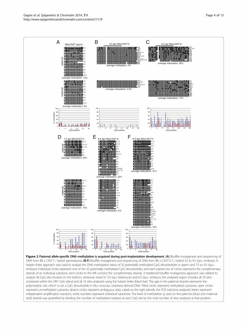
We confirmed that adult sperm DNA contains verylow levels of DNA methylation at the Dlk1-DMR(Figure 2A). Therefore, any paternal allele-specificmethylation observed in somatic tissues must be acquiredduring post-fertilization development. To determine whenDNA methylation is acquired at the Dlk1-DMR, weanalyzed the methylation status at the Dlk1-DMRduring early embryonic development. We were unableto scale down the hairpin linker approach for usewith the limited amount of material collected from3.5 d.p.c. blastocysts and 6.5 d.p.c. embryos. Therefore, forthese developmental stages we utilized a traditionalbisulfite mutagenesis approach to analyze the DNAmethylation status at 36 CpG sites, including all 29sites contained within the Dlk1 CpG island and all 16sites analyzed using the hairpin linker employed foranalysis of DNA derived from older embryonic, neonatal,and adult tissue (Figure 1). We observed an absence ofDNA methylation on both the paternal and maternalalleles in 3.5 d.p.c. blastocysts, indicating that the paternalDlk1 allele does not acquire methylation during pre-implantation development (Figure 2B). By 6.5 d.p.c.,the paternal Dlk1 allele has acquired DNA methylation(Figure 2C). We assessed the significance of these resultsusing a Mann–Whitney U test and found that there was astatistically significant difference in the median levelof DNA methylation on the paternal alleles of 3.5 vs.6.5 d.p.c. embryos (P <0.0001). Although the level of
DNA methylation on maternal alleles also increasessignificantly between 3.5 and 6.5 d.p.c. (P = 0.0023), thelevel of DNA methylation at the paternal Dlk1-DMR in6.5 d.p.c. embryos is significantly higher than the level ofmethylation on maternal alleles (P = 0.0025), illustratingthat differential DNA methylation has been established atthe Dlk1-DMR by 6.5 d.p.c. All of the raw data used toconduct the Mann–Whitney U tests can be found inAdditional file 1.We next assessed DNA methylation in 7.5 to 9.5 d.p.c.
embryos. While the average level of DNA methyla-tion is somewhat variable in 6.5 to 9.5 d.p.c. embryos(Figure 2C-F), neither the variation observed betweenthe paternal alleles at different developmental stagesnor between the maternal alleles at different develop-mental stages was significant, although the paternaland maternal alleles remain different from each other.Average levels of DNA methylation for each of theparental alleles at all developmental stages analyzedare presented in Table 1; this information, along withmedians and IQ ranges can be found in Additional file 2.These results demonstrate that the paternal allele-specific DNA methylation that is established at theDlk1-DMR prior to 6.5 d.p.c. is maintained duringearly embryonic development. The timing of DNAmethylation acquisition at the Dlk1-DMR is similar tothat which we and others have observed at the pater-nal Gtl2-DMR [11,12], suggesting that the acquisition
10,000 bp
Dlk1 Gtl2
Dlk1 Gtl2IG-DMR
*
5’- AACCCATGCGAGAA -3’3’- TTGGGTACGCTCTT -5’
5’- AACCCATGTGAGAA -3’3’- TTGGGTACACTCTT -5’
BL/6:
CAST:
C T
G A
CpGs
50 bp
*CpGs
A
B
C
50 bp
D
-DMR -DMR
hairpinlinker Dlk1-DMR
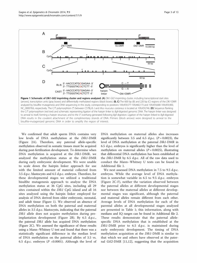
Figure 1 Schematic of Dlk1-Gtl2 imprinting cluster and regions analyzed. (A) Dlk1-Gtl2 imprinting cluster, including transcriptional start sites(arrows), transcription units (gray boxes) and differentially methylated regions (black boxes). (B, C) The 600 bp (B) and 220 bp (C) regions of the Dlk1-DMRanalyzed by bisulfite mutagenesis and DNA sequencing in this study, corresponding to positions 109,459,577-109,460,173 and 109,459,680-109,459,900,NC_000078.6, respectively. The C/T polymorphism (*) between C57BL/6 J and Mus musculus castaneus is located at 109,459,746. (D) Sequence flankingthe C/T polymorphism (red text) and schematic representing ligation of the hairpin linker to BglI-digested genomic DNA. The hairpin linker was designedto anneal to itself, forming a hairpin structure, and to the 3′ overhang generated following BglI digestion. Ligation of the hairpin linked to BglI-digestedDNA results in the covalent attachment of the complimentary strands of DNA. Primers (block arrows) were designed to anneal to thebisulfite-mutagenized genomic DNA in order to amplify the region of interest.
Gagne et al. Epigenetics & Chromatin 2014, 7:9 Page 3 of 13http://www.epigeneticsandchromatin.com/content/7/1/9
CpG position
8.5 dpc B6xCAST12E1-6
F1
F2
F3,7,11
F4
G1,8
G2
G7
J3,4,6,8,10,11,12K6
K4
M1,5
O3
P1
9.5 dpc B6xCAST12A1,4
A2
A10
A12
B6
B7
B12
C11
D11
J3
J7
J9
A8
B1,8
B2
C4,5,9,10,12D1,5,7D6
D10
I2
I3
I4I6
I1,7,12
J8
J10
I8
I9
I11
J4
J5
H1
H3
H6
H9
H10
L1-5
L7
N2,4
N5
O1
P2
P3
7.5 dpc B6xCAST12
C1,2,5
D1-3,5
E2
F2,3
J1
J2
pate
rnal
A2
A5
B1-5
C4
E1,3-5
F1
F4
C5
H1-5
I3,4
mat
erna
l
0
20
40
60
80
100
1 4 7 10 13 160
20
40
60
80
100
1 4 7 10 13 160
20
40
60
80
100
1 4 7 10 13 16
% m
ethy
latio
n
A2
C1
C2
C4
D1
E2
F1
G1
G2,3
H3
H4
A1
D2
D3
E1
F3,4
G4
H1
C3
D4
B6xCAST sperm
pate
rnal
mat
erna
l
A2
B5
M4
K15
K11
K5,7,9,12,16
D14
D1,3,11
B14
A11
B8
B6,10,15
N1N2-5,12
C1,3,8
C9
L11,12,14C17
B13
B9
3.5 dpc B6xCAST12
Y1
Y8
Y6
Z1-4,6Z5
W8
P6
X3
Q3
R6
Q6R9
W2W4
W3
W6
X1
W7
X2P2,5
P1
R7
Q4R10
S7
6.5 dpc B6xCAST12
0
20
40
60
80
100
1 4 7 10 13 16 0
20
40
60
80
100
1 6 11 16 21 26 31 360
20
40
60
80
100
1 6 11 16 21 26 31 36
% m
ethy
latio
n
CpG position CpG position
CpG position CpG position CpG position
A B C
D E F
average methylation, 12%
average methylation, 9%
average methylation, 4%
average methylation, 0.5%
average methylation, 36%
average methylation, 14%
average methylation, 35%
average methylation, 10%
average methylation, 22%
average methylation, 5%
average methylation, 37%
average methylation, 10%
Figure 2 Paternal allele-specific DNA methylation is acquired during post-implantation development. (A) Bisulfite mutagenesis and sequencing ofDNA from B6 x CAST F1 hybrid spermatozoa. (B-F) Bisulfite mutagenesis and sequencing of DNA from B6 x CAST12 F1 hybrid 3.5 to 9.5 d.p.c. embryos. Ahairpin linker approach was used to analyze the DNA methylation status of 32 potentially methylated CpG dinucleotides in sperm and 7.5 to 9.5 d.p.c.embryos. Individual circles represent one of the 32 potentially methylated CpG dinucleotides, and each paired row of circles represents the complimentarystrands of an individual subclone; semi-circles to the left connect the complimentary strands. A traditional bisulfite mutagenesis approach was utilized toanalyze 36 CpG sites located on the bottom, antisense strand in 3.5 d.p.c blastocysts and 6.5 d.p.c. embryos; the analyzed region includes all 29 sitescontained within the Dlk1 CpG island and all 16 sites analyzed using the hairpin linker (black bar). The gap in the paternal strands represents thepolymorphic site, which is not a CpG dinucleotide in Mus musculus castaneus-derived DNA. Filled circles represent methylated cytosines, open circlesrepresent unmethylated cytosines, absent circles represent ambiguous data. Labels to the right identify the PCR subclone analyzed; letters representindependent amplification reactions, while numbers represent individual subclones. The level of methylation (y axis) on the paternal (blue) and maternal(red) strands was quantified by dividing the number of methylated residues at each CpG site by the total number of sites analyzed at that position.
Gagne et al. Epigenetics & Chromatin 2014, 7:9 Page 4 of 13http://www.epigeneticsandchromatin.com/content/7/1/9
of DNA methylation across the Dlk1/Gtl2 locus may becoordinately controlled.
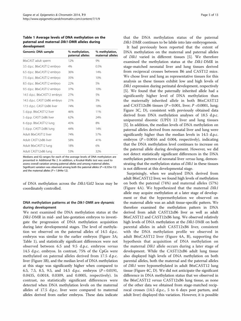
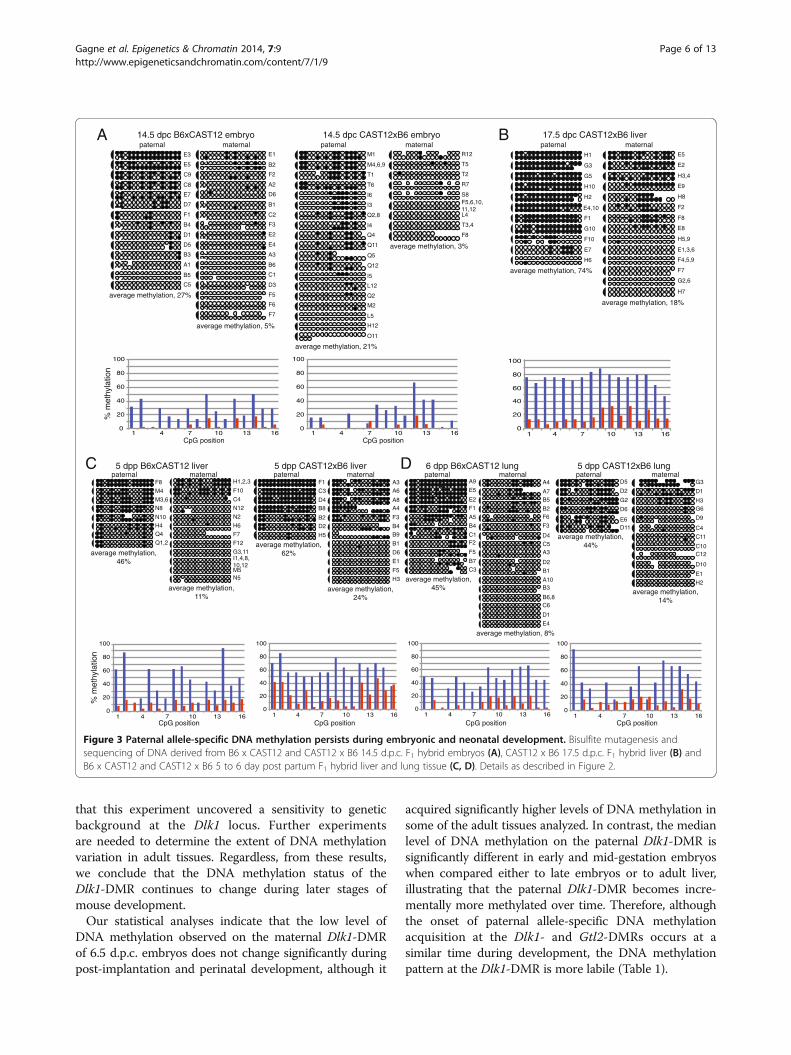
DNA methylation patterns at the Dlk1-DMR are dynamicduring developmentWe next examined the DNA methylation status at theDlk1-DMR in mid- and late-gestation embryos to investi-gate the progression of DNA methylation acquisitionduring later developmental stages. The level of methyla-tion we observed on the paternal alleles of 14.5 d.p.c.embryos was similar to the earlier embryos (Figure 3A;Table 1), and statistically significant differences were notobserved between 6.5 and 9.5 d.p.c. embryos versus14.5 d.p.c. embryos. In contrast, 75% of the CpGs weremethylated on paternal alleles derived from 17.5 d.p.c.liver (Figure 3B), and the median level of DNA methylationat this stage was significantly higher when compared to6.5, 7.5, 8.5, 9.5, and 14.5 d.p.c. embryos (P = 0.0191,0.0435, 0.0018, 0.0309, and 0.0005, respectively). Incontrast, no statistically significant differences weredetected when DNA methylation levels on the maternalalleles of 17.5 d.p.c. liver were compared to maternalalleles derived from earlier embryos. These data indicate
that the DNA methylation status of the paternalDlk1-DMR continues to be labile into late embryogenesis.It had previously been reported that the extent of
DNA methylation on the maternal and paternal allelesof Dlk1 varied in different tissues [5]. We thereforeexamined the methylation status at the Dlk1-DMR instage-matched neonatal liver and lung tissues derivedfrom reciprocal crosses between B6 and CAST12 mice.We chose liver and lung as representative tissues for thisanalysis as these tissues exhibit low and high levels ofDlk1 expression during perinatal development, respectively[5]. We found that the paternally inherited allele had asignificantly higher level of DNA methylation thanthe maternally inherited allele in both B6xCAST12and CAST12xB6 tissues (P = 0.001, liver; P <0.0001, lung;Figure 3C, D), consistent with previously obtained dataderived from DNA methylation analyses of 18.5 d.p.c.uniparental disomic (UPD) 12 liver and lung tissues[5]. In addition, the median levels of DNA methylation onpaternal alleles derived from neonatal liver and lung weresignificantly higher than the median levels in 14.5 d.p.c.embryos (P = 0.0016 and 0.004, respectively), indicatingthat the DNA methylation level continues to increase onthe paternal allele during development. However, we didnot detect statistically significant differences in the DNAmethylation patterns of neonatal liver versus lung, demon-strating that the methylation status of Dlk1 in these tissuesis not different at this developmental stage.Surprisingly, when we analyzed DNA derived from
adult B6xCAST12 liver, we found high levels of methylationon both the paternal (74%) and maternal alleles (57%)(Figure 4A). We hypothesized that the maternal Dlk1allele may acquire methylation at a later stage of develop-ment or that the hypermethylation we observed onthe maternal allele was an adult tissue-specific pattern. Wetherefore examined the methylation pattern in DNAderived from adult CAST12xB6 liver as well as adultB6xCAST12 and CAST12xB6 lung. We observed relativelyhigh levels of DNA methylation at the Dlk1-DMR on bothparental alleles in adult CAST12xB6 liver, consistentwith the DNA methylation profile we observed inadult B6xCAST12 liver (Figure 4A, B), supporting thehypothesis that acquisition of DNA methylation onthe maternal Dlk1 allele occurs during a later stage ofdevelopment. While the CAST12xB6 adult lung tissuealso displayed high levels of DNA methylation on bothparental alleles, both the maternal and the paternal allelesof Dlk1 were hypomethylated in adult B6xCAST12 lungtissue (Figure 4C, D). We did not anticipate the significantdifference in DNA methylation status that we observed inthe B6xCAST12 versus CAST12xB6 lung tissue, as noneof the other data we obtained from stage-matched recip-rocal crosses (14.5 d.p.c., 5 to 6 days post partum, andadult liver) displayed this variation. However, it is possible
Table 1 Average levels of DNA methylation on thepaternal and maternal Dlk1-DMR alleles duringdevelopment
Genomic DNA sample % methylation,paternal alleles
% methylation,maternal alleles
B6xCAST adult sperm 12% 9%
3.5 d.p.c. B6xCAST12 embryo 4% 0.5%
6.5 d.p.c. B6xCAST12 embryo 36% 14%
7.5 d.p.c. B6xCAST12 embryo 35% 10%
8.5 d.p.c. B6xCAST12 embryo 22% 5%
9.5 d.p.c. B6xCAST12 embryo 37% 10%
14.5 d.p.c. B6xCAST12 embryo 27% 5%
14.5 d.p.c. CAST12xB6 embryo 21% 3%
17.5 d.p.c. CAST12xB6 liver 74% 18%
5 d.p.p. B6xCAST12 liver 46% 11%
5 d.p.p. CAST12xB6 liver 62% 24%
6 d.p.p. B6xCAST12 lung 45% 8%
5 d.p.p. CAST12xB6 lung 44% 14%
Adult B6xCAST12 liver 74% 57%
Adult CAST12xB6 liver 58% 41%
Adult B6xCAST12 lung 18% 6%
Adult CAST12xB6 lung 53% 32%
Medians and IQ ranges for each of the average levels of DNA methylation arepresented in Additional file 2. In addition, a Kruskal-Wallis test was used toassess overall variation among paternal alleles and among maternal alleles.Significant variation was observed among both the paternal alleles (P = 8.335e-13)and the maternal alleles (P = 1.844e-12).
Gagne et al. Epigenetics & Chromatin 2014, 7:9 Page 5 of 13http://www.epigeneticsandchromatin.com/content/7/1/9
that this experiment uncovered a sensitivity to geneticbackground at the Dlk1 locus. Further experimentsare needed to determine the extent of DNA methylationvariation in adult tissues. Regardless, from these results,we conclude that the DNA methylation status of theDlk1-DMR continues to change during later stages ofmouse development.Our statistical analyses indicate that the low level of
DNA methylation observed on the maternal Dlk1-DMRof 6.5 d.p.c. embryos does not change significantly duringpost-implantation and perinatal development, although it
acquired significantly higher levels of DNA methylation insome of the adult tissues analyzed. In contrast, the medianlevel of DNA methylation on the paternal Dlk1-DMR issignificantly different in early and mid-gestation embryoswhen compared either to late embryos or to adult liver,illustrating that the paternal Dlk1-DMR becomes incre-mentally more methylated over time. Therefore, althoughthe onset of paternal allele-specific DNA methylationacquisition at the Dlk1- and Gtl2-DMRs occurs at asimilar time during development, the DNA methylationpattern at the Dlk1-DMR is more labile (Table 1).
5 dpp B6xCAST12 liver 5 dpp CAST12xB6 liver 6 dpp B6xCAST12 lung 5 dpp CAST12xB6 lung
F8
N8
N10
M4
M3,6
H4Q4
Q1,2
C4
F12
G3,11
F7
F10
I1,4,8,10,12
H1,2,3
N5
N12
H6
N2
M5
B2
B8
F1
H5
C3
D4
D2
A3
A4
A6
A8
B1
B4B9
E1
F3
D6
F5
H3
D5
E6
G2
D6
D2
D11
D10
C12
H2
C10
C4
D9
G6
C11
H3
D1
E1
G3C
A5
F5
F2
F1
E5
E2
C3
C1
B7
B4
A9
B6,8
B5
B3
B2
B1
A10
A7
A4
A3C5
F6
F3
E4
D4
D2
D1
C6
0
20
40
60
80
100
1 4 7 10 13 16 0
20
40
60
80
100
1 4 7 10 13 16 0
20
40
60
80
100
1 4 7 10 13 16 0
20
40
60
80
100
1 4 7 10 13 16
paternal maternal paternal maternal paternal maternal paternal maternal
% m
ethy
latio
n
CpG position CpG position CpG position CpG position
paternal maternal14.5 dpc B6xCAST12 embryo
A1
B3
B4
B5
D1
D5
D7
E3
E5
E7
C5
C8
C9
F1
A2
A3
B1
B2
B6
D3
D6
E1
E2
E4
C1
C2
F2
F3
F5
F6
F7
paternal maternal
F5,6,10,11,12
F8
L4
R7
R12
S8
T2
T3,4
T5
I4
H12
T6
T1
Q11
Q12
Q5
Q4
Q2,8
O11
M4,6,9
M1
Q2
L12
L5
I5
I3
I6
M2
14.5 dpc CAST12xB6 embryo
0
20
40
60
80
100
1 4 7 10 13 16 0
20
40
60
80
100
1 4 7 10 13 16
% m
ethy
latio
n
CpG position CpG position
A
C D
average methylation, 27%
average methylation, 5%
average methylation, 21%
average methylation, 3%
average methylation,11%
average methylation,46%
average methylation,24%
average methylation, 8%
average methylation,14%
average methylation,62%
average methylation,44%
average methylation,45%
E4,10
E7
F1
F10
G3
G5
G10
H1
H2
H6
H10
E1,3,6
E2
E5
E8
F2
F4,5,9
F7
F8
G2,6
H3,4
H5,9
H7
H8
E9
paternal maternal17.5 dpc CAST12xB6 liverB
0
20
40
60
80
100
1 4 7 10 13 16
average methylation, 18%
average methylation, 74%
Figure 3 Paternal allele-specific DNA methylation persists during embryonic and neonatal development. Bisulfite mutagenesis andsequencing of DNA derived from B6 x CAST12 and CAST12 x B6 14.5 d.p.c. F1 hybrid embryos (A), CAST12 x B6 17.5 d.p.c. F1 hybrid liver (B) andB6 x CAST12 and CAST12 x B6 5 to 6 day post partum F1 hybrid liver and lung tissue (C, D). Details as described in Figure 2.
Gagne et al. Epigenetics & Chromatin 2014, 7:9 Page 6 of 13http://www.epigeneticsandchromatin.com/content/7/1/9

Placental tissue displays biallelic methylation at theDlk1-DMRTo complete our analysis of the developmental dynamics ofDNA methylation at Dlk1, we investigated the methylationstatus in 14.5 d.p.c. B6xCAST12 placenta. Fifty-eightpercent of the CpGs were methylated on the paternalalleles and 53.5% were methylated on the maternal alleles,suggesting that both parental alleles are partially methylatedin mouse placenta (Figure 5A). These data are consistentwith those previously obtained using a methylation-sensitivesouthern blot to assess DNA methylation levels on the
parental alleles of the Dlk1-DMR in 16.5 d.p.c. placentae[22]. While the median level of DNA methylation onthe parental alleles in 14.5 d.p.c. placenta is significantlydifferent from the level observed in the corresponding14.5 d.p.c. embryo (27% and 5% for the paternal andmaternal alleles, respectively; P <0.0005), previous re-search has shown that the Gtl2-DMR is methylated onboth parental alleles in 6.5, 7.5, and 16.5 d.p.c. extraem-bryonic tissues, and it has been suggested that both theregulation of the non-coding RNAs and the maintenanceof DNA methylation in the Dlk1-Dio3 imprinting cluster
C3
C4
D2,3
D6
D7
D8
G1
G4
H9
C1,2
C5
C7
D1
G8
H1
H2
H3
G3
H8
I4
I3,5,8
I10
I12
A2
A5
A8
A12
D8
I2
D9
F10
A3,4
D3,4
D12
F8,9
G1,5,6,7,8,10
A1
C8
C11
F1
F2
F3
F5
F6
F7
C4
C9
G2
G3
G4
G44
I1
I6
I7
I9
D4
D5
D6
D7
D8
D10
E1
E3,6
E4
E5
E8
E11
I1
I3
I6
I9
I11
G3
G4
G5
G7
G9
G10
G12
H1
H2,3,7,10,11,12
H5
H6
H8
H9
F1
F3
F4
F12
J6
J12
I4,10
I5
I8
G2
G2
B6xCAST12adult liver
CAST12xB6adult liver
B6xCAST12adult lung
pate
rnal
mat
erna
l
B4
A9
C1,9
B1
D2
A6
D8
B2
C6
D6
B8
B3
C3,10
A2,4
B6
A1
B5
C5
C4
A10
C9
D4
C2
A8
D5,7
D1
A5
C7
A3
A7
B7
D3
0
20
40
60
80
100
1 4 7 10 13 16 0
20
40
60
80
100
1 4 7 10 13 16 0
20
40
60
80
100
1 4 7 10 13 16
CpG position
CAST12XB6adult lung
0
20
40
60
80
100
1 4 7 10 13 16 CpG positionCpG position CpG position
% m
ethy
latio
n
average methylation, 18%
average methylation, 53%
average methylation, 58%
average methylation, 74%
average methylation, 57%
average methylation, 41% average methylation, 32%
average methylation, 6%
Figure 4 DNA methylation is acquired on the maternal Dlk1-DMR in adult tissues. Bisulfite mutagenesis and sequencing of DNA from B6 xCAST12 adult liver (A), CAST12 x B6 adult liver (B), B6 x CAST12 adult lung (C), and CAST12 x B6 adult lung (D). Details as described in Figure 2.
Gagne et al. Epigenetics & Chromatin 2014, 7:9 Page 7 of 13http://www.epigeneticsandchromatin.com/content/7/1/9
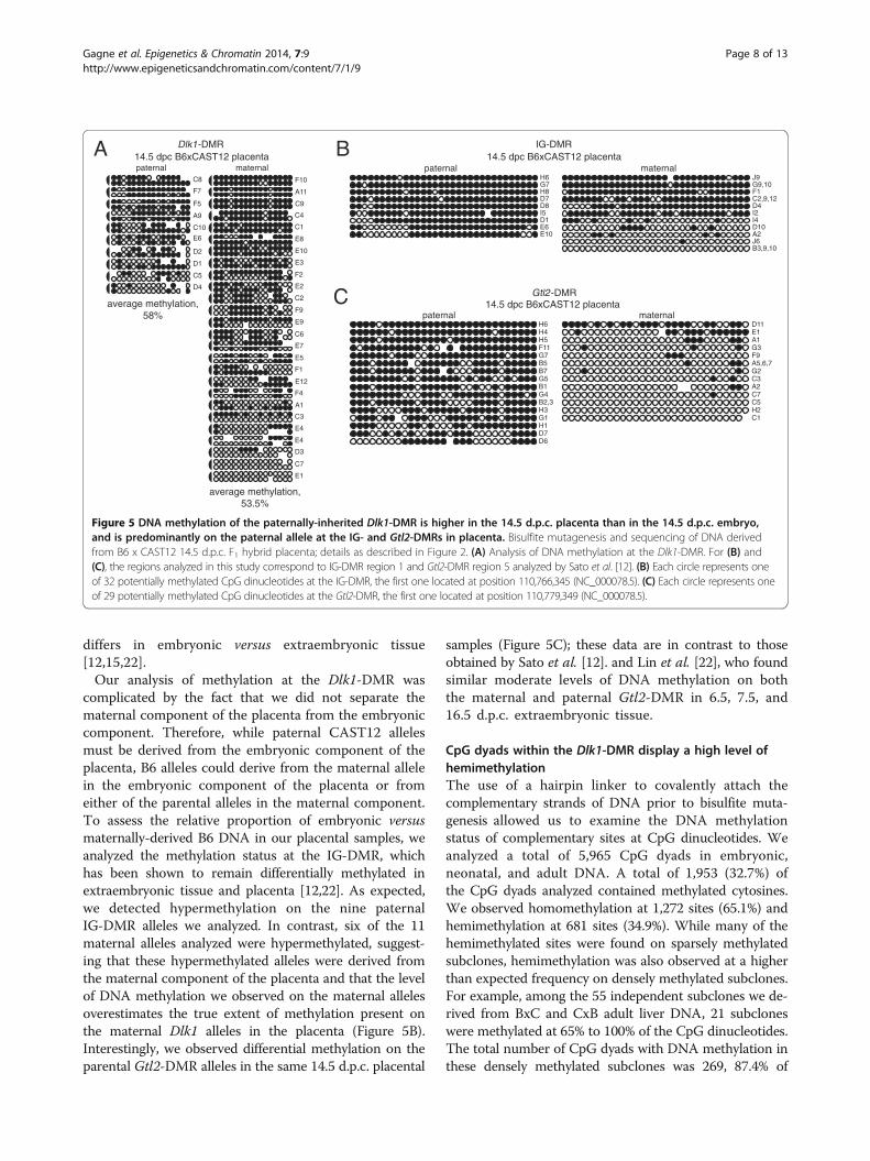
differs in embryonic versus extraembryonic tissue[12,15,22].Our analysis of methylation at the Dlk1-DMR was
complicated by the fact that we did not separate thematernal component of the placenta from the embryoniccomponent. Therefore, while paternal CAST12 allelesmust be derived from the embryonic component of theplacenta, B6 alleles could derive from the maternal allelein the embryonic component of the placenta or fromeither of the parental alleles in the maternal component.To assess the relative proportion of embryonic versusmaternally-derived B6 DNA in our placental samples, weanalyzed the methylation status at the IG-DMR, whichhas been shown to remain differentially methylated inextraembryonic tissue and placenta [12,22]. As expected,we detected hypermethylation on the nine paternalIG-DMR alleles we analyzed. In contrast, six of the 11maternal alleles analyzed were hypermethylated, suggest-ing that these hypermethylated alleles were derived fromthe maternal component of the placenta and that the levelof DNA methylation we observed on the maternal allelesoverestimates the true extent of methylation present onthe maternal Dlk1 alleles in the placenta (Figure 5B).Interestingly, we observed differential methylation on theparental Gtl2-DMR alleles in the same 14.5 d.p.c. placental
samples (Figure 5C); these data are in contrast to thoseobtained by Sato et al. [12]. and Lin et al. [22], who foundsimilar moderate levels of DNA methylation on boththe maternal and paternal Gtl2-DMR in 6.5, 7.5, and16.5 d.p.c. extraembryonic tissue.
CpG dyads within the Dlk1-DMR display a high level ofhemimethylationThe use of a hairpin linker to covalently attach thecomplementary strands of DNA prior to bisulfite muta-genesis allowed us to examine the DNA methylationstatus of complementary sites at CpG dinucleotides. Weanalyzed a total of 5,965 CpG dyads in embryonic,neonatal, and adult DNA. A total of 1,953 (32.7%) ofthe CpG dyads analyzed contained methylated cytosines.We observed homomethylation at 1,272 sites (65.1%) andhemimethylation at 681 sites (34.9%). While many of thehemimethylated sites were found on sparsely methylatedsubclones, hemimethylation was also observed at a higherthan expected frequency on densely methylated subclones.For example, among the 55 independent subclones we de-rived from BxC and CxB adult liver DNA, 21 subcloneswere methylated at 65% to 100% of the CpG dinucleotides.The total number of CpG dyads with DNA methylation inthese densely methylated subclones was 269, 87.4% of
maternal
A1
A2
A5,6,7
H2
G3
G2
F9
C5
D11E1
C7
C3
C1
paternal
B1
B2,3
B5B7
D6D7
F11
G1
G4
G5
G7
H1
H3
H4H5
H6
D1I5
H8
H6G7
E10E6
D8D7
A2
J9
J6
I4I2
G9,10F1
D10
D4C2,9,12
B3,9,10
IG-DMR
maternalpaternal
Gtl2 -DMR
C8
F7
F5
A9
C10
E6
D2
D1
C5
D4
F2
C4
C6
C2
C1
A11
F10
F9
E10
E9
E8
E2
E3
C9
F1
F4
E4
D3
C3
E7
E5
E1
E4
E12
A1
C7
paternal maternal 14.5 dpc B6xCAST12 placenta
Dlk1-DMR 14.5 dpc B6xCAST12 placenta
14.5 dpc B6xCAST12 placentaaverage methylation,58%
average methylation,53.5%
Figure 5 DNA methylation of the paternally-inherited Dlk1-DMR is higher in the 14.5 d.p.c. placenta than in the 14.5 d.p.c. embryo,and is predominantly on the paternal allele at the IG- and Gtl2-DMRs in placenta. Bisulfite mutagenesis and sequencing of DNA derivedfrom B6 x CAST12 14.5 d.p.c. F1 hybrid placenta; details as described in Figure 2. (A) Analysis of DNA methylation at the Dlk1-DMR. For (B) and(C), the regions analyzed in this study correspond to IG-DMR region 1 and Gtl2-DMR region 5 analyzed by Sato et al. [12]. (B) Each circle represents oneof 32 potentially methylated CpG dinucleotides at the IG-DMR, the first one located at position 110,766,345 (NC_000078.5). (C) Each circle represents oneof 29 potentially methylated CpG dinucleotides at the Gtl2-DMR, the first one located at position 110,779,349 (NC_000078.5).
Gagne et al. Epigenetics & Chromatin 2014, 7:9 Page 8 of 13http://www.epigeneticsandchromatin.com/content/7/1/9
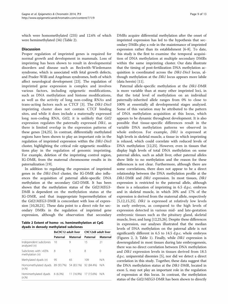
which were homomethylated (235) and 12.6% of whichwere hemimethylated (34) (Table 2).
DiscussionProper regulation of imprinted genes is required fornormal growth and development in mammals. Loss ofimprinting has been shown to result in developmentaldisorders and disease such as Beckwith-Wiedemannsyndrome, which is associated with fetal growth defects,and Prader-Willi and Angelman syndromes, both of whichaffect neurological development [23]. The regulation ofimprinted gene expression is complex and involvesvarious factors, including epigenetic modifications,such as DNA methylation and histone modifications,as well as the activity of long non-coding RNAs andtrans-acting factors such as CTCF [3]. The Dlk1-Dio3imprinting cluster does not contain CTCF bindingsites, and while it does include a maternally expressedlong non-coding RNA, Gtl2, it is unlikely that Gtl2expression regulates the paternally expressed Dlk1, asthere is limited overlap in the expression patterns ofthese genes [24,25]. In contrast, differentially methylatedregions have been shown to play an important role in theregulation of imprinted expression within the Dlk1-Dio3cluster, highlighting the critical role epigenetic modifica-tions play in the regulation of genomic imprinting.For example, deletion of the imprinting control region,IG-DMR, from the maternal chromosome results in itspaternalization [19].In addition to regulating the expression of imprinted
genes in the Dlk1-Dio3 cluster, the IG-DMR also influ-ences the acquisition of paternal allele-specific DNAmethylation at the secondary Gtl2-DMR. It has beenshown that the methylation status of the Gtl2/MEG3-DMR is dependent on the methylation status at theIG-DMR, and that inappropriate hypermethylation ofthe Gtl2/MEG3-DMR is concordant with loss of expres-sion [10,20,21]. These data point to a direct role for sec-ondary DMRs in the regulation of imprinted geneexpression, although the observation that secondary
DMRs acquire differential methylation after the onset ofimprinted expression has led to the hypothesis that sec-ondary DMRs play a role in the maintenance of imprintedexpression rather than its establishment [6-8]. To date,this study is the first to examine the temporal acquisi-tion of DNA methylation at multiple secondary DMRswithin the same imprinting cluster. Our data illustratethat the timing of post-fertilization DNA methylation ac-quisition is coordinated across the Dlk1-Dio3 locus, al-though methylation at the Dlk1 locus appears more labile(data herein) [11].Paternal allele-specific methylation at the Dlk1-DMR
is more variable than at many other imprinted loci, inthat the total level of methylation on an individualpaternally-inherited allele ranges from 0% to close to100% at essentially all developmental stages analyzed.Some of this variation may be attributed to the patternof DNA methylation acquisition at this locus, whichappears to be dynamic throughout development. It is alsopossible that tissue-specific differences result in thevariable DNA methylation patterns we observed inwhole embryos. For example, Dlk1 is expressed athigh levels in skeletal muscle, a tissue in which imprintingis relaxed, which could correlate with reduced levels ofDNA methylation [12,25]. However, even in tissues thatdisplay high levels of total DNA methylation on somepaternal alleles, such as adult liver, other paternal allelesshow little to no methylation and the reason for thesedifferences is not clear. Furthermore, although there aresome correlations, there does not appear to be a directrelationship between the DNA methylation profile at theDlk1-DMR and Dlk1 expression. In most tissues, Dlk1expression is restricted to the paternal allele, althoughthere is a relaxation of imprinting in 6.5 d.p.c. embryosand in skeletal muscle, in which 20% and 17% of theexpression is derived from the maternal allele, respectively[5,12,15,25]. Dlk1 is expressed at relatively low levelsin early embryos, as compared to the high levels ofexpression detected in various mid- and late-gestationembryonic tissues such as the pituitary gland, skeletalmuscle, liver, and lung [12,25,26]. Despite these differencesin expression, our analyses illustrated that the medianlevels of DNA methylation on the paternal allele is notsignificantly different in 6.5 to 14.5 d.p.c. whole embryos(Figures 2, 3; Table 1). Finally, while Dlk1 expression isdownregulated in most tissues during late embryogenesis,there was no direct correlation between DNA methylationand Dlk1 expression levels in tissues derived from 18.5d.p.c. uniparental disomies [5], nor did we detect a directcorrelation in this study. Together, these data suggest thatthe DNA methylation status at the Dlk1-DMR, located inexon 5, may not play an important role in the regulationof expression at this locus. In contrast, the methylationstatus of the Gtl2/MEG3-DMR has been shown to directly
Table 2 Extent of homo- vs. hemimethylation at CpGdyads in densely methylated subclones
BxC9C12 adult liver C9C12xB adult liver
Paternal Maternal Paternal Maternal
Independent subclonesanalyzed (n)
10 9 15 21
Subclones with >65%methylation (n)
8 5 8 0
Methylated dyads (n) 95 65 109 N/A
Homomethylated dyads(n,%)
89 (93.7%) 54 (83.1%) 92 (84.4%) N/A
Hemimethylated dyads(n,%)
6 (6.3%) 11 (16.9%) 17 (15.6%) N/A
Gagne et al. Epigenetics & Chromatin 2014, 7:9 Page 9 of 13http://www.epigeneticsandchromatin.com/content/7/1/9

influence expression of Gtl2 in cis, consistent with itslocation at the Gtl2 promoter [5,10,20,21]. The criticalregulatory role of the Gtl2-DMR may explain themaintenance of high average DNA methylation levelsat this locus once it has been established [5,11,12]. Itis possible that DNA methylation at the Dlk1-DMRmay reflect a broader, locus-wide epigenetic profilethat encompasses both Gtl2 and Dlk1.
The Dlk1-DMR displays low methylation fidelityThe approach we utilized allowed us to analyze the methy-lation pattern for complementary CpG dinucleotides withinthe Dlk1-DMR. To the best of our knowledge, this is thefirst study to comprehensively examine the methylationstatus of complementary CpG dinucleotides at an imprintedgene during development. Of the 1,953 methylated CpGdyads, 1,272 (65.1%) were homomethylated, while 681(34.9%) were hemimethylated. This result was unexpected,as the fidelity with which the maintenance DNA methyl-transferase in mouse, Dnmt1, has been shown to be greaterthan 95% [27,28]. There are several possible reasonsto explain some of the hemimethylation we detected.It is likely that some of the hemimethylated sites weobserved are a result of hybrid subclones, which havebeen shown to result as an artifact of PCR amplificationfollowing bisulfite mutagenesis [29]. It is also possiblethat some of the observed hemimethylation is a resultof Taq-induced PCR error during amplification. However,these artifacts are unlikely to account for the high level ofhemimethylation we detected. Rather, the high level ofhemimethylation we observed challenges the idea thatDnmt1 functions with high fidelity at all genomic locations.A large-scale study analyzing the in vivo regulation of
CpG methylation by DNA methyltransferases was recentlyconducted by Arand et al. [30]. In this study, the authorsfound relatively high levels of hemimethylated CpGsin embryonic liver, ranging from 16.2% to 30.6% of themethylated CpG dyads. Interestingly, this work illustratedthe relative stability of homomethylation at the imprintedSnprn and H19 genes, but demonstrated high levels ofhemimethylation at the imprinted Igf2 gene (22%).Analyses of DNA methylation profiles in Dnmt-mutantembryonic stem cells indicated that the DNA methylationprofiles at Snprn and H19 were dependent on the ac-tivity of Dnmt1 alone, while maintenance of DNAmethylation at Igf2 required the coordinated activity ofDnmt1, Dnmt3a, and Dnmt3b, a possible consequence of5-hydroxymethylcytosine enrichment at the Igf2 DMR[30]. It is therefore possible that the high level of hemi-methylation we observed at the Dlk1-DMR may be due tothe presence of 5-hydroxymethylcytosine at this locus,preventing high levels of fidelity via Dnmt1. An analysis ofmethylcytosine versus 5-hydroxymethylcytosine levels atthe Dlk1-DMR will address this possibility.
An alternative hypothesis to explain the high level ofhemimethylation we observed at the Dlk1-DMR is thatthere may be a lower level of fidelity associated with themaintenance of DNA methylation at secondary DMRs.Consistent with this hypothesis, a study by Vu et al. [31]examined DNA methylation on the top and bottomstrands of the human Igf2/H19 imprinted region. Vu andcolleagues analyzed DNA methylation on the top andbottom strands separately and found uniform levels ofmethylation present at the primary DMR. In contrast,they observed less uniformity in the methylation of thetop and bottom strands at the H19 promoter, which iscategorized as a secondary DMR as it loses and thenregains paternal allele-specific methylation during pre-and post-implantation development, respectively [32,33].Additionally, a more recent survey of differentially methyl-ated regions associated with imprinted genes in humanssupport this hypothesis. Woodfine et al. [34] reported ahigher level of stability for DNA methylation at gameticDMRs than at secondary DMRs. Further examination ofCpG dyad methylation patterns at imprinted loci may pro-vide additional insight into the mechanisms responsiblefor the acquisition and maintenance of DNA methylationat these sites.
ConclusionsOur analysis of DNA methylation at the mouse Dlk1-DMRillustrates that the acquisition of paternal allele-specificDNA methylation initiates between 3.5 and 6.5 d.p.c., sug-gesting that epigenetic modifications across the Dlk1-Dio3imprinting cluster may be coordinately regulated duringpost-implantation development. The range of DNA methy-lation levels on individual alleles at the same developmentalstage as well as the additional acquisition of DNA methyla-tion on the maternal Dlk1 allele in adult tissues suggest thatthe DNA methylation profile of this secondary DMR ismore variable than is commonly seen at imprinted loci. Wefurther observed a high level of hemimethylation at theDlk1-DMR: 35% of CpG dyads containing methylated resi-dues were methylated on only one of the two complemen-tary strands. This result is significant because it challengesthe idea that Dnmt1 functions with high fidelity at allgenomic locations. We hypothesize that the low DNAmethylation fidelity we observed is related to the variableDNA methylation profiles at the Dlk1-DMR, and may bea consequence of high levels of 5-hydroxymethylcytosineat this locus. These data provide insight into a novelepigenetic profile that may distinguish primary DMRsfrom secondary DMRs.
MethodsMiceC57BL/6 J (B6) and Mus musculus castaneus (CAST)mice were purchased from the Jackson Laboratory. To
Gagne et al. Epigenetics & Chromatin 2014, 7:9 Page 10 of 13http://www.epigeneticsandchromatin.com/content/7/1/9

facilitate the isolation of F1 hybrid mice, a strain ofmice that served as the source of the M. m. castaneusallele (CAST12) was constructed as previously de-scribed [11]. Natural matings between B6 and CASTwere used to generate F1 hybrid males for spermato-zoa collection; all other F1 hybrid tissues used for bi-sulfite analyses were generated from natural matingsbetween B6 and CAST12 mice. For all F1 hybridtissues, the maternal allele is located on the left.Ethical approval for procedures involving animals wasgranted by the Bryn Mawr College Institutional AnimalCare and Use Committee, PHS Welfare AssuranceNumber A3920-01.
DNA purification and bisulfite analysisFor bisulfite analysis of 3.5 and 6.5 d.p.c. DNA, two to fourembryos were pooled prior to digestion with proteinase K.The resulting DNA was subjected to bisulfite mutagenesisusing an EZ DNA methylation-direct kit (Zymo Research,cat# D5020). For all other tissues, genomic DNAextractions were performed either from a pool (four7.5 d.p.c. embryos) or from single embryos, fetuses,or tissues according to the DNeasy protocol (Qiagen)or using a series of phenol/chloroform extractions asdescribed previously [33], and the complementary strandswere covalently attached prior to bisulfite mutagenesis asfollows: 0.5 μg of genomic DNA was digested with 1 μLBglI (NEB, cat# R0143S) and ligated to 1 μg of a phos-phorylated hairpin linker (5′-AGCGATGCGTTCGAGCATCGCTCCC−3′) [35]. A total of 0.5 μg of hairpinlinked-ligated DNA was denatured by incubating in freshlyprepared 3 M NaOH for 20 min at 42°C, then subjected tobisulfite mutagenesis using an EZ DNA methylation-directkit, as above. All mutagenized DNAs were subjected tomultiple independent PCR amplifications to ensure analysisof different strands of DNA; subclones derived fromindependent PCR amplifications are distinguished bydifferent letters of the alphabet. Data from multipleindividuals at the same developmental stage werecombined, as we did not detect variation betweenbiological replicates. The following primer pairs wereused for nested amplification of the mutagenizedDNA, and were designed to incorporate both theSNP and at least 50% of the CpG dinucleotides withinthe CpG island. All base pair numbers are from GenBankAccession Number NC_000078.6. For the first round ofamplification of mutagenized 3.5 and 6.5 d.p.c. DNA, twocycles of 94°C for 2 min, 52°C for 1 min, 72°C for 1 minfollowed by 30 cycles of 94°C for 30 s, 52°C for 1 min, 72°Cfor 1 min using primers RDlke5BF3 (5′-CCCCATCTAACTAATAACTTACA-3′)/RDlke5BR3 (5′-GTGTTTAGTATTATTAGGTTGGTG-3′). For the second round ofamplification, 35 cycles of 94°C for 30 s, 52°C for1 min, 72°C for 1 min using primers RDlke5BF4 (5′-
ATTTCTACTACTCTATCCTAACCC-3′)/RDlke5BR4 (5′-TTAGGATGGTGAAGTAGATGGT-3′) yielded a 597 bpproduct. To amplify mutagenized DNA treated withthe hairpin linker, the same reaction conditions were usedwith the following primers to yield a 464 bp product:first round, RDlke5BR4 (5′-TTAGGATGGTGAAGTAGATGGT-3′)/Dlk1e5BR1 (5′-AACTCTTTCATAAACACCTTCAA-3′); second round, HPDlk1e5F (5′-GTTTATTTGGGTGTGTTGGAGG-3′)/HPDlk1e5R (5′-AAACTCACCTAAATATACTAAAAAC-3′). The following pri-mer pairs were used for nested or semi-nested amplifica-tion of IG- and Gtl2-DMRs, as previously described [11].All base pair numbers are from NC_000078.5. Gtl2IG-DMR, with the first nucleotide of IG-BS-F1 correspond-ing to position 110,766,235: 30 cycles of 94°C for30 s, 52°C for 1 min, 72°C for 1 min, using primersIG-BS-F1/IG-BS-R, followed by 35 cycles using IG-BS-F2/IG-BS-R and the same cycling conditions as above.Identical reaction conditions were used to amplify theGtl2-DMR, with the first nucleotide of Gtl2BI4F1 corre-sponding to position 110,779,293: Gtl2BI4F1/Gtl2BI4R1followed by Gtl2BI4F2/Gtl2BI4R2. Primer sequencesfollow. IG-BS-F1, 5′-GTATGTGTATAGAGATATGTTTATATGGTA-3′; IG-BS-F2, 5′-GTGTTAAGGTATATTATGTTAGTGTTAGGA-3′; IG-BS-R, 5′-GCTCCATTAACAAAATAATACAACCCTTCC-3′; Gtl2BI4F1, 5′-GAAGAATTTTTTATTTGGTGAGTGG-3′; Gtl2BI4F2,5′-GTTTGAAAGGATGTGTAAAAATG-3′; Gtl2BI4R1,5′-CAACACTCAAATCACCCCCC-3′; Gtl2BI4R2, 5′-GCCCCCCACATCTATTCTACC-3′. Subcloning of amplifiedproducts was achieved using a pGEM-T Easy vector(Promega Corporation, Madison, WI, USA). Sequencingreactions were performed using a Thermo SequenaseCycle Sequencing Kit (USB Corporation, Cleveland,OH, USA), and reactions were analyzed on a 4300DNA Analyzer (LI-COR Biosciences, Lincoln, NE, USA).Percent methylation was calculated based on data obtainedfrom both complementary strands.
Identification of CpG islandThe extent of the CpG island identified by Paulsen et al.[24] was determined using the EMBOSS CpGPlotanalyzer (http://www.ebi.ac.uk/Tools/emboss/cpgplot/index.html), with the following parameters: program =cpgplot, window= 200, step = 1, obs/exp = 0.6, MinPC= 50,length = 200. The position of the CpG island corre-sponds to nucleotides 109,459,650-109,460,035 (GenBank:NC_000078.6).
Additional files
Additional file 1: Data used for statistical analyses of DNAmethylation levels at the Dlk1-DMR during different stages ofmouse development. This file contains the numerical data used to
Gagne et al. Epigenetics & Chromatin 2014, 7:9 Page 11 of 13http://www.epigeneticsandchromatin.com/content/7/1/9

perform Kruskal-Wallis and Mann–Whitney U tests. Data from each of thedevelopmental stages are presented in chronological order, as they are inthe Results, Figures, and Table 1. Each dataset presents the informationfor a specific tissue, cross (maternal allele x paternal allele), and parentalallele analyzed, as indicated in columns B-D.% methylation (column E)was calculated by dividing the number of methylated CpG sites observedin a given subclone (column A) by the total number of CpG sitesanalyzed within the subclone; the raw data used to make thesecalculations are found in Figures 2, 3, 4, and 5.
Additional file 2: Average levels of DNA methylation on thepaternal and maternal Dlk1-DMR alleles during development,including median values and IQ ranges. This file expands on theinformation presented in Table 1. In addition to presenting the averagelevels of DNA methylation at each developmental stage, Additional file 2contains median values and IQ ranges. Data from each developmentalstage are presented in chronological order, as they are in the Results andFigures.
AbbreviationsB6: C57BL/6; C or CAST: Mus musculus castaneus; C12 or CAST12: Musmusculus castaneus chromosome 12 on a C57BL/6 background;DMR: Differentially methylated region; d.p.c.: Days post coitum; d.p.p.: Dayspost partum; ICR: Imprinting control region; PCR: Polymerase chain reaction;UPD: Uniparental disomy.
Competing interestsThe authors declare that they have no competing interests.
Authors’ contributionsMQ participated in experimental design and carried out molecular geneticstudies. AG, AH, CT, JA, and KM carried out molecular genetic studies. TLDconceived of the study and experimental design, carried out moleculargenetic studies, and wrote the manuscript. All authors read and approvedthe final manuscript.
AcknowledgementsWe thank Jeanette Bates for her contributions towards this work, JoshuaShapiro for assistance with the statistical analyses, and Michelle Wien andJoshua Shapiro for thoughtful discussion. This work was supported byawards from the Bryn Mawr College Faculty Research Fund and NationalScience Foundation grant 1157819 to TLD. In addition, AG, AH, MQ, CT, JA,and KM were supported in part by the Bryn Mawr College Summer ScienceResearch program.
Received: 11 February 2014 Accepted: 20 May 2014Published: 29 May 2014
References1. Morison IM, Ramsay JP, Spencer HG: A census of mammalian imprinting.
Trends Genet 2005, 21:457–465.2. Williamson CM, Blake A, Thomas S, Beechey CV, Hancock J, Cattanach BM,
Peters J: World Wide Web Site, Mouse Imprinting Data and References.Oxfordshire: MRC Harwell; 2011. http://www.har.mrc.ac.uk/research/genomic_imprinting/.
3. Bartolomei MS, Ferguson-Smith AC: Mammalian genomic imprinting.Cold Spring Harb Perspect Biol 2011, 3:a002592.
4. Hanel M, Wevrick R: Establishment and maintenance of DNA methylationpatterns in mouse Ndn: implications for maintenance of imprinting intarget genes of the imprinting center. Mol Cell Biol 2001, 21:2384–2392.
5. Takada S, Paulsen M, Tevendale M, Tsai C-E, Kelsey G, Cattanach BM,Ferguson-Smith AC: Epigenetic analysis of the Dlk1-Gtl2 imprinteddomain on mouse chromosome 12: implications for imprinting controlfrom comparison with Igf2-H19. Hum Mol Genet 2002, 11:77–86.
6. Bhogal B, Arnaudo A, Dymkowski A, Best A, Davis TL: Methylation at mouseCdkn1c is acquired during postimplantation development and functionsto maintain imprinted expression. Genomics 2004, 84:961–970.
7. John RM, Lefebvre L: Developmental regulation of somatic imprints.Differentiation 2011, 81:270–280.
8. Kobayashi H, Sakurai T, Sato S, Nakabayashi K, Hata K, Kono T: ImprintedDNA methylation reprogramming during early mouse embryogenesis at
the Gpr1-Zdbf2 locus is linked to long cis-intergenic transcription.FEBS Lett 2012, 586:827–833.
9. Lopes S, Lewis A, Hajkova P, Dean W, Oswald J, Forné T, Murrell A,Constância M, Bartolomei M, Walter J, Reik W: Epigenetic modifications inan imprinting cluster are controlled by a hierarchy of DMRs suggestinglong-range chromatin interactions. Hum Mol Genet 2003, 12:295–305.
10. Kagami M, O’Sullivan MJ, Green AJ, Watabe Y, Arisaka O, Masawa N,Matsuoka K, Fukami M, Matsubara K, Kato F, Ferguson-Smith AC, Ogata T:The IG-DMR and the MEG3-DMR at human chromosome 14q32.2:hierarchical interaction and distinct functional properties as imprintingcontrol centers. PLoS Genet 2010, 6:1–13.
11. Nowak K, Stein G, Powell E, He LM, Naik S, Morris J, Marlow S, Davis TL:Establishment of paternal allele-specific DNA methylation at theimprinted mouse Gtl2 locus. Epigenetics 2011, 6:1012–1020.
12. Sato S, Yoshida W, Soejima H, Nakabayashi K, Hata K: Methylationdynamics of IG-DMR and Gtl2-DMR during murine embryonic andplacental development. Genomics 2011, 98:120–127.
13. Stöger R, Kubicka P, Liu C-G, Kafri T, Razin A, Cedar H, Barlow DP:Maternal-specific methylation of the imprinted mouse Igf2r locus identifiesthe expressed locus as carrying the imprinting signal. Cell 1993, 73:61–71.
14. Takada S, Tevendale M, Baker J, Georgiades P, Campbell E, Freeman T,Johnson MH, Paulsen M, Ferguson-Smith AC: Delta-like and Gtl2 arereciprocally expressed, differentially methylated linked imprinted geneson mouse chromosome 12. Curr Biol 2000, 10:1135–1138.
15. Schmidt JV, Matteson PG, Jones BK, Guan X-J, Tilghman SM: The Dlk1 andGtl2 genes are linked and reciprocally imprinted. Genes Dev 2000,14:1997–2002.
16. Yevtodiyenko A, Carr MS, Patel N, Schmidt JV: Analysis of candidateimprinted genes linked to Dlk1-Gtl2 using a congenic mouse line.Mamm Genome 2002, 13:633–638.
17. Seitz H, Royo H, Bortolin ML, Lin SP, Ferguson-Smith AC, Cavaille J: A largeimprinted microRNA cluster at the mouse Dlk1-Gtl2 domain. Genome Res2004, 14:1741–1748.
18. Hagan JP, O’Neill BL, Stewart CL, Kozlov SV, Croce CM: At least ten genesdefine the imprinted Dlk1-Dio3 cluster on mouse chromosome 12qF1.PLoS One 2009, 4:e4352.
19. Lin S-P, Youngson N, Takada S, Seitz H, Reik W, Paulsen M, Cavaille J,Ferguson-Smith AC: Asymmetric regulation of imprinting on the maternaland paternal chromosomes at the Dlk1-Gtl2 imprinted cluster on mousechromosome 12. Nat Genet 2003, 35:97–102.
20. Sekita Y, Wagatsuma H, Irie M, Kobayashi S, Kohda T, Matsuda J, Yokoyama M,Ogura A, Shuster-Gossler K, Gossler A, Ishino F, Kaneko-Ishino T: Aberrantregulation of imprinted gene expression in Gtl2lacZ mice. Cytogenet GenomeRes 2006, 113:223–229.
21. Steshina EY, Carr MS, Glick EA, Yevtodiyenko A, Appelbe OK, Schmidt JV:Loss of imprinting at the Dlk1-Gtl2 locus caused by insertionalmutagenesis in the Gtl2 5′ region. BMC Genet 2006, 7:44.
22. Lin S-P, Coan P, da Rocha ST, Seitz H, Cavaille J, Teng P-W, Takada S,Ferguson-Smith AC: Differential regulation of imprinting in the murineembryo and placenta by the Dlk1-Dio3 imprinting control region.Development 2007, 134:417–426.
23. Robertson KD: DNA methylation and human disease. Nat Rev Genet 2005,6:597–610.
24. Paulsen M, Takada S, Youngson NA, Benchaib M, Charlier C, Segers K,Georges M, Ferguson-Smith AC: Comparative sequence analysis of theimprinted Dlk1-Gtl2 locus in three mammalian species reveals highlyconserved genomic elements and refines comparison with the Igf2-H19region. Genome Res 2001, 11:2085–2094.
25. da Rocha ST, Tevendale M, Knowles E, Takada S, Watkins M, Ferguson-Smith AC:Restricted co-expression of Dlk1 and the reciprocally imprinted non-codingRNA, Gtl2: implications for cis-acting control. Dev Biol 2007, 306:810–823.
26. Yevtodiyenko A, Schmidt JV: Dlk1 expression marks developingendothelium and sites of branching morphogenesis in the mouseembryo and placenta. Dev Dynam 2006, 235:1115–1123.
27. Hirasawa R, Chiba H, Kaneda M, Tajima S, Li E, Jaenisch R, Sasaki H: Maternaland zygotic Dnmt1 are necessary and sufficient for the maintenance ofDNA methylation imprints during preimplantation development.Genes Dev 2008, 22:1607–1616.
28. Vilkaitis G, Suetake I, Klimasauskas S, Tajima S: Processive methylation ofhemimethylated CpG sites by mouse Dnmt1 DNA methyltransferase.J Biol Chem 2005, 280:64–72.
Gagne et al. Epigenetics & Chromatin 2014, 7:9 Page 12 of 13http://www.epigeneticsandchromatin.com/content/7/1/9

29. Warnecke PM, Stirzaker C, Song J, Grunau C, Melki JR, Clark SJ: Identificationand resolution of artifacts in bisulfite sequencing. Methods 2002, 27:101–107.
30. Arand J, Spieler D, Karius T, Branco MR, Meilinger D, Meissner A, Jenuwein T,Xu G, Leonhardt H, Wolf V, Walter J: In vivo control of CpG and non-CpGDNA methylation by DNA methyltransferases. PLoS Genet 2012,8:e1002750.
31. Vu TH, Li T, Nguyen D, Nguyen BT, Yao X-M, Hu J-F, Hoffman AR: Symmetricand asymmetric DNA methylation in the human IGF2-H19 imprintedregion. Genomics 2000, 64:132–143.
32. Tremblay KD, Duran KL, Bartolomei MS: A 5′ 2-kilobase-pair region of theimprinted mouse H19 gene exhibits exclusive paternal methylationthroughout development. Mol Cell Biol 1997, 17:4322–4329.
33. Davis TL, Trasler JM, Moss SB, Yang GJ, Bartolomei MS: Acquisition of theH19 methylation imprint occurs differentially on the parental allelesduring spermatogenesis. Genomics 1999, 58:18–28.
34. Woodfine K, Huddleston JE, Murrell A: Quantitative analysis of DNAmethylation at all human imprinted regions reveals preservation ofepigenetic stability in adult somatic tissue. Epigenetics Chromatin 2011, 4:1.
35. Laird CD, Pleasant ND, Clark AD, Sneeden JL, Hassan KMA, Manley NC, Vary JC,Morgan T, Hansen RS, Stöger R: Hairpin-bisulfite PCR: assessing epigeneticmethylation patterns on complementary strands of individual DNAmolecules. Proc Natl Acad Sci U S A 2004, 101:204–209.
doi:10.1186/1756-8935-7-9Cite this article as: Gagne et al.: Analysis of DNA methylation acquisitionat the imprinted Dlk1 locus reveals asymmetry at CpG dyads. Epigenetics& Chromatin 2014 7:9.
Submit your next manuscript to BioMed Centraland take full advantage of:
• Convenient online submission
• Thorough peer review
• No space constraints or color figure charges
• Immediate publication on acceptance
• Inclusion in PubMed, CAS, Scopus and Google Scholar
• Research which is freely available for redistribution
Submit your manuscript at www.biomedcentral.com/submit
Gagne et al. Epigenetics & Chromatin 2014, 7:9 Page 13 of 13http://www.epigeneticsandchromatin.com/content/7/1/9