analysis of pleural fluid: differentiating transudate from
TRANSCRIPT

IP Archives of Cytology and Histopathology Research 2019;4(3):228–233
Content available at: iponlinejournal.com
IP Archives of Cytology and Histopathology Research
Journal homepage: www.innovativepublication.com
Original Research Article
Analysis of pleural fluid: Differentiating transudate from exudate
Karen Jaison1, M Sridevi1,*1Dept. of Pathology, Saveetha Medical College & Hospital, Tamil Nadu, India
A R T I C L E I N F O
Article history:Received 06-08-2019Accepted 19-08-2019Available online 01-10-2019
Keywords:ExudativePleural fluidTransudativeThoracocentesis
A B S T R A C T
Introduction: Pleural fluid is defined as the fluid that is found between the two layers of pleura, (i.e.the parietal and visceral pleura) the membranes which line the cavity and surround the lungs. Normally,10-20ml of pleural fluid, similar in composition to plasma but lower in protein (<1.5g/dl) is thinly spreadover the layers of pleura, facilitating the movement between lungs and chest wall. The pleural fluid entersthe systemic capillaries in the parietal pleura and exits via the parietal pleural stoma and lymphatics. (1)Pleural fluid accumulates when too much fluid enters or too little fluid exits the pleural cavity. Pleuralfluid analysis is used to diagnose the cause of accumulation of pleural fluid in the chest cavity (Pleuraleffusion). Pleural effusion may be Transudative or Exudative. Analysis is done in three broad spectrums(1) Biochemical analysis (2) Microbiological analysis (3) Clinical pathology or cytology analysis.Materials and Methods: Pleural fluid is obtained by Thoracocentesis. Thoracocentesis or pleural tap isan invasive procedure to remove fluid or air from the pleural space for diagnostic or therapeutic purposes.The fluid obtained is sent for analysis. Pleural fluid analysis is a group of tests used to diagnose the causeof fluid build-up such as CHF, Cirrhosis, Infection etc.Result: Study shows that pleural effusions are common among 40-60 years of age group with a slight malepredominance. The present study suggests that exudative effusions are more common than transudativeeffusions with a percentage of 61%.Conclusion: Pleural fluid analysis helps in diagnosing the etiology of the effusion based on the appearanceand biochemical parameters. The common etiology for pleural effusion reported in India is Tuberculosis.WHO statistics shows India accounted for 27% of the total new infections in 2017, which is the highestamong the top 30 high TB burden countries in the world.
© 2019 Published by Innovative Publication.
1. Introduction
Pleural effusion is the excessive accumulation of fluid inthe pleural space, indicating an imbalance between thepleural fluid formation and removal. Pleural effusion isnot considered as an individual disease, but it is often acomplication of an underlying pathology. It may resultfrom the pathology of lung, heart or any system. Potentialmechanisms of increased interstitial fluid in the lungs areprobably due to increased pulmonary capillary pressure(eg:- heart failure) or permeability (eg :- pneumonia);decreased intrapleural pressure (eg:- atelectasis); decreasedplasma oncotic pressure (eg:- hypoalbuminemia); increasedpleural membrane permeability and obstructed lymphatic
* Corresponding author.E-mail address: [email protected] (M. Sridevi).
flow (eg:- pleural malignancy or infection); diaphragmaticdefects (eg:- hepatic hydrothorax); and thoracic duct rupture(eg :- chylothorax).1
The mean amount of pleural fluid in a normal individualis as small as 8.4±4.3 ml, which is increased in the case ofpleural effusion.2 Pleural effusion may be broadly classifiedas (a) Transudative and (b) Exudative pleural effusion.To treat pleural effusion appropriately, it is important todetermine its cause. For which the nature of the fluid (i.e.Transudate or Exudate) has to be determined.
1.1. Transudative Effusion
This type is caused by fluid leaking into the pleural spaceas a result of either low blood protein content in the bloodvessel or increased hydrostatic pressure in the blood vessels.
https://doi.org/10.18231/j.achr.2019.0432581-5725/© 2019 Published by Innovative Publication. 228

Jaison and Sridevi / IP Archives of Cytology and Histopathology Research 2019;4(3):228–233 229
Its most common cause is congestive heart failure. It haslow protein content.
1.2. Exudative Effusion
This type is caused by blocked lymph or blood vessels,inflammation, tumours, lung injury. Common conditionsthat could result in this type of pleural infusion includepulmonary embolisms, pneumonia, and fungal infections.It has high protein content.
The following research aims at determining the cause ofpleural effusion based on laboratory characteristics of thefluid and bring out the diagnostic accuracy of materials usedin ascertaining the nature of the fluid to approach a case ofpleural effusion.
2. Materials and Methods
This study was done in the Saveetha medical college,Thandalam in 10 0 patients having pleural effusion. Theperiod of study was from July 2018 to December 2018.The patient were completely examined and a clear historywere taken. Clinical history, signs and symptoms gives anidea about the etiology and the probable system involved.Further investigations were done through.
Chest Radiography – Bilateral/Unilateral EffusionsThoracic ultrasonography (TUS)Computed TomographyMagnetic Resonance ImagingPositron emission tomography scanOnce the provisional diagnosis has been made, the
pleural fluid is drawn for laboratory investigations andanalysis.
2.1. Thoracocentesis
Diagnostic thoracocentesis must be performed if thethickness of the pleural fluid on decubitus radiograph, TUSor the CT scan is greater than 10mm.
1Thoracentesis should
be concerned under the following conditions: not bilateraland comparable sized effusions; pleuritic chest pain; febrile;and no responses to diuretics.
The main contraindication of thoracentesis is a haemor-rhagic diathesis.
Thoracocentesis is performed at eighth, ninth or tenthintercostal space along the midaxillary line. The person ispositioned sitting upright with arms raised and supported.A local anaesthetic is applied and then the healthcarepractitioner inserts the needle into the chest (pleural) cavityand the sample is removed. Approximately 20 − 40 mLof fluid that is aspirated should be immediately placed intoappropriate anticoagulant (EDTA or heparin) coated tubesfor biochemistry (5 mL), microbiology (5−10 mL), clinicalpathology (10−25 mL), and heparin coated syringe for thepH measurement. Pleural fluids should be analysed within4 hours of extraction.1
2.2. Inclusion Criteria
Patients with pleural effusion determined by clinical and orradiological means, who yield a minimum amount of fluidenough to carry out routine tests, were included in the study.
2.3. Exclusion Criteria
Patients with pleural effusion with non aspirable fluidquantity decided clinically or radiologically, were excluded.All these patients underwent detailed clinical examinationand routine laboratory examination like blood test forhaemoglobin, total WBC count, differential WBC count,erythrocyte sedimentation rate, random blood sugar, serumproteins, urine examination, sputum examination andtuberculin test.
Further classification into Exudates and Transudatesamples were done based on the following criteria.
Light ’s Criteria for Exudate (Gold standard):3
Pleural fluid protein to serum protein ratio > 0.5Pleural fluid LDH to serum LDH ratio > 0.6Pleural fluid level > 2/3 of upper value for serum LDHAt least one or more of the above criteria must be present.
2.4. Research Criteria
Transudative and Exudative effusions were classified basedon the biochemical analysis using the values of Totalproteins in g/dl. Transudate and exudate have the totalprotein values of <3 g/dl and >3 g/dl respectively.[Table 1] This served as the major criteria in classifying thefluids. Light’s criteria is the gold standard to differentiatethe transudate and exudate over the last three decades.However, the concentrations of the biochemical components(protein, LDH, and albumin) in pleural fluids increaseprogressively during the diuretic therapy of patients withcongestive heart failure according to a recent study.2 Hencethere tends to be a misclassification.
The gross appearance of pleural fluid also serves indistinguishing fluids. A reddish pleural fluid indicatesthat the blood is present (malignant disease, trauma, orpulmonary embolization). Reddish colour of pleural fluid isgenerally seen in Exudative effusions while Pale yellowishfluid is seen in Transudative effusions. However the findingstend to vary and therefore colour of the pleural fluid servesonly as a minor criteria for the above classification.
Other variations in colour can also be observed. Forexample; Black pleural fluids are pleural infections withAspergillus niger or Rhizopus oryzae, or following massivebleeding due to metastatic carcinoma and melanoma.1
3. Results
The study was performed at Saveetha Medical College,Tamil Nadu. The study aims at comparing betweentransudative and exudative effusions and also determining

230 Jaison and Sridevi / IP Archives of Cytology and Histopathology Research 2019;4(3):228–233
the predominant age group and gender prevalence amongthe cases of pleural effusion.
The age and gender co-relation has been tabulated forhundred cases of pleural effusion taking into considerationall the causes of effusion. Results showed a malepredominance. Maximum number of cases were reportedbetween age group of 40-60 years of age. [Table 2]
The age group 40-60 years accounts for 46% of the totalnumber of cases. [Figure 1]
Percentage of male pleural effusion cases was found to be64% compared tothat of females which was 36%. [Figure 2]
The total number of cases chosen for the study is 100. Ofwhich nine cases had unknown causes of pleural effusion.The rest ninety one cases were confirmed cases of pleuraleffusion with an underlying pathology. [Table 3]
Of which, 34% of cases (i.e. 34 cases) were ofPulmonary Tuberculosis. Tuberculous pleural effusion(TPE) results from Mycobacterium tuberculosis infectionof the pleura and is characterized by an intense chronicaccumulation of fluid and inflammatory cells in pleuralspace. TPE is the second most common form ofextrapulmonary tuberculosis and a common cause of pleuraleffusions in endemic tuberculosis areas.4 Ziehl neelsonstain is used to demonstrate the acid fast tubercle bacilli inthe pleural fluid. Mycobacterium tuberculosis appears asreddish-pink coloured rods against a blue background. Puscells are also visualised. [Figure 3]
The probable causes of pleural effusion for the nineunknown cases of pleural effusion were done based on theresearch criteria and were categorized as Transudate andExudate. Of which three were transudative and six wereexudative.
The exudative effusions can be further classified basedon the differential counts as Lymphocyte predominant,Neutrophil predominant and Eosinophil predominant, andthe probable diagnosis was made out. If exudate isconfirmed, further testing is required to evaluate the causeof exudate. Causes of exudate was evaluated on the basisof Differential Cell Count. (predominance of white cells)[Table 4]
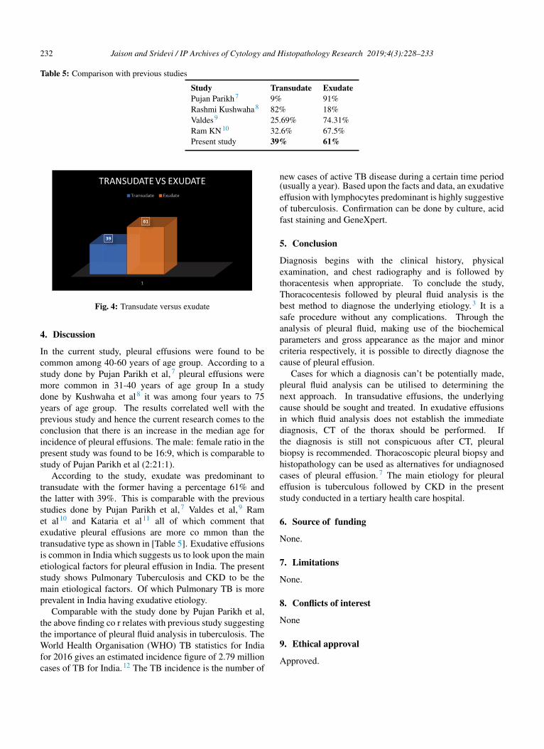
According to the data, thirty nine cases of transudativepleural effusions and sixty one cases of exudative pleuraleffusions were recorded. [Figure 4]
From the known causes of pleural effusion, certain rareconditions like Meigs Syndrome was observed. Meigssyndrome is a triad of benign ovarian tumour, ascites andpleural effusion. Out of hundred cases, 1% cases werereported with this condition. Although the prevalence of thesyndrome is low, it has an important clinical implication.5
According to the research the second most commoncase of pleural effusion was reported in CKD patientswith about 28% of the total number of cases. Pleuraldisease is a common problem in patients with chronicrenal insufficiency.6 There are several reasons why pleural
disease may be common in patients with chronic kidneydisease. These include congestive heart failure, fluidoverload, an increased risk of infection (Especiallytuberculosis), the presence of diseases associated withrenal and pleural manifestations (e.g., systemic lupuserythematosus), uremic pericarditis, an increased risk forcertain malignancies and pulmonary embolism.
The common etiology for pleural effusion includeTuberculosis, Malignancy and CKD according to the currentstudy.
Fig. 1: Age group prevalence
Fig. 2: Gender prevalence
Fig. 3: Acid fast bacilli in pleural fluid (1000X, Ziehl - Neelsenstain)

Jaison and Sridevi / IP Archives of Cytology and Histopathology Research 2019;4(3):228–233 231
Table 1: Research criteria
Type of effusion Total protein (g/dl) [Major] Colour [Minor]Transudate <3 Pale yellowExudate >3 Reddish
Table 2: Age and gender co-relation
Age group Male Female No. of Cases0-20 1 5 620-40 14 6 2040-60 30 16 4660-80 19 7 2680-100 0 2 2Total 64 36 100
Table 3: Total cases of pleural effusion
Clinical Diagnosis No.of casesUnknown causes of pleural effusion 9CKD 28Pulmonary TB 34GU TB 1Potts spine 1TB abdomen 1COPD 2Hydropneumothorax 2SAH 1Haemothorax 1Pneumonia 3Liver Cirrhosis 1Diffuse axonal injury 1CAD 1Anaemia 1Staphylococcal Scalded Skin Syndrome 1Meigs syndrome 1CCF 2DKA 1Ca oropharynx 1Ca Rectum 1Ca Breast with Lung metastasis 1Acute necrotising pancreatitis 1Calcific pericarditis 1Lower respiratory tract infection 1Pyelonephritis 1Pulmonary HTN 1Total = 100
Table 4: Classification of exudate based on DLC
DLC No. of cases Probable causesLymphocyte predominant 55 Malignancy, TBNeutrophil predominant 36 Pulmonary thromboembolism, Pancreatitis, Pneumonia, EmpyemaEosinophil predominant 9 Pneumothorax, Haemothorax, asbestosis
232 Jaison and Sridevi / IP Archives of Cytology and Histopathology Research 2019;4(3):228–233
Table 5: Comparison with previous studies
Study Transudate ExudatePujan Parikh7 9% 91%Rashmi Kushwaha8 82% 18%Valdes9 25.69% 74.31%Ram KN10 32.6% 67.5%Present study 39% 61%
Fig. 4: Transudate versus exudate
4. Discussion
In the current study, pleural effusions were found to becommon among 40-60 years of age group. According to astudy done by Pujan Parikh et al,7 pleural effusions weremore common in 31-40 years of age group In a studydone by Kushwaha et al8 it was among four years to 75years of age group. The results correlated well with theprevious study and hence the current research comes to theconclusion that there is an increase in the median age forincidence of pleural effusions. The male: female ratio in thepresent study was found to be 16:9, which is comparable tostudy of Pujan Parikh et al (2:21:1).
According to the study, exudate was predominant totransudate with the former having a percentage 61% andthe latter with 39%. This is comparable with the previousstudies done by Pujan Parikh et al,7 Valdes et al,9 Ramet al10 and Kataria et al11 all of which comment thatexudative pleural effusions are more co mmon than thetransudative type as shown in [Table 5]. Exudative effusionsis common in India which suggests us to look upon the mainetiological factors for pleural effusion in India. The presentstudy shows Pulmonary Tuberculosis and CKD to be themain etiological factors. Of which Pulmonary TB is moreprevalent in India having exudative etiology.
Comparable with the study done by Pujan Parikh et al,the above finding co r relates with previous study suggestingthe importance of pleural fluid analysis in tuberculosis. TheWorld Health Organisation (WHO) TB statistics for Indiafor 2016 gives an estimated incidence figure of 2.79 millioncases of TB for India.12 The TB incidence is the number of
new cases of active TB disease during a certain time period(usually a year). Based upon the facts and data, an exudativeeffusion with lymphocytes predominant is highly suggestiveof tuberculosis. Confirmation can be done by culture, acidfast staining and GeneXpert.
5. Conclusion
Diagnosis begins with the clinical history, physicalexamination, and chest radiography and is followed bythoracentesis when appropriate. To conclude the study,Thoracocentesis followed by pleural fluid analysis is thebest method to diagnose the underlying etiology.3 It is asafe procedure without any complications. Through theanalysis of pleural fluid, making use of the biochemicalparameters and gross appearance as the major and minorcriteria respectively, it is possible to directly diagnose thecause of pleural effusion.
Cases for which a diagnosis can’t be potentially made,pleural fluid analysis can be utilised to determining thenext approach. In transudative effusions, the underlyingcause should be sought and treated. In exudative effusionsin which fluid analysis does not establish the immediatediagnosis, CT of the thorax should be performed. Ifthe diagnosis is still not conspicuous after CT, pleuralbiopsy is recommended. Thoracoscopic pleural biopsy andhistopathology can be used as alternatives for undiagnosedcases of pleural effusion.7 The main etiology for pleuraleffusion is tuberculous followed by CKD in the presentstudy conducted in a tertiary health care hospital.
6. Source of funding
None.
7. Limitations
None.
8. Conflicts of interest
None
9. Ethical approval
Approved.
Jaison and Sridevi / IP Archives of Cytology and Histopathology Research 2019;4(3):228–233 233
References1. Diagnostic Tools of Pleural Effusion. Tuberc Respir Dis.
2014;76(5):199–210.2. Noppen M, M DW, R L, Gucht KV, J DH, et al. Volume and cellular
content of normal pleural fluid in humans examined by pleural lavage.Am J Respir Crit Care Med. 2000;162(3):1023–1029. Pt 1.
3. Karkhanis VS, Joshi J. Pleural effusion: diagnosis, treatment, andmanagement. Open Access Emerg Med. 2012;4:31–52.
4. Zhai K, Lu Y, Shi HZ. Tuberculous pleural effusion. J Thorac Dis.2016;8(7):486–494.
5. Krenke R, Maskey-Warzechowska M, Korczynski P, Zielinska-Krawczyk M, Klimiuk J, Light CR. Pleural Effusion in MeigsSyndrome-Transudate or Exudate. Med. 2015;94(49).
6. Pleural effusion in a patient with end-stage renal disease. Ann SaudiMed. 2006;26(2):145–146.
7. Parikh P, Odhwani J, Ganagajalia C. Study of 100 cases of pleuraleffusion with reference to diagnostic approach. Int J Adv Med.2016;3(2):328–359.
8. Rashmi K, Shashikala P, Hiremath S, Basavaraj HG. Cells in pleuralfluid and their value in differential fluid diagnosis. J Cytol. 2008;25(4).
9. Valdes L, Alvarez D, Valle JM, Pose A, Jose ES. The etiology ofpleural effusions in an area with high incidence of tuberculosis. Chest.1996;109(1):158–162.
10. Diagnostic value of cholesterol in pleural effusions. JAPI.1995;43(11):748–750.
11. Kataria YP, Khurshid I. Adenosine deaminase in the diagnosis oftuberculous pleural effusion ; 2001,.
12. TB India report 2018: Ministry of Health and Family welfare. Chapter2: TB Disease. Burden & Surveillance in India ; 2018,.
Author biography
Karen Jaison Student
M Sridevi Associate Professor
Cite this article: Jaison K, Sridevi M. Analysis of pleural fluid:Differentiating transudate from exudate. Arch Cytol Histopathol Res2019;4(3):228-233.