analytical biochemistry of nickel biochemistry of nickel* f. william sunderman, jr. departments of...
TRANSCRIPT
Pure & Appi. Chain., Vol.52, pp.527—544. 0033—4545/80/0201—0527 $02.00/OPergamon Press Ltd. 1980. Printed in Great Britain.© IUPAC
INTERNATIONAL UNION OF PUREAND APPLIED CHEMISTRY
CLINICAL CHEMISTRY DIVISION
COMMISSION ON TOXICOLOGY
SUBCOMMITTEE ON ENVIRONMENTAL AND OCCUPATIONAL
TOXICOLOGY OF NICKEL*
ANALYTICAL BIOCHEMISTRYOF NICKEL
Prepared for publication byF. W. SUNDERMAN, JR.
University of Connecticut School of Medicine,Farmington, Connecticut, USA
*Membership of the Subcommittee during the period 1977-79 in which the report wasprepared was as follows:
Chairman: F. W. SUNDERMAN, JR. (USA); Members: D. B. ADAMS (UK), R. T.BARTON (USA), C. BOUDENE (France), S. S. BROWN (UK), J. H. CLARY (USA),A. J. COLLINGS (UK), A. HOGETVEIT (Norway), K. S. KASPRZAK (Poland),5.0. LUXON (UK), E. MASTROMATTEO (Canada), M. D. D. McNEELY (Canada),J. MEININGER (France), M. MIKAC DEVIC (Yugoslavia), L. MORGAN (UK),S. NOMOTO (Japan), T. NORSETH (Norway), C. ONKELINX (USA), B. R. ROY(USA), J. SJOHOLM (Sweden), M. STOEPPLER (FRG), H. ZACHARIASEN(Norway).
ANALYTICAL BIOCHEMISTRY OF NICKEL*
F. William Sunderman, Jr.
Departments of Laboratory Medicine and Pharmacology, University of Con—necticut School of Medicine, Farmington, Connecticut 06032, U.S.A.
Abstract — Measurements of nickel in biological materials are reviewed,with emphasis on (a) preliminary steps for oxidation or removal of organic
matter; (b) pre—concentration and separation procedures; (c) instrumentaltechniques for quantitation of nickel; and (d) reference values for nickelconcentrations in human body fluids, tissues, and excreta. Electrothermalatomic absorption spectrometry is currently the most sensitive, conven—ient, and reliable technique for determination of nickel in biologicalmaterials, but it is rivalled in sensitivity by three other techniques
(differential pulse polarography, particle—induced x—ray emission spec—trometry, and gas chromatography). Isotope dilution gas chromatography—mass spectrometry may eventually become the definitive method for analysisof nickel in biological materials. Until such a definitive method hasbeen developed, the IUPAC provisional reference method (electrothermalatomic absorption spectrometry) will serve for comparative evaluations ofother procedures for clinical measurements of nickel concentrations.
INTRODUCTION
Within the past decade, clinical chemistry and toxicology laboratories throughout the worldhave begun to measure nickel concentrations in human body fluids, tissues, and excrete inorder to monitor exposures to nickel compounds which may result in acute or chronic toxic-
ity. The IUPAC Subcommittee on Environmental and Occupational Toxicology of Nickel hasconducted two interlaboratory surveys in which urine specimens were distributed to labora-tories in seven countries for measurements of nickel concentrations by atomic absorptionspectrometry. Both surveys disclosed wide discrepancies in the nickel concentrations thatwere obtained by participating laboratories. Adams et al (1) reported these findings andpointed out the serious need for improved accuracy of nickel determinations in body fluids.The interlaboratory comparisons showed that atomic absorption procedures with preliminaryoxidation and extraction steps were generally superior to direct electrothermal atomization
techniques in (a) analytical sensitivity; (b) recovery of added nickel; (c) interlaboratoryprecision; and (d) concordance of ranking of urine samples in order of increasing nickelconcentrations. Adams et al (1) suggested that a reference procedure for analysis of nickelin biological materials was needed to harmonize the discordant results of nickel determina-tions.
The IUPAC Subcommittee on Environmental and Occupational Toxicology of Nickel sponsored anInternational Conference on Nickel Toxicology which was held in Kristiansand, Norway in May1978 (2). The Working Group on Nickel Analysis at the Kristiansand Conference agreed upon aprovisional reference method for analysis of nickel in serum and urine. This method is cur-rently being evaluated in laboratories in the United States, Canada, England, West Germany,Finland, and Japan. Contingent upon the outcome of these trials, the provisional referencemethod will be published for critique by scientists throughout the world. Supporting docu-ments for the provisional reference method include a report by Ader and Stoeppler (3) onradiochemical measurements of the recovery and analysis of nickel in urine and the presentreview on the analytical biochemistry of nickel. This article is intended to fill a void inthe scientific literature, since methods for analysis of nickel in biological materials havenot previously been reviewed. In contrast, the metabolism, clinical biochemistry, radio—chemistry, toxicology, and carcinogenicity of nickel have been summarized in several recentarticles and monographs (4—9).
* This paper was presented at the 27th IUPAC Congress in Helsinki, Finland, on August 28,1979. It was written on behalf of the Subcommittee on Environmental and OccupationalToxicology of Nickel, Commission on Toxicology, Division of Clinical Chemistry, Inter-national Union of Pure and Applied Chemistry. This work was supported by research grantsEV—O314O from the U.S. tpartment of Energy and ES—O1337 from the U.S. National Instituteof Environmental Health Sciences.
PAAC 52:2—T 529
530 COMMISSION ON TOXICOLOGY
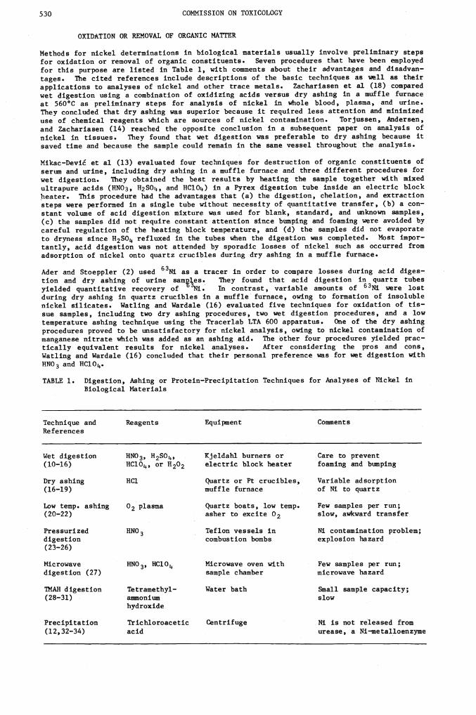
OXIDATION OR REMOVAL OF ORGANIC MATTER
Methods for nickel determinations in biological materials usually involve preliminary stepsfor oxidation or removal of organic constituents. Seven procedures that have been employedfor this purpose are listed in Table 1, with comments about their advantages and disadvan—tages. The cited references include descriptions of the basic techniques as well as theirapplications to analyses of nickel and other trace metals. Zachariasen et al (18) comparedwet digestion using a combination of oxidizing acids versus dry ashing in a muffle furnaceat 560°C as preliminary steps for analysis of nickel in whole blood, plasma, and urine.They concluded that dry ashing was superior because it required less attention and minimizeduse of chemical reagents which are sources of nickel contamination. Torjussen, Andersen,and Zachariasen (14) reached the opposite conclusion in a subsequent paper on analysis ofnickel in tissues. They found that wet digestion was preferable to dry ashing because itsaved time and because the sample could remain in the same vessel throughout the analysis.
Mikac—Devi et al (13) evaluated four techniques for destruction of organic constituents ofserum and urine, including dry ashing in a muffle furnace and three different procedures forwet digestion. They obtained the best results by heating the sample together with mixedultrapure acids (HNO3, H2SOL,, and HClOz) in a Pyrex digestion tube inside an electric blockheater. This procedure had the advantages that (a) the digestion, chelation, and extractionsteps were performed in a single tube without necessity of quantitative transfer, (b) a con-stant volume of acid digestion mixture was used for blank, standard, and unknown samples,(c) the samples did not require constant attention since bumping and foaming were avoided bycareful regulation of the heating block temperature, and (d) the samples did not evaporateto dryness since H2SO refluxed in the tubes when the digestion was completed. Most impor-tantly, acid digestion was not attended by sporadic losses of nickel such as occurred fromadsorption of nickel onto quartz crucibles during dry ashing in a muffle furnace.
Ader and Stoeppler (2) used 63Ni as a tracer in order to compare losses during acid diges-tion and dry ashing of urine sam]es. They found that acid digestion in quartz tubes
yielded quantitative recovery of 'Ni. In contrast, variable amounts of 63Ni were lostduring dry ashing in quartz crucibles in a muffle furnace, owing to formation of insolublenickel silicates. Watling and Wardale (16) evaluated five techniques for oxidation of tis-sue samples, including two dry ashing procedures, two wet digestion procedures, and a lowtemperature ashing technique using the Tracerlab LTA 600 apparatus. One of the dry ashingprocedures proved to be unsatisfactory for nickel analysis, owing to nickel contamination ofmanganese nitrate which was added as an ashing aid. The other four procedures yielded prac-tically equivalent results for nickel analyses. After considering the pros and cons,Watling and Wardale (16) concluded that their personal preference was for wet digestion with
HNO3 and HClO,.
TABLE 1. Digestion, Ashing or Protein—Precipitation Techniques for Analyses of Nickel inBiological Materials
Technique andReferences
Reagents Equipment Comments
Wet digestion(10—16)
HNO3, H2SO,HC1O4, or H202
KjeldaKl burners orelectric block heater
Care to prevent
foaming and bumping
Dry ashing(16—19)
BCl Quartz or Pt crucibles,muffle furnace
Variable adsorptionof Ni to quartz
Low temp. ashing(20—22)
02 plasma Quartz boats, low temp.asher to excite 02
Few samples per run;slow, awkward transfer
Pressurized
digestion(23—26)
HNO3 Teflon vessels incombustion bombs
Ni contamination problem;explosion hazard
Microwave
digestion (27)HNO3, HCl0 Microwave oven with
sample chamberFew samples per run;microwave hazard
ThAH digestion(28—31)
Tetramethyl—ammoniumhydroxide
Water bath Small sample capacity;slow
Precipitation(12,32—34)
Trichloroaceticacid
Centrifuge Ni is not released from
urease, a Ni—metalloenzyme
Analytical biochemistry of nickel 531
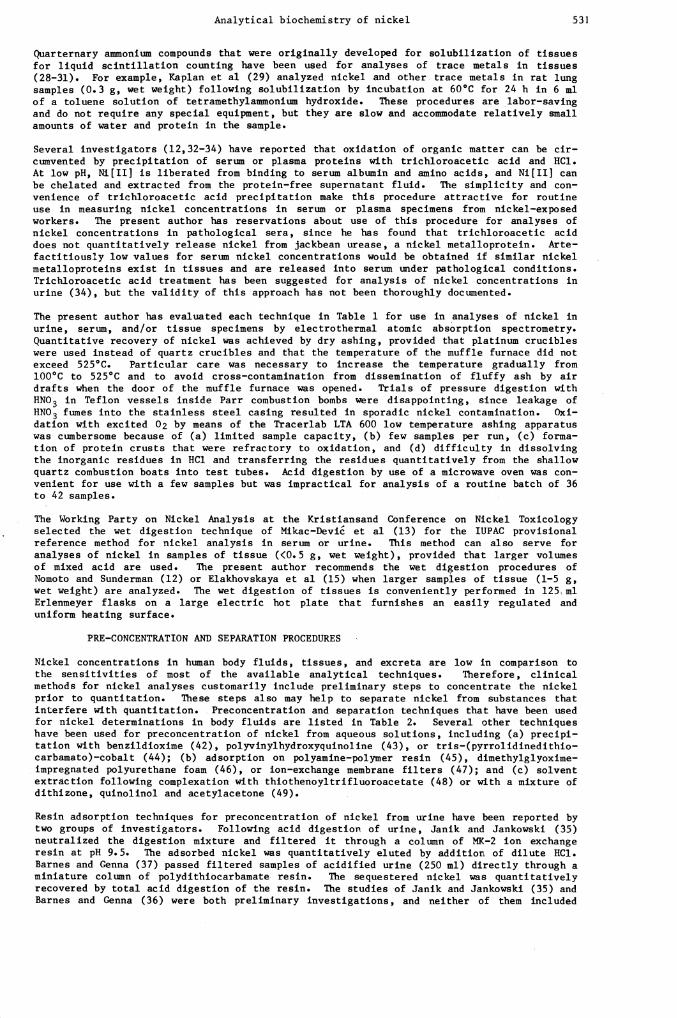
Quarternary ammonlum compounds that were originally developed for solubilization of tissuesfor liquid scintillation counting have been used for analyses of trace metals in tissues(28—31). For example, Kaplan et al (29) analyzed nickel and other trace metals in rat lungsamples (0.3 g, wet weight) following solubilization by incubation at 60°C for 24 h in 6 mlof a toluene solution of tetramethylammonium hydroxide. These procedures are labor—savingand do not require any special equipment, but they are slow and accommodate relatively smallamounts of water and protein in the sample.
Several investigators (12,32—34) have reported that oxidation of organic matter can be cir—cumvented by precipitation of serum or plasma proteins with trichloroacetic acid and HC1.At low pH, Ni[IIJ is liberated from binding to serum albumin and amino acids, and Nifil] canbe chelated and extracted from the protein—free supernatant fluid. The simplicity and con—venience of trichloroacetic acid precipitation make this procedure attractive for routineuse in measuring nickel concentrations in serum or plasma specimens from nickel—exposedworkers. The present author has reservations about use of this procedure for analyses ofnickel concentrations in pathological sera, since he has found that trichloroacetic aciddoes not quantitatively release nickel from jackbean urease, a nickel metalloprotein. Arte—factitiously low values for serum nickel concentrations would be obtained if similar nickelnetalloproteins exist in tissues and are released into serum under pathological conditions.Trichloroacetic acid treatment has been suggested for analysis of nickel concentrations inurine (34), but the validity of this approach has not been thoroughly documented.
The present author has evaluated each technique in Table 1 for use in analyses of nickel in
urine, serum, and/or tissue specimens by electrothermal atomic absorption spectronetry.Quantitative recovery of nickel was achieved by dry ashing, provided that platinum crucibleswere used instead of quartz crucibles and that the temperature of the muffle furnace did notexceed 525°C. Particular care was necessary to increase the temperature gradually from100°C to 525°C and to avoid cross—contamination from dissemination of fluffy ash by airdrafts when the door of the muffle furnace was opened. Trials of pressure digestion withHNO3 in Teflon vessels inside Parr combustion bombs were disappointing, since leakage ofHNO3 fumes into the stainless steel casing resulted in sporadic nickel contamination. Oxi—dation with excited 02 by means of the Tracerlab LTA 600 low temperature ashing apparatuswas cumbersome because of (a) limited sample capacity, (b) few samples per run, (c) forma—tion of protein crusts that were refractory to oxidation, and (d) difficulty in dissolvingthe inorganic residues in HC1 and transferring the residues quantitatively from the shallowquartz combustion boats into test tubes. Acid digestion by use of a microwave oven was con-venient for use with a few samples but was impractical for analysis of a routine batch of 36to 42 samples.
The Working Party on Nickel Analysis at the Kristiansand Conference on Nickel Toxicologyselected the wet digestion technique of Mikac—Devii et al (13) for the IUPAC provisionalreference method for nickel analysis in serum or urine. This method can also serve foranalyses of nickel in samples of tissue (<0.5 g, wet weight), provided that larger volumesof mixed acid are used. The present author recommends the wet digestion procedures ofNomoto and Sunderman (12) or Elakhovskaya et al (15) when larger samples of tissue (1—5 g,wet weight) are analyzed. The wet digestion of tissues is conveniently performed in 125 mlErlenmeyer flasks on a large electric hot plate that furnishes an easily regulated anduniform heating surface.
PRE—CONCENTRATION AND SEPARATION PROCEDURES
Nickel concentrations in human body fluids, tissues, and excreta are low in comparison tothe sensitivities of most of the available analytical techniques. Therefore, clinicalmethods for nickel analyses customarily include preliminary steps to concentrate the nickelprior to quantitation. These steps also may help to separate nickel from substances thatinterfere with quantitation. Preconcentration and separation techniques that have been usedfor nickel determinations in body fluids are listed in Table 2. Several other techniqueshave been used for preconcentration of nickel from aqueous solutions, including (a) precipi-tation with benzildioxime (42), polyvinylhydroxyquinoline (43), or tris—(pyrrolidinedithio—carbamato)—cobalt (44); (b) adsorption on polyanine—polymer resin (45), dimethylglyoxime—impregnated polyurethane foam (46), or ion—exchange membrane filters (47); and (c) solventextraction following complexation with thiothenoyltrifluoroacetate (48) or with a mixture ofdithizone, quinolinol and acetylacetone (49).
Resin adsorption techniques for preconcentration of nickel from urine have been reported bytwo groups of investigators. Following acid digestion of urine, Janik and Jankowski (35)neutralized the digestion mixture and filtered it through a column of ME—2 ion exchangeresin at pH 9.5. The adsorbed nickel was quantitatively eluted by addition of dilute HC1.Barnes and Cenna (37) passed filtered samples of acidified urine (250 ml) directly through aminiature column of polydithiocarbamate resin. The sequestered nickel was quantitativelyrecovered by total acid digestion of the resin. The studies of Janik and Jankowski (35) andBarnes and Cenna (36) were both preliminary investigations, and neither of them included
532 COMMISSION ON TOXICOLOGY
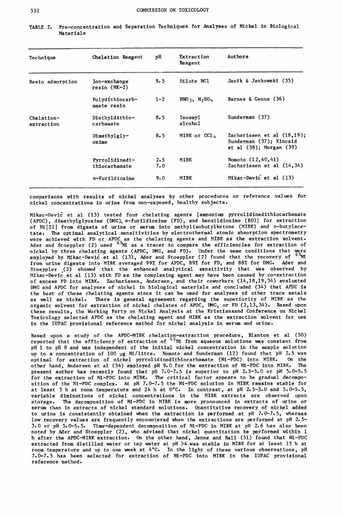
TABLE 2. Pre—concentration and Separation Techniques for Analyses of Nickel in BiologicalMaterials
Technique Chelation Reagent pH Extraction Authors
Reagent
Resin adsorption Ion—exchangeresin (MK—2)
9.5 Dilute HC1 Janik & Jankowski (35)
Polydithiocarb—amate resin
1—2 HNO3, H2SO Barnes & Genna (36)
Chelation— Diethyldithio— 8.5 Isoamyl Sunderman (37)
extraction carbaniate alcohol
Dimethylgly—oxime
8.5 MIBK or CCl Zachariasen et al (18,19);Sundemman (37); Kincaidet al (38); Morgan (39)
Pyrrolidinedi—thiocarbamate
2.5
7.0MIBK Nomoto (12,40,41)
Zachariasen et al (14,34)
ct—Furildioxime 9.0 MIBK Mikac—Devic et al (13)
comparisons with results of nickel analyses by other procedures or reference values fornickel concentrations in urine from non—exposed, healthy subjects.
Mikac—Devi et al (13) tested four chelating agents [ammonium pyrrolidinedithiocarbamate(APDC), dimethylglyoxmme (DMG), ct—furildioxmme (FD), and benzildioxime (BD)] for extractionof Ni[II] from digests of urine or serum into methylisobutylketone (MIBK) and n—butylace—tate. The optimal analytical sensitivities by electrothermal atomic absorption spectrometrywere achieved with FD or APDC as the chelating agents and MIBI( as the extraction solvent.Ader and Stoeppler (2) used 63 as a tracer to compare the efficiencies for extraction ofnickel by three chelating agents (APDC, DMG, and FD). Under the sane conditions that wereemployed by M1kac—Devi et al (13), Ader and Stoeppler (2) found that the recovery of 6Nifrom urine digests into MIRK averaged 99% for APDC, 89% for FD, and 88% for DMG. Ader andStoeppler (2) showed that the enhanced analytical sensitivity that was observed byMikac—Devic et al (13) with FD as the conplexing agent may have been caused by co—extractionof excess FD into MIBK. Zachariasen, Andersen, and their coworkers (14,18,19,34) evaluatedDMG and APDC for analyses of nickel in biological materials and concluded (34) that APDC isthe best of these chelating agents since it can be used for analyses of other trace metalsas well as nickel. There is general agreement regarding the superiority of MIBK as theorganic solvent for extraction of nickel chelates of APDC, DMG, or FD (2,13,34). Based uponthese results, the Working Party on Nickel Analysis at the Kristiansand Conference on NickelToxicology selected APDC as the chelating agent and MIBK as the extraction solvent for usein the IUPAC provisional reference method for nickel analysis in serum and urine.
Based upon a study of the APDC—MIBK chelation—extraction procedure, Blanton et al (50)reported that the efficiency of extraction of 63Ni from aqueous solutions was constant frompH 1 to pH 8 and was independent of the initial nickel concentration in the sample solutionup to a concentration of 100 pg Ni/litre. Nomoto and Sundemman (12) found that pH 2.5 wasoptimal for extraction of nickel pyrrolidinedithiocarbamate (Ni—PDC) into MIBK. On theother hand, Andersen et al (34) employed pH 9.0 for the extraction of Ni—PDC into MIBK. Thepresent author has recently found that pH 7.0—7.5 is superior to pH 2.5—3.0 or pH 5.0—5.5for the extraction of Ni—PDC into MIBK. The critical factor appears to be gradual decompo-sition of the Ni—PDC complex. At pH 7.0—7.5 the Ni—PDC solution in MIBK remains stable forat least 3 h at room temperature and 24 h at 0°C. In contrast, at pH 2.5—3.0 and 5.0—5.5,variable diminutions of nickel concentrations in the MIBK extracts are observed uponstorage. The decomposition of Ni—PDC in MIBK is more pronounced in extracts of urine orserum than in extracts of nickel standard solutions. Quantitative recovery of nickel addedto urine is consistently obtained when the extraction is performed at pH 7.0—7.5, whereaslow recovery values are frequently encountered when the extractions are performed at pH 2.5—3.0 or pH 5.0—5.5. Time—dependent decomposition of Ni—PDC in MIBK at pH 2.6 has also beennoted by Ader and Stoeppler (2), who advised that nickel quantitation be performed within 1h after the APDC—MIBK extraction. On the other hand, Jenne and Ball (51) found that Ni—PDCextracted from distilled water or tap water at pH >4 was stable in MIBK for at least 15 h atroom temperature and up to one week at 4°C. In the light of these various observations, pH7.0—7.5 has been selected for extraction of Ni—PDC into MIBK in the IUPAC provisionalreference method.
Analytical biochemistry of nickel 533
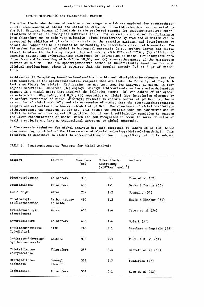
SPECTROPHOTOMETRIC AND FLUOROMETRIC METHODS
The molar lineic absorbances of various color reagents which are employed for spectrophoto—metric measurements of nickel are listed in Table 3. cz—Furildioxime has been selected bythe U.S. National Bureau of Standards as the preferred reagent for spectrophotometric deter—minations of nickel in biological materials (61). The extraction of nickel furildioximateinto chloroform can be made very selective, since interference by iron and aluminium can beprevented by addition of citrate or tartrate to the reaction mixture, and interference bycobalt and copper can be eliminated by backwashing the chloroform extract with ammonia. TheNBS method for analysis of nickel in biological materials (e.g., orchard leaves and bovineliver) involves the following steps: (a) wet ashing with HNO3 and HC1O4; (b) addition ofammonium citrate and cL—furildioxime solutions; (c) extraction of nickel furildioximate intochloroform and backwashing with d•ilute NHOH; and (d) spectrophotometry of the chloroformextract at 435 nm. The NBS spectrophtometric method is insufficiently sensitive for mostclinical applications, since it requires that the samples contain 0.5 to 4 g of nickel(61).
Zephiranine (l,2—naphthoquinonedioxime—4—sulfonic acid) and diethyldithiocarbamate are themost sensitive of the spectrophotometric reagents that are listed in Table 3, but they bothlack -specificity for nickel. Zephiramine has not been used for analyses of nickel n bio-logical materials. Sunderman (37) employed diethyldithiocarbamate as the spectrophotometricreagent in a nickel assay that involved the following steps: (a) wet ashing of biologicalmaterials with HNO3, H2SO, and H202; (b) separation of nickel from interfering elements bychloroform extraction of nickel c{imethylglyoximate in citrate buffer at pH 8.5; (c) back—extraction of nickel with HC1; and (d) conversion of nickel into the diethyldithiocarbamatecomplex and extraction into isoamyl alcohol at pH 8.5. The absorbance of nickel bisdiethyl—dithiocarbamate was measured at 325 nm. This method was suitable when the concentrations ofnickel in serum or urine exceed 10 pg/litre, but it was insufficiently sensitive to measurethe lower concentrations of nickel which are now recognized to occur in serum or urine ofhealthy subjects who have no occupational exposures to nickel compounds.
A fluorometric technique for nickel analysis has been described by Schenk et al (62) basedupon quenching by nickel of the fluorescence of aluminium—l—(2—pyridylazo)—2—naphthol. Thisprocedure is sensitive to nickel in concentrations as low as 1 iig/litre, but it is subject
TABLE 3. Spectrophotometric Reagents for Nickel Analysis
Reagent Solvent Abs. Max.
(mm)
Molar LineicAbsorbance(xlO6A.&1 mol 1)
Authors
Dimethylglyoxime Chloroform 335 0.5 Kuse et al (52)
Benzildioxime Chloroform 406 1.1 Banks & Barnum (53)
KCN & NHOH Water 267 1.2 Scoggins (54)
Thiothenoyl—trifluoroacetone
Carbon tetra—chloride
480 1.2 Muyle & Khopkar (55)
Cyclohexane—l,2—dionedioxime
Water 460 1.4 Perez et al (56)
c*—Furildioxime Chloroform 435 1.6 Bodart (57)
6—Nitroquinoxaline—2, 3—dithiol
MIBK 710 2.1 Bhaskare & Jagadale (58)
3—Nitroso—4—hydroxy—5, 6—benzocoumarin
Acetone 395 2.5 Kohli & Singh (59)
Thiotrifluoro—
acetylacetoneChloroform 256 3.4 Barratt et al (60)
Diethyldithio—carbamate
Isoamylalcohol
325 3.7 Sunderman (37)
Zephiramine Chloroform 307 5.1 Kuse et al (52)
534 COMMISSION ON TOXICOLOGY
to interference by other metals and by chloride, sulfate, and phosphate ions. Therefore, Ithardly appears to be suitable for nickel analysis of biological materials.
REACTION RATE METHODS
Mealor and Townshend (63) developed a kinetic method for microdetermination of nickel basedupon its catalytic effect on the decomposition of permanganate in alkaline solution in thepresence of acetodiphosphoric acid. The rate of the reaction is proportional to the squareof the nickel concentration. This reaction has been employed by Hadjiioannou et al (64) foran automated spectrophotometric reaction—rate system. Amounts of nickel in the range of0.3—2.1 jig/sample can be determined withcoefficients of variation of 2.5% within measure—ment times of 10 to 50 seconds (64). Kurzawa and Kubaszewski (65) described a kineticmethod for nickel analysis based on a reaction of sodium azide with iodine that is catalyzed
by sodium diethyldithiocarbamate. Nickel diethyldithiocarbamate does not catalyze theiodine—azide reaction. Therefore, the concentration of nickel in the reaction mixture isinversely related to the velocity of the iodine—azide reaction. Kurzawa an4 Kubaszewski
(65) applied the reaction system to analyses of samples containing 0.14—14 tg of nickel,including measurements of nickel concentrations in margarine and drugs.
DIFFERENTIAL PULSE POLAROGRAPHY
Polarographic techniques for nickel analysis have been described by several workers (49,66—68), but these techniques have lacked sufficient sensitivity to be employed for nickeldeterminatiois in biological materials. Recently Flora and Nieboer (69) found that additionof dimethylglyo2dme to ammoniacal tartrate or citrate buffers enhances by a factor of 15 thesensitivity of derivative polarography of nickel at a dropping mercury electrode. This
enhancement phenometwn has also been noted by. Vinogradova and Prokhorova (70) and Astafevaet al (71). By means of dimethylglyoxime—sensitized differential pulse polarography, Floraand Nieboer (69) detected nickel concentrations as low as 2—3 ig/litre in buffered reactionmixtures. In a preliminary study, Nieboer et al (72) applied their procedure to measure—ments of nickel in human urine and blood following oxidation of organic constituents by dry
ashing. Good agreement was observed between nickel analyses in body fluids by pulse polar—ography and by electrothermal atomic absorption spectrometry. Nieboer (personal communica—tion) is currently refining the dimethylglyoxime—sensitized polarographic technique foranalysis of nickel in biological materials, and he anticipates that the technique cam serveas a means for independent assessment of the IUPAC provisional reference method.
X—RAY FLUORESCENCE SPECTROMETRY
Concentrations of nickel that are found in human body fluids, tissues, and excreta are too
low to permit direct measurements of nickel by standard techniques of x—ray fluorescence
spectrometry. Forssen (17) ashed human tissues in a muffle furnace and compressed 30 mg
aliquots of the ash into a wafer (17 mm in diameter) by means of a hydraulic press. The
wafer served as the target of the incident x—ray beam. The spectral emission lines of 13elements including nickel were scanned with a lithium fluoride detector. Forssen (13)reported that the detection limit for nickel was approximately 10 pg/gram of ash. She wasable to measure nickel in only 20 of 665 tissue samples. Kessler and Mitchell (73) substan-tially increased the sensitivity of x—ray fluorescence spectrometry by preliminaryco—precipitation of trace metals by addition of titanium in the presence of diethyldithio—carbamate. The precipitate was confined to a microdot (1.3 mm in diameter) on a filter discwhich served as the x—ray target. Nickel was detected in aqueous solution in amounts assmall as 0.6 mg/sample, equivalent to a nickel concentration of approximately 2 11g/litre.Kessler and Mitchell (73) did not employ their procedure for measurements of nickel inbiological materialg.
PARTICLE—INDUCED X—RAY EMISSION SPECTROMETRY
Particle—induced x—ray emission ("PIXE") spectrometry is more sensitive than x—ray fluores-cence spectrometry, and it has recently been used for detection and quantitation of varioustrace metals in tissues and body fluids (73—76). In practice, a 2—4 MeV proton beam from aVan de Graff generator is focused magnetically upon a dried sample inside a vacuum chamber.The sample is deposited as a spot (10—20 mm in diameter) on a target that is composed of a
thin organic film (e.g., "Mylar," "Nucleopore," or carbon—impregnated polycarbonate). Theproton beam dislodges inner shell electrons from atoms in the sample, and the inner shellvacancies are immediately filled by outer shell electrons. This process induces release ofx—rays with energies that are characteristic of the element from which they were derived.The intensity of x—ray emission at each specific energy level is detected by a silicondetector and is quantified with a multi—channel analyzer. The intensity of x—ray emissionat a specific energy level is proportional to the concentration of the corresponding elementin the sample. Quantitation of PIXE analyses is accomplished either by use of internalstandards such as strontium, or by spiking the sample with known amounts of the element(s)to be analyzed (74—77).
Analytical biochemistry of nickel 535
Three groups of Investigators (78—80) have attempted to perform direct measurements ofnickel concentrations In dried human blood sertun by the PIXE technique, but the analyticalsensitivities were barely sufficient to detect the presence of nickel. Campbell (77)reviewed these data and concluded that ashing and pre—concentration will be required forquantitation of nickel in human serum by the PIXE technique. Chen et al (81) successfullyused PIXE for measurements of nickel concentrations in tissue specimens obtainedat autopsyfrom control patients and from patients who died of Legionnaires' disease. Samples (0.1—0.5 g, wet weight) were digested in 1*103 and 10 p1 aliquots were evaporated to dryness andanalyzed by PIXE with a 2 MeV proton beam. The x—ray intensity at the 7.472 Key energylevel that is characteristic of nickel atoms was used to estimate nickel concentrations bythe standard additions method. The correlation coefficient for nickel concentrationsobtained by then et al (81) using the PIXE method and those obtained by the present author'slaboratory using the IUPAC provisional reference method was 0.936, based upon paired meas—urements of 5 samples of lung tissue. These limited data suggest that PIXE is a promisingapproach to analysis of nickel in tissues for laboratories that possess the requisiteinstrumentation.
NEUTRON AND CHARGED PARTICLE ACTIVATION ANALYSIS
The practical usefulness of neutron activation analysis of nickel in biological materials islimited by the relative insensitivity of this technique for nickel. Lux and Zeisler (82)employed activation analysis using reactor irradiation and y—spectrometry with a Ce(Li)well—type detector for measurements of trace metals in human connective tissue samples (0.1—0.2 g, wet weight). The detection limit for nickel was 0.5 ig/g (wet weight). Lux andZeisler (82) did not detect nickel in normal connective tissue, but they did demonstrate thepresence of nickel in connective tissue samples taken near nickel—containing metal implants.Swanson and Truesdale (83) used neutron activation for analysis of nickel and other metalsin human lenses which had been lyophilized after quenching in liquid nitrogen. Swanson andTruesdale (83) speculated that nickel accumulation might be involved in the pathogenesis ofsenile cataracts, since nickel was inconstantly detected in lenses from young patients, butit was present in readily measured concentrations in cataractous lenses from senilepatients. Swindle and Schweikert (84) described a procedure for analysis of nickel bycharged particle activation analysis using an 88—inch cyclotron, based upon the reaction58Ni(p,pn)57Ni (t½ = 36 h). Post—irradiation chemical separation of 57Ni resulted in adetection limit for nickel of approximately I pg/g in inorganic reference materials.Versieck et al (85) measured 58Co produced by the reaction 5Ni(p,n) 58Co (t½ 71 da) tostudy the influence of contamination from needles and scalpels upon the nickel concentrationin human liver.
ISOTOPE DILUTION MASS SPECTROMETRY
Paulsen et al (86) and Moore et al (87) described techniques for nickel analysis by stableisotope dilution and spark source mass spectrometry. In the procedure of Moore et al (87)one aliquot of a dissolved sample was spiked with stable 62Ni, and nickel was extracted asthe dimethylglyoxime complex from an amnoniacal solution into chloroform and back extractedfrom the chloroform with dilute HNO3. Nickel was separated by cation exchange chromatogra-phy, and the ratios of 58Ni/62Ni and 60Ni/62Ni were determined by mass spectrometry with athermal ionization technique at 2060°C. A rhenium ribbon filament was used to reduce nickelbackground. The concentration of nickel was calculated from the relative abundances of58Ni, 60Ni, and 62Ni in the spiked and natural samples. Moore et al (87) applied this pro-cedure to measurements of nickel in fuel oil, coal, and fly ash. To date, isotope dilutionmass spectrometry has not been used for measurements of nickel in biological materials, butsuch measurements should be feasible in view of the recent success of Marino (88) andVeillon et al (89) in application of stable isotope dilution to determination of chromium inbiological materials. Veillon et al (89) ashed lyophilized urine samples in an 02 plasmadischarge and spiked the samples with 50Cr. A volatile, thermally stable trifluoroacetyl—acetone chelate of chromium was isolated and the isotope ratio of 50Cr/52Cr was measured by
combined gas chromatography—mass spectrometry using a Finnigan quadrupole mass spectrometer.The concentration of chromium in pooled urine from healthy adults averaged 0.32±0.02 ug/litre, which was about an order of magnitude lower than previously believed (89). Theinvestigations of Veillon et al (89) and Marino (88) have shown that isotope dilution massspectrometry with gas chromatographic separation is an extremely powerful analytical methodfor trace metals in biological materials. Attempts to adapt the method of Veillon et al(89) for analysis of nickel in body fluids are currently in progress in the present author'slaboratory.
RADIODISPLACEMENT ANALYSIS
German et al (90) developed a radioactive tracer displacement technique for determination ofsmall quantities of nickel. Nickel is first isolated by dimethylglyoxime extraction andthen determined by the displacement reaction between Ni[IIJ and 65ZnEDTA. The 65Zn which isdisplaced from 65ZnEDTA is extracted into a dithizone—CCl solution, and 65Zn is measured byy—spectrometry. German et al (90) demonstrated that samples containing as little as 0.5 ig
536.
COMMISSION ON TOXICOLOGY
of nickel could be analyzed by this relatively simple technique. This method has not beenused for analyses of nickel in biological materials.
.
GAS CHROMATOGRAPHY
Gas chromatography of nickel complexes, particularly with ligands of the —diketone type,has been a topic of intensive investigation during the past decade (91—99). The —diketonespossess thermal stability and volatility which are favorable for gas chromatography andtheir solubility in polar organic soluents is an advantage for preliminary solvent extrac—tion. Substitution with fluoro or thiol groups increases the sensitivity of these compoundsfor electron capture detection. The gas chromatographic reagents that are listed in Table 4are all —diketone derivatives, excepting dipropyldithiocarbamate, which is also an attrac—tive reagent for it yields excellent separations of nickel from copper and zinc (99).
larratt et al (94) and Uden et al (95) employed gas chromatography for measurements ofnickel in biological materials. For analysis of nickel and copper in mouse liver, lung, andkidney, TJden et al (95) ashed the samples in a muffle furnace and dissolved the residue inacid (0.3 g, wet weight, of tissue/ml of acid). Aliquots (100 p1) of the dissolved ash warealkalinized with gaseous NH3 and 1 ml of an ethanolic solution of the ligand [H2(enTFA2)1was added. After addition of 20 ml of H20, Ni(enTFA2) and Cu(enTFA2) were extracted into 1ml of benzene. Gas chromatography was performed under conditions indicated in Table 4. Thedetection limit was 20 pg/sample (1 p1) injected onto the column, with a 63Ni electroncapture detector and 4 pg/sample with a scandium tritide electron capture detector. Thenickel detection limit achieved with the scandium tritide detector was equivalent to approx-imately 15 pg/kg (wet weight) of tissue. Nickel concentrations in the mouse ticsues warenot specified, but the authors noted that the results agreed with values obtained by atomicabsorption spectrometry. Barratt et al (94) used a similar procedure to measure nickel insamples of instant tea and hydrogenated triglycerides which contained from 4—13 pg/g. Thus,it appears that gas chromatography can serve as a practical technique for measurements ofnickel in biological samples. To date, no thorough evaluations have been performed of gaschromatography of nickel in body fluids or excreta.
HIGH PERFORMANCE LIQUID CHROMATOGRAPHY
Uden and Walthers (100) reported that the nickel complex_with N,N'—ethylenebis(salicylaldi—mine) has a molar lineic absorbance of 5.OxlO6Am1mol 1 at 254 mm, and they showed thatthe Ni(enSal2) complex can be separated from the copper complex, Cu(enSal2), by high perfor-mance liquid chromatography on microparticulate silica with a solvent system consisting of20% acetonitrile in methylene chloride. By use of an ultraviolet detector at 254 mm and aflow cell with a volume of 8 p1, Uden and Walthers (100) achieved a nickel detection limit
TABLE 4. Chelation Reagents for Nickel Analyses by Gas Chromatography with ElectronCapture Detection
Reagent Abbrev—iation
ExtractionSolvent
Column Packing Column
Temp.(°C)
Authors
Trifluoro—
acetylacetone
TFA Benzene 2% Silicone SE—30on Chromosorb WHP
165—230 Tamura
(93)
et al
Monothiotrifluoro—
acetylacetone
T—TFA n—Hexane 5% Silicone E—350on Universal B
140—170 Barratt
(94)
et al
Bis(trifluoroacetyl—acetone)—ethylene—diimine
H2(enTFA2) Benzene
n—Hexane
1.5% OV—101 onChromosorb W
3% Silicone QF—1on Varaport 30
225
150
Uden et
(95)
Belcher
(96)
al
et al
N,N'—propylenebis—trifluoroacetyl—ace tone mine
H2(pnTFA2) Benzene 1.5% Dexsil 200on Chromosorb W
260 Uden et
(97)
al
Bis(acetylpivalyl—methane)—ethylene—diimine
H2(en(APM)2) Cyclo—hexane
5% Silicone E—350on Universal B
285 Belcher
(98)
et al
Dipropyldithio—carbamate
DPDTC Chloro—form
1% Dexsil 300on Chromosorb WHP
245 Cemmer—Colos& Neeb (99)
Analytical biochemistry of nickel 537
of approximately 5 ng/sample. Linear relationship between absorbance and Ni(enSal2) concen—tration was maintained up to the pg level. Liska et al (101—103) investigated the separa—tion of metal complexes of N—substituted dithiocarbamic acids by high performance liquid
chromatography. In their latest paper, Liska et al (103) showed that nickel bisdiethyldi—thiocarbamate can be separated from the corresponding complexes of Zn, Cu, Mn, Pb, Co, Cd,and Fe on microparticulate silica with 10% chloroform in cyclohexane as the solvent. Liskaet al (101) noted that the limit of detection of nickel bisdiethydithiocarbamate at 325 nis lO9—lO'10mol by use of a UV detector attached to the high performance liquid chromato—
graph. ?bre sensitive detection of nickel can probably be achieved by use of flame orelectrothermal atomic absorption detectors, as described by Jones et al (104), Koizumi et al
(105), and Vickrey et al (106). The usefulness of high performance liquid chromatographyfor trace analysis of nickel in biological materials has not yet been demonstrated.
ATOMIC EMISSION SPECTROMETRY
During the decade from 1955 to 1964, several investigators surveyed the concentrations oftrace metals in human blood and autopsy tissues by emission spectrography, and measurementsof nickel concentrations were frequently included in the tabulated results of these studies
(107—113). Little reliance can be placed upon these measurements of nickel, since thenickel concentrations were either below or barely above the detection limits.. When moresensitive atomic absorption procedures were developed during the mid—196O's, measurements ofnickel in biological materials by emission spectrography were generally abandoned. Renewedinterest in atomic emission techniques for nickel analysis has been evoked by the recentdevelopment of inductively coupled plasma—atomic emission spectrometry (36,114). Haas et al(114) have described an instrument for direct multi—element analysis of urine based upon (a)ultrasonic nebulization of the sample; (b) aspiration of the sample vapor by an argon streaminto a luminous plasma produced by an induction coil; and (c) simultaneous detection ofphotoemission at 20 wavelengths by a polychromator. Emission intensities of added amountsof internal reference elementswere used to compensate for variations in nebulization effi—
ciency. However, with the simple procedure for sample preparation that was used, theinstrument lacked sufficient sensitivity to detect nickel in normal urine. Haas et al (114)stated that the detection limit for nickel was approximately 4—9 pg/litre of urine. Barnesand Genna (36) overcame this limitation by pre—concentration of metals in urine by a factorof 125 by use of a poly(dithiocarbanate) resin. Samples of urine (250 ml) were passedthrough a resin column, and the sequestered metals were recovered by digestion of the resinto achieve a final sample volume pf 2 ml. The sample was then aspirated into the induc—tively coupled plasma for determination of 10 trace metals including nickel. Barnes andGenna (36) noted that the detection limit for nickel in urine was 0.06 pg/litre. This tech-nique appears to offer advantages for routine analyses of trace metal concentrations inurine specimens.
ATOMIC FLUORESCENCE SPECTROMETRY
Armentrout (115) pointed out the superior analytical sensitivity of nickel determinations byatomic fluorescence compared to atomic absorption spectrometry. Consistent with this obser-vation, Matousek and Sychra (116) found that the detection limit for nickel analysis at232.0 mm by flame atomic fluorescence spectrometry was approximately 3 pg/litre compared to20 pg/ liter by atomic absorption spectrometry with the same spectral source and spectrom-eter. Use of an organic dye laser as the excitation source for atomic fluorescence spec—trometry has enhanced analytical sensitivity and convenience. By use of a tunable dyelaser, Weeks et al (117) obtained a detection limit of 2 pg/litre for nickel analysis byflame atomic fluorescence spectrometry at 352.4 mm. However, since lower detection limitsfor nickel can easily be obtained by electrothermal atomic absorption, there has been littleinterest in flame atomic fluorescence spectrometry of nickel in biological materials.
ATOMIC ABSORPTION SPECTROMETRY
In 1960, Allan (118) first reported the use of flame atomic absorption spectrometry fornickel analysis in aqueous solutions. Applications of the technique to measurements ofnickel concentrations in human body fluids, tissues, and excreta were soon described by
several investigators (12,32,39,40,41,119,120). In the procedure of Nomoto and Sunderman(12) samples of urine (50 ml) were digested with HNO3, H2SO, and HClO, and samples ofserum (10 ml) were deproteinized with trichloroacetic acid. Nickel was extracted as nickelbisdiethyldithiocarbamate into MIBK (3 ml), and the concentration of nickel in the MIBKextract was determined by atomic absorption with an acetylene—air flame. This procedureachieved a detection limit for nickel of 0.1 pg/litre of urine or 0.5 pg/litre of serum(12). The coefficients of variation of replicate nickel analyses in urine and serum sampleswere 10% and 9%, respectively (12). The large sample requirement for serum and the pro-tracted digestion required for analysis of urine made this flame atomic absorption methodcumbersome for routine use. In order to achieve greater sensitivity, most laboratories thatwere engaged in nickel analyses for clinical purposes shifted to electrothermal atomicabsorption spectrometry as soon as graphite electrothermal atomizers became commerciallyavailable (1,13,18,19,33,34,121,122). In the electrothermal atomic absorption method of
538 CONNISSION ON TOXICOLOGY
Mikac—Devi et al (13) 1 ml samples of serum or urine are digested with HNO3, H2SO, andHClO, and nickel is extracted as the a—furildioxime complex into MIBIC (0.7 ml). Aliquots(50 pl) of the MIBK extract are pipetted into the graphite tube furnace, and the temperatureprogram for drying (up to 120°C), ashing (up to 950°C), and atomization (2600°C) is per—formed. This procedure achieves a detection limit for nickel in serum or urine of 0.4g/litre and coefficients of variation of ±10% and ±7% for analyses of serum and urine,
respectively (13).
Recent refinements in instrumentation for electrothermal atomic absorption spectrometry[e.g., (a) automatic sampling devices (123,124); (b) temperature ramping during the dryingand charring cycles (125); (c) more sudden heating for atomization (126,127); (d) opticalsensors to regulate the atomization temperature (125,128,129); (e) pyrolytic hardening ofgraphite tubes (123,130—132); (f) improved optical alignment of the D2—background correctionsystem (125,133); and (g) integration circuitry to measure peak areas instead of peakheights (134—136)] have significantly improved the sensitivity and precision of nickel anal—ysis in serum and urine. The IUPAC provisional reference method for nickel analysis, ascurrently employed in the present author's laboratory, involves: (a) digestion of 2 mlsamples of serum or urine as described by Mikac—Devic et al (13); (b) chelation of nickelaccording to the protocol of Mikac—Devi et al (13) but with substitution of 2% APDC forc*—furildioxime and adjustment to pH 7.2 for extraction of the Ni—PDC into 0.7 ml of MIBK;(c) analysis of 20 pl aliquots of MIBK extract by use of a Perkin—Elmer model 5000 atomicabsorption spectrometer with model HGA—500 electrothermal atomizer, optical sensing tempera—ture control system, D2—background corrector, and automatic sampling system. The temperatureprogram for the graphite tube furnace will be given below. The nickel concentrations thatare present in ultrapure reagents are the factors that determine the detection limit, ratherthan the sensitivity of the analytical instrument. The detection limit for nickel isapproximately 0.3 pg/litre of serum or urine. The coefficient of variation of nickel analy—sis in urine is 7.8%, based upon 21 analyses on consecutive working days of a single urinespecimen from a healthy subject, with a mean nickel concentration of 4.2 pg/litre. The
recovery of nickel averages 98% (SD±3.4%), based upon additions of nickel in a concentrationof 5 pg/litre to 12 specimens of urine from healthy subjects (mean nickel concentration3.9 pg/litre).
Dudas (137) studied the effects of drying parameters upon the sensitivity of electrothermalatomic absorption spectronetry of Ni—PDC in MIBK extracts. He noted that MIBK slowly spreadlaterally and up the walls of the graphite tube, provided that the drying cycle was delayedfor at least 1 mm after sample injection. Otherwise, boiling of MIBK caused sputtering ofthe sample and decreased the analytical sensitivity and reproducibility. The present authorhas achieved greatest sensitivity and precision for detection of Ni—PDC in 20 p1 samples ofMIBK by the following drying program for the Perkin—Elmer HGA—500 furnace: 70 sec linearramp from 25°C to 120°C and 10 sec plateau at 120°C.
Fuller (138) and Findlay et al (139) investigated the loss of nickel during the pre—atomi—zation heating period in electrothermal atomic absorption spectrometry. Fuller (138) foundthat heating at 750°C for 60 sec was associated with minimal loss of nickel, whereas heatingat 1100°C for 30 sec caused 10% to 35% loss of nickel, depending upon the sample matrix.Findlay et al (139) reported that heating at 900°C for 30 sec caused loss of less than 5% ofnickel by volatilization. The present author recommends the following program for thePerkin—Elmer HGA—500 furnace: 45 sec linear ramp from 120°C to 1000°C and 15 sec plateau at1000°C. Argon flow of 300 ml/min is continuous during the drying and ashing cycles in orderto sweep the vapors and combustion products out of the graphite tube. Ultrapure argon(99.999%) is used as the purge gas as recommended by Stoeppler et al (123) to achieve maxi-mum reproducibility and prolong the working life of the graphite tube. In the presentauthor's opinion (140) argon is preferable to nitrogen or helium, since the diffusion con-stants of vaporized metals in argon are lower than in helium (141), and the specific heatand thermal conductivity are lower for argon than for helium or nitrogen (142). Moreover,when nitrogen is used, there is a possibility of forming traces of cyanogen, which has anabsorption band in the ultraviolet spectrum (140). On the other hand, Cruz and Van Loon(143) prefer nitrogen as the purge gas for nickel analysis, since the background absorbanceat 232.0 mm is slightly lower for nitrogen than for argon. Beaty and Cooksey (125) sug-gested introducing air into the graphite tube for 10 sec during ashing at 800°C in order tooxidize the organic matrix of serum. They claimed to analyze nickel directly in serum by
this technique without preliminary digestion, deproteinization, chelation, or extractionsteps. Insufficient experimental details were provided to evaluate their procedure.
The boiling point of nickel is 1453°C (139). Kantor et al (144) studied the vaporization ofnickel at temperatures from 1330°C to 2100°C by measuring the atomic absorption at 232.0 mm.Minimal vaporization of nickel occurred at 1330°C, and the plateau of maximum absorption wasreached at 2000°C. Kzobik and Matousek (145) found that the plateau of maximum atomicabsorption of nickel was reached at 1710°C. The present author has found that maximum sen-sitivity for nickel in APDC—MIBK extracts and quantitative recovery of nickel added to serumor urine are achieved by the following atomization program for the Perkin—Elmer HGA—500furnace: 7 sec plateau at 2700°C, with argon flow reduced to 10 mi/mm. The advantage of
Analytical biochemistry of nickel 539
atomization at the relatively high temperature of 2700°C apparently derives from avoidanceof interference by other metals that are present in APDC—MIBK extracts of biologicalmaterials. Metal interference in atomic absorption spectronetry of nickel is more trouble—some with flame atomization than with electrothermal atomization. Nomoto (40) observedsignificant interference by Cu (10 mg/litre) and Au, Pt and Cd (2.5 mg/litre) upon atomicabsorption of nickel (50 jig/litre) at 232.0 nm in an acetylene—air flame. Sundberg (146)found that Fe, Mn, Cu, and Co (2 g/litre) suppressed atomic absorption of Ni (20 mg/liter)in oxidizing and reducing acetylene—air flames. The interferences re greatly influencedby observation height, and they could be eliminated by careful adjustment of the distancebetween the optical beam and the burner. Kantor et al (144) and Kzobik and Matousek (145)have both reported that >50 fold excess of copper suppresses atomic absorption of nickel atelectrothermal atomization temperatures below 20000 C. Kantor et al (144) did not observeany significant interference by copper on electrothermal atomic absorption of nickel at2100°C. Mikac—Devi et al (13) found that Fe (30 mg/litre) suppresses the electrothermalatomic absorption of Ni (10 pg/litre) in the furildioxime—MIBK extraction procedure, andthey cautioned that Fe might cause interference in measurements of nickel in whole blood ortissues. No interference was noted when Fe was tested at a concentration of 10 mg/litreunder the same conditions. Jackson and West (147) observed >15% suppression of electro—thermal atomic absorption of nickel by Cr, Be, Sn, Fe, Mg, Mn, Co, u, Al, and Ca when thesemetals were present in a concentration 100 times that of nickel, based upon analyses withcarbon. filament atomization. Interferences by these metals were reduced to an acceptablelevel by collimating the optical path with a small retangular slit so that the light beampassed immediately above the carbon filament.
Emara et al (148) found that HNO3, H2S04, and HC1O4 each caused suppression of nickel mea—surements at 232.0 run by flame atomic absorption. Julshamm (149) reported that HClO (1mol/litre) caused 18% suppression of the atomic absorption of nickel (1 mg/litre) at 232.0nm as determined by electrothermal atomization at 2500°C. This inhibitory effect could beprevented by preliminary evaporation of the HC1O4 solution. Sutter and LeRoy (150) foundthat the effects of Fe upon electrothermal atomic absorption of nickel were strongly influ—enced by the concentration of HNO3 in the sample. At a low concentration of HNO3 (1.5 mmol/litre), addition of Fe (50 mg/litre) slightly increased the atomic absorption of nickel (40pg/litre), whereas at a high concentration of HNO3 (1.5 mol/litre), similar addition of Festrongly suppressed the atomic absorption of nickel (150). The inhibitory effects of acidsupon atomic absorption of nickel are avoided in the IUPAC provisional reference method bychelation and extraction of the Ni—PDC complex into MIBK at pH 7.2. However, it is impor—tant that HCl0 be completely evaporated during the preliminary digestion step in order toavoid subsequent oxidation of the APDC reagent.
During an investigation of the nickel content of ureases, Grove and Sunderman (151) observedthat tris(hydroxymethylamino)methane ("tris") buffer suppressed electrothermal atomic ab-sorption spectrometry of nickel in aqueous standard solutions but did not interfere in anal-yses of nickel in urease. Thus addition of tris (2 mmol/litre) to an aqueous solution ofN1NO3 (0.9 ijmol/litre) caused 57% suppression of atomic absorption of nickel at 232 nm underthe instrumental conditions described by Mikac—Devi et al (13). Additions of tris (2—50mmol/litre) to an aqueous solution of jackbean urease (that contained 0.3 imol/litre ofprotein—bound nickel) did not affect the atomic absorption of nickel under the same condi-tions. Grove and Sunderman (151) suggested that the phenomenon of tris inhibition of elec-trothermal atomic absorption spectrometry of nickel might serve as a rapid and sensitivemethod to distinguish nickel which is free in solution from nickel which is tightly bound toprotein.
NICKEL CONCENTRATIONS IN HUMAN BODY FLUIDS, TISSUES, AND EXCRETA
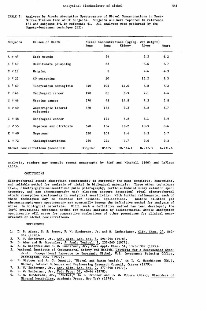
Measurements of nickel concentrations in serum or urine specimens from healthy adult inhabi-tants of several regions of the world are listed in Table 5. The analyses were all per-formed by atomic absorption spectrometry, and the subjects did not have any occupational ex-posures to nickel compounds. Excellent agreement was observed between the nickel concentra-tions in serum or urine from the subjects in West Germany, Japan, Spain, and the UnitedStates (2,33,40,152,155). Significant increases were noted in urine and serum nickel con-centrations in inhabitants of Sudbury, Canada, which is a site of large nickel deposits andnickel refineries (153). The mean nickel concentration and the range of concentrations thatwere reported in urine of inhabitants of Kristiansand, Norway (154) appear to be greaterthan have been observed in inhabitants of West Germany, Japan, and USA. This finding may berelated to the presence of a nickel refinery in Kristiansand, Norway. Plasma nickel concen-trations in the residents of Kristiansand, Norway were comparable to serum nickel concentra-tions in residents of Japan, Spain, and USA. In Table 6 are listed reference values fornickel concentrations in body fluids, excreta, and biopsy tissues from living, nonexposed,adult persons, based upon atomic absorption analyses by the present author and his col-leagues (12,156—160) and by Torjussen and coWorkers (14,154). In Table 7 are listed refer-ence values for nickel concentrations in human, postmortem tissues obtained at autopsy,based upon analyses by Nomoto (41) and Sunderman et al (161).

540 COMMISSION ON TOXICOLOGY
TABLE 5. Analyses by Atomic Absorption Spectrometry of Nickel Concentrations in Serumand Urine of Healthy Adult Subjects without Occupational Exposures to NickelCompounds. Each value is the mean ±SD. The numbers of subjects are listedin brackets.
Locations Nickel ConcentraSerum or Plasma
tions (pg/litre)Urine
Authors
Julich, West Germany 2.6±1.2 [21] Ader & Stoeppler (2)
Matsumoto, Japan 2.1±1.1 [24] 2.7±1.1 (73] Nomoto (40,152)
Santiago, Spain 2.5±0.5 [5] Gonzalez et al (33)
Hartford, U.S.A. 2.6±1.0 [26] 2.0±0.9 [20] McNeely et al (153)
Sudbury, Canada 4.6±1.4 [25] 7.2±3.9 (19] McNeely et al (153)
Kristjansand, Norway 1.9±1.4 [57] 4.9±4.2 [57] Torjussen & Andersen (154)
TABLE 6. Analyses by Atomic Absorption Spectrometry of Nickel Concentrations in Specimensfrom Healthy Adult Subjects without Occupational Exposures to Nickel Compounds(l55)
Specimen NickelMean±SD
Concentrat
Range
ionsNo.
in SpecimensUnits
Authors
Whole blood 4.8±1.3 2.9—7.0 [17] pg/litre Nonoto & Sunderman (12)
Serum 2.6±0.9 0.8—5.2 [80] pg/litre Sunderman (156)
Urine 2.2±1.22. 6±1.4
0.7—5.20.5—6.4
[50] pg/litrepg/day
Sunderman (156)
Feces 14.2±2.7258±126
10.8—18.780—540
[10] pg/g (dry)pg/day
Horak & Sunderman (157)
Scalp hair 220±80 130—510 [20] pg/kg Nechay & Sunderman (158)
Arm sweat 52±36 7—180 [33] pg/litre Hohnadel et al (159)
Parotid saliva 2. 2±1.2 0.8—4.5 (20] pg/litre Catalanatto & Sunderman (160)
Palatine tonsils 140±70 30—280 [15] pg/kg (wet) Torjussen et al (14)
Nasal mucosa 130±200 (57] pg/kg (wet) Torjussen & Andersen (154)
AVOIDANCE OF NICKEL CONTAMINATION
Contamination during specimen collection is a troublesome problem for measurements of nickelconcentrations in body fluids, excreta, and tissues. Sweat from the fingers and palms ofthe hands is rich in nickel (159) and is a common source of contamination of pipets andspecimen containers. Procedures to minimize nickel contamination during collection andanalysis of serum and urine have been described in detail by Mikac—Devif et al (13) andSunderman (140). Plastic cone—tips that are used for micropipetting instruments (e.g.,"Eppendorf" or "Oxford" pipettors) are frequently contaminated with metals (162,163) andshould invariably be cleansed before use by soaking in ultrapure HNO3 followed by multiplerinses with demineralized water which has been distilled in a quartz still (140). Commer-cially available evacuated tubes for collection of blood (e.g., "Vacutainer" tubes) areoften contaminated with trace metals (164,165) unless the tubes and stoppers have beenwashed with HNO3. Versieck et al (85) demonstrated that use of Menghini biopsy needles orsurgical scalpel blades can cause many—fold increases in the nickel concentrations of smallbiopsies of normal human liver. Segments of glass tubing and plastic knives that have beenwashed in ultrapure HNO3 are satisfactory tools for collection of postmortem tissuesspecimens for nickel analysis. For helpful advice on contamination control in trace metal
Analytical biochemistry of nickel 541
TABLE 7. Analyses by Atomic Absorption Spectrometry of Nickel Concentrations in Post-Mortem Tissues from Adult Subjects. Subjects A—D were reported in reference161 and subjects E—L in reference 41. All analyses were performed by theNomoto—Sunderman technique (12).
Subjects Causes of Death NickelBone
Concentra
Lung
tions (jig/kg,
Kidneywet weight)Liver Heart
A d' 44 Stab wounds 24 5.2 6.2
B 40 Barbiturate poisoning 22 8.6 5.7
C 18 Hanging 8 7.6 4.3
D 22 CO poisoning 10 13.2 8.3
E 60 Tuberculous meningitis 340 104 11.0 8.8 7.2
F Cf 48 Esophageal cancer 190 81 6.9 7.1 4.4
C 46 Uterine cancer 270 48 14.8 7.3 5.8
H 40 Amyotrophic lateralsclerosis
360 132 9. 2 5. 8 6.7
I 58 Esophageal cancer 121 6.8 6.1 4.9
J Cf 55 Hepatoma and cirrhosis 640 134 18.2 10.9 8.6
K 49 Hepatoma 290 109 9. 6 8. 3 5. 7
L 72 Cholangiocarcinoma 240 221 7.7 9.6 9.3
Nickel Concentrations (mean±SD): 333±147 85±65 10.5±4.1 8.2±2.3 6.4±1.6
analysis, readers may consult recent monographs by Zief and Mitchell (166) and LaFleur(167).
CONCLUSIONS
Electrothermal atomic absorption spectrometry is currently the most sensitive, convenient,and reliable method for analysis of nickel in biological materials. Three other techniques
(i.e., dinethylglyoxime—sensitized pulse polarography, particle—induced x—ray emission spec—trometry, and gas chromatography with electron capture detection) rival electrothermalatomic absorption spectrometry in analytical sensitivity. With further refinements, each ofthese techniques may be suitable for clinical applications. Isotope dilution gaschromatography—mass speçtrometry may eventually become the definitive method for analysis ofnickel in biological materials. Until such a definitive method has been developed, theIUPAC provisional reference method for nickel analysis by electrothermal atomic absorptionspectrometry will serve for comparative evaluations of other procedures for clinical meas-urements of nickel concentrations.
REFERENCES
1. D. B. Adams, S. S. Brown, F. W. Sundernan, Jr. and H. Zachariasen, Clin. Chem. 24, 862—867 (1978).
—2. F. W. Sunderman, Jr., Ann. Clin. Lab. Sci. 8, 491—494 (1978).3. D. Ader and M. Stoeppler, J. Anal. Toxicol. 1, 252—260 (1977).4. K. S. Kasprzak and F. W. Sundernan, Jr., Pure Appl. Chem. 51, 1375—1389 (1979).5. National Institute of Occupational Safety and Health, Criteria for a Recommended
dard: Occupational Exposure to Inorganic Nickel, U.S. Government Printing Office,Washington, D.C. (1977).
6. E. Nieboer and A. C. Cecutti, "Nickel and human health," in T. C. Hutchinson (Ed.),Nickel, Natural Sciences and Engineering Research Council, Ottawa (1979).
7. F. W. Sundernan, Jr., Ann. Clin. Lab. Sci. 7, 377—398 (1977).8. F. W. Sunderman, Jr., Fed. Proc. 37, 40—46 (1978).9. F. W. Sunderman, Jr., "Nickel," in F. Bronner and J. W. Coburn (Eds.), Disorders of
Mineral Metabolism, Academic Press, Hew York (1979).

(c961) cou—oTT 'I.E i'v ''qno ci a 99 (9L61) cLc—c9c 'i iuv •waq 'pjszazsqn) pU Zifl)j 'Z c9
(u61) orc—coc '1 :joy mT4301tT!4 '1flOAE){ •N Ta43TH 3 'noInodoTITN L 'SOISTS 'V d 'flOUUOTT1p}{ d •L •19
(6T) z-cc •uçq •[8U 'puaqsut.oj •y pu ioj •j •9 (6961) ic—oic 'It? UI14 •Tuy 'la:1-rnco 'S •f PU At'1oTTTU 'd •:a ')tu34,s H • •Z9
(LL6I) •D.':1 'uo:2uPtsM 'a3TO 2UflUTId UWU19AOfJ S'fl '6 UOTZTIfld TToadS SN 'STT1:N T3TUJO[ pu TB3TOTOT UT SUaUaIa aij pa:&zatas
aUJWIaeQ o SP1BPUBS c fl1fl TUUTN paso S1flP3O1d 'flUaUTpO1A 'j 19
— _________________ (1L61)
till_LOT 'c :iov WT4D •TUV 'Uapri d PU uaqda:g i 'i°ia i ''a s i 09 (EL6TicT—ziT 'i TS -"D '42UTS d 'L PU T14°)I N 6c
(LL6T) 6EC—cEE 'C6 WTq 'TUV 'a1p2Uf 'a n pue aijsq • • •ç —
. (6961) 9E—ZE '-z;i •W94D [Uy z ':iPoa 'a d Lc (9L61) LEZ—.CZ 'L9 UTq3 TUv 'ZaJ3d S W pu zag •j •'j 'Za1d o 9c
(zL61) 019—c09 'L TS U1daS 'iejdoqj • s pu apn • • •çç (6L61) EOE—TOE 'Z? 4D 1'V 'SUT22OZ •f, •j •iç
(8c61) L9L:O8_°s •iuaq •u1y r 'WflU1 •M •a PUB S)jUU A '3 •Ec (siL6T) 9L—c9 'OL WT4D 'TV 'TTL. )L pu flZTWOO S '8Sfl) S zc
(zL61) 16—06 'II 4TSN •sq OTUOW 'tTa tt r pu aUUa •y • •ç 'SSald T1UOSSfl4 30 ATUfl 'hA TA 'WflaH TUU1UO1TAUUT suisqn
a3U.U '(Pa) qdWFI H c1 UT •'1np3O1d iii-xiw ap 8UTSfl [II] T8I3TU snoanb
Jo £ZUT3T3Ja uooixa UT SUOTT1A.. 'UUUIT4 '1' "vT PUIN .11 •'I 'UOUBTU •f •D oc (6L61) IL '6? UaI4D •TUUy Z SflTUS1d 'ocUoH •L 6i
(oL6T) TcL—6tiL 'i 0UqL TS UOITAUa ':saM •M d PUU A8P43U ' •S •9$ (2L61) 8L8'LL8' °N U9d 'Sfl 'O13eA j )j UU 400318)j '[ Li
(9L61) w[zz—iiTz '•i UaqD TUUV 'UUUIT •14 UU 9'J •M 'U '917
. (ZL6T) LcET—IccT '1q17 U1943 TeUV ')j303ST}{ 'I ')L UB UO41 ' 'T22TS 'S '1f 'UU2UTU f •çi
(LL6T) 9ZZ—ZZ 'i UqD 'TUUV 'iCpp •j4 •W LflUIU4ST1) A )L •117
(cL6T) 0E61—9Z61 'j Waq IUUV '2UT43SU • r pu OUOfl •3 •j' 'OUOfl •y r E17
(9L61) cz17—1z17 '8 1ZV U43 TUUV 'UBij 'a •ci UU UOp.IUfl 0 Z17
(17L61) 171i—6 'i TqSz n)I2I fl4SUT4S 'OOUON S 117
(17L61) LE—c 'zz TqssZ 'II flI4SUT4S '010W0N 'S '017
(o961) ZTZ—60Z 'Z1 'paw •lsnpUI r '&T& 'U21OW '0 '1 6E
________________ (9c61) 611—LOT '9Z 'T°'Ii
UTTD f '19U1f 'Uw1apUnS ' J pUU q1Ot)j3afl H 'D '4a1U4S "1 a 'PTOUT)I 'd 1 'SE '(L961) cT—cTT 'çi •tuaij 'UTID ''If 'UBllUapUfl5 'M 'd 'LE
'(6L61) OLOT—E901 'ic 'Ua4D 'TBUV 'UUafJ '5 f jUB S3U1 'H 'I '9E '(EL6I) Z—l 'c 'B1Ua TUd 'PtS10)IUBf 'f P-' )ITUBI ' •cc
'(8L61) ZOZI—8611 'liZ •Wa143 'UTI3 'UaSBTIBI4OBZ H UB uassnIoj • 'uasiapUy • •1 ____________
'(9L61) 17TE—LOC 'OC •IBUV 'UTflb 'SOUB5—BflB5 '3 pUB i iOd—ZBTBZU0O 'y 'zalBzuoo "1 'D 'H 'EE
'(8961) 091—cd ' '0 UB iaUl4fl)j 'V 'iaf(B435 'H 'i 'zc
_______________ (cL61)
ZLE—c9E 'Ec 'Wa430TS 'IBUV '2UTiaad '0 H UB .laTTa 'N 'd 'apa 'a 'a 'i"w 'i 'Ic '(17L61) 801—LOT 'El a1SN 'sqy OTUOW 'UOSUTiBd 'S 'H pUB SSO1 '5 '5 'OE
'(cL61) 68E—L8E 'qlB9H 'UOITAUa 'alBPt3TU 'N UB aU04S)IZ)B15 'H 'UBTdB)( '(I 'ci '6Z '(ZL6T) d901—%'901 '1717 'q) 'TBUV 'iaq3BWflq35 'f •H PUB Tal3TH '} 'UOs))Bf 'f 'V 'SZ
____________ '(8L61) EZOT—TZOT
'() 'waq3 TBUV 'pUBladc)D 'j 'J PUB OIBUBd 'M ')I "if 'PlSL0PTAB(I 'f ' 'a11B5 'ci LZ '(cL6l) 061—1781 '171 'PH 'U3430Ta 'OSflIB3 'y 'f PUB UBAO)IBU ' '5 'sUqqo>j '5 'M '9Z '(17L61) coc—66z '17 'TS 'qr 'UTT3 •UUy 'T)ISUT3B 'L 'a UB 'if 'UBUUBpUfl5 'M 'a 'cz
'(8L61) OZT—911 '16Z '111343 'TBUV 'Z SflTUBSBIL 'SflBIpj3B5 '[ pUB iaTddaOlS 'w 'liZ
'(8961) 9891—Z891 '0i 'U343 'TBUV 'SBUIBSJ 'j 'ç '(6L6l) TEZ—dZZ '1701 B3 'WTqD 'IBUV '3)jZ)CTj 'f ' (cL6T) Z9—Ld '? 'f '3OSSy '2LH 'SflpUI 'uzy 'O3BT111T'I ' ' PUB OOtO(Tj 'H 'J 'IZ
'(z96l) Lclil—liclil "iE '111343 'T"V 'pUB(j 'Cl '11 UB 1T310 'a '3 'OZ
____________ '(9L61)
ELT—ZLT 'ZZ 'l'I 'flZiv 'UOI1B5 'J 'j pUB T°9°)l '3 'U3S13PW ' 'U3SBTIB43BZ 'H '61
____________ '(cL6T)
L9d—Z9d 'TZ •wa143 'UTID 'UOiB5 'J 'J pUB 1°°X '3 'UesiapUy '[ 'U3SBTIB43BZ 'H '81 '(zL6l) Z91—66 ' 'UU3 'l°TH 'dxa 'paw 'UUy 'U3SSiO1 'V 'LI
'(6L61) iO3XØ 'sSaid UOIuB2iaci 'sTBTi3Bw IBOT8OTOTH O STSACTBUV aqj '('Pa) iapn ' ' "I UT •,'LdO3SOij3ads Uo3diosqB OTUiOB Lq STBT13IBU TB3T2OTOTq
JO SSLBUB aq iOJ 2UT4SB Lip UB JO UOSTI?dWOO.. '3IBPIBM 'N 'I PUB SUTpB 'I 'H '91
'(8L61) L9—179 'ZE TUBS '2T0 'BAO)jS}j '[ "j 'BAO14Si 'ci ')j 'BLB)ISAOIPIBIa 'ci 'N 'di
'(LL6I) ZZOI—810l 'EZ '°43 'UTI3 'UasBTiBqzBz 'H puB U3Si3U ' 'UassnçioJ 'M '171
'(LL6I) 9c6—8176 'EZ '43 'UTID 'O4OWOj '5 PUB 'if 'UBuzi3Ufl5 '14 'a 'TA3U—)LTw 'N 'El '(oL6l) d8t1LLl1 '91 'waq 'UTIO "if 'UBuU3Un5 ' 'a pUB 010W0N 'S 'ZI
'(oL6I) PJOJXO 'sSaici UOU1B2I3d 'i34BN 3TUB2IQ JO UOT43n1S3Q 34J 'q3flSiO3 J 'J '(( '(6d61) ZIZ—60Z 'Z BUB(BJ '4PUS 'a '0 PUB T4TI 'H '01
A00'IODIXOI NO NOISSINNOD Zlid
(8L61) 8jEc '•ZI •:i:aisLa •sqy 3TWOW 'Acas)t000 •14 •M PU 1c:aa 'ci •i czT 0'L61) 6T—cT '91 •maq •iuy 'a r pu WSOd ci a 'uassa •ç • r •a •tlZI
(9L61) 81E—69E 'E8Z "4D iuy • 'zTat'i 'a Pu T9dm)L w 'iaiddao ii
(cL61) z8c—6c t01Bd UBUIflH '11 'U1IU9pUfl M d 'ZZT (zL61) oo 'T 30d Pd ''H D P1W I91)1d S ' 1Z1
(c96r) 88T—Z81 'titi T0WBd UTTD .1 'my '•iç 'uUU3pun M 'd 'OZT
.
(z961) iT9 'tiE •W9 •TBuV 'ci •f 611
— ___________ (o96T) orrr -zi:. uiw •a '1' 811
(8L61) 89E—09C 'O "'1D 'TUV '19UpLO;aUTM .a 'r pu p43n21Br •H 'sjaa • s 'LIT '(6961) zzc—81c 'ii 'TUV A )SflOJ4 'f '911
(9961) Lcz1—cczI ' 'tc 'i'v 'noiuawiy 'N 'ci 'cli (6L61) 111—16 'ZLI 'S '1D
'Ap\r 'pU'[1aLjfl 'I •.M PU aiasiux 'N •I 'Al nqei ' '1assd 'V A "1f 'S}j 'f 'fri 'iiII
('961) cL—99 W1B0H U01TAUa 'P1V '1aJ1$ 'f 'f pu uodjj 'H 'I 'Eli '(E961) cT—Eo1 '6 '91(qd WTH OO3 f 'H pU uoidp H 'I 'zrr
'(E961) 101—68 '6 Sq qJaH 'P01FS 'V H UB '.lf 'Lilac! 'N H '0fl '3 '1aUTaS 'j 'j ')jOC 'f 'J4 'uodp 'H 'I 'in
— ___________________ '(z961) ccz—cz
'09 'PJ4 'UTID q1 '1 ')j003 .1' '14 19O.l4Z 'çf 'H 'uod 'H 'I "If '11d 'N 'H '011 '(6c61) 1c—Loc ' °v "'t 'UTID 'aoA 'H '1 OXTd 'N "1 '601
'(9c61) 8c—LLc '1 °v wjq 'u 'ao' 'H 'r pu 'H 'TIIaoBuow i '801 '(9c61) 11c—66 '6 13UD '1OUUO 'f pu dwiu 'j 'N '41P"S 'I 'a 'ioi 'r 'H 'LOT
'(6L61) E881—0881 'Ic 'ulaq 'iuv 'asTpB1d 'j 'j pirn atoj ' 'H 'ca)ITA ' 'J '901 '(6L61) Z6E—L8E '1 '"1D '1'V 'T4TPH 'L P' U42njZ 'çj '- 'punzjo 'H 'cot
'(9L61) coc—zoc '8 'i'v 'uqu ' 'g pu Al SaUO ' 'j 'oi
______________ '(6L61) L8E—%'8E
'ZLI '2OWOL143 'I' 'UJ1CJ 'H pu UOOTW '3 'AO1aaSp1rn1 ' 'oqaj 'j 'syj '0 'COT ' (6L61)
6c1—EcI 'ILl 2O4IUO14D '1 'UOq3Oflf '3 jU AOtaajSUB1 ' 'ABOqa'j 'j 'jsyj ' 'oi '(6L61) ici—ci 'TZi '$o:4uIo1ID t 'uitoo H PU uoqoopj ' 'ijsyj '0 '101 '(cL6I) E81—cLT '6L '1ii 'i'v 'siaq 'H ' pu uapfl ' 'd 00i
'(8L61) i6Z—06 'E6Z 'UIj3 .z SflJU3S1 'qaa ' pUB SO[O3—IBWUIBfJ 'A '66 '(8L61) 1c—Eoc 'ööï "m 'IBuv 'uaqda 'i pu anbJTBq 'V ''L'1H 'I '86
'(L61) 818—L08 'L ''1 '1"V 'a1flj 'y '3 UB uosiapuj 'a 'a 'Uapfl '3 'd 'L6 '(EL6I) EOZT—L611 'ci, 'q3 '1'V
'Upfl '3 'd UB PBZJ[BUB){ 'y 'uosapuaij 'a 'ci 'uaqdais 'I 'A 'uflJB 'ç " 'iia 'I '96 '(L6I) 86c—16c 'Z1 'T3S '2011101lj3 'I' 'PTT")1 ' jUB UoS1apUa{ ' 'Q 'Upfl '3 'd '6
_________________ '(zL6r)
EL—6c '6c °V "P-tO '1UV 'Uapfj '3 'd PUB uaqdai 'I 'A '4Z)1H '1 '11tt 'S L '4?6
'(LL6I) i99Z—T99Z 'oc 'udç '309 'UII3 '1lra 'B.IBqwB) 'j pue pjBZWJ ')l "'-"""BA 'A 'E6 '(zL61) 8ZI—11 '99 '20WW01q3 'f 'suuiouj, '3 pu loTBnb3Bf 'd 'Z6
'(zL61) E4?E—cEE '09 B43V 'WPtO '1'V 'SBWOq] '3 pUB B11TB4 'd '1 'l0Tanb3Bf 'd '16 '(cL6I) 199—8c9 'Li? "'Tl3 'IBUV 'U0UB 'd 'H UB UO1TUIBII 'i 'a 'UBUUB3 'V 'I '06
'(6L61) 4?01—ZZ01 'Ic 'WBt3 'IBUV 'a11W1110 'a 'a PUB flOA ' 'j '3 '68 '(9L61) £61—ELI '4?I 0[flBd OBS 'ATUci 'luinbOla 'liuBa 'ABH 'OUJBj4 'V 'ci '88
___________ '(iL61)
6801—Z801 '94 'UB43 'TBUV 'IBU.1B3 '-i 'a PUB spioqs 't 'A 'uBIq 'V "1 'aLoow 'r 'i 'L8 '(oL6I) cL9—EL9 'Ztl ''13 'IBUV '1aanl4 '4 '3 pUB ZBIBAW 'j 'UBSIflBd 'f 'd '98
'(EL6I) cL4?—ZL4? '61 'U813 'UTTO 'Laq1Bc 'j UB asc •f 'aj3aad5 'v ')I3BTSIBA '1' 'c8 (EL6I) c1i—iiiz 'c4? "(3 'IBUV '1Iit43S 'V 'a PUB B1U9 'i 'a '4?8
_______________________________ '(1L61)
964?1—884?T 'ç4? 'UflLUUIO3 'SBJ 'SAOdOTH 'U1a1t3OTa 'BIBPSanA 'A V UB U0SUBIS 'V 'V E8 '(zL6r)_8ZE—iIE '19Z "'-t3 'IBUV 'Z '1ISTZ ' pUB XTYI 'a 'Z8
'(LL6I) 806—906 '961 B3UBT3S 'laIInH 'a 'A UB O3ST3UBId 'a ' 'Uq3 'H '1 '18 '(LL6I) 6c1 'Zti1 '4H 'SU[ '1N 'IfleItlaA 'H PUB tUB)( IBP UBA 'V 'H 'd 'STA 'U 'H '08
_____ '(9L6r) 681 '4?ET 'qH 'SUI
'PEN 'OLBUOH '5 pUB STPB1Bd 'd 'aiulooai 'j 'aqe' 'a 'XflB91fl0WB ' '9lB11B5 'H '6L '(4?L61) IL—89 'I 'sAqj 'paj '33AO21j{ 'H PUB 2U943 '-i 'a '2110 'S 'd
'sdpi,j '3 '3 '3T0tIBA 'A 'd 'H 'BB 'A 'tiaqaj 'a 'H 'IBIBB4A 'H 'H '(6L6I) 99E1—E9EI '9—SN 'TS '1N 'SUBIA aaai 'Bqd1UB3 " 'f
_______________________________ '(6L61) iz—ci
'LI '1U9Tl3OTa 'UTID "'43_'UTID_'1'_'LflPTu 'A 'd UB HTUBO)I 'A 'BPO5 '3 'C '1uTW 'a '9L '(LL6I) 0I4?—E04? 'OZ 'IBIW IcBH—x 'APV 'SWBW 'H A PUB I9ASUP1 UBA 'V 'H 'IBBrj 'a 'a 'cL
'(9L61) EL4? 'LET 'qH '1SI '[N 'uosuuuqof 'a 'A UB UOSUUBI4Of 'a 'V 'S '4?L
'(8L61) L4?91—rn9I 'oc 'u'to 'IBUV 'I1'I°lTW 'A 'f UB IBISSB)1 'a 'C 'EL
____________________ '(8L6r) L64?
'8 'T5 'qu-j 'uTID •UUV 'Tt"O 'a 'V UB TUTSSBWOA 'ci 'a 'B101J 'I '3 '1B9 'a 'ZL
__________________ '(9L61) 09Z
'IC 'WTtDL T1BUV 'qz 'BAOUB4ZpLpITIBS d 'H 'H PUB BAO1OIptOld 'A '3 'BAa;BlsT 'A 'A 'tL '(8961) 9991 'EZ 'WTIOI 'TIBUV "Z 'BAOIOIPtOId 'A '3 UB BAOPBI2OUTA 'N 'a 'OL
'(ssaid UT) 'iq 'IBUV 'iaoqa 'a PUB B10II 'C '3 '69 '(zL61) 88—E8Z '8 B3 'WT43 'IBUV 'a1ao5 '3 ' PUB 'I 'H '89
'(6961) 9—1 '84?Z '1uaq 'IBuV 'Z 'pjst.eqso ' PUB I I3SBTcI 'V 'OHIBa 'H 'L9
Ia)IOTU jo LtisxIat3otq IB3TIBUV
(9L61) 3'a 'UO2UT4SBM 'azT3
—Jo 2UTUT1d :uawuiaco sn 'Z'i sp1pus ;o neainq TUOTN 'Z pirn I SIOA 'STSI(TUV '2UTIpUH aIdWS '2uTIdtuBS :sTa&Iuy 31L UT £31n33y ''Id'I 'ci d 'L91
(9L61) jio AI 'SUO p1W AaTTL Uqof 'STSIIUV UaWaIa UT IO14UO UOTUTW4UO 'aqZ4T M 1 PU 3TZ '4 '991
'(9L61) Z69—169 'Z "43 UTTD '1aIX0L '0 'I PU ' Q 'saq2nj 'o i 'c91 (IL6I) 19 'LI '1D 'UTIa 'PIO2UTI 'N '[ 1[ ')j 'a 'UBuillafi 'a 'a "i91
(cL6I) ZE—IE 'tiI 'I'N 'Sq OTWOW 'Uas 'a I PW aAcYI 'a 'L 'PI.T1a1111110S 'I 'N 'E91 '(zL61) 9Lc—L9c '09 'PN UTID f 'PUPTUTL '2 ' 1JW UOSOU 'd 'f Z9I
'(L6I) 9ç—ç 'dd '? 1°A 'qWno) 'Tfl0SSTN 0 'AUfl 'qlBaH 1UWUO1TAU2 UT S93UWS
—qn '('Pa) IITqdu1I 'a 'a UI 'A(Wq) 'J4 UB OOU1Oj 'S "if 'UUUUPUflS 'M 'a '191 '(LL6I) IcI—9'7I 'L 'Ps 'q'i 'UTID 'UUy' "if 'UBuIiapUn5 'M 'd UU OU3 'V 'd '091
____________ '(EL6I) Z6I—88ZI
'61 "4D 'UTID 'aIaaNzJN 'a 'w UU If 3 'f 'fi "if 'UBuupUn5 '1 'i 'I3PeU40H 'D 'a '6cr '(0L6I) c—oE ' 'PS 'q'i 'UTID 'UUV "if 'UlUiWUn a UW Lq3a 'M 'w '8cr
'(CL6T) 0c7—6z7 '61 '"'4D 'UTID "if 'UBUUPUI1S 'M 'a UB BiCfl 'a 'LcI '(LL6r) 86E—LLC 'L TS 'qI 'UTID 'UUV "if 'UBUUBpUfl5 'M 'a '9c1
•(ssaid UT) 'PS •Bj 'UTID 'UU "if 'UBUUBpUn5 ' pUB Z3O21C 'a 'PPUlfl 'f 2 'ccl '(6L6I) 86Z—68Z '6 'PS 'q'i 'UTID UUV 'uasiapUy '[ pUB uBssncioJl 't 'i?cl
'(zL6l) c66—Z66 'fit '111D 'UTID ''if 'UBUUBpUfl5 'Lf 'a UB 'lN ' N 'aINzN a 'N 'Ect ___________________'(cL6I) 86 'OE 'SXH 'f 'Udf 'ofouIo 's 'zci
(8L61) c6'i '8 TS 'q 'UTID 'UUV "if 'UBU1iBpUfl5 'ii 'a P BAOi3 '}f 'A 'Icr '(8L6r) 6i?Z—EiZ '96 'WTq 'IBUV '°t'i 'a 'f 'N P 'N 'N '2 'oct
'(LL6r) ocl—6i?I '91 '4aI8L'aN 'sq 3TWOW 'WWBI4SInf ')I '6i?I
'(flL6l) 'i81—I2I 'ZOl fV 'WTfD 'IV 'qTO •V 'a 'V PUB TIN N 'N 'Bi2 'N N '8il '(zL6r) 961—L81 '6c ZV 'WTTLD 'IBUV '4saM 'S 'A PU UOS)f3Bf 'M ')L 'L1i1
(cL6I) i?9il—09'il 'cii 'uiaq 'IBUV 'SiaqpUn5 "1 '1 '9i?l
'(8L61) 01—Z 'oc 'waq 'IBUV ')1B9Th0N 'd '1 PUB )jTOZD 'f 'a 'cl '(i?L6l) ciz—cozz '9t1 'W9143 'IBUV 'UOIITBA 'D PUB UiflqA 'V 'S 'IO8UB)( •j '1tfl
'(i?L61) Ei?Z—TEZ 'ZL B3V 'WTILD 'IBUV 'UOO'f UBA 'D f UB Zfl13 '5 'j •Ei?l
'(ZL6I) IZOZ—810Z 'i?i? 'WBLf3 'IBUV 'T°°IIfl 'V 'd UB iBB)fCfl 'f '3 '2UBtI}j 'A 'C 'i?I '(i?L61) i?tli?T_6E111 '9i? 'Wa143 'IBUV 'BWSOd 'a 'a UB UBSSBBfl 'f 'N 'f 'a 'Pit
'(cL6r) i?Ei?—IZi? 'S 'PS ''1 'UTID 'UUy ''if 'UBWIBpUTI5 'M 'a '0171
'(i?L6l) i?9E—ccC 'L '4Bf '3BOifOBdg 'ia)IzTflb 'N UB pjSLBçO1pZ 'V 'aBIPUTa 'f '11 '6E1 '(zL6I) ci?i?—zi?i? 'Z9 B3V 'WTq3 'IBUV 'iauna 'M '3 '8E1
'(i?L6I) 69—L9 'El 'BlSBN •sq 3TWOW 'sBpna 'C 'N 'LET '(i?L61) 0E6—6Z6 'E9 'PS 'nBWiBTLd 'f ';Bi2pUB '3 ' pUB 3BiB5 'j 'f '9EI
'(cL6l) LcZI—ocZT 'Li? 'WBq3 'IBUV 'Sifl '3 'd PUB iBBi)fBI3 'f '3 'UOB2ifl5 'a 'a 'cEl '(i?L6I) 8Ii?'-i?li? 'U B43\T 'UITIjO 'IBUV 'awB1q35 'd 'i?EI
'(flL6l) Z061—0061 'oc 'Wa43 'IBUV 'UOIITBA '3 pUB I°L'l 'I 't4 'aiq1nf 'a ' 'EEl '(9L6r) i?i?—Zi? 'cr 'alszaN 'sqxj 3TWOW 'ia2Tp 'a 'i UB 2UTUUBN 'D 'a 'ZEI
'(i?L6l) clz—Elzz '911 '1D 'IBUV 'UOIITBA '3 pUB iOUB{ ' 'Uinqa3 'V S 'IEI
____________ '(zL61)
OZLI—81L1 "it 'IBUV "if '1a21B15 'd 'N UB TiBBiB 'f '3 'BidSV 'I ')L 'OEI ji?L61) 'i9z—Lcz 'l? B4UBIBA 'UOSSUBIjOf '0 PUB UBi2PUIYI '0 '6Z1
'('iL6l) IEOI—8Z01 '91? 'W43 'IBUV 'UOSSUBqOf '0 UB )jiBWpUIYj 'j 'Uai2pUtrj '0 '8Z1 '(cL6r) ci?—8E '1)1 'ma43 'TBUV 'If3flO3 'f '5 PUB IBSB4Uq4 'V 'LZI
'(EL6r) 9I8I—Z181 'c'i 'm9tD 'IBUV 'TiBSSBA '3 UB TSiOA '0 '9Z1
A0OIO3IXOI NO NOISSINNOD