analytical currents: the whole proteome picture
TRANSCRIPT
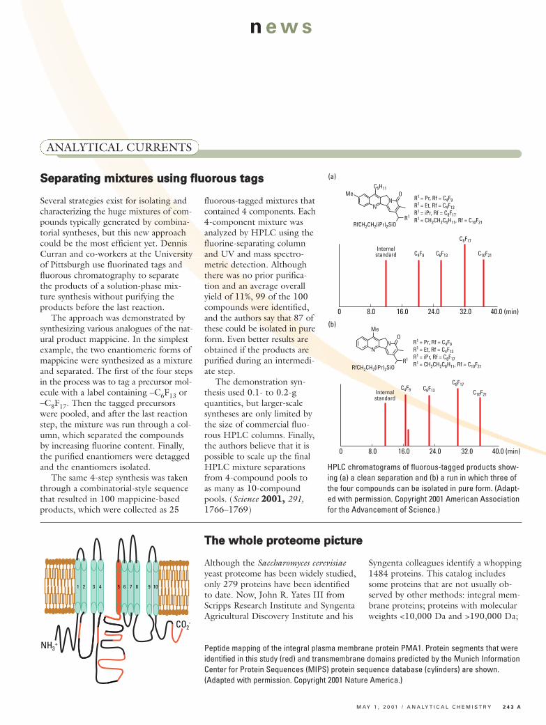
Several strategies exist for isolating andcharacterizing the huge mixtures of com-pounds typically generated by combina-torial syntheses, but this new approachcould be the most efficient yet. DennisCurran and co-workers at the Universityof Pittsburgh use fluorinated tags andfluorous chromatography to separatethe products of a solution-phase mix-ture synthesis without purifying theproducts before the last reaction.The approach was demonstrated by
synthesizing various analogues of the nat-ural product mappicine. In the simplestexample, the two enantiomeric forms ofmappicine were synthesized as a mixtureand separated. The first of the four stepsin the process was to tag a precursor mol-ecule with a label containing –C6F13 or–C8F17. Then the tagged precursorswere pooled, and after the last reactionstep, the mixture was run through a col-umn, which separated the compoundsby increasing fluorine content. Finally,the purified enantiomers were detaggedand the enantiomers isolated.The same 4-step synthesis was taken
through a combinatorial-style sequencethat resulted in 100 mappicine-basedproducts, which were collected as 25
fluorous-tagged mixtures thatcontained 4 components. Each4-component mixture wasanalyzed by HPLC using thefluorine-separating columnand UV and mass spectro-metric detection. Althoughthere was no prior purifica-tion and an average overallyield of 11%, 99 of the 100compounds were identified,and the authors say that 87 ofthese could be isolated in pureform. Even better results areobtained if the products arepurified during an intermedi-ate step.The demonstration syn-
thesis used 0.1- to 0.2-gquantities, but larger-scalesyntheses are only limited bythe size of commercial fluo-rous HPLC columns. Finally,the authors believe that it ispossible to scale up the finalHPLC mixture separationsfrom 4-compound pools toas many as 10-compoundpools. (Science 22000011,, 291,1766–1769)
news
C5H11Me O
R1
NN
RfCH2CH2(iPr)2SiO
MeO
R1
NN
RfCH2CH2(iPr)2SiO
R1 = Pr, Rf = C4F9R1 = Et, Rf = C4F13R1 = iPr, Rf = C8F17R1 = CH2CH2C6H11, Rf = C10F21
8.0 16.0 24.0 32.0 40.0 (min)0
Internalstandard C4F9 C6F13
C8F17
C10F21
8.0 16.0 24.0 32.0 40.0 (min)0
Internalstandard
C4F9 C6F13
C8F17
C10F21
R1 = Pr, Rf = C4F9R1 = Et, Rf = C4F13R1 = iPr, Rf = C8F17R1 = CH2CH2C6H11, Rf = C10F21
(a)
(b)
HPLC chromatograms of fluorous-tagged products show-ing (a) a clean separation and (b) a run in which three ofthe four compounds can be isolated in pure form. (Adapt-ed with permission. Copyright 2001 American Associationfor the Advancement of Science.)
SSeeppaarraattiinngg mmiixxttuurreess uussiinngg fflluuoorroouuss ttaaggss
ANALYTICAL CURRENTS
TThhee wwhhoollee pprrootteeoommee ppiiccttuurree
Although the Saccharomyces cerevisiaeyeast proteome has been widely studied,only 279 proteins have been identifiedto date. Now, John R. Yates III fromScripps Research Institute and SyngentaAgricultural Discovery Institute and his
Syngenta colleagues identify a whopping1484 proteins. This catalog includessome proteins that are not usually ob-served by other methods: integral mem-brane proteins; proteins with molecularweights <10,000 Da and >190,000 Da;
M AY 1 , 2 0 0 1 / A N A LY T I C A L C H E M I S T R Y 2 4 3 A
1 2 3 4 5 6 7 8 9 10
NH3+
CO2-
Peptide mapping of the integral plasma membrane protein PMA1. Protein segments that wereidentified in this study (red) and transmembrane domains predicted by the Munich InformationCenter for Protein Sequences (MIPS) protein sequence database (cylinders) are shown.(Adapted with permission. Copyright 2001 Nature America.)
2 4 4 A A N A LY T I C A L C H E M I S T R Y / M AY 1 , 2 0 0 1
news
proteins with pIs <4.3 and >11; andlow-abundance proteins such as tran-scription factors and kinases.To identify these proteins, the group
optimized conditions for an establishedmethod that couples two-dimensionalLC, in which a microcapillary column ispacked with two chromatography phases,with MS/MS. The researchers analyzed
complex peptide mixtures from three frac-tions of an S. cerevisiae lysate. After eachfraction was loaded onto its own micro-column off-line, the columns were insert-ed into the instrumental setup, and no ad-ditional sample handling was required.Peptides were eluted in an iterative
process in which the columns were re -equilibrated and increasingly high con-
centrations of salt were added. The pep -tides were fed directly from the columnsinto an ion trap mass spectrometerequipped with a nano-LC electrosprayionization source. Like the chromato -graphy and MS steps, the subsequentdatabase comparison and identificationof proteins were automated. For eachclass of protein, the number predictedagreed well with the number found.Thus, the authors conclude that theirmethod is largely unbiased and is a steptoward a comprehensive, automated,high-throughput system. (Nat. Biotech-nol. 22000011,, 19, 242–247)
ANALYTICAL CURRENTS
A protein’s role can be predicted by its con-
formational state and location inside a cell.
Green fluorescent protein labeling is often
used to determine cellular locations in vivo,
but this technique gives no information on
three-dimensional (3-D) structures. On the
other hand, NMR provides this 3-D informa-
tion, but to date, in vivo studies required the
external addition of isotopes. Now, Volker
Dötsch and colleagues at the University of
California–San Francisco describe a new
and easier approach to examining a protein
in the cytoplasm of bacterial cells with in
vivo NMR.
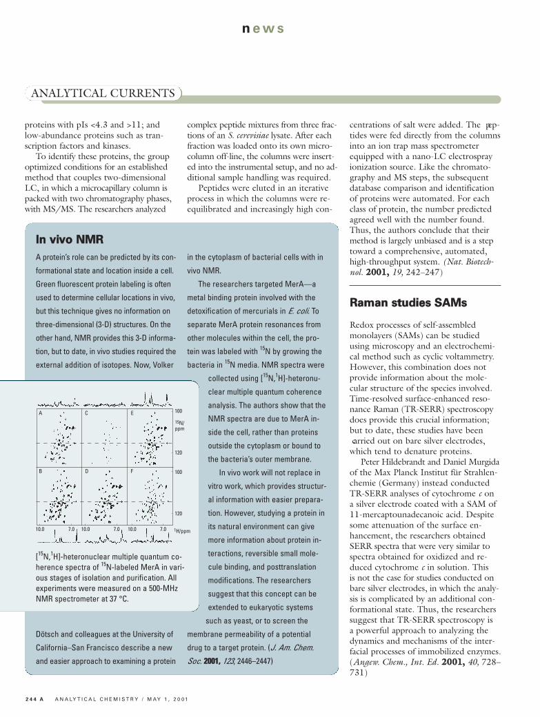
The researchers targeted MerA—a
metal binding protein involved with the
detoxification of mercurials in E. coli. To
separate MerA protein resonances from
other molecules within the cell, the pro-
tein was labeled with 15N by growing the
bacteria in 15N media. NMR spectra were
collected using [15N,1H]-heteronu-
clear multiple quantum coherence
analysis. The authors show that the
NMR spectra are due to MerA in-
side the cell, rather than proteins
outside the cytoplasm or bound to
the bacteria’s outer membrane.
In vivo work will not replace in
vitro work, which provides structur-
al information with easier prepara-
tion. However, studying a protein in
its natural environment can give
more information about protein in-
teractions, reversible small mole-
cule binding, and posttranslation
modifications. The researchers
suggest that this concept can be
extended to eukaryotic systems
such as yeast, or to screen the
membrane permeability of a potential
drug to a target protein. (J. Am. Chem.
Soc. 22000011,, 123, 2446–2447)
IInn vviivvoo NNMMRR
Redox processes of self-assembledmonolayers (SAMs) can be studiedusing microscopy and an electrochemi-cal method such as cyclic voltammetry.However, this combination does notprovide information about the mole -cular structure of the species involved.Time-resolved surface-enhanced reso-nance Raman (TR-SERR) spectroscopydoes provide this crucial information;but to date, these studies have been carried out on bare silver electrodes,which tend to denature proteins.Peter Hildebrandt and Daniel Murgida
of the Max Planck Institut für Strahlen-chemie (Germany) instead conductedTR-SERR analyses of cytochrome c ona silver electrode coated with a SAM of11-mercaptounadecanoic acid. Despitesome attenuation of the surface en-hancement, the researchers obtainedSERR spectra that were very similar tospectra obtained for oxidized and re-duced cytochrome c in solution. Thisis not the case for studies conducted onbare silver electrodes, in which the analy-sis is complicated by an additional con-formational state. Thus, the researcherssuggest that TR-SERR spectroscopy isa powerful approach to analyzing thedynamics and mechanisms of the inter-facial processes of immobilized enzymes.(Angew. Chem., Int. Ed. 22000011,, 40, 728–731)
RRaammaann ssttuuddiieess SSAAMMss
100
10.0 7.0 10.0 10.07.0 7.0
120
100
120
15N/ppm
A C E
B D F
1H/ppm
[15N,1H]-heteronuclear multiple quantum co-herence spectra of 15N-labeled MerA in vari-ous stages of isolation and purification. Allexperiments were measured on a 500-MHzNMR spectrometer at 37 °C.
M AY 1 , 2 0 0 1 / A N A LY T I C A L C H E M I S T R Y 2 4 5 A
n ews
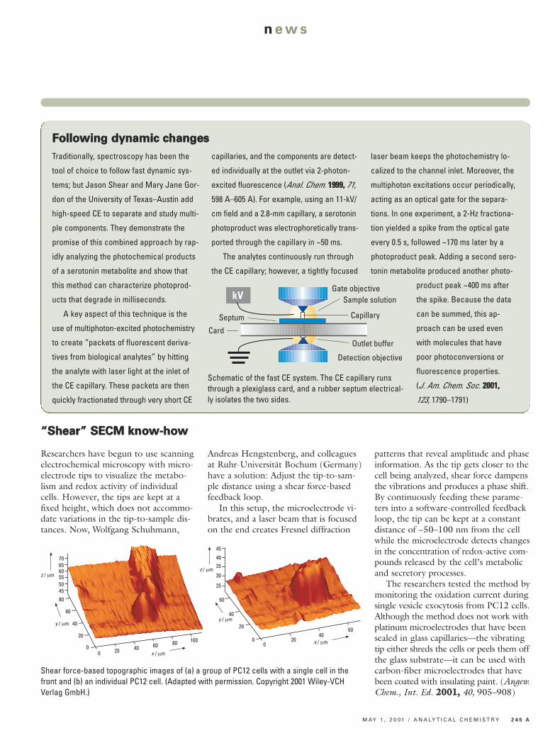
Researchers have begun to use scanningelectrochemical microscopy with micro-electrode tips to visualize the metabo-lism and redox activity of individualcells. However, the tips are kept at afixed height, which does not accommo-date variations in the tip-to-sample dis-tances. Now, Wolfgang Schuhmann,
Andreas Hengstenberg, and colleaguesat Ruhr-Universität Bochum (Germany)have a solution: Adjust the tip-to-sam-ple distance using a shear force-basedfeedback loop.In this setup, the microelectrode vi-
brates, and a laser beam that is focusedon the end creates Fresnel diffraction
patterns that reveal amplitude and phaseinformation. As the tip gets closer to thecell being analyzed, shear force dampensthe vibrations and produces a phase shift.By continuously feeding these parame-ters into a software-controlled feedbackloop, the tip can be kept at a constantdistance of ~50–100 nm from the cellwhile the microelectrode detects changesin the concentration of redox-active com-pounds released by the cell’s metabolicand secretory processes.The researchers tested the method by
monitoring the oxidation current duringsingle vesicle exocytosis from PC12 cells.Although the method does not work withplatinum microelectrodes that have beensealed in glass capillaries—the vibratingtip either shreds the cells or peels them offthe glass substrate—it can be used withcarbon-fiber microelectrodes that havebeen coated with insulating paint. (Angew.Chem., Int. Ed. 22000011,, 40, 905–908)
““SShheeaarr”” SSEECCMM kknnooww--hhooww
200
z / µm
70656055504580
60
40
20
0
y / µm
x / µm40 60 80 100 20
0
z / µm
45
4035
30
25
60
40
20
0
y / µm
x / µm40
60
Shear force-based topographic images of (a) a group of PC12 cells with a single cell in thefront and (b) an individual PC12 cell. (Adapted with permission. Copyright 2001 Wiley-VCH Verlag GmbH.)
Traditionally, spectroscopy has been the
tool of choice to follow fast dynamic sys-
tems; but Jason Shear and Mary Jane Gor-
don of the University of Texas–Austin add
high-speed CE to separate and study multi-
ple components. They demonstrate the
promise of this combined approach by rap-
idly analyzing the photochemical products
of a serotonin metabolite and show that
this method can characterize photoprod-
ucts that degrade in milliseconds.
A key aspect of this technique is the
use of multiphoton-excited photochemistry
to create “packets of fluorescent deriva-
tives from biological analytes” by hitting
the analyte with laser light at the inlet of
the CE capillary. These packets are then
quickly fractionated through very short CE
capillaries, and the components are detect-
ed individually at the outlet via 2-photon-
excited fluorescence (Anal. Chem. 11999999,, 71,
598 A–605 A). For example, using an 11-kV/
cm field and a 2.8-mm capillary, a serotonin
photoproduct was electrophoretically trans-
ported through the capillary in ~50 ms.
The analytes continuously run through
the CE capillary; however, a tightly focused
laser beam keeps the photochemistry lo-
calized to the channel inlet. Moreover, the
multiphoton excitations occur periodically,
acting as an optical gate for the separa-
tions. In one experiment, a 2-Hz fractiona-
tion yielded a spike from the optical gate
every 0.5 s, followed ~170 ms later by a
photoproduct peak. Adding a second sero-
tonin metabolite produced another photo-
product peak ~400 ms after
the spike. Because the data
can be summed, this ap-
proach can be used even
with molecules that have
poor photoconversions or
fluorescence properties.
(J. Am. Chem. Soc. 22000011,,
123, 1790–1791)
FFoolllloowwiinngg ddyynnaammiicc cchhaannggeess
Gate objectiveSample solution
Capillary
Outlet buffer
Detection objective
Septum
Card
kV
Schematic of the fast CE system. The CE capillary runsthrough a plexiglass card, and a rubber septum electrical-ly isolates the two sides.
2 4 6 A A N A LY T I C A L C H E M I S T R Y / M AY 1 , 2 0 0 1
news
PPrrootteeiinn oorriieennttaattiioonn sslleeuutthhss
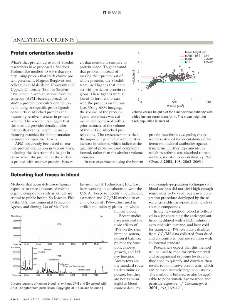
What’s that protein up to now? Swedishresearchers have proposed a SherlockHolmes-like method to solve that mys-tery, using probes that track elusive pro-tein placement. Magnus Bergkvist andcolleagues at Mälardalen University andUppsala University (both in Sweden)have come up with an atomic force mi-croscopy (AFM)-based approach tostudy a protein molecule’s orientationby binding site-specific probe ligandsonto surface-adsorbed proteins andmeasuring relative increases in proteinvolume. The researchers suggest thatthis method provides detailed infor -mation that can be helpful in manu -facturing materials for bioimplantationor immunodiagnostic devices.AFM has already been used to ana-
lyze protein orientation in various ways,including the detection of a height in-crease when the protein on the surfaceis probed with another protein. Howev-
er, that method is sensitive toprotein shape. To get aroundthat problem, instead ofmaking their probes out ofwhole proteins, the Swedishteam used ligands that inter-act with particular protein re-gions. These ligands were al-lowed to form complexeswith the proteins on the sur-face. Using AFM imaging,the volume of the protein–ligand complexes was esti-mated and compared with aprior estimate of the volumeof the surface-adsorbed pro-tein alone. The researchers note thatthe important parameter is the relativeincrease in volume, which indicates thequantity of protein–ligand complexesformed, rather than the absolute volumeestimates.In two experiments using the human
protein transferrin as a probe, the re-searchers studied the orientations of dif-ferent monoclonal antibodies againsttransferrin. Further experiments, inwhich transferrin was adsorbed to twosurfaces, revealed its orientation. (J. Phys.Chem. B 22000011, 105, 2062–2069)
ANALYTICAL CURRENTS
Heig
ht [n
m]
5
0 500 1000Volume [nm3]
4
3
2
1
0
Mean height for:mAb1 + hST 2.44mAb1 2.25 nmhST 1.85 nm
Volume versus height plot for a monoclonal antibody withadded human serum transferrin. The mean height foreach population is marked.
DDeetteeccttiinngg ffuueell ttrraacceess iinn bblloooodd
Methods that accurately assess humanexposure to trace amounts of volatileorganic compounds such as jet fuel arecritical to public health. So Joachim Pleilof the U.S. Environmental ProtectionAgency and Siming Liu of ManTech
Environmental Technology, Inc., havebeen working in collaboration with theU.S. Air Force to modify a liquid–liquidextraction and GC/MS method to ex-amine levels of JP-8—a fuel used incivilian and military planes—in whole
human blood.Recent studies
have indicated thetoxic effects ofJP-8 on the skin,immune system,postural balance,pulmonary func-tion, embryogrowth, and kid-ney function.Breath tests arethe standard routeto determine ex-posure, but theyare not as mean-ingful as bloodcontent data. Pre-
vious sample-preparation techniques forblood analysis did not yield high enoughsensitivities to be valid, but a new prep -aration procedure developed by the re-searchers yields parts-per-trillion levels ofvolatile compounds.In the new method, blood is collect-
ed in a jar containing the anticoagulantheparin, diluted with a NaCl solution,extracted with pentane, and kept coldfor transport. JP-8 levels are calculatedfrom GC/MS data collected from driedand concentrated pentane solution withan internal standard.Researchers expect that this method
will be used to monitor environmentaland occupational exposure levels, andthey hope to quantify and correlate theseresults to noninvasive breath tests, whichcan be used to study large populations.The method is believed to also be appli-cable to polyaromatic hydrocarbon andpesticide exposure. (J. Chromatogr. B22000011, 752, 159–171)
10.00 30.00 40.00 50.00
Abundance
3000000
20.00
ITSD2000000
1000000
0Time 60.00 70.00
(a)
10.00 30.00 40.00 50.00
Abundance
3000000
20.00
ITSD2000000
1000000
0Time 60.00 70.00
(b)200000
150000
100000
50000
10.00 15.
Tolu
ene
Benz
ene
NonaneDecane
Undecane
DodecaneTridecane Tetradecane
Pentadecane
Chromatograms of human blood (a) without JP-8 and (b) spiked withJP-8. (Adapted with permission. Copyright 2001 Elsevier Science.)

M AY 1 , 2 0 0 1 / A N A LY T I C A L C H E M I S T R Y 2 4 7 A
n ews
TTwwoo ssppeecciieess,, oorr nnoott ttwwoo ssppeecciieess??
Anyone who studies lateral diffusion atchemical interfaces such as biologicalmembranes and chromatography sys-tems will be interested in knowing thatsingle molecule spectroscopy can differ-entiate a system that has two differentdiffusing species from a system in whicha single species is undergoing both dif-fusion and strong absorption.Mary Wirth and Derrick Swinton at
the University of Delaware examinedlateral transport of individual oligonu-cleotides (oligos)—made from the SP6promoter sequence labeled with tetra -methylrhodamine—at an interface ofwater and silica modified with chlo -rodimethyloctadecylsilane, which is thepreferred stationary phase for HPLC
analysis of oligonucleo tides. Unlike flu-orescence correlation spectroscopy, thismethod resolves radial trajectories ofspecies passing through the beam. Thecount rate (I) depends on the radial po-sition (r) of the molecule in the beam.I varies with r for diffusing molecules,but I is constant for strongly adsorbedmolecules.The researchers considered various
models to explain the SP6 data, includ-ing beam distortion and self-hybridiza-tion of SP6. After removing long bursts,the researchers determined that the bestfit for the remaining data was the fastlateral diffusion of a single species. Thelong burst data were best explained bystrong, temporary adsorption of ~10%
of SP6 molecules to the surface—a phe-nomenon that produces the ubiquitouspeak tailing of hydrogen-bonding speciesin HPLC.Assuming that the strong adsorption
is due to hydrogen bonding of the oligo’sbases to the surface, not to adsorptionof the fluorescent label, the results haveimplications for hybridization. The re-searchers note that oligos captured bythe surface would undergo fast diffusion,which would facilitate hybridization, butthe strong adsorption would lower thehybridization rate by ~10%. (J. Phys.Chem. B 22000011, 105, 1472–1477)
TTuunniinngg iinn ttoo eelleeccttrroonniicc mmoolleeccuullaarr aannaallyyssiiss
Researchers in Texas are on a mission tofine-tune a new method for analyzing theelectronic properties of molecules in self-assembled monolayers (SAMs). AllenBard, James Tour, and co-workers at the University of Texas–Austin and RiceUniversity show that it’s possible to rap-idly characterize and screen moleculeswith differing electronic properties using atuning fork-based scanning probe micro-scope and current–voltage measurements.The researchers say that the methodshows promise for the high-throughputanalysis of molecular electronic devices.In this method, the sharpened micro-
scope tip is attached to a tuning fork,which is excited by a piezoelectric ele-ment oscillating at 33–100 kHz. A de-crease in the amplitude and frequencyof the oscillation indicates that the tiphas made contact with the SAM, whichhas been deposited on a gold substrate. This permits a rapid approach to the
surface. After contact is made, the po-tential across the SAM attains a charac-teristic bias voltage, and current beginsto flow through the film. The magni-tude of the current is a function of theconductance of the molecules. The tipcan then be retracted slightly and movedto another location.The researchers tested the method
on various SAMs and reproduced somepreviously observed phenomena, includ-ing a negative differential resistance effecton a 2´-ethyl-4,4´-bis(phenylethynyl)-1-benzenethiolate SAM. In addition, dur-ing the analysis of a SAM containingdinitro substituents, the researchers sawdistinctive current peaks in the negativebias region, which are usually attributedto the multielectron redox activity ofdinitro compounds. (J. Am. Chem. Soc.22000011, 123, 2454–2455)
Organic fluorochemicals have been ter-
rific for commercial applications, but
their increased use since the 1980s
raises questions about the levels of flu-
orine in humans. At 3M, Kristen Hansen
and colleagues have developed a
method of quantitative detection by
negative ion electrospray MS/MS that
quantifies specific fluorinated organic
compounds in biological matrixes.
The new method includes extraction
with an ion-pairing reagent and analysis
of the extract by HPLC and electrospray
MS/MS. With this method, four organic
fluorochemicals—perfluoroocta noate,
perfluorooctanesulfonate, perfluorooc-
tanesulfonylamide, and perfluorhexane -
sulfonate—were identified and quanti-
tatively analyzed from the sera and
liver tissue of 65 people. These studies
yielded estimates of 27 ng/mL of total
fluorine, which compares well with the
earlier estimates of 26 ng/mL. (Environ.
Sci. Technol. 22000011,, 35, 766– 770)
OOrrggaanniicc fflluuoorriinnee iinn hhuummaannss
Dithering piezoelectricelement
STM tip (Pt)Tuning fork
SAM
V
IAu
A schematic of measurement and formationof the metal–molecule–metal junction with atuning fork-based scanning probe microscopetip contacting the SAM on a gold surface.

2 4 8 A A N A LY T I C A L C H E M I S T R Y / M AY 1 , 2 0 0 1
news
ANALYTICAL CURRENTS
TToottaall--aannaallyyssiiss ““ttaassttee”” cchhiipp
Are electronic cravings next? First we hadelectronic “noses”. Then came electron-ic “tongues”. Now, John McDevitt anda host of colleagues at the University ofTexas–Austin bring us a chemi-cal/optical array-based detection systemthat they call a total-analysis “tastechip” sensory system.The system is based around a silicon
wafer that is dotted with inverted pyra-mid cavities, which serve as tiny reactionvessels and analysis chambers. Minusculeholes in the cavities allow reagents to
flow through. The array elements aremicrospheres made from polystyrene–poly(ethylene glycol) or agarose that sitin the cavities. The microspheres are la-beled with various indicator molecules—for example, fluorescent tags, enzymes,or proteins—and are individually ad-dressable. Once the silicon wafer is loadedwith microspheres, the unit is sealed in acus tom ized flow cell that allows optical detection. A charge-coupled device col-lects absorbance and fluorescence signalsfor rapid digital analysis.
The system wastested by analyzingpH, metal cations,sugars, and antibod-ies from beverages,biological samples,and other fluids. Inaddition, the re-searchers evaluatedthe system’s repro-ducibility, reversibili-ty, concentrationthresholds, and re-sponse times in stat-ic and flow-basedexperiments. Thedata acquisition ratewas 30 Hz, and thesite-to-site opticalresponse variancewas 2–4%. Now, the re-
searchers say theyare developing“theme” chips forviral agents, bacteria,DNA, redox- activecompounds, andother analytes. Thismay lead to the de-velopment of “pro-grammable” tastechips, in which spe-cialized beads are an -al ogous to softwaremodules. (J. Am.Chem. Soc. 22000011,,123, 2559–2570)
Schematic of antigen–antibody assay. (a) Antigens are bound tobeads. If a solution containing the corresponding antibody is pre-sented, antigen–antibody complexes form. (b) A fluorescently la-beled secondary antibody is added. (c) Experimental results. Onlythe third column contains beads labeled with the proper antigen.
CClleeaavviinngg ddiiggeessttiioonn--rreessiissttaanntt pprrootteeiinnss
Coercing proteins to yield their secretsis a time-consuming process of purifyingand identifying their digested fragments.The process is particularly cumbersomewhen proteins resist digestion. StevenBark and colleagues at the Scripps Re-search Institute present a new tactic thatovercomes many formerly nondigestibleproteins in a short time and requiresfewer processing steps.Noting that mobility is a key factor
in proteolysis and that proteins are moreflexible at higher temperatures, the re-searchers find that digesting proteins at65 °C speeds up cleavage and conquersresistant structures. The secret is to usethe protease thermolysin, which prefersto operate at high temperatures. Noextra sample preparation or purificationis required because the substrates arethermally denatured. After cleavage, thesamples are analyzed by MALDI-TOFMS or LC/MS/MS, with only a singleon-plate washing step to remove saltsbefore MALDI.The researchers digested bovine
serum albumin in <15 min and ribonu-clease A in <5 min without chemical de-naturants. In addition, the notoriouslytough interlinked capsid from the HongKong 97 virus showed significant cleav-age in <2 h, as opposed to minimal di-gestion with trypsin at the usual tem-perature after 24 h. On the basis ofthese results, the researchers suggestthat different thermophilic enzymes willprovide a range of selectivity and in-creased reaction rates for a variety ofbiochemical processes. (J. Am. Chem.Soc. 22000011,, 123, 1774–1775)
1263
1263~
500 m/z 4000
246716361522
1137821994
Hong Kong 97 virus capsid and spectrumafter thermolysin cleavage for 2 h at 65 °C.
C
R1
R2
R3
C1 C2 C3
B
A
M AY 1 , 2 0 0 1 / A N A LY T I C A L C H E M I S T R Y 2 4 9 A
n ews
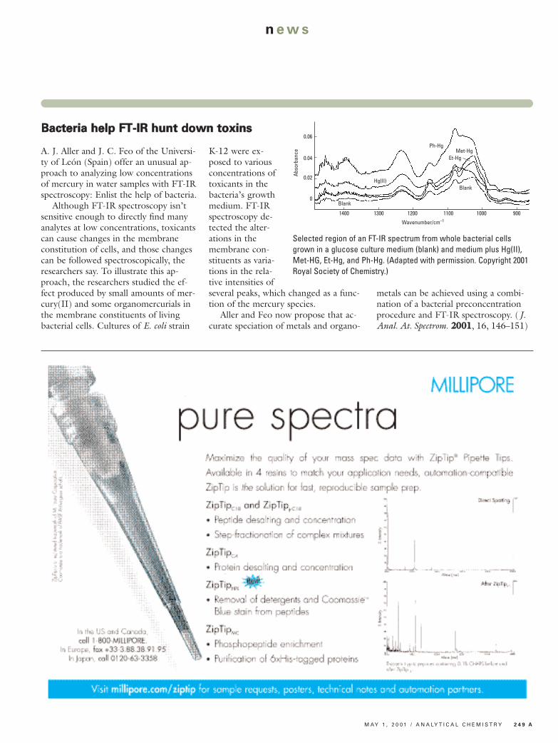
BBaacctteerriiaa hheellpp FFTT--IIRR hhuunntt ddoowwnn ttooxxiinnss
A. J. Aller and J. C. Feo of the Universi-ty of León (Spain) offer an unusual ap-proach to analyzing low concentrationsof mercury in water samples with FT-IRspectroscopy: Enlist the help of bacteria.Although FT-IR spectroscopy isn’t
sensitive enough to directly find manyanalytes at low concentrations, toxicantscan cause changes in the membraneconstitution of cells, and those changescan be followed spectroscopically, theresearchers say. To illustrate this ap-proach, the researchers studied the ef-fect produced by small amounts of mer-cury(II) and some organomercurials inthe membrane constituents of livingbacterial cells. Cultures of E. coli strain
K-12 were ex-posed to variousconcentrations oftoxicants in thebacteria’s growthmedium. FT-IRspectroscopy de-tected the alter-ations in themembrane con-stituents as varia-tions in the rela-tive intensities ofseveral peaks, which changed as a func-tion of the mercury species.Aller and Feo now propose that ac-
curate speciation of metals and organo -
metals can be achieved using a combi-nation of a bacterial preconcentrationprocedure and FT-IR spectroscopy. (J.Anal. At. Spectrom. 22000011, 16, 146–151)
Abso
rban
ce
0.06
1400 1300 1200 1100 900
0.04
0.02
0
1000
Wavenumber/cm–1
Blank
Hg(II)
Ph-HgMet-Hg
Et-Hg
Blank
Selected region of an FT-IR spectrum from whole bacterial cellsgrown in a glucose culture medium (blank) and medium plus Hg(II),Met-HG, Et-Hg, and Ph-Hg. (Adapted with permission. Copyright 2001Royal Society of Chemistry.)
2 5 0 A A N A LY T I C A L C H E M I S T R Y / M AY 1 , 2 0 0 1
news
PPrrooffiilliinngg eennzzyymmeess ttoo ddiiaaggnnoossee ggeenneettiicc ddiisseeaassee
Even in the age of high-powered geneticscreening labs, enzyme analyses play acrucial role in the study of biologicalsystems. Despite advanced DNA testing,many of the mutations that compromiseenzymatic function remain undiscoveredor poorly understood.In the April 15 issue of
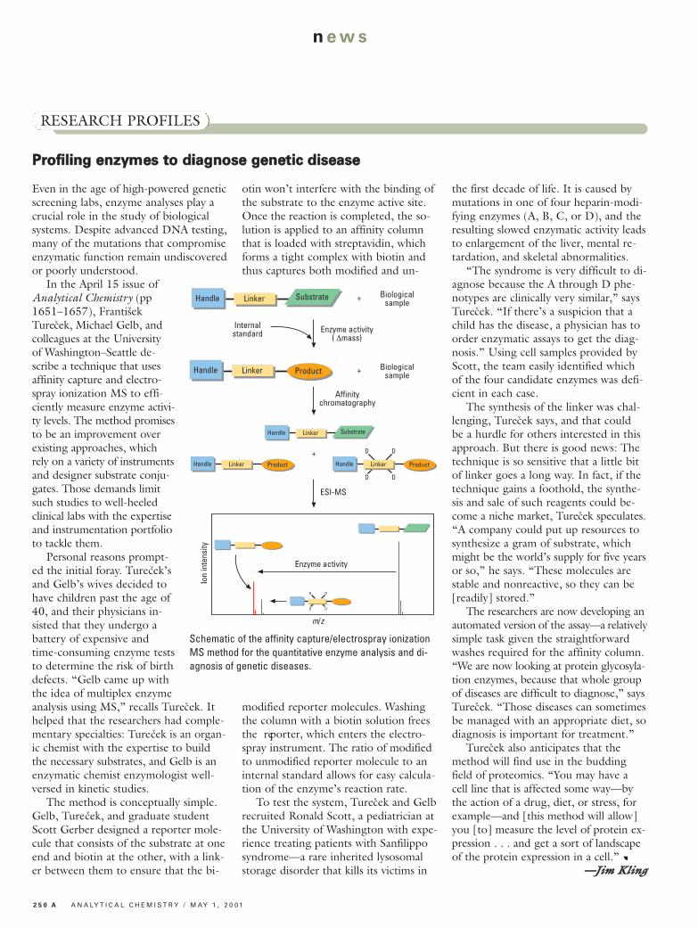
Analytical Chemistry (pp1651–1657), FrantisekTurecek, Michael Gelb, andcolleagues at the University of Washington–Seattle de-scribe a technique that usesaffinity capture and electro-spray ionization MS to effi-ciently measure enzyme activi-ty levels. The method promisesto be an improvement overexisting approaches, whichrely on a variety of instrumentsand designer substrate conju-gates. Those demands limitsuch studies to well-heeledclinical labs with the expertiseand instrumentation portfolioto tackle them.Personal reasons prompt-
ed the initial foray. Turecek’sand Gelb’s wives decided tohave children past the age of40, and their physicians in-sisted that they undergo abattery of expensive andtime-consuming enzyme teststo determine the risk of birthdefects. “Gelb came up withthe idea of multiplex enzymeanalysis using MS,” recalls Turecek. Ithelped that the researchers had comple-mentary specialties: Turecek is an organ-ic chemist with the expertise to buildthe necessary substrates, and Gelb is anenzymatic chemist enzymologist well-versed in kinetic studies.The method is conceptually simple.
Gelb, Turecek, and graduate studentScott Gerber designed a reporter mole-cule that consists of the substrate at oneend and biotin at the other, with a link-er between them to ensure that the bi-
otin won’t interfere with the binding ofthe substrate to the enzyme active site.Once the reaction is completed, the so-lution is applied to an affinity columnthat is loaded with streptavidin, whichforms a tight complex with biotin andthus captures both modified and un-
modified reporter molecules. Washingthe column with a biotin solution freesthe reporter, which enters the electro-spray instrument. The ratio of modifiedto unmodified reporter molecule to aninternal standard allows for easy calcula-tion of the enzyme’s reaction rate.To test the system, Turecek and Gelb
recruited Ronald Scott, a pediatrician atthe University of Washington with expe-rience treating patients with Sanfilipposyndrome—a rare inherited lysosomalstorage disorder that kills its victims in
the first decade of life. It is caused bymutations in one of four heparin-modi-fying enzymes (A, B, C, or D), and theresulting slowed enzymatic activity leadsto enlargement of the liver, mental re-tardation, and skeletal abnormalities.“The syndrome is very difficult to di-
agnose because the A through D phe-notypes are clinically very similar,” saysTurecek. “If there’s a suspicion that achild has the disease, a physician has toorder enzymatic assays to get the diag-nosis.” Using cell samples provided byScott, the team easily identified whichof the four candidate enzymes was defi-cient in each case.The synthesis of the linker was chal-
lenging, Turecek says, and that couldbe a hurdle for others interested in thisapproach. But there is good news: Thetechnique is so sensitive that a little bitof linker goes a long way. In fact, if thetechnique gains a foothold, the synthe-sis and sale of such reagents could be-come a niche market, Turecek speculates.“A company could put up resources tosynthesize a gram of substrate, whichmight be the world’s supply for five yearsor so,” he says. “These molecules arestable and nonreactive, so they can be[readily] stored.”The researchers are now developing an
automated version of the assay—a relativelysimple task given the straightforwardwashes required for the affinity column.“We are now looking at protein glycosyla-tion enzymes, because that whole groupof diseases are difficult to diagnose,” saysTurecek. “Those diseases can sometimesbe managed with an appropriate diet, sodiagnosis is important for treatment.”Turecek also anticipates that the
method will find use in the buddingfield of proteomics. “You may have acell line that is affected some way—bythe action of a drug, diet, or stress, forexample—and [this method will allow]you [to] measure the level of protein ex-pression . . . and get a sort of landscapeof the protein expression in a cell.”
——JJiimm KKlliinngg
RESEARCH PROFILES
Internalstandard Enzyme activity
( ∆mass)
Affinitychromatography
+ Biologicalsample
+ Biologicalsample
SubstrateHandle Linker
ProductHandle Linker
+
ESI-MS
ProductHandle Linker
SubstrateHandle Linker
D D
ProductHandle Linker
D D
D D
D D
Enzyme activity
Ion
inte
nsity
m/z
Schematic of the affinity capture/electrospray ionizationMS method for the quantitative enzyme analysis and di-agnosis of genetic diseases.
M AY 1 , 2 0 0 1 / A N A LY T I C A L C H E M I S T R Y 2 5 1 A
n ews
QQuuaannttiittaattiivvee pprrootteeoommiiccss ggooeess gglloobbaall
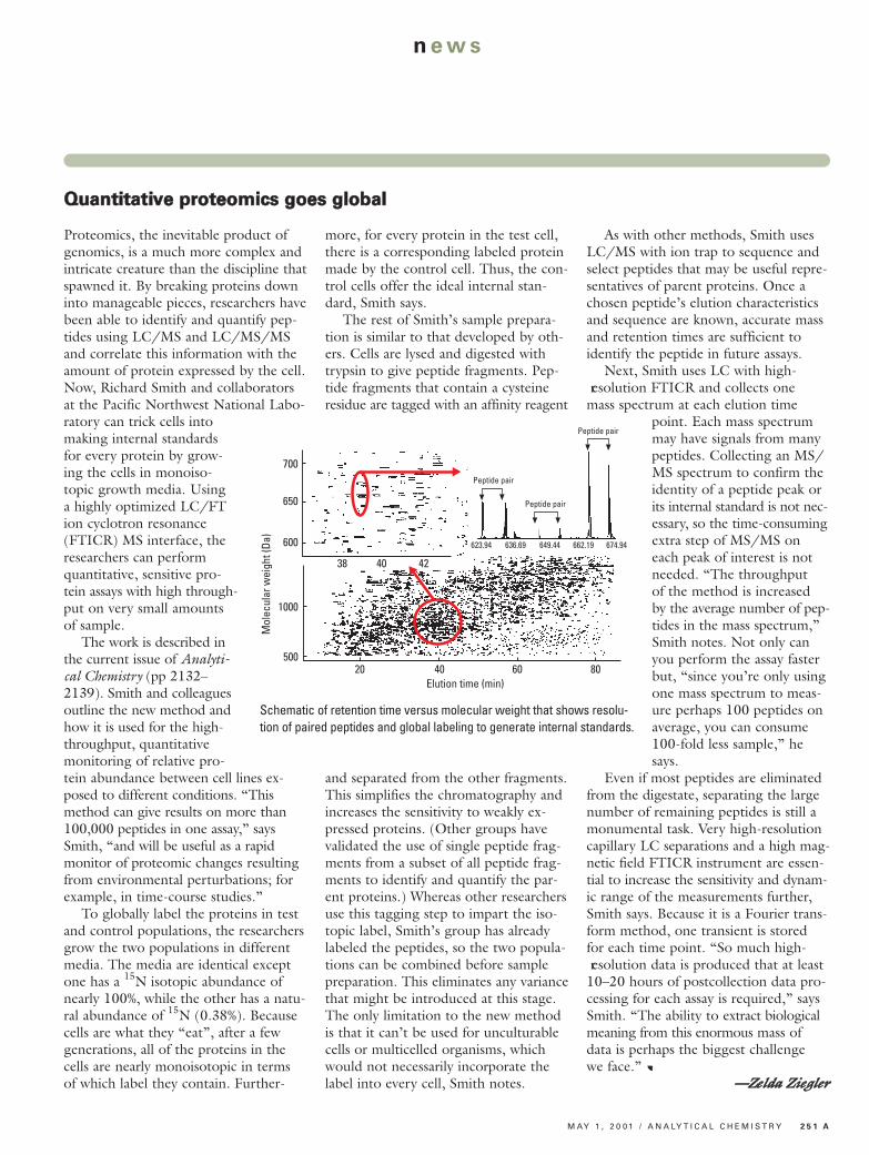
Proteomics, the inevitable product ofgenomics, is a much more complex andintricate creature than the discipline thatspawned it. By breaking proteins downinto manageable pieces, researchers havebeen able to identify and quantify pep-tides using LC/MS and LC/MS/MSand correlate this information with theamount of protein expressed by the cell.Now, Richard Smith and collaboratorsat the Pacific Northwest National Labo-ratory can trick cells intomaking internal standardsfor every protein by grow -ing the cells in monoiso -topic growth media. Usinga highly optimized LC/FTion cyclotron resonance(FTICR) MS interface, theresearchers can performquantitative, sensitive pro -tein assays with high through-put on very small amountsof sample.The work is described in
the current issue of Analyti-cal Chemistry (pp 2132–2139). Smith and colleaguesoutline the new method andhow it is used for the high-throughput, quantitativemonitoring of relative pro-tein abundance between cell lines ex-posed to different conditions. “Thismethod can give results on more than100,000 peptides in one assay,” saysSmith, “and will be useful as a rapidmonitor of proteomic changes resultingfrom environmental perturbations; forexample, in time-course studies.”To globally label the proteins in test
and control populations, the researchersgrow the two populations in differentmedia. The media are identical exceptone has a 15N isotopic abundance ofnearly 100%, while the other has a natu-ral abundance of 15N (0.38%). Becausecells are what they “eat”, after a fewgenerations, all of the proteins in thecells are nearly monoisotopic in termsof which label they contain. Further-
more, for every protein in the test cell,there is a corresponding labeled proteinmade by the control cell. Thus, the con-trol cells offer the ideal internal stan-dard, Smith says.The rest of Smith’s sample prepara-
tion is similar to that developed by oth-ers. Cells are lysed and digested withtrypsin to give peptide fragments. Pep-tide fragments that contain a cysteineresidue are tagged with an affinity reagent
and separated from the other fragments.This simplifies the chromatography andincreases the sensitivity to weakly ex-pressed proteins. (Other groups havevalidated the use of single peptide frag-ments from a subset of all peptide frag-ments to identify and quantify the par-ent proteins.) Whereas other researchersuse this tagging step to impart the iso-topic label, Smith’s group has alreadylabeled the peptides, so the two popula-tions can be combined before samplepreparation. This eliminates any variancethat might be introduced at this stage.The only limitation to the new methodis that it can’t be used for unculturablecells or multicelled organisms, whichwould not necessarily incorporate thelabel into every cell, Smith notes.
As with other methods, Smith usesLC/MS with ion trap to sequence andselect peptides that may be useful repre-sentatives of parent proteins. Once achosen peptide’s elution characteristicsand sequence are known, accurate massand retention times are sufficient toidentify the peptide in future assays.Next, Smith uses LC with high-
resolution FTICR and collects onemass spectrum at each elution time
point. Each mass spectrummay have signals from manypeptides. Collecting an MS/MS spectrum to confirm theidentity of a peptide peak orits internal standard is not nec-essary, so the time-consumingextra step of MS/MS oneach peak of interest is notneeded. “The throughputof the method is increasedby the average number of pep-tides in the mass spectrum,”Smith notes. Not only canyou perform the assay fasterbut, “since you’re only usingone mass spectrum to meas-ure perhaps 100 peptides onaverage, you can consume100-fold less sample,” hesays.
Even if most peptides are eliminatedfrom the digestate, separating the largenumber of remaining peptides is still amonumental task. Very high-resolutioncapillary LC separations and a high mag-netic field FTICR instrument are essen-tial to increase the sensitivity and dynam-ic range of the measurements further,Smith says. Because it is a Fourier trans-form method, one transient is storedfor each time point. “So much high- resolution data is produced that at least10–20 hours of postcollection data pro-cessing for each assay is required,” saysSmith. “The ability to extract biologicalmeaning from this enormous mass ofdata is perhaps the biggest challengewe face.”
——ZZeellddaa ZZiieegglleerr
700
650
600
38 40 42
1000
500
Mol
ecul
ar w
eigh
t (Da
)
20 40 60 80Elution time (min)
623.94 636.69 649.44 662.19 674.94
Peptide pair
Peptide pair
Peptide pair
Schematic of retention time versus molecular weight that shows resolu-tion of paired peptides and global labeling to generate internal standards.
2 5 2 A A N A LY T I C A L C H E M I S T R Y / M AY 1 , 2 0 0 1
news
55tthh SSyymmppoossiiuumm oonn tthhee IInntteerrffaaccee ooff RReegguullaattoorryy aanndd AAnnaallyyttiiccaall SScciieenncceess ffoorr BBiiootteecchhnnoollooggyy HHeeaalltthh PPrroodduuccttss — Judith Handley and Elizabeth Zubritsky report from Washington, DC
BBlloooodd--bbaasseedd pprriioonn tteessttDespite the recent efforts to detectbovine spongiform encephalopathy(BSE), commonly known as mad cowdisease, and stop its transmission, oneglaring problem remains: The currenttests must be performed on dead ani-mals. Thus, there is no feasible way toroutinely screen livestock herds beforethe animals exhibit symptoms—so-calledpreclinical screening.
The crux of the problem is that thelevels of prion protein (PrP)—the abnor-mal form of which is thought to be re-sponsible for BSE and related diseases—are too dilute to be detected reliably inbodily fluids such as blood. Thus, testsare limited to brain and spinal tissue,where the abnormal prions are muchmore abundant.Now, Mary Jo Schmerr and Eddie
Takahashi at the U.S. Department ofAgriculture’s National Animal DiseaseCenter and Andrew Alpert at PolyLC,Inc., describe a blood-based test for arange of transmissible spongiform en-cephalopathies in livestock and humans.In this test, a blood sample is mixedwith a fluorescently labeled peptidefrom PrP and peptide antibodies. Using
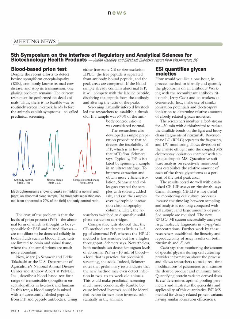
either free-zone CE or size-exclusionHPLC, the free peptide is separatedfrom antibody-bound peptide, and thepeak areas are compared. If the bloodsample already contains abnormal PrP,it will compete with the labeled peptide,displacing the peptide from the antibodyand altering the ratio of the peaks. Screening naturally infected livestock
led the researchers to establish a thresh-old: If a sample was >70% of the anti-
body control ratio, itwas considered normal.The researchers also
developed a sample prepa-ration procedure that ad-dresses the insolubility ofPrP, which is as low asthat of Teflon, Schmerrsays. Typically, PrP is iso-lated by spinning a samplein an ultracentrifuge. Toimprove extraction andobtain more efficient iso-lation, Schmerr and col-leagues treated the sam-ples with solvent, addedsalt, and ran the samplesover hydrophilic-interac-tion chromatographycolumns. Later, the re-
searchers switched to disposable solid-phase extraction cartridges.Comparative tests showed that the
CE method can detect as little as 1–2pg of abnormal PrP, whereas the HPLCmethod is less sensitive but has a higherthroughput, Schmerr says. Nevertheless,both methods can detect femtogram levelsof abnormal PrP in ~10 mL of blood—a level that is practical for preclinicalscreening, she adds. Indeed, Schmerrnotes that preliminary tests indicate thatthe new method may even detect infec-tion in two- to six-week-old animals.This could make preclinical screeningmuch more economically feasible be-cause infected livestock could be identi-fied before farmers have invested sub-stantially in the animals.
EESSII qquuaannttiiffiieess ggllyyccaann mmooiieettiieessHow would you like a one-hour, in-process method to identify and quantifythe glycoforms on an antibody? Work-ing with the recombinant antibody rit-uximab, Jerry Cacia and co-workers atGenentech, Inc., make use of similarionization potentials and electrosprayionization to determine relative amountsof closely related glycan moieties.The researchers incubate a feed-stream
for ~30 min with dithiothreitol to reducethe disulfide bonds on the light and heavychain fragments of rituximab. Reversed-phase LC (RPLC) separates the fragments,and UV monitoring allows diversion ofthe analyte effluent into the coupled MSelectrospray ionization chamber with sin-gle quadrupole MS. Quantitative soft-ware analysis on selectively monitoredions establishes the relative amount ofeach of the three glycoforms as a per-cent of the total peak area.The results correlate well with estab-
lished CE-LIF assays on rituximab, saysCacia, although CE-LIF is not usefulfor monitoring cell culture processes because the time lag between samplingand analysis is too long compared withcell culture, and large amounts of puri-fied sample are required. The newRPLC/ MS system successfully analyzedlarge molecule fragments in <5-µg/mLconcentrations. Further work by theseresearchers established the linearity andreproducibility of assay results on bothrituximab and E. coli.Cacia says that monitoring the amount
of specific glycans during cell culturingprovides information about the processand allows researchers to make real-timemodifications of parameters to maximizethe desired product and minimize time.Quantifying protein variants derived fromE. coli determines optimal pooling para -meters and illustrates the generality andapplicability of this quantitative ESI-MSmethod for closely related protein variantshaving similar ionization efficiencies.
MEETING NEWS
Antibody controlRatio = 1.03
Normal sheepRatio = 0.97
Scrapie infected sheepRatio = 0.48
Electropherograms showing peaks in (middle) a normal and(right) an abnormal blood sample. The threshold separating nor-mal from abnormal is 70% of the (left) antibody control ratio.
M AY 1 , 2 0 0 1 / A N A LY T I C A L C H E M I S T R Y 2 5 3 A
n ews
990000--MMHHzz NNMMRR iiss hheerree
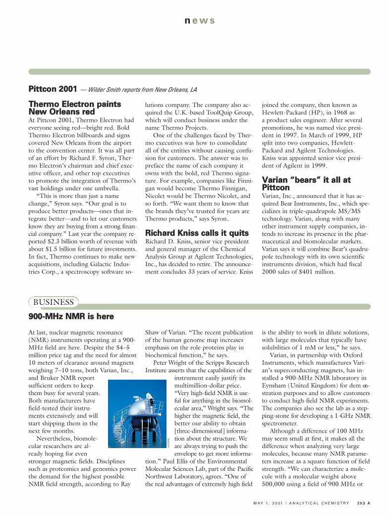
At last, nuclear magnetic resonance(NMR) instruments operating at a 900-MHz field are here. Despite the $4–5million price tag and the need for almost10 meters of clearance around magnetsweighing 7–10 tons, both Varian, Inc.,and Bruker NMR reportsufficient orders to keepthem busy for several years.Both manufacturers havefield-tested their instru-ments extensively and willstart shipping them in thenext few months.Nevertheless, biomole-
cular researchers are al-ready hoping for evenstronger magnetic fields. Disciplinessuch as proteomics and genomics powerthe demand for the highest possibleNMR field strength, according to Ray
Shaw of Varian. “The recent publicationof the human genome map increasesemphasis on the role proteins play inbiochemical function,” he says.Peter Wright of the Scripps Research
Institute asserts that the capabilities of theinstrument easily justify itsmultimillion-dollar price.“Very high-field NMR is use-ful for anything in the biomol-ecular area,” Wright says. “Thehigher the magnetic field, thebetter our ability to obtain[three-dimensional] informa-tion about the structure. Weare always trying to push theenvelope to get more informa-
tion.” Paul Ellis of the EnvironmentalMolecular Sciences Lab, part of the PacificNorthwest Laboratory, agrees. “One ofthe real advantages of extremely high field
is the ability to work in dilute solutions,with large molecules that typically havesolubilities of 1 mM or less,” he says.Varian, in partnership with Oxford
Instruments, which manufactures Vari-an’s superconducting magnets, has in-stalled a 900-MHz NMR laboratory inEynsham (United Kingdom) for dem on -stration purposes and to allow customersto conduct high-field NMR experiments.The companies also see the lab as a step-ping-stone for developing a 1-GHz NMRspectrometer. Although a difference of 100 MHz
may seem small at first, it makes all thedifference when analyzing very largemolecules, because many NMR parame-ters increase as a square function of fieldstrength. “We can characterize a mole-cule with a molecular weight above500,000 using a field of 900 MHz or
BUSINESS
PPiittttccoonn 22000011 — Wilder Smith reports from New Orleans, LA
TThheerrmmoo EElleeccttrroonn ppaaiinnttssNNeeww OOrrlleeaannss rreeddAt Pittcon 2001, Thermo Electron hadeveryone seeing red—bright red. BoldThermo Electron billboards and signscovered New Orleans from the airportto the convention center. It was all partof an effort by Richard F. Syron, Ther-mo Electron’s chairman and chief exec-utive officer, and other top executivesto promote the integration of Thermo’svast holdings under one umbrella.“This is more than just a name
change,” Syron says. “Our goal is toproduce better products—ones that in-tegrate better—and to let our customersknow they are buying from a strong finan-cial company.” Last year the company re-ported $2.3 billion worth of revenue withabout $1.5 billion for future investments.In fact, Thermo continues to make newacquisitions, including Galactic Indus-tries Corp., a spectroscopy software so-
lutions company. The company also ac-quired the U.K.-based ToolQuip Group,which will conduct business under thename Thermo Projects.One of the challenges faced by Ther-
mo executives was how to consolidateall of the entities without causing confu-sion for customers. The answer was topreface the name of each company itowns with the bold, red Thermo signa-ture. For example, companies like Finni-gan would become Thermo Finnigan,Nicolet would be Thermo Nicolet, andso forth. “We want them to know thatthe brands they’ve trusted for years areThermo products,” says Syron.
RRiicchhaarrdd KKnniissss ccaallllss iitt qquuiittssRichard D. Kniss, senior vice presidentand general manager of the ChemicalAnalysis Group at Agilent Technologies,Inc., has decided to retire. The announce-ment concludes 33 years of service. Kniss
joined the company, then known asHewlett-Packard (HP), in 1968 asa product sales engineer. After severalpromotions, he was named vice presi-dent in 1997. In March of 1999, HPsplit into two companies, Hewlett-Packard and Agilent Technologies.Kniss was appointed senior vice presi-dent of Agilent in 1999.
VVaarriiaann ““bbeeaarrss”” iitt aallll aattPPiittttccoonnVarian, Inc., announced that it has ac-quired Bear Instruments, Inc., which spe-cializes in triple-quadrupole MS/MStechnology. Varian, along with manyother instrument supply companies, in-tends to increase its presence in the phar-maceutical and biomolecular markets.Varian says it will combine Bear’s quadru-pole technology with its own scientificinstruments division, which had fiscal2000 sales of $401 million.
VA
RIA
N
2 5 4 A A N A LY T I C A L C H E M I S T R Y / M AY 1 , 2 0 0 1
news
more,” says Wright. “With an 800-MHzfield, the S/N is just not high enough.Even in the 50,000 molecular weightrange, there is a lot of compressed data.The increased dispersion [of the 900-MHz field] helps resolve structural detailthat is otherwise not available.”The need for a resolution of at least
900 MHz is evident in the backlog oforders at both Bruker and Varian. Bruk-er expects to be able to deliver about six900-MHz instruments per year and hasat least eight already on order. Varianhas received about 10 orders and proj-ects a manufacturing capability of 4 in-struments per year. Customers for thehigh-field instruments are generally na-tional laboratories and large researchuniversities and institutes in the United
States, Europe, and Japan.Ideally, the Scripps scientists would
like an even more powerful magnet be-cause the institute is performing trans-verse relaxation-optimized spectroscopywith 15N—an approach that avoids theusual limits on the size of the biomacro-molecular structures that can be stud-ied. The optimal field for this type ofexperiment is 1 GHz, and the efficiencyfalls off very rapidly with lower fields.Bruker and Oxford say they have sev-
eral groups developing the next genera-tion of NMR magnets, but they expectthe introduction of 1-GHz NMR sys-tems to be at least four years away. Cur-rent instruments are reaching the limitsof existing superconducting magnettechnology, and further advances will
require significant research. “The stateof the art is pushing the technology,”says Mark Chaykowsky of the NMR di-vision at Bruker. “In a way, it’s amazingthat these instruments actually exist sincethe applications require not just highfield, but also high homogeneity.” Eventhe 900-MHz instrument was not possi-ble without recent advances in super-conductor current-carrying capability.Nevertheless, when the instrument
companies eventually succeed, the mar-ket appears certain. “Every time we getto these new fields, people say thatthere’s no way the cost will justify plac-ing the system,” says Shaw. “But theusers always find new applications thatrequire the additional capabilities.”
——SStteevvee MMiilllleerr
BUSINESS